Original Paper Neonatology 2008;93:236–240 DOI: 10.1159/000111102
formerly Biology of the Neonate
Received: April 16, 2007 Accepted after revision: September 3, 2007 Published online: November 16, 2007
Mode of Delivery – Effects on Gut Microbiota and Humoral Immunity Anu Huurre a Marko Kalliomäki a Samuli Rautava a Minna Rinne a Seppo Salminen b Erika Isolauri a a b
Department of Pediatrics, Turku University Central Hospital and University of Turku, and Functional Foods Forum, University of Turku, Turku, Finland
Key Words Caesarean section ! Vaginal delivery ! Gut microbiota ! Humoral immunity
Abstract Background: The rate of caesarean deliveries has increased 10-fold worldwide during the past decades. Objective: To evaluate differences in the establishment of gut microbiota in infants born by vaginal or caesarean delivery and its impact on mucosal immunity. Methods: Altogether, 165 consecutive children, prospectively followed from birth at our clinic in Turku, Finland, were gathered; 141 (85%) were born by vaginal delivery and 24 (15%) by caesarean section. Blood was drawn at physician visits for indirect evaluation of mucosal immunity by ELISPOT assay. Faecal samples were obtained for determination of the gut microbiota by fluorescence in situ hybridization of bacterial cells. Results: Infants delivered by caesarean section harboured fewer bifidobacteria at an early age and were shown to mount a stronger humoral immune response. At 1 month of age, the total gut bacterial cell counts per 1 g faeces were higher in vaginally delivered infants (9.9 ! 109, 95% CI 7.9 ! 109 –1.2 ! 1010) as compared to caesarean section delivered (3.1 ! 109, 95% CI 1.1 ! 109 –8.6 ! 109) (p = 0.001). This distinction was mainly
Funding sources: Sigrid Juselius Foundation, Academy of Finland.
Fax +41 61 306 12 34 E-Mail
[email protected] www.karger.com
© 2007 S. Karger AG, Basel 1661–7800/08/0934–0236$24.50/0 Accessible online at: www.karger.com/neo
due to the greater number of bifidobacteria in vaginally delivered infants (1.9 ! 109, 95% CI 6.3 ! 108–5.6 ! 109 vs. 1.5 ! 106, 95% CI 4.1 ! 102–5.7 ! 109, respectively) (p = 0.001). During the first year of life, the total number of IgA-, IgG- and IgM-secreting cells was lower (p = 0.03, p = 0.02, p = 0.11, respectively) in infants born by vaginal delivery than in those born by caesarean section, possibly reflecting excessive antigen exposure across the vulnerable gut barrier. Conclusions: Our findings demonstrate that the mode of delivery may have, possibly via gut microbiota development, significant effects on immunological functions in the infant (http://www.clinicaltrials.gov/ct/gui/show/NCT00167700). Copyright © 2007 S. Karger AG, Basel
Introduction
The establishment of the gut microbiota occurs rapidly after birth, initiating as the fetal membranes are ruptured. Intestinal colonization follows successive steps, dominated first by facultative anaerobes such as enterobacteria, coliforms and lactobacilli, followed by anaerobic genera such as Bifidobacterium, Bacteroides, Clostridium and Eubacterium [1–3]. After weaning, an adult-like gut microbiota gradually develops. This process is dependent on genetic factors, maternal microbiota, birth environment, feeding practices and particularly the mode of delivery. Anu Huurre Department of Pediatrics, Turku University Central Hospital, University of Turku Kiinanmyllynkatu 4–8 FI–20520 Turku (Finland) Tel. +358 2 313 0000, Fax +358 2 313 1460, E-Mail
[email protected]
Aberrant microbiota development may increase the risk of specific human diseases [4, 5]. Indeed, recent scientific data challenge the traditional thinking on commensalism, indicating an impartial coexistence of microbes and host, where a risk of disease is generated if translocation of specific intraluminal bacteria takes place. Interest in host-microbe interaction has been reawakened by the demonstration that the gut microbiota are critical for the host’s physiology. Major functions of the microbiota include metabolic activities which result in salvage of energy and absorbable nutrients, protection of the host against invasion by pathogenic microbes, trophic effects on the intestinal epithelium and intestinal epithelial homeostasis and regulation of immune responses [4]. The significance of the regulatory function culminates at an early age, when the mucosal barrier and immune system are immature. The rate of caesarean deliveries has increased 10-fold worldwide during the past few decades. We therefore evaluated differences in the establishment of microbiota in children born by vaginal or caesarean delivery. In particular, the possible effect of gut microbiota on mucosal immunity, the first line of host defence, was examined. Methods Subjects and Study Design For this study, 165 consecutive children from allergic families (i.e. mother, father or sibling with atopic disease) who have been prospectively followed from birth at our clinic were gathered, 141 (85%) of them born by vaginal delivery and 24 (15%) by caesarean section. Infants who had been exposed to probiotics directly or through the mother were excluded from the study. All studies were approved by The Ethical Committee of the Hospital District of Southwest Finland. Written informed consent was obtained from the children’s parents. All data in the study were treated confidentially. The infants were examined by a research nurse at the age of 1 month and by a physician at the ages of 3, 6 and 12 months. Blood was drawn at physician visits for evaluation of mucosal immunity. Faecal samples were obtained at every scheduled visit and stored at –20 ° C for later analyses. Skin prick tests were performed at the ages of 6 and 12 months as previously described [6]. Atopic eczema was diagnosed if atopic sensitization (i.e. positive skin prick test) was confirmed [7] and the following clinical features were detected: pruritus, facial and/or extensor involvement and chronic relapsing course [8]. Cow milk allergy was verified either by a combination of positive skin prick test and clinical symptoms related to cow milk or double-blind placebo-controlled cow milk challenge. The number of circulating immunoglobulin-secreting cells was assayed by the enzyme-linked immunospot (ELISPOT) assay, since it indirectly indicates immunological events in the gut
Effects of Mode of Delivery on Gut Microbiota
[9, 10]. Fluorescence in situ hybridization (FISH) was used for enumeration of four dominant bacterial groups of gut microbiota. A significant proportion of gut microbiota remains unidentified. For this purpose the total bacterial cell number from faecal samples was determined in addition of the numbers of bifidobacteria, clostridia, lactobacilli and bacteroides, which reflect the most frequently identified microbe species in the infant gut. Evaluation of Humoral Immunity: ELISPOT Assay In brief, mononuclear cells containing mainly lymphocytes were obtained by Ficoll-Paque (Pharmacia LKB Biotechnology AB, Uppsala, Sweden) centrifugation of lithium-heparinized blood. Isolated cells, which were washed three times in Hanks’ buffered salt solution, were subsequently suspended in RPMI 1640 medium (Gibco-BRL Life Technologies, Paisley, UK) containing 10% fetal calf serum (Gibco-BRL Life Technologies), and adjusted to a concentration of 1–2 ! 106 cells/ml. To determine the number of specific antibody-secreting cells against dietary antigens, coating was performed for 2 h at 37 ° C with the following concentrations of the antigens (both from Sigma Chemical Co., St. Louis, Mo., USA): !-lactoglobulin, 20 "g/ml (from bovine milk) and casein, 20 "g/ml (from bovine milk). Uncoated binding sites were blocked with 1% bovine serum albumin (BSA; Boehringer Mannheim GmbH, Germany) in phosphatebuffered saline (PBS; pH 7.4) for 30 min at 37 ° C. After washings the lymphocyte suspension was incubated on antigen-coated flatbottomed 96-well microtitre plates (Immunoplate R I!, Nunc A/S, Roskilde, Denmark) at 37 ° C for 2 h. The antibodies secreted during that time were detected with alkaline phosphatase-conjugated goat antisera to human IgA, IgG and IgM (all from Sigma Chemical Co.) diluted in 1% BSA-PBS (pH 7.4) incubated overnight at room temperature, followed by a substrate agarose overlay and observation of coloured spots. The total number of immunoglobulin-secreting cells was interpreted to reflect non-antigen-specific mucosal immune responsiveness. The wells were coated with rabbit anti-human IgA (Dako A/S, Glostrup, Denmark) and IgM (Dako A/S), and goat anti-human IgG (Sigma Chemical Co.) diluted 1/100 in PBS (pH 7.4). Subsequent steps were as described above. Determination of the Gut Microbiota: FISH The faecal samples (1 g sample, wet weight) were suspended in 0.1 M PBS (pH 7.0) to give a final concentration of 10% (w/v). The slurries were homogenized and centrifuged at low gravity (250 g for 2 min) to remove particulate matter. Bacterial cells were fixed and FISH analyses performed as previously described [11]. In brief, cells were fixed overnight in 4% (v/v) paraformaldehyde at 4 ° C, washed twice in PBS and stored at –20 ° C in a PBS:ethanol (1:1) solution. Subsamples of the fixed cells were hybridized overnight in hybridization buffer with a 5 ng/"l Cy3 indocarbocyanin-labelled oligonucleotide probe. Probes (sequence 5"]3") included were Bac303 (CCAATGTGGGGACCTT) [12] specific for bacteroides, Bif164 (CATCCGGCATTACCACCC) [11] for bifidobacteria, His150 (TTATGCGGTATTAA TCT(C/T)CCTTT) [13] for clostridia (perfringens/histolyticum subgroups) and Lab158 (GGTATTAGCA(T/C)CTGTTTCCA) [14] for lactobacilli and enterococci. Total cell numbers were counted with a nucleic acid stain 4",6-diamidino-2-phenylindole (DAPI). Cells were washed with the hybridization buffer, filtered through a 0.2-"m polycarbonate filter (Millipore Corp., Etten-Leur, The Nether-
Neonatology 2008;93:236–240
237
lands) and mounted on a slide with SlowFade! (Molecular Probes Inc., Eugene, Oreg., USA). They were counted visually with a Leica Laborlux D epifluorescence microscope mounted with Cy3 and DAPI-specific filters. Fifteen microscopic fields were counted per assay.
120 × 108 100 × 108
VD CS
80 × 108
Statistical Analysis Statistical analyses were conducted with StatView 4.57 (Abacus Concepts, Inc.). Data are presented as mean values with, when appropriate, 95% confidence interval (CI) or range. Statistical differences between the delivery groups were compared by ANOVA and Mann-Whitney U-test. The #2 test was used to determine the difference in proportions.
60 × 108 40 × 108 20 × 108 2 × 10⁶ 1 × 10⁶ 0 × 106
Results
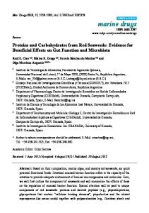
Clinical Data The vaginally delivered infants were born at a mean gestation of 39.5 weeks (range 35.0–42.1) and infants born by caesarean section at 39.2 weeks (range 34.0– 42.3). Birth weights were 3,558 g (range 2,140–4,890) and 3,611 g (range 2,215–4,800), correspondingly. The infants born by vaginal delivery were exclusively breast-fed until a mean age of 2.3 months (range 0–6.0) and the caesarean section babies until 2.1 months (range 0–5.5). The total duration of breastfeeding was 6.2 (range 0.3–19) and 5.8 (range 0.8–16) months, respectively. All of these characteristics were comparable between the groups. There was a trend towards lower frequency of atopic sensitisation in the vaginally delivered infants as compared to those born by caesarean section. At the age of 6 months, 12% of those delivered vaginally and 22% of those delivered by caesarean section had a positive skin prick test (p = 0.18). At 12 months of age, the respective frequencies were 16 and 17% (p = 0.86). The most common allergens responsible for positive reactions were egg white and milk. Atopic eczema was diagnosed in 14% of the vaginally delivered infants and 17% of those born by caesarean section (p = 0.71). Cow milk allergy was detected in 12 and 17% (p = 0.43) of the infants, respectively. Gut Microbiota At 1 month of age, no differences were found in the numbers of clostridia, lactobacilli or bacteroides. However, the number of bifidobacteria was 1,300-fold higher in vaginally delivered infants compared to caesarean section-born infants (p = 0.001). This was consequently reflected in the total bacterial cell numbers (fig. 1), which were 3-fold higher in infants born by vaginal versus caesarean delivery (p = 0.001). At the age of 6 months these differences were no longer observed. 238
Neonatology 2008;93:236–240
Bifidobacterium
Total bacterial cell number
Fig. 1. The number of bifidobacteria and total bacteria cells (per 1 g faecal wet mass) in infants born by vaginal delivery (VD) and caesarean section (CS) at 1 month of age. Data represent geometric means with 95% CI.
Humoral Immune Responses During the first year of life, as measured at ages of 3, 6 and 12 months, the total number of IgA-secreting cells was lower (p = 0.03, ANOVA) in infants born by vaginal delivery than in those born by caesarean section, as shown in figure 2. The total numbers of IgG- and IgMsecreting cells followed a similar pattern, p = 0.02 and 0.11 (ANOVA), respectively. However, in antigen-specific antibody-secreting cells in the IgA class against milk antigens (casein and !-lactoglobulin), no statistically significant difference was detected when compared with the vaginally delivered versus caesarean section-born infants: at 3 months, 12 (95% CI 7–16) versus 16 (95% CI 8–24), respectively (p = 0.45). The respective figures at 6 months of age were 10 (95% CI 7–13) and 16 (95% CI 7–25) (p = 0.20), and at 12 months 12 (95% CI 8–15) versus 11 (95% CI 5–15) (p = 0.77). Discussion
We detected a significant correlation between the mode of delivery, gut microbiota and mucosal immunity in a substantial cohort of young infants by culture-independent methods. Infants delivered by caesarean section harboured fewer bifidobacteria at an early age and were shown to mount a stronger non-specific humoral immune response. Huurre /Kalliomäki /Rautava /Rinne / Salminen /Isolauri
6,000
VD CS
IgA
5,000 4,000 3,000 2,000 1,000
a
0 3 months
6,000
VD CS
6 months
12 months
IgG
5,000 4,000 3,000 2,000 1,000
b
0 3 months
6,000
VD CS
6 months
12 months
IgM
5,000 4,000 3,000 2,000 1,000
c
0 3 months
6 months
12 months
Fig. 2. The number of (a) IgA-, (b) IgG- and (c) IgM-secreting cells per 106 mononuclear cells in infants born by vaginal delivery (VD) and caesarean section (CS) at ages of 3, 6 and 12 months. Data represent means with 95% CI.
Effects of Mode of Delivery on Gut Microbiota
It is noteworthy that bifidobacteria are the most abundant members of the gut microbiota during the first months of life. In fact, bifidobacteria compose up to 60– 90% of the gut microbiota of a healthy breastfed infant, whereas the number of lactobacilli seems to be less significant than earlier reported [15]. The present study substantiates these observations in documenting that vaginally delivered infants who showed 1,300-fold higher numbers of bifidobacteria in their gut microbiota compared to caesarean section-born infants also exhibited higher proportional numbers of bifidobacteria and 3-fold higher total bacterial cell numbers in their faeces. A distinct pattern of bacterial colonization in caesarean sectioned and vaginally delivered infants has previously been reported by Li et al. [16], namely in their study Streptococcus mutans colonization within the mouth was detected significantly later in vaginally delivered than in caesarean section-born infants. The lower concentration of bifidobacteria and the lower number of total bacterial cells are likely to hamper the successive establishment of the gut microbiota and thereby result in an altered collective composition and metabolic activity influencing the immune response during a critical period of development. In support of this suggestion, we have previously shown that differences in intestinal microbiota remain beyond infancy [17]. We demonstrated here that infants born by caesarean delivery have a higher level of immunoglobulin-producing cells in their peripheral blood compared to those born by vaginal delivery. While there was no difference in antigen-specific antibody-secreting cells against milk antigens, the difference in total number of antibody-secreting cells must be explained by other, non-specific factors such as excessive antigen exposure across the vulnerable gut barrier. Indeed, commensal bacteria have a major role in protecting the gut from injury by interaction with the gut epithelium and by forming the mucosal barrier [18]. The host-microbe interaction on the intestinal mucosa is mediated by specific toll-like receptors (TLRs). Under normal balanced conditions, commensal bacteria, as well as pathogens, are recognized by TLRs, but the ensuing immune responses, non-inflammatory versus inflammatory, differ between commensals and pathogens. If this crosstalk is disturbed, intestinal epithelial cells may proliferate at a more rapid rate, rendering the defence barrier vulnerable to intraluminal offending antigens [19], the major source of such antigens at an early age being derived from food and bacteria.
Neonatology 2008;93:236–240
239
It appears that the mucosa of young children, unlike that of adults, tends to be, through a TLR-dependent pathway, particularly responsive to microbial stimuli. In an in vitro study, where explanted nasal mucosa was cultured with bacterial lipopolysaccharide and allergen, lipopolysaccharide was able to prevent allergic inflammation in the nasal mucosa of atopic children, but not in adults, by transforming local immune responses from Thelper type 2 to T-helper type 1 and increasing the expression of interleukin-10 [20]. Furthermore, bifidobacteria, regularly present in the healthy breast-fed infant gut, may influence this process. For instance, production of interleukin-10 by intestinal dendritic cells appears to be up-regulated following stimulation with bifidobacte-
ria strains, not lipopolysaccharide [21, 22]. Taking these observations together, the crosstalk between infant as the host and gut microbiota at an early age serves the purpose of immune maturation. The results of the present study demonstrate that the mode of delivery may have, possibly via gut microbiota development, significant effects on immunological functions in the infant. While early infancy is a critical phase in the development of the immune system, aberrations in gut microbiota development and thereby in the immunologic homeostasis may have a long-term impact on the infants’ future health. The microbiota succession in caesarean section-born infants should thus be more thoroughly characterized and understood.
References 1 Isolauri E, Salminen S: Probiotics, gut inflammation and barrier function. Gastroenterol Clin North Am 2005; 34:437–450. 2 Isolauri E, Rautava S, Kalliomäki M, Kirjavainen P, Salminen S: Role of probiotics in food hypersensitivity. Curr Opin Allergy Clin Immunol 2002;2:263–271. 3 Edwards CA, Parrett AM: Intestinal flora during the first month of life: new perspectives. Br J Nutr 2002;88:S11–S18. 4 Guarner F, Malagelada JR: Gut flora in health and disease. Lancet 2003; 361: 512– 519. 5 Kirjavainen PV, Arvola T, Salminen S, Isolauri E: Aberrant composition of gut microbiota of allergic infants: a target of bifidobacterial therapy at weaning? Gut 2002; 51: 51–55. 6 Isolauri E, Turjanmaa K: Combined skin prick and patch testing enhances identification of food allergy in infants with atopic dermatitis. J Allergy Clin Immunol 1996;97: 9–15. 7 Johansson SGO, Bieber T, Dahl R, Friedmann PS, Lanier BQ, Lockey RF, Motala C, Ortega Martell JA, Platts-Mills TAE, Ring J, Thien F, Van Cauwenberge P, Williams HC: Revised nomenclature for allergy for global use: Report of the nomenclature review committee of the World Allergy Organization, October 2003. J Allergy Clin Immunol 2004; 113:832–836. 8 Hanifin JM: Atopic dermatitis in infants and children. Pediatr Clin N Am 1991; 38: 763– 789.
240
9 Isolauri E, Virtanen E, Jalonen T, Arvilommi H: Local immune response measured in blood lymphocytes reflects the clinical reactivity of children with cow’s milk allergy. Pediatr Res 1990;28:582–586. 10 Forrest BD: Identification of an intestinal immune response by using peripheral blood lymphocytes. Lancet 1988;I:81–83. 11 Langendijk PS, Schut F, Jansen GJ, Raangs GC, Kamphuis GR, Wilkinson MHF, Welling GW: Quantative fluorescence in situ hybridisation of Bifidobacterium spp. with genus-specific 16S rRNA-targeted probes and its application in fecal samples. Appl Environ Microbiol 1995; 61:3069–3075. 12 Manz W, Amann R, Ludwig W, Vancanneyt M, Schleifer KH: Application of a suite of 16S rRNA-specific oligonucleotide probes designed to investigate bacteria of the phylum cytophaga-flavobacter-bacteroides in the natural environment. Microbiology 1996; 142:1097–1106. 13 Harmsen HJM, Gibson GR, Elfferich P, Raangs GC, Wildeboer-Veloo ACM, Argaiz A, Roberfroid MB, Welling GW: Comparison of viable cell counts and fluorescence in situ hybridisation using specific rRNA-based probes for the quantification of human fecal bacteria. FEMS Microbiol Lett 2000; 183: 125–129. 14 Harmsen HJ, Elfferich P, Schut F, Welling GW: A 16S rRNA-targeted probe for detection of lactobacilli and enterococci in faecal samples by fluorescent in situ hybridisation Microbiol Ecol Health Dis 1999; 11:3–12. 15 Favier CF, Vaughan EE, de Vos WM, Akkermans AD: Molecular monitoring of succession of bacterial communities in human neonates. Appl Environ Microbiol 2002; 68: 219–226.
Neonatology 2008;93:236–240
16 Li Y, Caufield PW, Dasanayake AP, Wiener HW, Vermund SH: Mode of delivery and other maternal factors influence the acquisition of Streptococcus mutans in infants. J Dent Res 2005;84:806–811. 17 Salminen S, Gibson GR, McCartney AL, Isolauri E: Influence of mode of delivery on gut microbiota composition in seven-year-old children. Gut 2004;53:1388–1389. 18 Kraehenbuhl JP, Corbett M: Keeping the gut microflora at bay. Science 2004; 303: 1624– 1625. 19 Rakoff-Nahoum S, Paglino J, Eslami-Varzaneh F, Edberg S, Medzhitov R: Recognition of commensal microflora by toll-like receptors is required for intestinal homeostasis. Cell 2004;118:229–241. 20 Tulic MK, Fiset PO, Manoukian JJ, Frenkiel S, Lavigne F, Eidelman DH, Hamid Q: Role of toll-like receptor-4 in protection by bacterial lipopolysaccharide in the nasal mucosa of atopic children but not adults. Lancet 2004;363:1689–1697. 21 Rigby RJ, Knight SC, Kamm MA, Stagg AJ: Production of interleukin (IL)-10 and IL-12 by murine colonic dendritic cells in response to microbial stimuli. Clin Exp Immunol 2005;139:245–256. 22 Hart AL, Lammers K, Brigid P, Vitali B, Rizzello F, Gionchetti P, Campieri M, Kamm MA, Knight SC, Stagg AJ: Modulation of human dendritic cell phenotype and function by probiotics bacteria. Gut 2004; 53: 1602– 1609.
Huurre /Kalliomäki /Rautava /Rinne / Salminen /Isolauri