Free Radical Research, December 2006; 40(12): 1284–1294
Mitochondrial DNA damage and the aging process – facts and imaginations* ´ BOR ZSURKA3,4, & WOLFRAM S. KUNZ3,4,† RUDOLF J. WIESNER1,2, GA 1
Faculty of Medicine, Institute of Vegetative Physiology, University of Ko¨ln, Ko¨ln, Germany, 2Center for Molecular Medicine Cologne (CMMC), University of Ko¨ln, Ko¨ln, Germany, 3Department of Epileptology, University of Bonn Medical Center, Bonn, Germany, and 4Platform NeuroCognition, Life & Brain Center, Bonn, Germany Accepted by Dr T. Grune
(Received 8 June 2006; in revised form 11 July 2006)
Abstract Mitochondrial DNA (mtDNA) is a circular double-stranded molecule organized in nucleoids and covered by the histone-like protein mitochondrial transcription factor A (TFAM). Even though mtDNA repair capacity appears to be adequate the accumulation of mtDNA mutations has been shown to be at least one important molecular mechanism of human aging. Reactive oxygen species (ROS), which are generated at the FMN moiety of mitochondrial respiratory chain (RC) complex I, should be considered to be important at least for the generation of age-dependent mtDNA deletions. However, the accumulation of acquired mutations to functionally relevant levels in aged tissues seems to be a consequence of clonal expansions of single founder molecules and not of ongoing mutational events.
Keywords: Mitochondrial DNA, TFAM, mtDNA, FMN moiety
“Mitochondrial DNA (mtDNA) is highly susceptible to damage produced by reactive oxygen species (ROS), which are generated in close proximity and in large concentrations by the respiratory chain (RC). This damage is aggravated because mtDNA is (i) not protected by histones, (ii) because there is little capacity for DNA repair in the mitochondrial compartment and (iii) because mutations of mtDNA lead to RC dysfunction, causing the release of even more ROS. Due to this vicious cycle, mutations of mtDNA accumulate over time and become an important cause of aging.”
relations between ROS release from the mitochondrial RC, the topology of the mtDNA molecule in the mitochondrial matrix, the proteins involved in its transcription, replication, maintenance and repair and about the accumulation of mutations during the normal aging process. In this review, we have tried our best to critically summarize the now available literature concerning these questions and to evaluate the pros and cons.
This is how many reviews on the role of mtDNA mutations in aging start. However, 50 years after Harman [1] has postulated the mitochondrial theory of aging, without knowing about the existence of mtDNA at that time, we know a lot more about the
Human mtDNA is a 16.569 bp, plasmid-like circular DNA molecule present in many 1000 copies in cell lines usually used in cell culture experiments and up to many hundreds of thousands of copies in large cells in the body. Vertebrate mtDNA has no introns and
Organization, transcription and replication of mtDNA
Correspondence: R. J. Wiesner, Faculty of Medicine, Institute of Vegetative Physiology, University of Ko¨ln, Robert-Kochstr. 39, D-50931 Ko¨ln, Germany. Tel: 49 221 478 3610. Fax: 49 221 478 3538. E-mail:
[email protected] *This article is dedicated to Prof. Dr Wolfgang Kunz (*6.5.1925 –5.6.2006) who made important contributions to biochemical studies of isolated mitochondria, in particular by identifying the role of adenine nucleotide translocase for flux control of mitochondrial respiration. † Tel: 49 2286885290. Fax: 49 2286885295. E-mail:
[email protected]. ISSN 1071-5762 print/ISSN 1029-2470 online q 2006 Informa UK Ltd. DOI: 10.1080/10715760600913168
Mitochondrial DNA damage and the aging process carries the genes encoding 13 inner membrane proteins of complex I, III and IV of the RC as well as ATP synthase, two ribosomal RNAs as structural parts of the mitochondrial ribosomes and 22 tRNAs, one for each amino acid, while serine and leucine can be linked to two different tRNAs [2]. It also contains a regulatory region (D-loop), which is about 1 kb long and encodes no known products, although open reading frames for small peptides exist and many transcripts from this region have been found at steady state [3,4]. MtDNA is transcribed in both directions from two initiation sites called light and heavy strand promoters, respectively to yield polycistronic RNAs which are processed by endonucleolytic cleavage to yield mature transcripts (reviewed recently by [5]). Replication of mtDNA has been thought initially to occur in an asymmetric mode starting with an RNA primer from the light strand promotor, which is converted into a DNA molecule further downstream at an origin of replication [6]. The nascent strand is polymerized further until, after spanning about two thirds of the molecule, a second origin is reached. Only then the second new strand is synthesized in the opposite direction using the template which has been left single stranded and which is covered by a high abundance single-strand DNA binding protein [7]. This model has been challenged in the last few years and based on complex analysis of replication intermediates an alternative model has been proposed [8]. This new model postulates a rather broad replication initiation zone in the regulatory region from which replication starts in two directions in a symmetric fashion [9,10]. DNA polymerase g, singlestrand DNA binding protein and the helicase Twinkle are necessary and sufficient for productive DNA synthesis in vitro [11]. The nascent strands are rich in ribonucleotides which are inserted as patches and which are probably removed only later and replaced by desoxyribonucleotides [12]. The exact mode of replication has important consequences for the mechanism responsible for large deletions, which are the best established mtDNA mutations accumulating over time in aging human cells and which are maybe created during replication (see below). Thus, it will be of great importance that this question will be solved in the near future. Is mtDNA naked or is it covered by histone-like proteins? There has been considerable evidence for a while that mtDNA molecules are not freely floating around within the mitochondrial matrix. In fact, it has long been known that mtDNA is somehow attached to the inner membrane by proteins [13] and recently studies have shown that in yeast, this is achieved by a complex composed of the mtDNA polymerase g (Mip1), the high-mobility group protein Abf2 and the Mgm101
1285
protein, spanning even both membranes [14]. In addition, several copies of mtDNA seem to reside within a three-dimensional nucleo-protein complex called the mitochondrial nucleoid [15,16], in analogy to the corresponding bacterial chromosome. This has been known for a long time for the yeast mitochondrial genome, however, is still ignored by many for vertebrate mtDNA. Many proteins have been found in the nucleoid or are associated to it, interestingly also enzymes involved in energy metabolism like the citric acid cycle enzyme aconitase and the adenine nucleotide translocator and these have been summarized recently [17]. Obviously, all mtDNA copies within the nucleoid are replicated in a rather synchronized manner, so the whole structure rather than single mtDNA copies seems to be the unit of inheritance [18]. This is important concerning the question of segregation over time of different “alleles” in the heteroplasmic state, i.e. copies with different sequences being present within one cell. The topology of mtDNA within the nucleoid is still unknown and especially the role of the mitochondrial transcription factor A (TFAM), a high-abundance protein belonging to the family of high-mobility group DNA-binding proteins [19,20], has been a matter of hot debate. This protein is the ideal candidate for an mtDNA histone. However, initially it was shown that TFAM, mitochondrial RNA polymerase [21] and one out of two alternative TFBM proteins bound to the latter [22] are needed in vitro to efficiently initiate transcription from the two mtDNA promoters at the correct start site. Since replication probably also starts with an RNA primer, irrespective of the replication mode, all these proteins are therefore also necessary for initiation of mtDNA synthesis. On the other hand, TFAM can indeed also unspecifically bind to and is able to bend and wrap DNA in vitro [23] and the yeast homolog Abf2p is abundant enough to cover the whole genome in yeast mitochondria [24]. However, in vitro Abf2p is not necessary to initiate transcription by yeast RNA polymerase from the multiple promoters dispersed over the whole yeast mtDNA, compared to one regulatory region in vertebrate mtDNA, so these proteins have obviously evolved to fulfil different functions in different phyla. A general knock-out of the TFAM gene results in complete loss of mtDNA, which is early embryonic lethal in homozygous knock-out animals. Heterozygous animals develop cardiomyopathy and the corresponding tissue specific TFAM knock-out results in severe neuropathy, myopathy, cardiomyopathy and diabetes, respectively according to the targeted tissue [25,26]. These experiments clearly underlined the vital importance of this protein, however, they could not distinguish between the two possible TFAM roles in mtDNA maintenance, namely its involvement in replication initiation vs. mtDNA binding. We have
1286 R. J. Wiesner et al. shown that import of TFAM into isolated mitochondria stimulates the rate of transcription and initiates synthesis of DNA [27,28] and that overexpression in HeLa and HEK cells increases mitochondrial transcript levels, but is not sufficient to increase mtDNA copy number [29], emphasizing its role in transcription regulation in situ. The concentration of TFAM and mtDNA was measured in mitochondria from human placenta [30] and mouse kidney [31] and the authors found enough TFAM to completely cover all mtDNA molecules. However, in HeLa cells conflicting data were reported: Kang and co-workers [32] measured 1000 molecules of mtDNA and 1.7 £ 106 TFAM molecules per cell, again rather supporting its role as a mitochondrial histone. We found that HeLa cells contain about 7500 molecules of mtDNA, in agreement with old data available for a wide variety of cell types [33] and 2.6 £ 105 molecules of TFAM, so there are only about 35 TFAM molecules per mtDNA. Also, in organello footprinting experiments, able to distinguish between DNA sequences free from or covered by protein, have shown that the control region is occupied in a regular protection pattern by some protein, probably TFAM. However, these data have also consistently shown that the binding sites close to the transcription start sites are less occupied than other sites [34]. Thus, we believe that TFAM is definitively somehow involved in structuring the mtDNA molecule into the nucleoid, however, still the sequences around the transcription initiation sites are available to regulation by increased or decreased binding of TFAM molecules, maybe from a separate pool. Another new and important aspect which has been controversally discussed for about 10 years is the relation between mtDNA, or better the nucleoids and mitochondrial structure and morphology. At least in cultured cells, mitochondria are certainly not the bean-shaped organelles as still depicted in many textbooks, but rather form a dynamic network, which constantly undergoes fusion and fission [35]. Whether there is exchange of genetic material during these processes or whether mitochondria remain distinct entities is still unclear. Fusion of two cell types bearing two different mtDNA mutations and each of them being unable to synthesize RC complexes led to complementation in one lab [36], but not in others [37,38]. A simple reason for this discrepancy might be that in the first case, after the cell fusion procedure the authors waited for several weeks for complementation to occur and then strongly selected for clones which were able to synthesize RC complexes, proving successful mixing of genetic material from the two complementing donor cells [39]. In Attardi’s lab, no selection pressure was used and the authors agreed that complementation may indeed occur, but that they consider this to be an extremely rare event. However, more arguments for the fusion and complementation
hypothesis were provided when mouse mitochondria carrying a large deletion were introduced into mouse zygotes, giving rise to “mito-mice”. If the mice survived, which was the case if the percentage of deleted molecules (heteroplasmy) was below 85%, they contained only one type of mitochondria expressing RC complexes, although at low absolute levels and not organelles being either purely wild type or carrying a heavy load of deletions, again pointing to mixing of genetic material [40]. We believe that exchange of genetic material indeed may occur, certainly in cultured cells, importantly maybe also in the oocyte and maybe also in other, not very specialized and structured cells. However, unfortunately in patients suffering from mtDNA diseases, there is little evidence for such complementation and this is especially seen in mitochondrial myopathy, where alternating segments along the skeletal muscle fibers can be above or below the threshold for expressing a pathological phenotype. A similar phenomenon is also observed in aged skeletal muscle seen as local thinning of the fiber (sarcopenia) [41]. So, obviously, mitochondria are not able to migrate long distances along muscle fibers, to fuse and complement each other.
Is there indeed little repair capacity? For a long time, mitochondrial proteins homologous to the complex systems involved in repair of DNA damage in the nucleus, which contain many dozens of different enzymes, were thought to be absent. Instead, the fact that large amounts of damaged mtDNA molecules were not found was thought to be due to the constant turnover of mtDNA, which even takes place in terminally differentiated cells, taking care of discarding defective molecules. Clayton and coworkers [42] showed that pyrimidine dimers, induced by UV irradiation in the nucleus and being one of the classic mutagenic models, cannot be removed from mtDNA. Probably this early report precluded researchers to further investigate this topic for a long time. Also, technical problems to isolate repair enzymes from mitochondria excluding contaminations from nuclear sources, especially from cultured cells where the mitochondrial compartment is small compared to the nucleus, made this a difficult task. However, clear evidence has been provided now for the mitochondrial presence of enzymes able to perform base excison repair (BER), namely 8oxoGglycosylase (OGG) [43], uracil glycosylase [44], thyminglycosylase [45] together with other necessary activities like an endonuclease specific for abasic sites [46] or a DNA ligase [47]. Interestingly, some of these enzymes are encoded by the same genes as their nuclear counterpart [44], with the mitochondrial form being translated from an upstream start codon giving
Mitochondrial DNA damage and the aging process rise to an N-terminal mitochondrial targeting sequence [48]. In addition, enzymes capable of repairing alkylated bases have also been identified [49,50]. Also, activities being able to perform double strand break repair (DSBR) have been found in mitochondrial extracts [51,52] and the fact that homologous recombination indeed occurs in the mitochondrial compartment [53,54] strongly supports the existence of a DSBR system. Finally, evidence for mismatch repair (MR), taking care of misincorporated nucleotides, was also provided [55], so that only mitochondrial nucleotide excision repair (NER) still awaits its discovery. In conclusion, it is clear now that DNA damage repair systems are well evolved in the mitochondrial compartment and help to maintain, together with a high mtDNA turnover, the integrity of the total cellular mitochondrial genome. How significant are mitochondrial sites of ROS generation for mtDNA damage? Considerable progress has been made in identifying the mechanisms how the ROS superoxide and hydrogen peroxide, which are toxic by-products of aerobic metabolism, damage cells. While the direct involvement of ROS in the process of aging remains still to be proven, with regard to human disease cell damage by ROS has been shown to play an important role in the pathology of several neurological diseases. Strong evidence for oxygen radical involvement has been provided for Parkinson’s disease [56], and familial amyotrophic lateral sclerosis, where point mutations in the Cu/Zn-SOD have been shown [57]. Despite the progress in characterising ROS effects on lipids (peroxidation, see chapter by Uchida, this volume), proteins (SHgroup oxidation and formation of carbonyls, see chapters by Stadtman and Grune, this volume) and DNA (8-OH guanosine (8-OH-G) and double strand break formation) [58,59], the mechanisms of cellular superoxide and hydrogen peroxide formation are less well understood. The ability of mitochondria to generate superoxide in the presence of the RC chain inhibitor antimycin at complex III of RC is a well documented fact [60,61], but the physiological significance of this and possible other individual superoxide generation sites and their location remains elusive. Molecular oxygen is a triplet species that can accept electrons one at a time from potential donors [62]. This prevents oxygen from spontaneously oxidizing reduced biomolecules with appropriate redox potentials (the midpoint potential of the O2/O2z 2 couple is 2 0.33 V [63], such as NAD(P)H, which are obligate two-electron donors. Many potential anti-oxidizing sites in mitochondria are located within the RC, which transfers electrons to oxygen. Within the RC
1287
complex I, the FMN moiety [64,65], iron – sulphur clusters [66,67] and semiquinones [68], all of which are competent for univalent redox reactions, have been suggested to be responsible for superoxide production. For RC complex III, the semiquinone at center “o” of the Q-cycle being stabilized by antimycin A treatment has been tentatively identified as the major site of superoxide production [60], however, it releases superoxide to the intermembrane space [69,70] and not to the matrix. Also, this site appears to be not relevant under normal conditions of uninhibited electron flow. Additionally, several mitochondrial flavoproteins, like the alipoamide dehydrogenase moiety of the a-ketoglutarate dehydrogenase complex [71] or the electron transfer flavoprotein of the b-oxidation pathway [69] are possible candidate sites for mitochondrial ROS production. Regarding possible damage to mitochondrial DNA, only intramitochondrially released superoxide appears to be a likely candidate for a ROS-mediated attack (Figure 1), since the superoxide anion is membraneimpermeable while the membrane-permeable H2O2 itself has an extremely low mutagenic potency. Therefore, according to present knowledge, only superoxide released by complex I fulfils the criteria of (i) being produced in the mitochondrial matrix [69] and (ii) of being generated under conditions of regular electron transport [65]. In addition, it has been shown that hydrogen peroxide treatment of brain mitochondria enhances the superoxide production by direct electron flow at the FMN site which appears to create a vicious cycle potentially relevant for neurodegeneration [65].
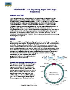
Figure 1. Several copies of mtDNA are packed into a nucleoprotein complex (nucleoid), closely associated with the membrane. High abundance DNA-binding proteins like TFAM or single stranded DNA binding protein partially cover the DNA, but have also important roles in transcription and replication, in concert with low copy proteins like the polymerases or the helicase Twinkle. MtDNA is still susceptible to damage by ROS, however, efficient repair systems are present.
1288 R. J. Wiesner et al. Do mutations of mtDNA accumulate during normal aging?
Figure 2. Intramitochondrial superoxide produced by the FMN moiety of RC complex I as potential ROS, relevant for the attack of mtDNA (modified according to [65]).
How much of the oxygen consumed by a cell is converted to superoxide first and only after the actions of Mn-SOD, Cu/Zn-SOD, glutathion peroxidase and catalase becomes water under physiological conditions is unclear. While initial experiments with isolated mitochondria estimated this up to 5%, more recent data indicate that it is maybe less than 0.2% [72] (Figure 2). Mechanism of ROS-mediated mtDNA damage The ROS-mediated attack of DNA leads basically to two major mutagenic alterations: the formation of 8OH-G (the oxidative modification of other bases is less frequent) [59] as well as the formation of single and double strand breaks. MtDNA appears to contain 10fold more 8-OH-G than nuclear DNA at steady state [73], thus 8-OH-G formation would have the potential of causing somatic point mutations leading to G ! T or C ! A transversions. These types of transversion are, however, observed rarely and have not been described so far among the known agingassociated point mutations [74,75]; details see next paragraph). Since mtDNA point mutations potentially induced by ROS appear not to contribute considerably to the somatic mutation profile of an aging tissue, it seems that the oxidative modifications of mtDNA are repaired rather efficiently (cf. paragraph about mtDNA repair capacity). This situation appears to be completely different with regard to double strand breaks. They are definitely a potential cause for deletions frequently observed in relation to aging [76,77]. Interestingly, in tissues with higher ROS turnover, like substantia nigra neurons which perform an intense dopamine metabolism associated with a monaminooxidase mediated catecholamine breakdown, considerable levels of mtDNA deletions were recently detected [78,79].
In the classic literature, somatic mutations in mitochondrial DNA have been hypothesized to be one likely reason for the aging process [80,81]. Reports on certain specific mutations accumulating with age strengthened this hypothesis, however, contradicting studies made an interpretation very difficult. To get a clearer picture, several questions should be answered: (i) Is there indeed an increasing amount of mutations in mitochondrial DNA during aging? (ii) If yes, what kinds of mutations accumulate? Do they represent a specific set of somatic changes typical for aging, or rather a broad spectrum of mutations? Do we need to assume aging-specific alterations of the mitochondrial genome, or can mutation accumulation be explained by other, more general mechanisms? (iii) Can the accumulation of deleterious mutations reach levels for which one can expect a true functional relevance? Conclusively answering these questions would clarify whether and how changes of the mtDNA affect the process of aging. In addition, in order to complete the vicious circle suggested by the mutational extension of the mitochondrial theory of aging, another important question is left open: Do functional changes that occur during aging, above all production of reactive oxygen species, also significantly contribute to the accumulation of more mtDNA mutations?
Point mutations Michikawa et al. [74] reported a number of mutations in the regulatory region (D-loop) being present in aging fibroblasts. The authors found these mutations, T414G, T285C, A368G and 383i to be more frequently present in aged individuals than in young ones. From three of the investigated individuals duplicate samples were taken with at least 15 years elapsed between the times of sampling. While in two individuals a clear increase in mutation loads was observed with age, the third individual had significant levels of the mutation both at young and at older ages and furthermore, even a considerable decrease was observed with age. As the authors point out, such an apparent decrease of mutation loads might be a consequence of tissue heterogeneity, which underlines the tremendous technical problems that can obscure the results when examining mutation loads at bulk tissue level. Similar studies have been performed in other tissues, among them muscle and brain. Interestingly, the set of D-loop mutations found in aged muscle, A189G and T408A [82], was different from that reported for fibroblasts, but no similar accumulation of D-loop mutations was detected in brain [83,84], suggesting tissue-specific processes influencing either
Mitochondrial DNA damage and the aging process the generation or the propagation of the observed mutations. In opposition to the latter findings, a higher burden of mtDNA mutations was detected in brain of patients with Alzheimer’s disease and elderly controls [85]. A different approach was taken by Nekhaeva et al. [75], when they examined single cells from buccal epithelium and the heart. Approximately 30% of the cells carried high levels of a single mutation (rarely two mutations) and importantly, different cells contained mostly different mutations. Similar to the bulk tissue studies mentioned above, mutational spectra in dividing buccal epithelium and post-mitotic heart muscle were clearly distinct. These findings on the single-cell level demonstrate that segregational drift may play a crucial role in shaping mutation patterns both in dividing and post-mitotic tissues. If the estimation by Khrapko et al. [86] is correct, newly generated mutations need decades to be able to segregate to levels high enough to be observed in single cells of older individuals. As depicted in Figure 3 this would mean that accumulation of a specific point mutation with age is not at all a consequence of ongoing mtDNA damage throughout life and, importantly, does not severely expand in a vicious circle in old tissue, but that it rather originates from tissue progenitor cells, or even from very early events in development [87]. The functional relevance of such somatically acquired mtDNA point mutations was demonstrated in colon crypts, the invaginations of the colon wall which are produced from a few rapidly dividing stem cells at the bottom [90]. In this study, colon crypts carrying a clonally expanded single mutation, multiple
Figure 3. Clonal expansion of somatic mitochondrial mutations. The upper panel represents a scenario where, in line with the mitochondrial theory of aging, the main source of mutation accumulation is repeated mutational events. In such a case one would expect a mixed set of mutations in each affected cell. Single cell observations contradict this assumption [88,89]. An alternative scenario is shown in the lower panel. Once a mitochondrial mutation is created, it undergoes intracellular or divisional segregation and can reach high levels within individual cells ultimately resulting in phenotypical changes.
1289
mutations, or no mutations at all were found in comparable numbers, causing a severe functional defect in the mitochondrial RC in 3 – 30% of the crypts, with the incidence being proportional to the age of the patients.
Deletions Large scale deletions of the mitochondrial DNA are much more frequently found to cause disease than mitochondrial point mutations. Accordingly, several studies investigated the possible role of mtDNA deletions in aging. Early studies concentrated on quantification of specific deletions in bulk tissues, most frequently the so-called “common deletion”. Although a clear increase in the proportions of deleted mitochondrial genomes with age was consistently demonstrated, the measured amounts of deleted molecules were always much below any functionally relevant level. However, such somatic mtDNA deletions are distributed non-uniformly between different tissues [91] and between individual cells within the same tissue. Particularly in brain, some areas, like substantia nigra, harbor a few orders of magnitude more deletions than others [76,77]. Similar to mtDNA point mutations, an important breakthrough was achieved by single-cell studies. In heart muscle of old patients, up to 25% of the myocytes were found to contain high amounts of large scale deletions [88,89]. In most cases, a cell either contained no or a single species out of a wide spectrum of different deletions. This again emphasises the importance of intracellular clonal expansion vs. ongoing de novo mutational events. In substantia nigra, the primary site of neurodegeneration in Parkinson disease, 30% of the neurons had defects in mitochondrial respiratory activity when examining old individuals. Similar to heart muscle cells, defective neurons were found to carry high levels of clonally expanded deletions [78,79]. In opposition to heart and colon crypts, no accumulation of point mutations was found in substantia nigra neurons [78]. In summary, recent single cell studies greatly contributed to our understanding of the role of somatic mtDNA mutations in aging. By this approach, it was possible to solve the apparent problem that even old tissues contain only a very low overall amount of specific mutations. The question is not anymore if the degree of an accumulating mutation is high enough in a tissue to cause a functional defect, but rather whether the number of cells disabled by clonally expanded mtDNA point mutations or deletions is high enough to significantly disturb the overall performance of the tissue. This is especially important in muscle, heart and the nervous system, notably those tissues most dramatically deteriorating during aging, where only a few
1290 R. J. Wiesner et al. non-functional cells (or segments in muscle fibers) might severely disturb whole organ function. A further important conclusion is that significant differences exist between the dynamics of mutation accumulation between different tissues and different areas of the same tissue. This suggests that cell typespecific mechanisms underlying either acquisition or expansion of mtDNA mutations (or both) have to be taken into consideration. Last, the fact that single cells of older individuals in most cases contain a single clonally expanded species of a mutation demonstrate the importance of intracellular segregational drifts. It shows that once a mutation has been generated, its propagation does not require further mutational events. Therefore, whether a mutation will interfere with mitochondrial function of a cell does not depend on repeated mutational events (as one would expect in the case of a local DNA maintenance defect or increased free radical production), but rather on how successfully the mutation segregates (Figure 3). When and how the original mutational events occur and if oxidative damage plays a role in this initial process, still remains a key question. Functional consequences of aging on mitochondrial oxidative phosphorylation (OXPHOS) Several biochemical investigations which have been carried out in various laboratories on pieces of tissues or cells from animal sources taken into culture [92 – 94] or from human tissues [95 – 98] have shown a mild aging-related decrease in the activities of the mitochondrial OXPHOS apparatus. These findings appear to contradict others, which did not find age related changes [99] and some of those reports mentioned above are inconsistent, since they show, e.g. maintained amounts of prosthetic groups, like cytochromes b and c þ c1, but decreased enzyme activities with aging [97]. Generally, the results of biochemical investigations on bulk tissues or whole cell populations, which only yield average values for the cells being investigated and neglect cell-to-cell differences, show a large variability. The extensive analysis of the activities of respiratory enzymes in mitochondria isolated from biopsy samples of human skeletal muscle from more than 200 “normal” subjects 10 – 90-year-old illustrates this problem of functional investigations of bulk tissue samples [100]. Although a linear regression analysis showed an aging-related decline in the activities of some of the investigated enzymes, others did not change. However, whenever the analysis has been carried out on tissue sections by histochemical or immunohistochemical methods, by in situ hybridization or laser capture microdissection and PCR, convincing results have clearly pointed also to a mosaic-like cellular distribution of the age-related decline of
mitochondrial respiratory enzyme activities [101,102] and also of associated mutations of mtDNA [41,102]. In conclusion, on a single-cell level the functional effects of aging on OXPHOS appear to be by far more impressive than on the bulk tissue level. In line with this consideration, the importance of age-acquired deletions for the mitochondrial function of single S. nigra neurons has been directly shown [78,79]. Only neurons with deletion levels above 60% show a bioenergetic impairment as evidenced by a defect of cytochrome c oxidase staining. It remains to be proven, however, whether, the functional problems of individual cells are relevant for the physiological function of the entire tissue. Mouse models apparently proving the relation between mtDNA mutations and aging The generation of mice which randomly accumulate point mutations and deletions of mtDNA over time in all cells of the body provided an important tool to investigate the relevance of mtDNA mutations for the aging process [103]. This was done by homozygous knock-in of a mtDNA polymerase engineered to be defective in proofreading by combined efforts of the Karolinska and Tampere teams and a second group published results on a similar strain of mice about 1 year later [104]. The mice had a dramatically shortened life span and showed many features typical for old animals and humans like weight loss, hair loss, osteoporosis, ventricular hypertrophy, anemia and reduced fertility, so the author teams claimed this to be a strong support for the mitochondrial mutation theory of aging. Kujoth et al. postulated that an increased rate of apoptosis, leading to depletion of stem cells and thus regenerative capacity, is the reason for this phenotype and ultimately the aging process [104]. However, hair loss for example is typical for old human beings, but not for “healthy”, normal aging mice, in which this is rather a sign of “illness”. Definitively, the mutator mice accumulate very high burdens of mtDNA point mutation (10 –15 mutations per 104 nucleotides), which is more than 10-fold higher than the mutation load found in aged human tissues (0.5 – 1 mutations per 104 nucelotides, Khrapko et al., Mut Research, in press). So, whether these mice show indeed premature aging or a severe multisystem mitochondrial disease may become a semantic problem at the end, if we consider aging to be a progressive multisystem mitochondrial disease. Indeed, another mouse model accumulating deletions and no point mutations by expressing an engineered mutator form of the mtDNA helicase Twinkle develops late onset mitochondrial myopathy and neuropathy, yet the authors claim that there is no premature aging [105]. Finally, Kujoth and also Trifunovic and colleagues, in a follow-up study, showed that although they consider their mice to age
Mitochondrial DNA damage and the aging process
1291
References
Figure 4. Vicious cycle concept of mitochondrial aging. While increased ROS production after ROS damage of complex I has been shown [65] increased mitochondrial ROS production by higher mtDNA mutation loads has not been observed [104,106].
faster, this is not due to increased ROS production [106]. This seems to contradict one important aspect of the mitochondrial theory of aging. However, this interpretation requires caution, since the results contradict only the “classical” self-sustaining ROSmaintained vicious cycle concept (line with question mark depicted in Figure 4). The data do not, however, discard ROS as potential inducers of age-relevant mutation in man. Also, the cells Trifunovic used were embryonic fibroblasts from mitochondrial polymerase mutator mice with almost complete deficiency of the RC, thus the amount of ROS produced by these cells per oxygen consumed was actually much higher compared to control cells [106].
Conclusions The accumulation of mitochondrial DNA mutations appears to be a least one important molecular mechanism of human aging. Reactive oxygen species are generated at the FMN moiety of mitochondrial RC I and should be considered to be important at least for the initial generation of age-dependent mtDNA deletions. However, the accumulation of acquired mutations to functionally relevant levels in aged tissues seems to be a consequence of clonal expansions of single founder molecules and not of ongoing mutational events.
Acknowledgements Work in our laboratories was supported by grants of the University of Bonn (BONFOR to W.S.K.), Deutsche Forschungsgemeinschaft (Ku 911/11-3 to W.S.K) and Center for Molecular Medicine Cologne, University of Ko¨ln (ZMMK, to R.J.W. Germany).
[1] Harman D. Aging: A theory based on free radical and radiation chemistry. J Gerontol 1956;11:298–300. [2] Attardi G, Schatz G. Biogenesis of mitochondria. Annu Rev Cell Biol 1988;4:289 –333. [3] Sbisa E, Tullo A, Nardelli M, Tanzariello F, Saccone C. Transcription mapping of the Ori L region reveals novel precursors of mature RNA species and antisense RNAs in rat mitochondrial genome. FEBS Lett 1992;296:311 –316. [4] Selwood SP, McGregor A, Lightowlers RN, ChrzanowskaLightowlers ZM. Inhibition of mitochondrial protein synthesis promotes autonomous regulation of mtDNA expression and generation of a new mitochondrial RNA species. FEBS Lett 2001;494:186–191. [5] Montoya J, Lopez-Perez MJ, Ruiz-Pesini E. Mitochondrial DNA transcription and diseases: Past, present and future. Biochim Biophys Acta 2006. [6] Clayton DA. Replication of animal mitochondrial DNA. Cell 1982;28:693–705. [7] Shadel GS, Clayton DA. Mitochondrial DNA maintenance in vertebrates. Annu Rev Biochem 1997;66:409–435. [8] Holt IJ, Lorimer HE, Jacobs HT. Coupled leading- and lagging-strand synthesis of mammalian mitochondrial DNA. Cell 2000;100:515–524. [9] Bowmaker M, Yang MY, Yasukawa T, Reyes A, Jacobs HT, Huberman JA, Holt IJ. Mammalian mitochondrial DNA replicates bidirectionally from an initiation zone. J Biol Chem 2003;278:50961– 50969. [10] Yasukawa T, Yang MY, Jacobs HT, Holt IJ. A bidirectional origin of replication maps to the major noncoding region of human mitochondrial DNA. Mol Cell 2005;18:651–662. [11] Korhonen JA, Pham XH, Pellegrini M, Falkenberg M. Reconstitution of a minimal mtDNA replisome in vitro. EMBO J 2004;23:2423 –2429. [12] Yang MY, Bowmaker M, Reyes A, Vergani L, Angeli P, Gringeri E, Jacobs HT, Holt IJ. Biased incorporation of ribonucleotides on the mitochondrial L-strand accounts for apparent strand-asymmetric DNA replication. Cell 2002;111:495–505. [13] Ojala D, Attardi G. Precise localization of the origin of replication in a physical map of HeLa cell mitochondrial DNA and isolation of a small fragment that contains it. J Mol Biol 1978;122:301– 319. [14] Meeusen S, Nunnari J. Evidence for a two membranespanning autonomous mitochondrial DNA replisome. J Cell Biol 2003;163:503– 510. [15] Spelbrink JN, Li FY, Tiranti V, Nikali K, Yuan QP, Tariq M, Wanrooij S, Garrido N, Comi G, Morandi L, Santoro L, Toscano A, Fabrizi GM, Somer H, Croxen R, Beeson D, Poulton J, Suomalainen A, Jacobs HT, Zeviani M, Larsson C. Human mitochondrial DNA deletions associated with mutations in the gene encoding Twinkle, a phage T7 gene 4-like protein localized in mitochondria. Nat Genet 2001;28:223–231. [16] Garrido N, Griparic L, Jokitalo E, Wartiovaara J, van der Bliek AM, Spelbrink JN. Composition and dynamics of human mitochondrial nucleoids. Mol Biol Cell 2003;14:1583–1596. [17] Chen XJ, Butow RA. The organization and inheritance of the mitochondrial genome. Nat Rev Genet 2005;6:815–825. [18] Jacobs HT, Lehtinen SK, Spelbrink JN. No sex please, we’re mitochondria: A hypothesis on the somatic unit of inheritance of mammalian mtDNA. Bioessays 2000;22:564–572. [19] Fisher RP, Topper JN, Clayton DA. Promoter selection in human mitochondria involves binding of a transcription factor to orientation-independent upstream regulatory elements. Cell 1987;50:247– 258.
1292 R. J. Wiesner et al. [20] Parisi MA, Clayton DA. Similarity of human mitochondrial transcription factor 1 to high mobility group proteins. Science 1991;252:965 –969. [21] Fisher RP, Clayton DA. A transcription factor required for promoter recognition by human mitochondrial RNA polymerase. Accurate initiation at the heavy- and light-strand promoters dissected and reconstituted in vitro. J Biol Chem 1985;260:11330 –11338. [22] Falkenberg M, Gaspari M, Rantanen A, Trifunovic A, Larsson NG, Gustafsson CM. Mitochondrial transcription factors B1 and B2 activate transcription of human mtDNA. Nat Genet 2002;31:289–294. [23] Fisher RP, Clayton DA. Purification and characterization of human mitochondrial transcription factor 1. Mol Cell Biol 1988;8:3496– 3509. [24] Diffley JF, Stillman B. DNA binding properties of an HMG1related protein from yeast mitochondria. J Biol Chem 1992;267:3368–3374. [25] Larsson NG, Wang J, Wilhelmsson H, Oldfors A, Rustin P, Lewandoski M, Barsh GS, Clayton DA. Mitochondrial transcription factor A is necessary for mtDNA maintenance and embryogenesis in mice. Nat Genet 1998;18:231–236. [26] Silva JP, Larsson NG. Manipulation of mitochondrial DNA gene expression in the mouse. Biochim Biophys Acta 2002;1555:106–110. [27] Gensler S, Weber K, Schmitt WE, Perez-Martos A, Enriquez JA, Montoya J, Wiesner RJ. Mechanism of mammalian mitochondrial DNA replication: Import of mitochondrial transcription factor A into isolated mitochondria stimulates 7S DNA synthesis. Nucleic Acids Res 2001;29:3657–3663. [28] Garstka HL, Schmitt WE, Schultz J, Sogl B, Silakowski B, Perez-Martos A, Montoya J, Wiesner RJ. Import of mitochondrial transcription factor A (TFAM) into rat liver mitochondria stimulates transcription of mitochondrial DNA. Nucleic Acids Res 2003;31:5039 –5047. [29] Maniura-Weber K, Goffart S, Garstka HL, Montoya J, Wiesner RJ. Transient overexpression of mitochondrial transcription factor A (TFAM) is sufficient to stimulate mitochondrial DNA transcription, but not sufficient to increase mtDNA copy number in cultured cells. Nucleic Acids Res 2004;32:6015– 6027. [30] Alam TI, Kanki T, Muta T, Ukaji K, Abe Y, Nakayama H, Takio K, Hamasaki N, Kang D. Human mitochondrial DNA is packaged with TFAM. Nucleic Acids Res 2003;31: 1640–1645. [31] Ekstrand MI, Falkenberg M, Rantanen A, Park CB, Gaspari M, Hultenby K, Rustin P, Gustafsson CM, Larsson NG. Mitochondrial transcription factor A regulates mtDNA copy number in mammals. Mol Genet 2004;13:935–944. [32] Takamatsu C, Umeda S, Ohsato T, Ohno T, Abe Y, Fukuoh A, Shinagawa H, Hamasaki N, Kang D. Regulation of mitochondrial D-loops by transcription factor A and singlestranded DNA-binding protein. EMBO Rep 2002;3: 451–456. [33] King MP, Attardi G. Human cells lacking mtDNA: Repopulation with exogenous mitochondria by complementation. Science 1989;246:500–503. [34] Ghivizzani SC, Madsen CS, Nelen MR, Ammini CV, Hauswirth WW. In organello footprint analysis of human mitochondrial DNA: Human mitochondrial transcription factor A interactions at the origin of replication. Mol Cell Biol 1994;14:7717 –7730. [35] Bereiter HJ. Behavior of mitochondria in the living cell. Int Rev Cytol 1990;122:1–63. [36] Takai D, Inoue K, Goto Y, Nonaka I, Hayashi JI. The interorganellar interaction between distinct human mitochondria with deletion mutant mtDNA from a patient with mitochondrial disease and with HeLa mtDNA. J Biol Chem 1997;272:6028–6033.
[37] Yoneda M, Miyatake T, Attardi G. Complementation of mutant and wild-type human mitochondrial DNAs coexisting since the mutation event and lack of complementation of DNAs introduced separately into a cell within distinct organelles. Mol Cell Biol 1994;14:2699–2712. [38] Enriquez JA, Cabezas-Herrera J, Bayona-Bafaluy MP, Attardi G. Very rare complementation between mitochondria carrying different mitochondrial DNA mutations points to intrinsic genetic autonomy of the organelles in cultured human cells. J Biol Chem 2000;275:11207–11215. [39] Sato A, Nakada K, Hayashi JI. Mitochondrial dynamics and aging: Mitochondrial interaction preventing individuals from expression of respiratory deficiency caused by mutant mtDNA. Biochim Biophys Acta 2006;1763:473–481. [40] Nakada K, Inoue K, Ono T, Isobe K, Ogura A, Goto YI, Nonaka I, Hayashi JI. Inter-mitochondrial complementation: Mitochondria-specific system preventing mice from expression of disease phenotypes by mutant mtDNA. Nat Med 2001;7:934–939. [41] Wanagat J, Cao Z, Pathare P, Aiken JM. Mitochondrial DNA deletion mutations colocalize with segmental electron transport system abnormalities, muscle fiber atrophy, fiber splitting, and oxidative damage in sarcopenia. FASEB J 2001;15:322–332. [42] Clayton DA, Doda JN, Friedberg EC. The absence of a pyrimidine dimer repair mechanism in mammalian mitochondria. Proc Natl Acad Sci USA 1974;71:2777–2781. [43] Nishioka K, Ohtsubo T, Oda H, Fujiwara T, Kang D, Sugimachi K, Nakabeppu Y. Expression and differential intracellular localization of two major forms of human 8-oxoguanine DNA glycosylase encoded by alternatively spliced OGG1 mRNAs. Mol Biol Cell 1999;10:1637– 1652. [44] Slupphaug G, Markussen FH, Olsen LC, Aasland R, Aarsaether N, Bakke O, Krokan HE, Helland DE. Nuclear and mitochondrial forms of human uracil-DNA glycosylase are encoded by the same gene. Nucleic Acids Res 1993;21:2579–2584. [45] Miller H, Fernandes AS, Zaika E, McTigue MM, Torres MC, Wente M, Iden CR, Grollman AP. Stereoselective excision of thymine glycol from oxidatively damaged DNA. Nucleic Acids Res 2004;32:338–345. [46] Pinz KG, Bogenhagen DF. Efficient repair of abasic sites in DNA by mitochondrial enzymes. Mol Cell Biol 1998; 18:1257–1265. [47] Lakshmipathy U, Campbell C. Mitochondrial DNA ligase III function is independent of Xrcc1. Nucleic Acids Res 2000;28:3880–3886. [48] Lakshmipathy U, Campbell C. The human DNA ligase III gene encodes nuclear and mitochondrial proteins. Mol Cell Biol 1999;19:3869– 3876. [49] Myers KA, Saffhill R, O’Connor PJ. Repair of alkylated purines in the hepatic DNA of mitochondria and nuclei in the rat. Carcinogenesis 1988;9:285 –292. [50] Satoh MS, Huh N, Rajewsky MF, Kuroki T. Enzymatic removal of O6-ethylguanine from mitochondrial DNA in rat tissues exposed to N-ethyl-N-nitrosourea in vivo. J Biol Chem 1988;263:6854– 6856. [51] Thyagarajan B, Padua RA, Campbell C. Mammalian mitochondria possess homologous DNA recombination activity. J Biol Chem 1996;271:27536– 27543. [52] Coffey G, Lakshmipathy U, Campbell C. Mammalian mitochondrial extracts possess DNA end-binding activity. Nucleic Acids Res 1999;27:3348 –3354. [53] Kraytsberg Y, Schwartz M, Brown TA, Ebralidse K, Kunz WS, Clayton DA, Vissing J, Khrapko K. Recombination of human mitochondrial DNA. Science 2004;304:981. [54] Zsurka G, Kraytsberg Y, Kudina T, Kornblum C, Elger CE, Khrapko K, Kunz WS. Recombination of mitochondrial DNA in skeletal muscle of individuals with multiple
Mitochondrial DNA damage and the aging process
[55]
[56] [57]
[58]
[59]
[60]
[61]
[62] [63] [64]
[65]
[66]
[67]
[68]
[69]
[70]
[71]
[72]
[73]
mitochondrial DNA heteroplasmy. Nat Genet 2005; 37:873–877. Mason PA, Matheson EC, Hall AG, Lightowlers RN. Mismatch repair activity in mammalian mitochondria. Nucleic Acids Res 2003;31:1052–1058. Halliwell B. Reactive oxygen species and the central nervous system. J Neurochem 1992;59:1609 –1623. Rosen DR, Siddique T, Patterson D, Figlewicz DA, Sapp P, Hentati A, Donaldson D, Goto J, O’Regan JP, Deng HX. Mutations in Cu/Zn superoxide dismutase gene are associated with familial amyotrophic lateral sclerosis. Nature 1993;362:59– 62. Liochev SI, Fridovich I. The role of O2z 2 in the production of HOz: In vitro and in vivo. Free Radic Biol Med 1994;16: 29–33. Klungland A, Rosewell I, Hollenbach S, Larsen E, Daly G, Epe B, Seeberg E, Lindahl T, Barnes DE. Accumulation of premutagenic DNA lesions in mice defective in removal of oxidative base damage. Proc Natl Acad Sci USA 1999;96:13300–13305. Boveris A, Cadenas E, Stoppani AO. Role of ubiquinone in the mitochondrial generation of hydrogen peroxide. Biochem J 1976;156:435 –444. Turrens JF, Boveris A. Generation of superoxide anion by the NADH dehydrogenase of bovine heart mitochondria. Biochem J 1980;191:421 –427. Naqui A, Chance B, Cadenas E. Reactive oxygen intermediates in biochemistry. Annu Rev Biochem 1986;55:137–166. Wood PM. The potential diagram for oxygen at pH 7. Biochem J 1988;253:287 –289. Liu Y, Fiskum G, Schubert D. Generation of reactive oxygen species by the mitochondrial electron transport chain. J Neurochem 2002;80:780–787. Kudin AP, Bimpong-Buta NY, Vielhaber S, Elger CE, Kunz WS. Characterization of superoxide-producing sites in isolated brain mitochondria. J Biol Chem 2004;279: 4127–4135. Votyakova TV, Reynolds IJ. DeltaPsi(m)-dependent and -independent production of reactive oxygen species by rat brain mitochondria. J Neurochem 2001;79:266– 277. Genova ML, Ventura B, Giuliano G, Bovina C, Formiggini G, Parenti Castelli G, Lenaz G. The site of production of superoxide radical in mitochondrial complex I is not a bound ubisemiquinone but presumably iron–sulfur cluster N2. FEBS Lett 2001;505:364 –368. Lambert AJ, Brand MD. Inhibitors of the quinone-binding site allow rapid superoxide production from mitochondrial NADH: Ubiquinone oxidoreductase (complex I). J Biol Chem 2004;279:39414–39420. St-Pierre J, Buckingham JA, Roebuck SJ, Brand MD. Topology of superoxide production from different sites in the mitochondrial electron transport chain. J Biol Chem 2002;277:44784– 44790. Kudin AP, Debska-Vielhaber G, Kunz WS. Characterization of superoxide production sites in isolated rat brain and skeletal muscle mitochondria. Biomed Pharmacother 2005;59:163– 168. Starkov AA, Fiskum G, Chinopoulos C, Lorenzo BJ, Browne SE, Patel MS, Beal MF. Mitochondrial alpha-ketoglutarate dehydrogenase complex generates reactive oxygen species. J Neurosci 2004;24:7779 –7788. St-Pierre J, Buckingham JA, Roebuck SJ, Brand MD. Topology of superoxide production from different sites in the mitochondrial electron transport chain. J Biol Chem 2002. Richter C, Park JW, Ames BN. Normal oxidative damage to mitochondrial and nuclear DNA is extensive. Proc Natl Acad Sci USA 1988;85:6465 –6467.
1293
[74] Michikawa Y, Mazzucchelli F, Bresolin N, Scarlato G, Attardi G. Aging-dependent large accumulation of point mutations in the human mtDNA control region for replication. Science 1999;286:774– 779. [75] Nekhaeva E, Bodyak ND, Kraytsberg Y, McGrath SB, Van Orsouw NJ, Pluzhnikov A, Wei JY, Vijg J, Khrapko K. Clonally expanded mtDNA point mutations are abundant in individual cells of human tissues. Proc Natl Acad Sci USA 2002;99:5521–5526. [76] Corral-Debrinski M, Horton T, Lott MT, Shoffner JM, Beal MF, Wallace DC. Mitochondrial DNA deletions in human brain: Regional variability and increase with advanced age. Nat Genet 1992;2:324–329. [77] Soong NW, Hinton DR, Cortopassi G, Arnheim N. Mosaicism for a specific somatic mitochondrial DNA mutation in adult human brain. Nat Genet 1992;2:318 –323. [78] Bender A, Krishnan KJ, Morris CM, Taylor GA, Reeve AK, Perry RH, Jaros E, Hersheson JS, Betts J, Klopstock T, Taylor RW, Turnbull DM. High levels of mitochondrial DNA deletions in substantia nigra neurons in aging and Parkinson disease. Nat Genet 2006;38:515–517. [79] Kraytsberg Y, Kudryavtseva E, McKee AC, Geula C, Kowall NW, Khrapko K. Mitochondrial DNA deletions are abundant and cause functional impairment in aged human substantia nigra neurons. Nat Genet 2006;38:518–520. [80] Harman D. The biologic clock: The mitochondria? J Am Geriatr Soc 1972;20:145–147. [81] Linnane AW, Marzuki S, Ozawa T, Tanaka M. Mitochondrial DNA mutations as an important contributor to ageing and degenerative diseases. Lancet 1989;1:642 –645. [82] Wang Y, Michikawa Y, Mallidis C, Bai Y, Woodhouse L, Yarasheski KE, Miller CA, Askanas V, Engel WK, Bhasin S, Attardi G. Muscle-specific mutations accumulate with aging in critical human mtDNA control sites for replication. Proc Natl Acad Sci USA 2001;98:4022– 4027. [83] Murdock DG, Christacos NC, Wallace DC. The age-related accumulation of a mitochondrial DNA control region mutation in muscle, but not brain, detected by a sensitive PNA-directed PCR clamping based method. Nucleic Acids Res 2000;28:4350–4355. [84] Chinnery PF, Taylor GA, Howell N, Brown DT, Parsons TJ, Turnbull DM. Point mutations of the mtDNA control region in normal and neurodegenerative human brains. Am J Hum Genet 2001;68:529–532. [85] Lin MT, Simon DK, Ahn CH, Kim LM, Beal MF. High aggregate burden of somatic mtDNA point mutations in aging and Alzheimer’s disease brain. Hum Mol Genet 2002;11:133–145. [86] Khrapko K, Nekhaeva E, Kraytsberg Y, Kunz W. Clonal expansions of mitochondrial genomes: Implications for in vivo mutational spectra. Mutat Res 2003;522:13– 19. [87] Coller HA, Khrapko K, Bodyak ND, Nekhaeva E, HerreroJimenez P, Thilly WG. High frequency of homoplasmic mitochondrial DNA mutations in human tumors can be explained without selection. Nat Genet 2001;28:147–150. [88] Khrapko K, Bodyak N, Thilly WG, van Orsouw NJ, Zhang X, Coller HA, Perls TT, Upton M, Vijg J, Wei JY. Cell-by-cell scanning of whole mitochondrial genomes in aged human heart reveals a significant fraction of myocytes with clonally expanded deletions. Nucleic Acids Res 1999;27:2434– 2441. [89] Bodyak ND, Nekhaeva E, Wei JY, Khrapko K. Quantification and sequencing of somatic deleted mtDNA in single cells: Evidence for partially duplicated mtDNA in aged human tissues. Hum Mol Genet 2001;10:17 –24. [90] Taylor RW, Barron MJ, Borthwick GM, Gospel A, Chinnery PF, Samuels DC, Taylor GA, Plusa SM, Needham SJ, Greaves LC, Kirkwood TB, Turnbull DM. Mitochondrial DNA mutations in human colonic crypt stem cells. J Clin Invest 2003;112:1351–1360.
1294 R. J. Wiesner et al. [91] Cortopassi GA, Shibata D, Soong NW, Arnheim N. A pattern of accumulation of a somatic deletion of mitochondrial DNA in aging human tissues. Proc Natl Acad Sci USA 1992;89:7370 –7374. [92] Hagen TM, Yowe DL, Bartholomew JC, Wehr CM, Do KL, Park JY, Ames BN. Mitochondrial decay in hepatocytes from old rats: Membrane potential declines, heterogeneity and oxidants increase. Proc Natl Acad Sci USA 1997; 94:3064– 3069. [93] Bowling AC, Mutisya EM, Walker LC, Price DL, Cork LC, Beal MF. Age-dependent impairment of mitochondrial function in primate brain. J Neurochem 1993;60: 1964–1967. [94] Barazzoni R, Short KR, Nair KS. Effects of aging on mitochondrial DNA copy number and cytochrome c oxidase gene expression in rat skeletal muscle, liver, and heart. J Biol Chem 2000;275:3343–3347. [95] Trounce I, Byrne E, Marzuki S. Decline in skeletal muscle mitochondrial respiratory chain function: Possible factor in ageing. Lancet 1989;1:637–639. [96] Isobe K, Ito S, Hosaka H, Iwamura Y, Kondo H, Kagawa Y, Hayashi JI. Nuclear-recessive mutations of factors involved in mitochondrial translation are responsible for age-related respiration deficiency of human skin fibroblasts. J Biol Chem 1998;273:4601–4606. [97] Boffoli D, Scacco SC, Vergari R, Solarino G, Santacroce G, Papa S. Decline with age of the respiratory chain activity in human skeletal muscle. Biochim Biophys Acta 1994;1226:73 –82. [98] Greco M, Villani G, Mazzucchelli F, Bresolin N, Papa S, Attardi G. Marked aging-related decline in efficiency of oxidative phosphorylation in human skin fibroblasts. FASEB J 2003;17:1706 –1708. [99] Allen RG, Keogh BP, Tresini M, Gerhard GS, Volker C, Pignolo RJ, Horton J, Cristofalo VJ. Development and ageassociated differences in electron transport potential and
[100]
[101]
[102]
[103]
[104]
[105]
[106]
consequences for oxidant generation. J Biol Chem 1997;272:24805–24812. Papa S. Mitochondrial oxidative phosphorylation changes in the life span. Molecular aspects and physiopathological implications. Biochim Biophys Acta 1996;1276:87–105. Muller-Hocker J. Cytochrome-c-oxidase deficient cardiomyocytes in the human heart—an age-related phenomenon. A histochemical ultracytochemical study. Am J Pathol 1989;134:1167–1173. Muller-Hocker J. Cytochrome c oxidase deficient fibres in the limb muscle and diaphragm of man without muscular disease: An age-related alteration. J Neurol Sci 1990;100: 14– 21. Trifunovic A, Wredenberg A, Falkenberg M, Spelbrink JN, Rovio AT, Bruder CE, Bohlooly YM, Gidlof S, Oldfors A, Wibom R, Tornell J, Jacobs HT, Larsson NG. Premature ageing in mice expressing defective mitochondrial DNA polymerase. Nature 2004;429:417–423. Kujoth GC, Hiona A, Pugh TD, Someya S, Panzer K, Wohlgemuth SE, Hofer T, Seo AY, Sullivan R, Jobling WA, Morrow JD, Van Remmen H, Sedivy JM, Yamasoba T, Tanokura M, Weindruch R, Leeuwenburgh C, Prolla TA. Mitochondrial DNA mutations, oxidative stress, and apoptosis in mammalian aging. Science 2005;309:481 –484. Tyynismaa H, Mjosund KP, Wanrooij S, Lappalainen I, Ylikallio E, Jalanko A, Spelbrink JN, Paetau A, Suomalainen A. Mutant mitochondrial helicase Twinkle causes multiple mtDNA deletions and a late-onset mitochondrial disease in mice. Proc Natl Acad Sci USA 2005;102:17687 –17692. Trifunovic A, Hansson A, Wredenberg A, Rovio AT, Dufour E, Khvorostov I, Spelbrink JN, Wibom R, Jacobs HT, Larsson NG. From the Cover: Somatic mtDNA mutations cause aging phenotypes without affecting reactive oxygen species production. Proc Natl Acad Sci USA 2005;102: 17993–17998.