Journal of Anesthesia and Perioperative Medicine
Review Article
Mineralocorticoid Receptor, A Promising Target for Improving Management of Low Back Pain by Epidural Steroid Injections Shaimaa I.A. Ibrahim 1,2 , Judith A. Strong1 , and Jun-Ming Zhang1
ABSTRACT Aim of review: Low back pain is a major health problem in United States and worldwide. In this review, we aim to show that mineralocorticoid receptor (MR) activation has a critical role in the initiation of immune and inflammatory responses, which in turn can impact the effectiveness of the currently used steroids for epidural injections in low back pain management since most steroids activate MR in addition to the primary target, glucocorticoid receptor (GR). Moreover, we would like to determine some of the benefits of blocking the MR- induced negative effects. Overall, we propose a novel therapeutic approach for low back pain management by using a combination of a MR antagonist and a GR agonist in the epidural injections. Method: We will first introduce the societal cost of low back pain and discuss how epidural steroid injections became a popular treatment for this condition. We will then describe several preclinical models used for the study of low back pain conditions and the findings with respect to the role of MR in the development of inflam matory low back pain. Recent findings: MR has pro-inflammatory effects in many tissues which can counteract the anti- inflammatory effects induced by GR activation. Blocking MR using the selective MR antagonist eplerenone can reduce pain and sensory neuron excitability in experimental models of low back pain. Moreover, combining the MR antagonist with clinically used steroids is more effective in reducing pain behaviors than using the steroids alone. Summary: MR antagonists are promising candidates to increase the effectiveness of currently used steroids. Since the activation of the MR is evident in preclinical models of low back pain, blocking its deleterious effects can be beneficial in managing inflammatory pain conditions.
L
ow back pain is a major health problem in United States and worldwide. In many cases, it can become chronic. Low back pain can be caused by many disorders such as lum bar herniated discs and compression of nerve roots. The clinical use of epidural steroid injections for low back pain management has increased dramatically during the past decade. However, this popularity doesn't reflect effectiveness. Clin-
From 1 Pain Research Center, Department of Anesthesiology, University of Cincinnati College of Medicine, Cincinnati, USA; 2 Graduate Program in Molecular, Cellular, and Biochemical Pharmacology, University of Cincinnati, Cincinnati, USA. Correspondence to Dr. Jun- Ming Zhang at
[email protected]. Citation: Shaimaa I.A. Ibrahim, Judith A. Strong and Jun- Ming Zhang. Mineralocorticoid receptor, a promising target for improving management of low back pain by epidural steroid injections. J Anesth Perioper Med 2016; 3: 177-184.
ical trials reported mixed results regarding the effectiveness as not all patients achieve adequate pain relief. Preclinical studies, on the other hand, largely focus on the effects of various clinically used steroids on the glucocorticoid receptor (GR), whereas the potential implications of mineralocorticoid receptor (MR) activation in low back pain and other inflammatory pain conditions have been largely ignored.
This is an open-access article, published by Evidence Based Communications (EBC). This work is licensed under the Creative Commons Attribution 4.0 International License, which permits unrestricted use, distribution, and reproduction in any medium or format for any lawful purpose.To view a copy of this license, visit http://creativecommons.org/licenses/by/4.0/.
177
Review Article
Journal of Anesthesia and Perioperative Medicine
Chronic Back Pain Cost to Society Chronic pain continues to be an important worldwide heath problem. According to the International Association for the Study of Pain (IASP), pain is considered to be chronic if it lasts for at least 3 months (1). Generally, research studies use the standard of pain duration of 3 to 6 months to define chronic pain. The prevalence of chronic pain in US population is 30.7% , and is higher in females (34.3% ) than males (26.7% ). Importantly, the prevalence increases with age (2). Approximately 100 million adults in the United States are impacted by chronic pain. There are indirect costs as well as direct costs arising from chronic pain. For example, not only does pain require medical treatment, but it also decreases worker productivity, increasing the economic burden to society. It's reported that the total cost of chronic pain, including medical expenditures and reduced productivity, was approximately $560-$635 billion in 2010. Surprisingly, the annual costs of chronic pain has surpassed the annual costs of the diseases usually considered the most costly, such as heart disease, cancer and diabetes ($309 billion, $243 billion, and $188 billion, respectively) (3). Generally, the number of people affected by pain is greater than the number affected by cardiovascular disease, diabetes or cancer. Low back pain is the major type of chronic pain reported, being more common than osteoarthritis, rheumatoid arthritis and migraine headache. Low back pain is one of the leading causes of disability in the United States and other countries. It progresses to being chronic in about 30% of cases. The direct medical costs in the United States are estimated to be $34 billion, with over $100 billion in lost wages and productivity per year. Therefore, it is a major contributor to health care costs (2, 4, 5). Epidural Steroid Injections for Low Back Pain In 2002, interventional pain management was regarded as a specialty (6). Chronic pain management using interventional techniques has become very popular, including epidural steroid injections, facet joint interventions, disc proce-
178
JAPM
WWW.JAPMNET.COM
dures and nerve block. According to a Medicare analysis, there has been a dramatic increase in the use of interventional techniques. From 2000 to 2014, the increase was reported to be 153% in the Medicare population, with an annual rate of increase of 6.9% per 100,000 Medicare population (7). Does the popularity of epidural steroid treatment reflect actual effectiveness or are they not really useful for management of chronic low back pain? In fact, the current medications may not be working appropriately. Not all of them succeed in relieving pain in chronic pain conditions like chronic low back pain (5). Given that inflammation is usually involved in low back pain, local injections of anti- inflammatory corticosteroids are commonly used to relieve both inflammation and pain. However, several random ized clinical trials showed that these medications failed to achieve adequate relief in many patients, which led to controversy regarding the effectiveness of steroid injections in pain management (8- 12). For example, randomized clinical trials showed that epidural injections with local anesthetic alone improved chronic low back pain secondary to lumbar spinal stenosis in a comparable manner to the improvement due to epidural injections with steroids. These studies showed that there is no advantage for injecting steroids over local anesthetic (13). Other randomized clinical trials revealed that epidural steroid injections are only beneficial for a short time (8, 9). Preclinical Models for the Study of Low Back Pain Low back pain can be detrimental as it reduces patients' quality of life. It can be caused by different disorders. For example, compression of nerve roots due to spinal stenosis (narrowing of spinal canal) or displacement or degeneration of the intervertebral disc is a major contributor to low back pain. Inflammatory conditions such as arthritis can be also involved. Treatment strategies range from surgeries (if possible and in severe cases) to the less invasive procedure of epidural steroid injections (4, 5, 14). The increased use of interventional techniques in pain management and the controversial results in clinical trials led to the need for further investigation to
July, 2016
Volume 3
Number 4
Shaimaa I.A. Ibrahim et al.
confirm the safety and effectiveness of these steroid injections. Several preclinical models of back pain have been developed to simulate different clinical conditions for low back pain. For example, an early model was developed in rats by applying nucleus pulposus (NP, the inner jellylike material of the vertebral disc) into the lum bar dorsal root ganglia (DRG) (15). A different rat model is implemented by inducing local inflammation of the DRG (LID), which involves the local application of the immune stimulator zymosan in the vicinity of the L5 and/or L4 DRGs. These models can cause mechanical hypersensitivity, tactile allodynia and cold allodynia. In addition to the pain behaviors, these models cause upregulation of pro-inflammatory cytokines, satellite glial cell activation and increased sensory neuron excitability. As a result of the presence of materials not recognized as“ self ”elements by the immune system, such as the NP or the immune stimulator, robust inflammatory cell infiltration of the DRG occurs. This can last for a long time after the model is established (16, 17). Another rat model of chronic compression of the DRG (CCD) is implemented by com pressing the lumbar DRG through inserting Lshaped metal rods into the intervertebral foram ina. This rod remains in place for the duration of the experiment (18, 19). Despite the fact that an immune stimulus was not used in the CCD model, upregulation of pro- inflammatory cytokines, satellite glial activation, macrophage infiltration and behavioral sensitivity to anti- inflam matory drugs were observed. The cytokine profile was similar to that seen in the LID model (20-22). In addition, sensory neurons showed increased sensitivity to many pro-inflammatory cytokines after implementation of the CCD model (23, 24). It is documented that the NFκB pathway is involved in these models (25, 26) and one of the major mechanisms of steroid anti- inflammatory effects is suppression of this pathway. Non- Steroidal and Steroidal Anti- Inflammatory Drugs in Managing Low Back Pain Given that inflammation is a key player in inducing low back pain, it is expected that anti-inflam matory drugs can relieve some of the pain symp-
JAPM
WWW.JAPMNET.COM
Mineralocorticoid Receptor in Low Back Pain
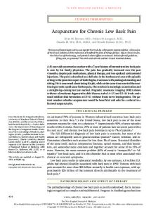
toms. In the aforementioned models, inflammatory responses were observed such as activation of satellite glia cells, infiltration of macrophages, increase in pro-inflammatory cytokines and activation of inflammatory signaling pathways (15, 16, 27- 30). In the models of ruptured disc, NP can compress the DRG or adjacent nerve roots and also act as an inflammatory stimulus. In addition, it acts as a source of pro- inflammatory cytokines. Interleukin- 1β (IL- 1β) and tumor necrosis factor α (TNF- α) have been found in the NP and are also thought to contribute to pain behaviors in different models such as the NP and the CCD models (28, 31-33). Non- steroidal anti- inflammatory drugs can alleviate pain behaviors in different rodent back pain models when given systemically (17, 30) or administered by local or epidural injections (29, 34). However, steroidal anti- inflammatory drugs are more often used clinically, and are usually given locally into the epidural space to manage different low back pain conditions. The application can be done through different routes such as intraforaminal, caudal or interlaminar routes. Different time points and different routes can have significant effects on the results. For exam ple, in a preclinical study using the rat CCD model, triamcinolone (a steroidal anti- inflammatory drug) decreased pain behaviors applied epidurally when given 3 days after the establishment of the model. However, it didn't reduce pain behaviors when given 10 days after the establishment of the model (35, 36). Steroid Receptors, Inflammation and Low Back Pain Clinically used steroids for back pain injections are meant to target the GR. GR is a member of the nuclear receptor family, for which the ligand diffuses into the cell and interacts with the receptor. Then, the ligand- receptor complex is translocated into the nucleus where it regulates gene expression. It is widely distributed in almost every tissue in the body. Its activation has an overall anti-inflammatory effect. As shown in Figure 1, GR activation stimulates type II inflam mation (including M2 polarized macrophages) which involves tissue remodeling and wound repair. At the same time, it depresses type I inflam -
July, 2016
Volume 3
Number 4
179
Review Article
Journal of Anesthesia and Perioperative Medicine
The MR belongs also to the nuclear receptor family. Its function has been well- studied in the kidney, heart and hippocampal neurons. Additionally, in the central nervous system, MR is expressed in the glia (40). It was originally viewed only as the target of aldosterone. Its activation was thought to be mainly involved in electrolyte balance, specifically sodium and water reabsorption in kidney. However, this receptor was detected in other cell types including cardiomyocytes (41), brain neurons (42) and DRG neurons (43). In tissues other than kidney, MR activation has a pro- inflammatory role (promotes type I inflammation) that may offset the anti- inflammatory effects of GR activation (44, 45). MR activation is thought to contribute to inflammation in kidney, heart and central nervous system. In tissues other than kidney, glucocorticoids (the primary glucocorticoid in humans is cortisol, and in rodents is corticosterone), act as the primary endogenous activators of the MR. It is worth noting that in kidney, aldosterone is considered to be the only activator for MR due to the inactivation of glucocorticoids by 11β- dehydrogenase type 2 enzyme. In kidney, this enzyme inactivates the corticosterone (in rodents) and cortisol (in humans) in order to ensure that aldosterone will be the nominal activator of the MR to maintain the electrolyte balance. However, in non-renal tissues, due to reduced activity of the glucocorticoid- inactivating enzyme and the high corticosterone plasma concentration than aldosterone, corticosterone is considered to be the primary activator of MR (46). Figure 1. Diagram of Hypothesis About the Role of GR and MR in Mediating Effects of Clinically Used Steroids. GR, glucocorticoid receptor; MR, mineralocorticoid receptor; DRG, dorsal root ganglia.
mation (including M1 polarized macrophages) which involves tissue damage, high levels of oxidative metabolites and pro- inflammatory cytokines. Microarray analysis showed that 6 out of 10 selected M1 markers were upregulated after 3 days of LID (37). Recently, it has been shown that some clinically used steroids (such as 6- α methylprednisolone and triamcinolone) can also activate the MR in vitro with significant potency (38, 39).
180
JAPM
WWW.JAPMNET.COM
Preclinical Studies of Mineralocorticoid Receptor in Inflammatory Low Back Pain Our focus in the laboratory is to explore the MR as a target for increasing the efficacy of clinically used steroids. We tested the hypothesis that MR is involved in the pro- inflammatory effects in the low back pain models, by combining a selective MR antagonist eplerenone with the current clinically used steroids, in order to block the MRmediated pro- inflammatory effects. In this way, MR activation and its antagonism of the desired anti- inflammatory effects caused by GR activation can be avoided.
July, 2016
Volume 3
Number 4
Shaimaa I.A. Ibrahim et al.
Mineralocorticoid Receptor in Low Back Pain
Figure 2. Changes in MR Expression and Effect of GR Agonists with and without MR Blocker, Eplerenone (EPL), on Pain Behaviors in Rats with Localized Inflammation of the DRG (LID). Two images above: Immunohistochemical staining showing nuclear translocation of activated MRs (red) in the inflamed DRG neurons (green) on POD 1. Scale bar=50 µm; A: Comparisons of the fraction of MRs in cytoplasm and nuclear between normal and LID neurons. *** p