dental materials Dental Materials 19 (2003) 199±205
www.elsevier.com/locate/dental
Microtensile bond strength between adhesive cements and root canal dentin Serge Bouillaguet a,*, Sabra Troesch b, John C. Wataha c, Ivo Krejci a, Jean-Marc Meyer b, David H. Pashley d a
Department of Cariology, Endodontics and Pediatric Dentistry, School of Dental Medecine, University of Geneva, 19 Rue BartheÂlemy-Menn, CH-1205 Geneva, Switzerland b Department of Biomaterials, School of Dental Medecine, University of Geneva, Geneva, Switzerland c Department of Oral Rehabilitation, School of Dentistry, Medical College of Georgia, Augusta, GA 30912, USA d Department of Oral Biology and Maxillofacial Pathology, School of Dentistry, Medical College of Georgia, Augusta, GA 30912, USA Received 11 April 2001; revised 16 October 2001; accepted 11 December 2001
Abstract Objectives: The hypotheses tested were that the bond strength of adhesive cements to root canal dentin (1) would be reduced as a function of con®guration factor, polymerization process and type of luting material and (2) would be lowered near the apex of the tooth. Methods: Human canines and premolars were prepared for post cementation using Single Bond/Rely X ARC, ED Primer/Panavia F, C and B Metabond, and Fuji Plus. The specimens were divided into two groups. For intact roots, the posts were luted using standard clinical procedures. For ¯at roots, the posts were applied directly into ¯at ground canals. All roots were sectioned into 0.6 mm thick slices, trimmed mesio-distally and stressed to failure at 1 mm/min. The mTBS of each slab was calculated as the force at failure divided by the bonded crosssectional surface area. The results were compared using a one-way ANOVA and Tukey multiple comparison intervals
a 0:05: Least squares linear regression analysis was used to assess the effect of dentin location on bond strength. Results: All cements showed signi®cantly
p # 0:05 lower bond strengths in intact vs. ¯at roots. The mTBS of posts to intact roots were not signi®cantly different for Single Bond/Rely X ARC and Panavia F, but both were signi®cantly lower
p # 0:05 than the bonds produced by C and B Metabond and Fuji Plus cements. For Single Bond/Rely X ARC and Fuji Plus a signi®cant decrease in bond strength was observed in dentin closer to the apex of the root. Signi®cance: Stresses from polymerization shrinkage and problems with adequate access to the root canal complicate the formation of high-strength bonds when cementing endodontic posts with resin cements. q 2003 Published by Elsevier Science Ltd on behalf of Academy of Dental Materials. Keywords: Root canal dentin; Adhesion; Post; Bond strength; Microtensile testing
1. Introduction Posts and cores are frequently used in endodontically treated teeth that suffered excessive loss of coronal tooth structure. In such cases, the cementation of a post inside the root canal is used to provide retention for the ®nal restoration [1]. However, reports have shown that root preparation for post insertion can result in additional loss of tooth substance, which, in turn, can lead to catastrophic root fracture under long-term clinical use [2,3]. Clinicians now use adhesive resins to place posts during the restoration of non-vital teeth. The rationale for using * Corresponding author. Fax: 141-22-38-29-990. E-mail address:
[email protected] (S. Bouillaguet).
adhesive cements is based on the premise that the use of adhesive cements for bonding posts to root canal dentin will reinforce the tooth and help retain the post and the restoration [4]. However, little is known about the bonding performance of adhesive cements applied under such conditions. Bonding to root canal dentin is affected by the endodontic procedures performed prior to post cementation. Nikaido et al. [5] reported that endodontic irrigants such as 5% sodium hypochlorite, or 3% H2O2 or their combination for as little as 60 s can signi®cantly reduce the bond strengths of resin bonded to overlying coronal dentin. More recently, Morris et al. [6] have demonstrated that the bond strength of C and B Metabond to root canal dentin was reduced by half when the dentin was previously treated with 5% NaOCl or 15% EDTA/10% urea peroxide (RC Prep). Other reports have
0109-5641/03/$30.00 + 0.00 q 2003 Published by Elsevier Science Ltd on behalf of Academy of Dental Materials. PII: S 0109- 564 1(02)00030- 1
200
S. Bouillaguet et al. / Dental Materials 19 (2003) 199±205
shown that the contamination of the dentin walls by eugenol diffusing from endodontic sealers can affect the retention of bonded posts [7]. Selecting the appropriate adhesive and luting procedure for bonding endodontic posts to root canal dentin is a further challenge. Different types of bonding systems can be used in combination with a number of different luting resins. These materials may be polymerized through a chemical reaction, a photopolymerization process, or a combination of both mechanisms. Total etching systems can produce high bond strengths to ¯at dentin surfaces. However, reports have shown that poor control of moisture or incomplete resin impregnation can signi®cantly reduce the dentin±resin bond [8,9]. It is more likely that bonding problems will occur within the con®nes of a post space because the post space cannot be visualized well. Further, it is dif®cult to control the amount of moisture in a root canal, since the narrow canal holds water by surface tension, making it dif®cult to displace that water with bonding agents [10]. The use of self-etching adhesives in combination with luting resins has been proposed for the cementation of endodontic posts. Because self-etching adhesives are generally used on dry dentin, and do not require rinsing of the etchant, they may represent a more successful approach. However, their ef®ciency at in®ltrating thick smear layers like those produced during post preparation remains a major concern [11,12]. Since the introduction of composite resins in the 70s, the problems of polymerization shrinkage and contraction stresses induced during polymerization have been well documented [13,14]. The composition of the material and its curing mode are both factors that can in¯uence the amount of shrinkage produced after polymerization. To decrease viscosity and to facilitate clinical handling, resin cements have low ®ller content. Therefore, they exhibit more volumetric shrinkage than heavy ®lled composite materials [15]. Further, most current resin cements have a dual-curing process that requires light exposure to initiate the reaction. However, it has been reported that photocured composites generate more polymerization shrinkage stress and exhibit less ¯ow than chemically cured composites [16]. Contraction stresses induced by polymerization also depend on the geometry of the cavity and the thickness of the resin layer [14,17]. Previous research has shown that the restriction of ¯ow of resin cements by the con®guration of the preparation can signi®cantly increase the contraction stress at the adhesive interface. According to Feilzer et al. [14], who described the C-factor, the cementation of endodontic posts to root canal dentin represents the worst case scenario. Alster et al. [17] also showed that when resin cements are applied in thin layers in con®ned spaces, the contraction stress produced by the polymerizing resin could exceed 20 MPa. This value approaches closely the bond strength values reported for several current adhesive systems on ideal ¯at dentin, and it exceeds the bond strengths provided by some adhesive systems [18].
The null hypothesis to be tested was that the bond strengths of adhesive cements to root canal do not vary with C-factor, polymerization chemistry, or type of luting material. This hypothesis was tested using different adhesive cements (including resin and resin-modi®ed glass ionomer cements) and by measuring the microtensile bond strength to uncon®ned ¯at dentin and in con®ned, intact canals. In the current study, the microtensile test was used to attempt to gain a clearer picture of the local bonding pattern inside the root canal. In this sense, the authors hoped that the microtensile test would yield more information than `push-out' or `pull-out' tests, which have been traditionally used to assess the retention of posts [19]. Finally, the authors also tested the null hypothesis that there are no regional differences in microtensile bond strengths within root canals due to intrinsic substrate differences or technical problems in the apical third. 2. Materials and methods Forty-eight extracted human canines and premolars without excessive root curvature (canal curvature 15±358) were selected for this study. The crown was sectioned below the cemento±enamel junction to obtain a 12 mm long root that was then prepared for endodontic treatment. During endodontic procedures, the canal space was mechanically enlarged using the Hero 6, 4,2 endodontic ®les (Micro Mega SA, Geneva, Switzerland) operated at 400 rpm under a constant irrigation with 3% NaOCl. The ®nal preparation had a 68 taper and a diameter of 0.3 mm at the apex. The canals were then rinsed with distilled water, dried with ethanol and paper points, and obturated with gutta percha cones and sealer (AH Plus, Dentsply De-Trey, Konstanz, Germany, and P.D. SA, Vevey, Switzerland). After 24 h, the roots were prepared for post insertion. The canal space of each root was enlarged with Parapost twist drills (ColteÁne AG, AltstaÈten, Switzerland) to a ®nal diameter of 1.7 mm and a depth of 8 mm from the cervical surface. The specimens were then divided into two groups: intact roots and ¯at roots. Roots in the ¯at group were ground longitudinally under binocular vision to expose the full length of half the canal. Before post cementation, the root canals were rinsed for 1 min with 3% NaOCl, rinsed with double distilled water for 2 min and dried with paper points. Custom-made endodontic posts (apical diameter: 1 mm, coronal diameter: 1.7 mm, length: 10 mm) fabricated with Z100 composite resin material (3M ESPE, St Paul, MN, USA). These prepolymerized posts were adhesively cemented to the roots. Composite posts were used because pilot studies showed less premature debonding of the posts during sectioning than with metallic posts. Furthermore, the primary focus in the current study was the strength of the bond between the root dentin and the adhesive cement. Prior to cementation, the posts were passively inserted inside the
S. Bouillaguet et al. / Dental Materials 19 (2003) 199±205
201
Table 1 Materials used in the study Material
Composition
Manufacturer
Single Bond Rely X ARC
Etchant: 35% phosphoric acid; adhesive: bis-GMA, HEMA, polyalkenoic acid copolymer, photoinitiators, ethanol, water; luting resin: bis-GMA, TEGDMA, zirconia/silica ®ller 68%, proprietary dimetacrylate monomer ED primer: HEMA, MDP, 5-NMSA sodium benzene sul®nate N,N-diethanol p-toluidine, water; Panavia F: silanated barium glass and silica powder sodium ¯uoride bis-phenol A polyethoxy demethacrylate 10metacryloyloxydecyl dihydrogen phosphate (MDP) hydrophobic and hydrophilic dimethacrylates enzoyl peroxide, photo sensitizer Conditioner: citric acid 10%, ferric chloride 2%, distilled water 88%; cement: powder: alumino-silicate glass; liquid: HEMA 37%, polyacrylic acid 22%, proprietary resins 10%, tartaric acid 6%, distilled water 25% Conditioner: 10% citric acid/3% ferric chloride; liquid: 95% MMA 1 5% 4META; powder: polymethyl methacrylate; catalyst: tri-n-butyl borane
3M ESPE St Paul, MN, USA
ED primer Panavia F
Fuji Plus C and B Metabond
root canal to verify ®t. Then, a silane coupling agent (ESPE Sil, 3M ESPE, St Paul, MN, USA) was applied for 5 min to the surface of the post and dried with air. For intact roots, the posts were luted using standard clinical procedures for either Single Bond/Rely X ARC (3M ESPE, St Paul MN, USA), ED Primer/Panavia F (Kuraray Co., Ltd, Osaka, Japan), C and B Metabond (Parkell, Farmingdale, NY, USA), or Fuji Plus (GC Co., Tokyo, Japan) (Table 1). For Single Bond/Rely X ARC luting cement (3M ESPE), the root canal dentin was etched for 15 s with a 35% phosphoric acid gel and rinsed for 1 min with water. Excess water was further eliminated with paper points without desiccating the dentin. One coat of Single Bond was applied inside the canal with a small sponge, thinned with a gentle air spray and polymerized for 10 s. The adhesive resin was also applied to the silanated post, thinned with air and polymerized for 10 s. Equal amounts of pastes A and B were dispensed onto a mixing pad, mixed for 10 s and inserted inside the canal by use of a lentulo spiral (size 40, PD SA, Vevey, Switzerland). Finally, the post was covered with luting cement, inserted in the canal and polymerized for 40 s through the composite post. For the Panavia F luting system, the dentin surfaces were primed and bonded following the manufacturer's instructions. Equal amounts of ED Primer liquids A and B were mixed together on the mixing dish, applied with a brush inside the canal and allowed to stand for 60 s. Excess liquid was eliminated with a paper point before completely drying the primer with a gentle air ¯ow. Equal amounts of Panavia F paste A and B were then mixed for 20 s on the mixing plate and applied with a brush to the silanated post. The post covered with cement was inserted into the root canal and polymerized for 20 s. Oxygen-excluding gel was applied to the margins of the ¯at dentin but not to the intact root. According to manufacturer's instructions, the C and B Metabond adhesive cement was applied to the canal after conditioning the dentin with dentin activator (10% citric acid with 3% ferric chloride). This conditioner was applied with a small sponge to the canal for 10 s, rinsed with water
Kuraray Dental Products Osaka, Japan
GC Co., Tokyo Japan Parkell, Farmingdale, NY, USA
thoroughly, and dried with paper points. The C and B Metabond resin was prepared by mixing four drops of liquid with one drop of catalyst in a cool mixing well and introduced with a brush inside the canal to wet the dentin walls. The same procedure was done on the composite post. Then two scoops C and B Metabond radio-opaque powder were added to a fresh mix of base and catalyst to prepare the luting cement, which was inserted inside the canal using a lentulo spiral. Finally the post was inserted into the post space and held in place for 10 min. For cementation of posts with Fuji Plus, the root canal dentin was conditioned for 20 s with the Fuji conditioner using a cotton pellet before rinsing with water. Care was taken to avoid excessive dehydration of the dentin. The Fuji Plus cement was prepared by mixing one scoop of powder with one drop of liquid for 15 s and introduced into the canal by use of a lentulo spiral. The post was then covered with cement and immediately inserted in the canal where it was chemically cured. For roots in the ¯at group, the procedure for cementation of the posts was identical, except that the composite post was applied directly into the exposed canal space and allowed to set. One hour after post cementation, all specimens were attached to the grips of a low speed saw (Isomet, Buehler Ltd, Lake Bluff, IL) and sectioned perpendicular to the tooth axis into 0.6 mm thick slabs (Fig. 1). The thickness of each slab was measured with a digital caliper. The diameter of the post in each slab was measured using a stereomicroscope. Each slab was further trimmed by an ultra-®ne diamond bur mounted in a high speed handpiece with water coolant. This procedure was performed under the microscope, to expose the composite post on the mesial and distal sides. The bonded surface area was approximately 1 mm 2. The trimmed specimens were attached to the grips of a custom-made holder with cyanoacrylate adhesive (Zapit, DVA Inc., Corona, CA, USA) and stressed to failure at 1 mm/min with a universal testing machine (Vitrodyne V1000 Universal Tester, John Chatillon and Sons, Greensboro,
202
S. Bouillaguet et al. / Dental Materials 19 (2003) 199±205
Fig. 1. Preparation of bonding substrate in intact and ¯at roots. For intact roots, the posts were luted using standard clinical procedures. Roots in the ¯at group were ground longitudinally to expose the full length of half the canal and the posts were applied directly into the exposed canals and allowed to set. After bonding and cementing the post, the roots were sectioned into 0.6 mm thick slices, trimmed mesio-distally and stressed to failure at 1 mm/min. The mTBS of each slab was calculated as the force at failure divided by the bonded cross-sectional surface area. For intact roots, the level of dentin inside the root was identi®ed by letters (from a: coronal to g: apical).
NC, USA). The tensile bond strength of each slice was calculated as the force at failure divided by the bonded cross-sectional surface area and expressed in MPa. Since the adhesive interface was curved, the exact length of the interface was calculated by measuring the cord (Fig. 2) and then calculating the length of the arc,
L 0 r £ 2 sin u21 £
L=2r; where u is the angle formed between the cord and center of the post. All specimens used for the microtensile test were observed with a stereomicroscope to assess the fracture mode. Each tooth yielded multiple bond strength measurements (ca. 8±9 specimens per root). The average composite± dentin bond strength was calculated for each tooth, and the means among teeth were compared using ANOVA. Since this ANOVA showed no statistically signi®cant differences among the means
p . 0:05; the individual specimens within each tooth were treated as independent measurements. This strategy was much more practical than using one root for each microtensile specimen. During the bond strength testing, several samples failed after sectioning but before trimming. Mean microtensile bond strengths of the composites to dentin were computed with and without including these prematurely failed specimens, where these specimens assigned a zero bond strength. The Table 2 Microtensile bond strengths to root dentin in MPa (values are mean tensile bond strength (SD) (number of tested specimens/total number of specimens). Asterisks indicate differences between ¯at and intact roots within each adhesive cement (t-test, a 0:05). Within the intact canal samples, means with the same letter are not statistically different
a 0:05) Flat dentin SB1/Rely X ARC ED Primer/Panavia C and B Metabond Fuji Plus
Intact canal p
23.2 (6.5) (40/40) 15.9 (6.4) (40/40) p 13.1 (4) (48/48) p 13.1 (5.7) (47/47) p
5.3 (6.3) (86/86) a 7.2 (8.7) (84/84) a 10.8 (5.3) (80/80) b 10.4 (5.7) (81/81) b
Fig. 2. The exact length of the interface was calculated by measuring the cord (L) and then calculating the length of the arc (L 0 ),
L 0 r £ 2 sin u21 £
L=2r; where u is the angle formed between the cord and center of the post.
bond strengths for intact roots and ¯at roots were compared using a two-sided t-tests with a 0:05 for each adhesive cement. The bond strengths among different cements in intact roots were compared using a one-way ANOVA and Tukey multiple comparison intervals
a 0:05 because this was the most clinically relevant comparison. To assess the effect of dentin location relative to the apex of the tooth on bond strength, a least squares linear regression analysis was used. In these analyses, all zero bond strength values were included. The appropriateness of the linear model was assessed using an R 2 value, and the presence of a non-zero slope was also tested
a # 0:05: 3. Results For the Single Bond/Rely X ARC system, a mean mTBS of 23.2 ^ 6.5 MPa was observed for the specimens bonded on ¯at root surfaces (Table 2, including zero values). Single Bond/Rely X ARC applied to intact canals showed signi®cantly lower mTBS (5.3 ^ 6.3 MPa, p , 0:001). All other cements also showed signi®cantly
p # 0:05 reduced bond strengths in intact vs. ¯at roots (Table 2). The mTBS of composite posts to intact root dentin fell into two groups when the four adhesive cements were compared (Table 2). The Single Bond/Rely X ARC and Panavia F were not signi®cantly different from each other
p . 0:05; but both were signi®cantly lower
p # 0:05 than the bonds produced by C and B Metabond and Fuji Plus cements. These latter two cements were not statistically different from each other. While no specimen failed before testing in the ¯at group for Single Bond/Rely X ARC, 41% of the specimens (51 out of 86) in the intact canals did not survive the preparation and failed prior to testing (Table 3). The mean mTBS for Single Bond/Rely X ARC without including the spontaneously debonded specimens was 9.0 ^ 5.8 MPa, which was signi®cantly
p # 0:05 lower than mean mTBS for the ¯at specimens. The rate of spontaneous failure in intact
S. Bouillaguet et al. / Dental Materials 19 (2003) 199±205 Table 3 Microtensile bond strengths to root dentin (MPa) not including specimens that failed during preparation (values are mean tensile bond strength (SD) (number of specimens tested/total number of specimens). Asterisks indicate differences between ¯at and intact roots within each adhesive cement (ttest, a 0:05). Within the intact canal samples, means with the same letter are not statistically different
a 0:05)
SB1/Rely X ARC ED Primer/Panavia F C and B Metabond Fuji Plus
Flat dentin
Intact canal
23.2 (6.5) (40/40) p 16.7 (5.3) (38/40) 13.1 (4.0) (48/48) 13.9 (5.0) (45/47) p
9.0 (5.8) (51/86) a 14.4 (6.7) (43/84) a 12.1 (4.1) (72/80) a 12.1 (4.3) (70/81) a
roots vs. ¯at roots was also greater for Panavia F (51% vs. 5%). However, the mean mTBS were statistically similar to those in both groups. For the C and B Metabond and Fuji Plus, the spontaneous failure rates in ¯at roots were approximately 5% and only increased to 10% in intact teeth. Due to this low pretreatment failure rate, the bond strengths were not signi®cantly different in the inclusion/exclusion groups (Tables 2 and 3) using C and B Metabond, but were signi®-
203
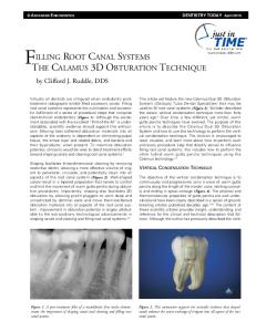
cantly higher in the ¯at specimens vs. intact roots for Fuji Plus (Tables 2 and 3). Least squares regression analyses were performed to determine if any relationship could be found between mTBS and distance from the apex of the tooth (Fig. 3). For Single Bond/Rely X ARC, a signi®cant decrease in bond strength was observed in dentin closer to the apex of the root (R2 0:65; p , 0:012). A similar relationship was observed for Fuji Plus (R2 0:87; p , 0:0001). However, no signi®cant correlation was seen for C and B Metabond or Panavia F, although there was some indication of a correlation for C and B Metabond
p 0:14: 4. Discussion The bene®ts of adhesive techniques used for dental restorations are well documented. Among the most important factors are the reinforcement of tooth structure and the esthetic aspects of the ®nal restoration [20]. For these reasons, the use of adhesive cements has been
Fig. 3. Mean microtensile bond strength in intact root canals plotted vs. level of dentin (from coronal to apical).
204
S. Bouillaguet et al. / Dental Materials 19 (2003) 199±205
proposed for cementing endodontic posts in non-vital teeth [21]. Push-out and pull-out tests have been traditionally used to assess the retention of endodontic posts in the root canal [19,22]. These tests are a clear improvement over simple SEM observational studies of adhesive failures in root canals [23,24]. Drummond et al. [25] measured pull-out strength of various endodontic posts and reported shear bond strengths to root canal dentin in the range of 10 MPa. They pointed out that the surface area of the post should be carefully evaluated to allow calculation of shear strength. However, the push-out and pull-out tests are probably heavily in¯uenced by ¯aws and non-uniform bonding in a manner similar to coronal bonding [26]. Thus, the microtensile test may give a better evaluation of the local bonding pattern inside the root canal when using adhesive cements [27]. Further, the microtensile test allowed the use of relatively ¯at surfaces, which served as a control not subjected to shrinkage stresses and accessibility problems, which dominate the intact canal. This type of control may not be possible in a push-out test. It is always debatable whether specimens that fail prematurely should be included in bond strength calculation in these types of studies. They were included because the authors wanted to present both inclusion and exclusion data sets. Further the authors believe that they were not simply caused by the sectioning technique or problems. The low incidence of premature failures in the ¯at or uncon®ned root specimens and, the relatively high incidence of premature failures in the intact canal (sometime over 50%) indicate that shrinkage stresses or access problems may have played a role in bonding posts for some materials (Tables 2 and 3). The con®guration factor has been well accepted as an important consideration in bonding procedures [13,14,16,17]. The C-factor is the ratio of the bonded to the unbonded surface areas of cavities. Whereas it typically varies from 1 to 5 in intracoronal restorations, it probably exceeded 200 in the case of the current study. This was estimated by dividing the free surface area of the 150 mm-thick luting cement (unbonded area) surrounding the 1.7 mm-diameter post by the total bonded area (the surface area of the post, 38.7 mm 2, and the dentinal surface area, 42.1 mm 2). In cases where the C-factor is high, slower setting materials may reduce stress at the bonding interface because the slow setting allows ¯ow of the material to relieve polymerization stress. This idea is supported in the current study because the two chemically cured cements (C and B Metabond and Fuji Plus), which are slower setting than dualcured materials showed the least incidence of spontaneous failure (Table 3). Additionally, bonding for some materials, such as the dual-cured Panavia F, tended to fail on either one side or the other at a given level in the intact canal. This observation supports the idea that shrinkage stresses in the con®nement of the intact root canal exceed the cement± dentin bond strength, causing debonding of the cement
from the dentin. Finally, the dual-cured materials are more complex to apply and may not be as well suited in the root canal environment because of problems with vision, access, and moisture level control. Our expectation was that the bond strength would be reduced nearer the apex because of the problems of accessibility mentioned above. Therefore, we expected that the materials requiring more bonding steps would show a signi®cant negative regression of bond strength as a function of distance to the apex. However, this was not completely supported by our results. Although the dual-cured Rely X ARC cement showed a signi®cant regression (Fig. 3), Panavia F, which is also dual-cured, did not show this relationship. Further, Fuji Plus, which is the simplest material to apply, showed the strongest regression relationship. Thus, although the regression of mTBS with proximity to the apex can be demonstrated for some materials, its causes are not clear from the results of the current study. Factors such as changes in the dentin structure could play a role in these relationships [28,29]. In summary, the use of adhesive resin to cement posts is an attractive clinical concept. Past studies have shown good clinical success for these procedures if suf®cient coronal dentin remains. When less than 2 mm of coronal dentin remained, failures were observed and debonding of the post was often seen [30]. The results of this study indicate that dentin bond strengths of resin cements to dentin are not very high inside intact canals, and that clinical failure is not seen when suf®cient coronal dentin is available because the restoration does not rely heavily on the bonding of the post to the root dentin. The current study indicates that obtaining high bond strengths of resin cements to root canal dentin is not straightforward because of polymerization stress and access problems. It is clear that extrapolation of coronal bonding procedures and results are not appropriate for the cementation of posts with adhesive cements. Lower risks of bonding failure may be realized if relatively short, loose ®tting posts are used and as much coronal dentin is preserved as possible. The use of reducing agents such as sodium ascorbate to correct for the negative effects of NaOCl on adhesive bond strength may be required to obtain bond strengths to root dentin that can resist polymerization stress [6]. These factors will all help ensure that the bonding in the root canal will be successful and that true sealing will occur. From the standpoint of simplicity, the resin-modi®ed glass ionomer cement was the best among those used in the current study. Acknowledgements The authors would like to thank Mrs Chantal Godin and Huguette Hernoux for their technical assistance with this project and all manufacturers for material support. This project was supported by the SSO (Swiss Dental Society) research fund #186.
S. Bouillaguet et al. / Dental Materials 19 (2003) 199±205
References [1] Robbins JW. Restoration of endodontically treated teeth. In: Summit JB, Robbins JW, Schwart RS, editors. Fundamentals of operative dentistryÐa contemporary approach, Illinois: Quintessence Publishing Co. Inc, 2001. p. 546±66. [2] Creugers NHJ, Mentink AJB, Kayser AF. An analysis of durability data on post and core restorations. J Dent 1993;21:281±4. [3] Stockton LW. Factors affecting retention of posts systems: a literature review. J Prosthet Dent 1999;81:380±5. [4] Duncan JP, Pameijer CH. Retention of parallel-sided titanium posts cemented with six luting agents: an in vitro study. J Prosthet Dent 1998;80:423±8. [5] Nikaido T, Takano Y, Sasafuchi Y, Burrow MF, Tagami J. Bond strengths to endodontically treated teeth. Am J Dent 1999;12:177±80. [6] Morris MD, Lee KW, Agee KA, Bouillaguet S, Pashley DH. Effects of sodium hypochlorite and RC-prep on bond strengths of resin cement to endodontic surfaces. J Endod 2001;27:753±7. [7] Tjan AH, Nemetz H. Effect of eugenol-containing endodontic sealer on retention of prefabricated posts luted with adhesive composite resin cement. Quint Int 1992;23:839±44. [8] Tay FR, Gwinnet AJ, Wei SHY. Variability in microleakage observed in a total-etch wet-bonding technique under different handling conditions. J Dent Res 1996;74:1168±78. [9] Pashley DH, Ciucchi B, Sano H, Horner JA. Permeability of dentin to adhesive agents. Quint Int 1993;24:618±31. [10] Helfer AR, Melnick S, Schilder H. Determination of the moisture content of vital and pulpless teeth. Oral Surg Oral Med Oral Pathol 1972;34:661±70. [11] Watanabe I, Saimi Y, Nakabayashi N. Effect of smear layer on bonding to ground dentinÐrelationship between grinding conditions and tensile bond strength. Jpn Soc Dent Mater Device 1994;13:101±8. [12] Miyasaka K, Nakabayashi N. Combination of EDTA conditioner and phenyl-P/HEMA self-etching primer for bonding to dentin. Dent Mater 1999;15:153±7. [13] Davidson CL, De Gee AJ, Feilzer A. The competition between the composite±dentin bond strength and the polymerization contraction stress. J Dent Res 1984;63:1396±9. [14] Feilzer A, De Gee AJ, Davidson CL. Setting stress in composite resin in relation to con®guration of the restoration. J Dent Res 1987;66: 1636±9. [15] Condon JR, Ferracane JL. Assessing the effect of composite formulation on polymerization stress. J Am Dent Assoc 2000;131:497±503.
205
[16] Feilzer A, De Gee AJ, Davidson CL. Setting stresses in composite for two different curing modes. Dent Mater 1993;9:2±5. [17] Alster D, Feilzer AJ, de Gee AJ, Davidson CL. Polymerization contraction stress in thin resin composite layers as a function of layer thickness. Dent Mater 1997;13:146±50. [18] Bouillaguet S, Gysi P, Wataha JC, Ciucchi B, Cattani M, Godin Ch, Meyer JM. Bond strength of composite to dentin using self-etching, conventional and one step adhesive systems. J Dent 2001;29:55± 61. [19] Mitchell CA, Orr JF, Connor KN, Magill JPG, Maguire GR. Comparative study of four glass ionomer luting cements during post pullout tests. Dent Mater 1994;10:88±9. [20] Morin D, DeLong R, Douglas WH. Cusp reinforcement by the acid etch technique. J Dent Res 1984;63:1075±8. [21] Paul SJ, Scharer P. Post and core reconstruction for ®xed prosthodontic restoration. Pract Period Aesthetic Dent 1997;9:513±20. [22] Pest LB, Cavalli B, Bertani P, Gagliani M. Adhesive post-endodontic restorations with ®ber posts. In: Tagami J, Toledano M, Prati C, editors. Advanced adhesive dentistry, Third International Kuraray Symposium2000. p. 49±58 ISBN 88-87961-00-X. [23] Dietschi D, Romelli M, Goretti A. Adaptation of adhesive posts and cores to dentin after fatigue testing. Int J Prosthod 1997;10:498±507. [24] Ferrari M, Mannoci F. A-one-bottle adhesive system for bonding a ®ber post into a root canal: an SEM evaluation of the post-resin interface. Int Endod J 2000;33:397±400. [25] Drummond JL. In vitro evaluation of endodontic posts. Am J Dent 2000;13:5B±8B. [26] Sano H, Shono T, Sonoda H, Takatsu T, Ciucchi B, Carvalho R, Pashley DH. Relationship between surface area for adhesion and tensile bond strengthÐevaluation of a micro-tensile bond test. Dent Mater 1994;10:236±40. [27] Pashley DH, Carvalho RM, Sano H, Nakajima M, Yoshiyama M, Shono Y, Fernandes C, Tay F. The microtensile bond test: a review. J Adhesive Dent 1999;1:299±309. [28] Carrigan PG, Morse DR, Furst L, Sinai JH. A scanning electron microscopic evaluation of human dentinal tubules according to age and location. J Endod 1984;10:359±63. [29] Tidmarsch BG, Arrowsmith MG. Dentinal tubules at the root ends of apicected teeth: a scanning electron microscopic study. Int Endod J 1989;22:184±9. [30] Ferrari M, Vichi A, Manocci F, Mason PN. Retrospective study of the clinical performance of ®ber posts. Am J Dent 2000;13:10B±3B.