Scientific series
Medical Aspects of Chemical and Biological Terrorism Chemical Terrorism and Traumatism
Alexander Monov and Christophor Dishovsky Editors
Sofia 2005 Publishing House of the Union of Scientists in Bulgaria
© Alexander Monov and Christophor Dishovski, 2005 All rights reserved. No reproduction, copy or transmission of this publication may be made without written permission from the authors. No paragraph of this publication may be reproduced, copied or transmitted save with written permission or in accordance with the provisions of the Copyright Act, 1993, Bulgaria. The authors have asserted their rights to be identified as the authors of this work in accordance with the Copyright Act. Authors bear full responsibility for their articles, presented in this publication. Authors will not receive honoraria for their contributions First published in April 2005 by the Publishing House of the Union of Scientists in Bulgaria, 39 Madrid Str., 1505, Sofia A catalogue record of this book is available from the National Library “St. St. Cyril and Methodius”, Sofia Alexander Monov and Christophor Dishovski Medical Aspects of Chemical and Biological Terrorism – Chemical Terrorism and Traumatism On the Cover: Electron microscopy of neuromuscular junction from diaphragm muscle of rat. Intoxication with Vx. 10 days after treatment with oxime reactivator of ChE. 60 000 ×. ( Dishovsky, 1975).
ISBN 954-8329-69-7 Publishing House of the Union of Scientists in Bulgaria 2
Table of Contents Contributors
...............................................................................................7
Preface .......................................................................................................11 About the Editors ........................................................................................14 Chapter 1 Mass Poisonings by Chemical Toxic Substances Alexander Monov .......................................................................................15 Chapter 2 Medical Issues of Chemical Terrorism Boris Filatov .............................................................................................73 Chapter 3 CW Terrorism: a Comparative Analysis of the Cases of the Revolutionary Armed Forces of Colombia (FARCs) (low tech) and Aum Shinrikio (high tech) Maria Jose Espona, Ignacio Alejo Aladro ..................................................91 Chapter 4 Aum Shirinkyo and Terrorist Use of Nerve Agent in Japan Milos Stojiljkovic, Milan Jokanovic ..........................................................101 Chapter 5 Toxicological Aspects of Investigation of Act of Chemical Terrorism in Matsumoto (Japan) Victor Shulga, Evgeni Fokin, Sergey Shokin ...............................................117 Chapter 6 Low-level Nerve Agent Exposure: Objectives of Future Research for Military and Civilian Populations David Moore ...........................................................................................121
3
Chapter 7 How to Confront Chemical Terrorism (Medical Management of Nerve Agent Casualties) Mostafa Ghanei, Shahriar Khateri .............................................................129 Chapter 8 Effects of Mustard Gas Exposure in Pediatric Patients: Long-Term Health Status of Mustard-Exposed Children, 14 Years After Chemical Bombardment of Sardasht Shahriar Khateri, Mustafa Ghanei, Mohammad Soroush, David Haines ....143 Chapter 9 Cholinesterase Blockers as Potential Agents for Chemical Terrorism and Contemporary Approaches to Therapy of Acute Poisonings Induced by Anti-Choliesterase Neuroparalytic Substances Natalia Kokshareva, Nikolai Prodanchuk, Peter Zhminko, Vladimir Krivenchuk .................................................................................153 Chapter 10 Organophosphate Poisoning: Possibilities of Prophylaxis Jiri Bajgar ................................................................................................183 Chapter 11 The Role of Oximes in the Antidotal Treatment of Chemical Casualties Exposed to Nerve Agents Jiri Kassa ................................................................................................193 Chapter 12 Some Aspects of the Mechanisms of Action of Oxime Reactivators of Cholinesterase Christophor Dishovsky .............................................................................209 Chapter 13 Paraoxonase 1 (PON1) as a Potential Catalytic Scavenger in the Prophylaxis and Treatment of Organophosphate Poisoning Dragomir Draganov .................................................................................227
4
Chapter 14 Biochemical Mechanisms of Biotransformation of Organophosphorus Compounds Milan Jokanovic, Milos Stojiljkovic ..........................................................247 Chapter 15 Organophosphate Induced Delayed Neurotoxicity Galina Makhaeva, Vladimir Malygin ........................................................271 Chapter 16 Immunochemical Procedures for Simple Analysis of Exposure to Sulfur Mustard G. P. van der Schans, R. H. Mars-Groenendijk, F. J. Bikker, D. Noort .........303 Chapter 17 Delayed Neuro-Endocrine Toxicity induced by Organophosphorus Compounds – Natural Consequence of Poisonous substances Application for Terrorist Purposes Victor Shulga ...........................................................................................315 Chapter 18 Application of IR-Spectroscopy for Identification of Mustard Gas and Lewisite in Bulk Containers to be disposed Oleg Strukov, Evgen Fokin ......................................................................325 Chapter 19 Micotoxins Heybatullah Kalantari Index
.............................................................................333
...............................................................................................345
5
6
Contributors Ignacio Alejo Aladro, BA University of Buenos Aires Rodriguez Peña 1464 Buenos Aires Argentina Jiri Bajgar,MD, D.Sc. Professor Department of Toxicology Purkyne Military Medical Faculty Hradec Kralove, University of Defence, Czech Republic Floris J. Bikker, Ph.D. TNO Prins Maurits Laboratory P.O.Box 45 2280 AA Rijswijk The Netherlands Haines David, Ph.D. Department of Epidemiology and Biostatistics The George Washington University 2121 Eye Street, N.W. Washington, D.C. 20052 | 202.994.1000 USA
Christophor Dishovsky, M.D., Ph.D., D.Sc. Department Military Toxicoloigy Military Medical Academy 3,St.G.Sofiisky Str. 1606 Sofia Bulgaria Dragomir Draganov, M.D., Ph.D. University of Michigan Department of Pharmacology MSRB 3, Room 1301 Ann Arbor, MI 48109-0632 USA Maria Jose Espona, Ph.D. Associate Professor of Science and Technology National Defense School, Maipú 262 Buenos Aires Argentina Boris N. Filatov, M.D., Ph.D. Professor, Director Research Institute of Hygiene, Toxicology and Occupational Pathology (RINTOP) 12 Zemlyachka Str., Volgograd 4000487 Russia
7
Evgeny A. Fokin Ph.D. Deputy Director General Department of Organic Chemistry State Research Institute of Organic Chemistry and Technology (GosNIIOKhT) 23, Entouziastov sch., Moscow, 111024, Russia
Shahriar Khateri, M.D. Director Chemical Warfare Victims Research Unit Janbazan Medical and Engineering Research Center (JMERC) 19615/616 Tehran Iran
Mostafa Ghanei, M.D. Professor, Pulmonologist Division of Respiratory diseases Department of Medicine Baqyiatallah University of Medical Sciences Tehran Iran
Natalia V. Kokshareva, Ph.D., D.Sc. Medved’s Institute of Ecohygiene and Toxicology 6, Heroiv Oborony str., Kiev 03022 Ukraine
Milan Jokanovic, Ph.D. Professor of Toxicology Galenika Pharmaceutical Co. Center for Biomedical Research Nehruova 57 11000 Belgrade Serbia & Montenegro Heibatullah Kalantari, Ph.D. Professor, School of Pharmacy Ahwaz University of medical Science Ahwaz Iran Jiri Kassa, M.D., Ph.D. Professor Department of Toxicology Purkyne Military Medical Faculty Hradec Králové Czech Republic
8
Vladimir E. Krivenchuk Medved’s Institute of Ecohygiene and Toxicology 6, Heroiv Oborony str., Kiev 03022 Ukraine Galina F. Makhaeva, Ph.D. Department of Pharmacology Institute of Physiologically Active Compounds Russian Academy of Sciences Chernogolovka Moscow Region, 142432 Russia Vladimir V. Malygin, M.D., Ph.D. Head, Department of Pharmacology Institute of Physiologically Active Compounds Russian Academy of Sciences Chernogolovka Moscow Region, 142432 Russia
Roos H. Mars-Groenendijk, Research Assistant TNO Prins Maurits Laboratory P.O.Box 45 2280 AA Rijswijk The Netherlands
Govert P. van der Schans, Ph.D. Department of Pharmacology TNO Prins Maurits Laboratory P.O.Box 45 2280 AA Rijswijk The Netherlands
Alexander Monov, M.D. Professor of Toxicology Scientific Consultant of Clinical Toxicology President “Medical Science” Section at the Union of Scientists in Bulgaria 24,Midjur Str., 1421 Sofia Bulgaria
Victor Y. Shulga Ph.D., D.Sc. Professor, Head of department Department of Toxicology State Research Institute of Organic Chemistry and Technology (GosNIIOKhT) 23, Entouziastov sch., Moscow, 111024 Russia
David H. Moore, D.V.M., Ph.D. DABT Vice President Defense Medical Technology Battelle Eastern Science and Technology Center 1204 Technology Drive Aberdeen, Maryland, 21001-1228 U.S.A.
Sergey N. Shokin Deputy Director State Research Institute of Organic Chemistry and Technology (GosNIIOKhT) 23, Entouziastov sch., Moscow, 111024 Russia
Daan Noort, Ph.D. TNO Prins Maurits Laboratory P.O.Box 45 2280 AA Rijswijk The Netherlands Mykola Prodanchuk, M.D., Ph.D., D.Sc. Professor, Director Medved’s Institute of Ecohygiene and Toxicology 6, Heroiv Oborony str., Kiev 03022 Ukraine
Mohammad R. Soroush, M.D. Director, medical and Engineering Reaseach Center (MERC) Janbazn Org. (Veterans affair) P.O. Box 196151616 Tehran Iran Milos P. Stojiljkovic, M.D. Associate Professor National Poison Control Center Military Medical Academy Belgrade Serbia & Montenegro 9
Oleg G. Strukov Professor, Head of department Department of Analytical Chemistry State Research Institute of Organic Chemistry and Technology (GosNIIOKhT) 23, Entouziastov sch., Moscow, 111024, Russia
10
Peter G. Zhminko, Ph.D., D.Sc. Head, Toxicology Department Medved’s Institute of Ecohygiene and Toxicology 6, Heroiv Oborony str., Kiev 03022 Ukraine
Preface Many countries and the international community made steady progress in the recent past in their attitude on how terrorist danger should be dealt with. The most important issues like vulnerability of the countries to terrorism, and how to minimize the damage and recover from terrorist attacks underwent improvement. Others, like the connection between counter-terrorism capabilities and investigations and the existing emergency management systems are under discussion. Nevertheless, mass traumatism and its sinister form - chemical terrorism, remain essential moments in National Strategies for Homeland Security. They are strategic points in the medical science due to the emergence of new types of pathology, which require development of adequate organizational, clinical and laboratory doctrines connected with the treatment of a large number of injured people. That includes also a new philosophy about scientific investigation. The Editors of the Scientific series “Medical Aspects of Chemical and Biological Terrorism” in collaboration with eminent scientists from different countries profess this new philosophy to help science combat terrorism throughout the world. They are applying the strategy of introducing different branches of knowledge in the aim of creating new methods and resources for antiterrorist action. The second issue of the series “Medical Aspects of Chemical and Biological Terrorism” is devoted to chemical terrorism and traumatism. The success of the first issue “ Biological Terrorism and Traumatism”(2004) encouraged the editors to involve more contributors from different countries like Argentina, Bulgaria, the Czech Republic, Iran, the Netherlands, Russia, Serbia & Montenegro, Ukraine and the USA. This highly competent international team covers different scientific and applied aspects of the up-to-date topics of chemical terrorism and traumatism. The primary focus of the proposed book is the introduction of scientific concepts and practical means for management of chemical agent casualties from terrorist attacks with emphasis on improving the medical treatment. The main topics include: – new approaches in pretreatment and prophylactics of chemical agent intoxication; – diagnosis of exposure to chemical agents; 11
– therapy of chemical agent intoxication; – medical treatment and delayed effect of intoxication with chemical agents. The chapter contributors are experts in the science for toxic chemical agents. Their contributions can be summarized as follows: Monov presents uniform studies from the clinical point of view including his doctrines and programs connected with the essence, diagnosis and medical treatment of the mass toxico-chemical terrorism and traumatism. He points out specialized organizational medical activities for these cases. Filatov analyses a large spectrum of chemical terrorism medical issues such as planning for medical response to terrorist acts, potentially dangerous chemical agents, chemical expertise of contaminated focal points, personal-protection equipment and personnel training. Espona and Aladro point out the main characteristics of terrorism with chemical weapons in Colombia and compare it to the well known case of Aum Shinrikio’s attacks to the Tokyo subway. Stojiljkovic and Jokanovic review the security and medical aspects of terrorist poisoning of civilian population with nerve gases sarin and VX performed by AUM Shinrikyo fanatic religious cult in 1994 and 1995 in Japan. In the second study they reviews current understanding of biochemical mechanisms of biotransformation of organophosphorus compounds (OPC) describing the role of the enzymes involved in this process Bajgar discusses different ways for the prophylaxis of Organophosphate Poisonings. Moor emphasizes requirements for accurate and reliable estimates of the effects of low-level CWA exposures on human performance as well as a need for more precise data for acute, long-term and delayed health effects. Draganov presents evidence for catalytic scavengers for nerve agents, like engineered bacterial PTEs or PON1 variants. He clarifies their advantage over the existing prophylactic and therapeutic drugs against OP poisonings. Kassa makes a short overview of antidotal activity of the oxime reactivators of cholinesterase activity like antidote of OPC intoxications. Kokshareva, Prodanchuk, Zhminko and Krivenchuk summarize both literature data about mechanisms of toxic action of OPC as potential agents for chemical terrorism and contemporary approaches to therapy of acute poisonings induced by blockers of cholinesterase. Dishovsky presents some mechanisms of action of the oxime reactivators 12
of cholinesterase such as recovery of neuromuscular transmission and changes in the pharmacokinetic parameters after intoxication with OPC. Makhaeva and Malygin discuss the problem of organophosphate-induced delayed neurotoxicity (OPIDN) in connection with possibility of using neuropathic OP compounds for chemical terrorism. Khateri presents the experience of Iranian medics and paramedics which confront the mass casualties management in large scale chemical attacks during the war with Iraq (1980-1988). In the paper with Ghanei, Soroush,and Haines he discusses long-term health status of mustard-exposed children, 14 years after chemical bombardment of Sardasht, Iran. Van der Schans, Mars-Groenendijk, Bikker and Noort describe a standard operating procedure for an immunoslotblot assay of sulfur mustard adducts to DNA in human blood and skin for use in a field laboratory. Shulga discusses in his paper the possibility for development of the typical delayed syndrome of the neuro-endocrine toxicity after intoxication with OPC. Shulga, Fokin and Shokin used clinical-and-toxicological method for analysis the case of act of chemical terrorism in Matsumoto (Japan). Strukov and Fokin, consider a possibility to use IR-spectroscopy for identification of lewisite, mustard gas, and various mixtures containing these substances. The stockpiles of these compounds, stored in depots are being destroyed under control of Organization for the Prohibition of Chemical Weapons (OPCW). Kalantary makes a general outline on mycotoxins and fusarium toxins as environmental toxicants, their mycotoxicosis, the historical background of trichothecene mycotoxins and toxicological aspects. The materials in the book are published as presented by the authors and they bear full responsibility for their articles in this publication. The Editors wish to thank all the contributors for their participation. The authors’ as well as the editors’ work to prepare this series was on a voluntary basis and is not remunerated. The editors strongly hope that the efforts involved will make of this book a contribution to the word’s struggle against terrorism. Alexander Monov, Christophor Dishovsky
13
About the Editors Alexander Monov is born in Pleven. Bulgaria in 1921. He studied medicine, graduated the medical faculty and took “doctor of medicine” degree in 1945 at the “St. Kliment Ohridsky” state university in Sofia, Bulgaria. Dr. Alexander Monov was elected assistant professor (1968), professor -research associate 1 st degree of toxicology (1973) and university professor of internal medicine and clinical toxicology (1976). Prof. Monov was director of the Clinic of Urgent Internal Medicine. He is founder, director and present patron of the National Clinical Poisoning Centre at the “Pirogov” Institute, Sofia, of the Bulgarian School of Clinical Toxicology and Chief republican toxicologist, more than 40 years. He is also honorary president of the Bulgarian Association of Clinical Toxicology, constant member of the Bulgarian National Academy of Medicine, founder-member of the European Association of Poison Control Centre, founder-member and exrcgional secretary for Europe of the World Federation of Clinical Toxicology, member of the Bulgarian Toxicological Society (EUROTOX and IUTOX); president of the “Medical Sciences” Section of the Union of Scientists in Bulgaria. Prof. Monov is one of the fathers of clinical toxicology as a separate discipline on a world scale. He is more than 50 years scientific researcher, clinicist and university lecturer in the field of toxicology, urgent medicine, mass traumatism and terrorism. In his books and publications he is author of doctrines treating all aspects of toxicology, important issues of the coma states, shock states, immunity pathology, clinical ecology and others. He is laureate of prestigious national and international awards. Christophor Dlshovsky was born in Sofia, Bulgaria, in 1940. He graduated in 1966 from Sofia Medical University. He obtained a PhD- (Kiev Medical Institute, Kiev, former USSR, 1971) and D.Sc. (Military Medical Academy, Sofia, 1989) degrees in toxicology for research of mechanism of action and development of new antidotes of nerve agents intoxications. Professor of Toxicology (1989) and Pharmacology (1996), with extensive experience of almost 36 years in Military Toxicology, Pharmacology and Chemical Defense. Included in the Golden Book of Bulgarian Discoverers and Inventors. President of Bulgarian Toxicological Society (member of EUROTOX and IUTOX). 14
1
Mass Poisonings by Chemical Toxic Substances
Alexander Monov CONTENS I. MASS POISONINGS BY CHEMICAL TOXIC COMPOUNDS .....................16 1. Factors pre-conditioning mass poisonings ...........................................16 2. Types of mass poisonings ....................................................................18 3. Basic treatment phases in mass poisonings .........................................19 3.1. Site of the incident - treatment activities .........................................19 3.2. Treatment in the transportation vehicle ...........................................20 3.3. Treatment in the clinic-stationary ..................................................20 II. CLINICAL ISSUES ON MASS TRAUMATISM AND TERRORISM. GENERAL PART .....................................................................................21 1. Introductory information ...................................................................21 2. Basic models of mass poisonings .......................................................22 3. Gas inhalation poisonings .................................................................24 3.1. Mass poisonings by mono- and poly-toxic gas combined and non-combined poisons .................................................................24 3.2. Clinical characteristics of gas inhaling mass poisonings ...............25 4. Mass nutritive poisonings ...................................................................26 4.1. Mechanisms of the damage and clinical characteristics .................26 5. General treatment strategy for mass acute poisonings .........................27 5.1. Preparatory programme ................................................................27 5.2. Therapeutic programme .................................................................27 III. CLINICAL ISSUES ON MASS TRAUMATISM AND TERRORISM. SPECIAL PART ....................................................................................30 1. Introductory information .....................................................................30 15
2. Mono-toxic industrial and other mass poisonings ...............................31 2.1. Pulmonary toxic type of monotoxic poisonings ...............................31 2.2. Pulmocerebral type of monotoxic poisonings ..................................37 2.3. Mass poisonings by rhenal cerebral type of toxic agents ................44 2.4. Acute mass poisonings with prevailing cerebral psycho-toxic effect ..44 2.5. Mass poisonings with polyorganic effect .........................................47 2.6. Poisonings by incapacitating agents with different effects ................59 2.7. Poisonings by highly toxic compounds with delayed effect ...............63 2.8. Mass poisonings by organophosphorus compounds in specific conditions .....................................................................................65 3. Mass polytoxic industrial poisonings ..................................................69 3.1. Poisonings by thermally-produced gas combinations from plastic compounds .....................................................................................69 3.2. Poisonings by explosion gases .......................................................70 4. Combined mass industrial and other poisonings ..................................71 I. MASS POISONINGS BY CHEMICAL TOXIC COMPOUNDS 1. Factors pre-conditioning mass poisonings The effect of mass poisoning is manifested upon infliction of the poison on a big number of people. Several factors (Table 1) pre-condition the mass poisoning and its aggressiveness on people [5]: – the type of incident, provoking release of poison; – the type of poison; – the chemical site and the conditions that caused the incident; – entry area of the poison. Table 1 Factors pre-conditioning mass poisonings Type of incident
Type of poison
Chemical site
Entry area in organism
The incidents that provoke poison release, most often are: – failure in the technological process of a factory or a laboratory; – fire in an industrial plant, storage premises or living quarters; 16
– blow up of an explosive product or installation, storing big quantities of poisonous com-pounds; – violation of the technology or safety measures against pesticide treatment of agricultural sites; – inclusion of poisonous compounds in food and other mass consumption product; – terrorist acts using strong poisons for quick mass affection; Toxic compounds that provoke mass poisoning vary according to their physical and chemical properties. They belong to three types of state: gaseous, powder from hard particles and liquid. Gaseous and liquid forms may sometimes combine simultaneously, thus extending the effect of the toxic aggression on man. The chemical site is a very important factor in mass intoxication. The following components play an important part here: a) The place of incident - this is the area where the process, that caused release of poison, occurred. It can be closed premises, open area or big containers or installations, storing or producing the poisonous product. b) The area of dissemination - this is the space penetrated by the toxic agent where damages are caused on people and other objects. This component is very important for the extent and the character of the poison-incurred effect. Specialists often underestimate it - many of them consider that the area of spreading is exhausted with the incident site. The spread area may sometimes take a huge size depending on the conditions for its occurrence: the whole building, an entire settlement or a group of villages; for mass food intoxication - the poison may spread from the production or trade enterprise around the entire, even out of the country. c) The toxicological characteristics include: the type of the poison, its quantity and concentration on the incident site and the dissemination area, as well as how long the poison withheld on the incident site. The mentioned toxicological components and toxicological status of the incident site interact and increase the effect of the toxicological aggression on the human organism. The type of the mentioned incident site components determines in a considerable degree the extent and the severity of the mass poisoning damages. The entry area where the poison penetrates the organism of the injured is very often the respiratory tract and the skin. Only for mass food intoxication – it 17
is the intestinal tract. Very often the poison penetrates simultaneously different entry areas, which pre-determines a heavier damage. Studies show that defined types of poisons act in specific conditions, depending on how they are formed and combined. Further to this statement, the following main types of mass intoxi-cation have been delineated (Table 2): mass poisonings by industrial poisons, mass poisonings by toxic agricultural substances, mass poisonings by domestic, terrorist and other toxic com-pounds. An important role in mass poisonings affection has the circumstance that a number of compounds, which are not toxic and are widely used in contemporary human environment, under specific conditions may become a source of mass intoxication, causing severe damages. Plastic goods and other analogue products are made of such compounds. Different products made of synthetic matter, under high temperature or during fire incidents, form highly toxic gases, special attention of which deserve the following: carbon oxide, nitric oxide, sulphur oxide, phenol, nitrobenzene and aminobenzene vapours, phosgene, formaldehyde, toluol gases etc. 2. Types of mass poisonings Table 2
Other pesticide types poisoning
Gramoxon poisoning
Halogen-carbon-hydrogen poisoning
Mono- and polytoxic mass poisoning by agricultural preparations (professional, domestic, terrorist)
Sulphur-organic poisoning
Combined industrial poisoning
Poisoning by explosion gas
Polytoxic mass poisoning (industrial, domestic, terrorist)
Poisoning by plastic gas
Poly-organ damages
Pulmo-cerebral damages
Pulmo-toxic damages
Monotoxic mass poisoning (industrial, domestic, terrorist)
Poisoning by highly toxic compounds
Types of mass poisoning
Very often in mass poisoning the toxic aggressor is combined with the following very powerful “allies”: mechanical agent (ashes and other types of powder); thermal agent (high temperature during fires; barofactor – high pressure of the toxic mass) – very often due to the blast wave after explosion. Thus com18
bined, the complex etiologic factor, has a much stronger and varied effect in mass intoxication on the affected contingent of victims. This complex etiologic factor is most often manifested in patients affected by mass industrial and domestic intoxication [5, 6, 8]. 3. Basic treatment phases in mass poisonings The mentioned features of the incidents led to mass intoxication, the specific clinical characteristics and the dynamics impose the application of a special treatment approach (after A. Monov) (Tables 3, 4, 5, 6) [5, 11]. This approach includes the following treatment phases and extent of activities: The basic treatment phases include the site of the incident, the transportation vehicle and clinical (stationary) treatment (Table 3). Table 3 Basic treatment phases in mass poisoning Site of incident
Transportation vehicle
Clinic – stationary
3.1. Site of the incident - treatment activities (Table 4); 3.1.1. Extremely severe and severe forms: reanimation, pulmo-protection, antidotes; 3.1.2. Medium severe forms: pulmo-protection, reanimation, antidotes - if necessary; 3.1.3. Mild forms: antidotes. Table 4 Site of incident Mild forms
Antidotes
Antidotes
Medium severe forms
Pulmo-protection, reanimation – if needed
Antidotes
Pulmo-protection
Reanimation
Extremely severe and severe forms
19
3.2. Treatment in the transportation vehicle (Table 5) 3.2.1. Extremely severe and severe forms: reanimation and pulmo-protection continue, as well as application of antidotes; 3.2.2. Medium severe forms: pulmo-protection and antidotes application, reanimation - for additionally appeared indications. 3.2.3. Mild forms: transportation in normal vehicle, if transportation takes long - application of antidotes continues. 3.3. Treatment in the clinic-stationary (Table 6) 3.3.1. Extremely severe and severe forms: treated at the reanimation quarters; the following procedures are applied: reanimation, detoxic depuration, antidotes, organ-protection of affected organs, symptomatic treatment. Table 5 Treatment in the transportation vehicle
Antidotes
Mild forms
Transportation in normal vehicle
Antidotes
Medium severe forms
Pulmo-protection, reanimation – if needed
Antidotes
Pulmo-protection
Reanimation
Extremely severe and severe forms
3.3.2. Severe and medium severe cases: can be treated at the toxicology, internal and child wards and clinics. Application of detoxic depuration, antidote, organ-protective, symptomatic treatment is undertaken. Upon indications - reanimation as well. 3.3.3. Mild forms: can be treated at the toxicology, skin, neurology, ophthalmology and similar wards of regional hospitals after urgent deplacement of the patients overtaken there and according to previously elaborated plans and alertness of the competent authorities.
20
Table 6 Treatment at the clinic - stationary
Symptomatic treatment
Detoxic depuration
Mild forms – at the dermatology, neurology, ophthalmology, otology, etc. ward
Antidotes
Symptomatic treatment
Organ-protection
Antidotes
Severe and medium severe forms – at the internal or child ward
Detoxic depuration
Symptomatic treatment
Organ-protection
Antidotes
Detoxic depuration
Reanimation
Extremely severe and severe forms – at the reanimation ward
II. CLINICAL ISSUES ON MASS TRAUMATISM AND TERRORISM GENERAL PART 1. Introductory information Toxic-chemical catastrophes are calamitous events in a specific area, occurred due to industrial, domestic, natural failures, during which high quantities of toxic compounds have been spread or highly toxic compounds have been used under specific conditions. The effect of the mono- or polytoxic agent affects many people. The conditions under which the incident has occurred and the mass character of the aggression determine the presence of specific characteristics of the clinical picture and the treatment, that differentiates them from individual toxic damages by the same type of noxa [2, 5, 6, 11, 12]. Mass traumatism is provoked by highly toxic chemical compounds that have been used purposefully in a specific region in order to provoke mass damages to people. They also require specific approaches for the diagnosis and the treatment. These specific characteristics will be presented in this chapter. Throughout such incidents, the mass acute poisonings in their essence are one of the most aggressive pathologies in present times. This statement is determined by the following circumstances: a) A huge number of people are simultaneously affected for a very short time (minutes and hours), in a manner no other damaging cause can act (earth21
quake, flood, industrial failures, terrorist act, etc.); b) The damaging effect of the agent is such that it provokes high mortality and disability of the injured people, if not adequately fought against. 2. Basic models of mass poisonings Our investigations show that group and mass acute poisonings can be classified as follows (Table 1), according to their type and conditions for occurrence (Al.Monov) [2, 5, 6]: Table 1 Basic model of mass poisonings (after Al. Monov)
Mass poisonings occurred under specific conditions terrorism, etc.
Drug mass poisonings
Medical mass poisonings
Hospital sanitary mass poisonings
Other types of mass poisonings
Domestic chemical mass poisonings
Domestic mass poisonings
Nutritional mass poisonings
Agro-industrial mass poisonings
Industrial mass poisonings
Professional mass poisonings
1. Professional: a) industrial; b) agro-industrial group and mass poisonings; 2. Domestic: a) nutritional; b) domestic chemical; c) other domestic group and mass poisonings. 3. Medical: a) hospital sanitary; b) drug group and mass poisonings. 4. Mass poisonings occurred in specific conditions - terrorism, etc. The type of damages in the acute poisonings of the mentioned groups is 22
determined by the toxic noxa, which has different chemical characteristics. The specific characteristics of these damages are presented in the chapters dedicated to the different types of poisonings. Due to the mass character of the inflicted damages during mass acute poisonings and their simultaneous occurrence, we have defined these as toxic-chemical traumatism. Existing data from the competent international institutions and organizations point that traumatic injuries are one of the most common diseases in our modern world, related to the chemical and technological character of our civilization. (Here we can differentiate the physical-mechanical traumatism, to be found mainly in road transport cases, neuropsychic traumatism and toxico-chemical traumatism). According to the author's investigations, the toxic-chemical traumatism is the most aggressive form of this type of pathology and data from the different research units of the World Health Organization point that it is increasing daily. Al. Monov's studies result in the following classification of the toxic-chemical traumatism (Table 2): 1. Monotoxic forms: a) Gas inhalation traumatism and terrorism; b) Nutritive; 2. Poly-toxic forms; 3. Combined forms: toxic-mechanical-bar-thermal types. Table 2 Basic model of mass traumatism and terrorism (after Al. Monov) Physical -mechanical
Toxic - chemical
Neuropsychic traumatism and terrorism Combined traumatism and terrorism
Poly-toxic traumatism and terrorism
Nutritive traumatism and terrorism
Gas inhalation traumatism and terrorism
Radiation traumatism
Thermal traumatism
Traumatism in natural calamities
Road transport terrorism
Monotoxic
Chemical catastrophes are most often caused by two types of agents: poisonous gases, penetrating the organism mainly through the respiratory system and less often through the skin and mucous membranes, and other types of poi23
sons, penetrating through the gastrointestinal tract from different types of food. According to the presented classification, the damaging agent is in some cases monotoxic - represents one toxic substance only, in other cases it is monoor poly-toxic, combined with other damaging causes: powder-like substances (ashes), heated gas-powder mixture affecting human organism under hyper pressure. These specific features of the etiological character of mass poisonings determine the heavier and wider spectrum of their aggression. Poisonous gases are formed most often under circumstances of catastrophes, fires, explosions. Thus causes for the mass poisonings are smoke gases and explosion gases, silo gases etc. Referring to the chemical nature, the mentioned types of gases are most often carbon oxide, chlorine, fluoric, sulphur, hydrogen sulphide, nitric and other gases, formaldehyde, toluene, amino- and nitrobenzene and phenol vapours, hydrocyanic gases, etc. The complex content of toxic gas agents contributes to the multiplication of their damaging effect on the injured organism. This effect is increased also by other circumstances, such as: a) The combination of the different elements of the mentioned mixtures forms new, still more toxic gas compounds (carbon oxide and chlorine in such combinations at high temperatures form in the process of the aggression phosgene, which is a still more toxic new element in the gas mixture); b) The simultaneous combination of the mono- and combined poisonous gases with the above-mentioned non-toxic harmful agents (thermal, powder, etc.) Chemical catastrophes occurred from nutritive causes, are most often provoked by liquid poisons, which have no taste or smell characteristics, or the latter have been corrected, especially in industrial food technologies, and affect in a short period of time a big number of people. To this type of damages belong the mass poisonings caused by toxic polluted sources of drinking water. The damaging mechanisms on human organism in mass poisonings are described below. 3. Gas inhalation poisonings 3.1.
Mass poisonings by mono- and poly-toxic gas combined and noncombined poisons
This type of chemical catastrophes develops local and general damaging mechanisms. The local ones affect mainly the respiratory system and are characterized by the following processes: a. Irritative or necrotic injuries of the mucous membrane and the walls of the respiratory tract with oedema and increased secretion in these organs; 24
b. Blocking up of different parts of the bronchial tree, as well as oedema of the mucus membrane and mucous secretions; c. Formation of multiple atelectasis areas resulting from the blocking up and the presence of bronchial spasm; d. Additional quickly superposing inflammatory processes in the so changed lung areas; e. Lung oedema in different degrees of severity and hemorrhages in the lung parenchyma. In the presence of a baro-factor, micro-ruptures occur in different spots on the bronchi or bronchioli walls, while in the presence of a thermofactor, burnings of different size appear on the surface of the respiratory tract. Less severe different types of toxic injuries are observed on the contact areas of the skin and eye mucous membranes. The described injuries of the respiratory tract determine the occurrence of a broncho-alveoli-capillary block, which prevents oxygen from penetrating in the respiratory organs and in the blood in sufficient quantities, which in its turn causes severe general affection. The general affections are different in character and depend on the chemical characteristics of the poison and the circumstances of the intoxication. The following basic mechanisms are formed in these cases: Hypoxia - affecting mainly the cells of the main brain, of the myocardum and other organs; Shock status - due to blood circulation disturbances; Direct inflictions on enzyme systems due to the penetration of the poison in the cell, disrupting the different cell substance circulation processes. In this manner, severe affections are developed in the parenchyma organs, in the blood and in the main balance processes in the organism, such as the acid-alkaline with heavy acidosis, the water electrolytic - with heavy dehydratation and haemoconcentration; protein, carbohydrate and lipid balances, etc. 3.2.
Clinical characteristics of gas inhaling mass poisonings
It is characterized by the described features of the etiological factors and of the resulting damaging processes. It includes the following main syndromes: General toxic - depending on the volume of the aggression, different degrees of damages of the general condition of injured people are observed; Pulmonary-toxic - different degrees of disturbances in the ventilation processes of the respiratory system are observed, including dispnea, painful coughing, pains behind the chest bone, severe cyanosis, acute respiratory insufficiency; the degree of severity of the affection depending on the severity of the poisoning; 25
Ophthalmic-toxic and rhinal-toxic - it appears with lacrimation, sneezing; strongly expressed conjunctivitis and rhinitis symptoms; Cerebral-toxic - it is manifested by different degrees of consciousness disorders, very often coma, sometimes convulsions as a result of the occurred hypoxia and direct toxic effect of some poisons; Haemotoxic - its expression includes carboxy-, meta-, sulpho-, cyan- haemoglobinaemia, depending on the toxic components of the gas mixture, haemolysis and disorders of blood electrolytic alkaline-acid parameters. Haepato- and rhenal-toxic syndrome - it is most often manifested as a result of general severe damage of the organism following hypoxia and other balance disturbances and less often as a direct injury on the mentioned organs cells depending on the poison. The clinical picture of the mentioned mass poisonings very often is worsened by bronchopneumonia, shock and the pointed respiratory insufficiency, which contributes to a speeded lethal outcome and irreversibility of the heavy forms. 4. Mass nutritive poisonings 4.1. Mechanisms of the damage and clinical characteristics Here too, local and general affections are present. The local affections include: a) different degrees of damage of the gastrointestinal tract mucous membrane, most often hyperaemia and increased secretion; b) fast disordering of the bacterial balance in the intestinal tract, resulting in saprophyte bacteria turning explosively into pathogen forms. The general processes are manifested by various mechanisms, determined by the type and characteristics of the toxic noxa. Most often met with are: a) heavy dehydratation by copious vomiting and diarrheas related to the local damages; b) affection of the acid-alkaline and water-electrolytic balance; c) acute blood circulation disorders as a consequence of the dehydratation, as well as of the direct injury by some poisons of the vasomotor centers; d) syndromes of the central nervous system (consciousness disorders, convulsions, coma, etc.), the liver, the kidneys, the blood, etc., depending on the chemical character of the poison. The clinical picture in mass nutritive poisonings is manifested by: Main gastrointestinal syndrome: vomiting, diarrheas, sometimes stomach pain. Other syndromes of different organs and systems depending on the type of intoxication, as mentioned above. The forms of this type of mass 26
poisonings are worsened by shock conditions, respiratory disorders, heavy metabolite acidosis, increased temperature, that determine a bad prognosis and a lethal outcome for in-adequately treated cases. 5. General treatment strategy for mass acute poisonings 5.1. Preparatory programme The unified preparatory programme includes the following groups of activities [5, 8]: 1. Preliminary information on possible noxa from industrial plants, agricultural farms, domestic cases from the different regions, which could have caused mass poisonings under specific circumstances. This information should be available currently and be presented to the leadership of health protection units in the region. 2. Availability and schedule of medical and other transport means and medical staff, in the specific region, which should be at disposal around the clock to carry out medical care and saving activities in the occurrence of mass poisoning. 3. Determining the number of beds in every hospital, where patients will be taken after mass poisonings, the severest cases being hospitalized in reanimation and intensive care units, the others - in children (for injured children), internal diseases and other hospital sections. 4. Preliminary filing of the medical staff (doctors, sisters, laboratory and sanitary personnel), that will take care of the incoming contingent of poisoned patients. 5. The leadership of the health protection units in the region should in advance have a reserve of buildings and premises to develop additional hospital facilities in cases of exceptionally mass character of the poisoning. 6. Prepare in advance a number of spare linen and drugs as well as other medical preparations, which should be at disposal of the medical personnel around the clock to serve a preliminary determined average number of poisoned. Such readiness is necessary in the preparatory chapter of the unified doctrine, to avoid the mess, loss of time in case of incident, which is usually on the account of the patients [1, 5, 6, 11]. 5.2. Therapeutic programme It defines two kinds of data: a) criteria for assessment of the type of intoxication and the degree of damage incurred by the poisoning; b) volume of the 27
health care during the three stages of the patients' assistance: on site of the incident, in the transport vehicle and in the hospital. The volume and the type of the health care are determined by the type of the intoxication, as well as by the degree of the damage [2, 3, 4, 5, 6, 7, 11, 13]. Main activities for the treatment of gas-inhaling type mass poisonings: 1. Retreat of the injured patients from the gased area. 2. Reanimation methods - applied for severe and marginally severe degrees of injuries throughout the three stages of attending the patients. They are: respiratory and cardiovascular reanimation and corrective-substitutive therapy. a) Respiratory reanimation. It is carried out with mobile or stationary respiratory equipment, or in better equipped circumstances and if necessary - in mobile or stationary baro-cameras; b) Cardio-vascular reanimation [9] and corrective-substitutive therapy. It is done by drop-infusion therapy venally, with water-electrolyte, monosaccharides and plasma substitute solutions, in cases of shock combined with glucocorticoid and vasopressure means; upon indication, cardiotonic drugs are added (Strophanthin, cardiac glucosides, etc.) 3. Pulmonary - protective means. They include aerosol and parenteral forms of the different medications (glucocorticoids, Novphyllin, Acetylcistein, etc.) which help overcome the bronchi-alveolli-capilliary block, described above, and bring to effectiveness the respiratory reanimation; diuretics, which help the toxic oedema etc. - in the three stages of the assistance. 4. Antidote means - drugs, corresponding to the type of the acting noxa, applied in aerosol forms, in fractions, repeatedly and by injecting (in poisoning by organophosphorus compounds (OPC) - Atropin sulph. amp., Toxogonin amp., etc.,) in the three stages of the assistance. 5. Antihypoxia - Pyramem or Nootropil and other antihypotoxic drugs and combinations (Pyramem, Centrophenoxin, vitamin B6, Serum Glucosae – after Al. Monov) during the three stages of the assistance. 6. Protective drugs and combinations for the effected by the active poisons organs and systems according to the Unified diagnostic and treatment programme (especially effective for liver and brain damages is Orocetam amp. or combination of Ac. Oroticum and Pyramem). 7. Blood detoxication and purgative methods: exchange blood transfusion and dialysis - if indicated by the stationary. 8. Antibiotics and immune protective means against the inflammatory processes in the lungs - in the stationary. 28
9. Symptomatic means - if indicated. Basic methods for treatment of mass food poisonings. 1. Detoxic purging of the gastrointestinal tract by the classic methods - applied at the beginning of the intoxication, if not contraindicated. 2. Venal drops infusion of water electrolytic, monosaccharide, aminoacid solutions and protein preparations, plasma substitutes with corrective and substitutive effect according to the indications of the Unified treatment programme during the three stages of the assistance. 3. Reanimation - applied in the marginally severe and severest forms of blood circulation and respiratory disorders according to the Unified treatment programme, during the three stages of the assistance. 4. Antidote means - according to the active poison type - during the three stages of the assistance. 5. Organ-protective treatment means, pointed at the damaged by intoxication parenchyma organs - at the stationary. 6. Antibiotics and symptomatic drugs - upon indications at the stationary. An important meaning for the mass poisonings affection has the circumstance that a number of compounds, which are not toxic and are widely applied in today's human environment, can become a toxic source under specific conditions and can cause severe damages in mass poisonings. Such compounds are plastic products and some of their derivatives. At different degrees of high temperature and fires, the different kinds of synthetic products form highly toxic gases, among which special attention deserve the carbon oxide, the nitric oxide, sulphur oxide, phenol, nitrobenzene and amino-benzene gases, phosgene, formaldehyde, toluene gases, etc. Allies Quite often in mass poisonings, the toxic aggressor combines with the following important "allies": mechanical agents (ashes and other types of powders); thermal agents - high temperatures in fires; barofactor - high pressure of the toxic mass, most often by the explosion wave. The formed etiologically complex factor affects more severely and in a variety of forms the injured group in cases of mass intoxication. This etiologically complex factor appears mainly in affections by industrial and urban mass intoxication.
29
III.
CLINICAL ISSUES ON MASS TRAUMATISM AND TERRORISM SPECIAL PART
1. Introductory information Mass poisonings occur in circumstances of failures in industrial plants and huge laboratories (table 1), most often of which are: 1. Incidents in the technical equipment, crashes in the poisonous gas supply pipes or crashes and explosions of gas storage tanks. 2. Incidents in the technological process during production. It can be expressed by: a) unexpected side reaction: when temperature values deviate from the standard temperature during synthesis of a specific product, when there is a break in the "pressure regime" of gas components during the chemical technological reaction, when the chemically reactive "partner" has not been completely cleaned up, etc.; b) wrong mixture of specific chemical partners during production operation due to misunderstanding or other reasons; c) chemical "conflict" between specific chemical compounds: due to technical mistake, internal process transport failures, corrosion damages, dehydrating and air sucking processes, etc. d) disintegrating processes occurring in the product during storage or as a result of unfavourable effects from unexpected physical or other factors. 3. Fires in the different sections of the industrial process or in the product storage sector. 4. Explosion of poisonous compounds stored under pressure in big containers or due to severe "chemical conflict" during technological operations. 5. Activities with toxic compounds in specific conditions: terrorist acts, war conflicts, fighting of riots, etc. The type of the failure situation is important for the volume and the degree of the effect. In some cases people, who have been in direct contact with the poisonous agent are affected, in others, people far from the incident site are also injured. Mass poisonings in industry are mainly provoked by gas poisons, which due to their physical and chemical properties can enter into contact with a greater number of people. Gas poison intoxication is the most widely spread form of this kind of affection. Up-to-date industrial conditions permit occurrence of the following types of mass industrial poisonings [4, 5, 6, 10, 13]: 1. Monotoxic mass poisonings by industrial poisons. They are caused by one type of toxic agent. 30
2. Polytoxic mass poisonings by industrial poisons. They are caused by a mixture of more than one toxic agent. Our studies show that, beside the initial components of the gas toxic combination, the interaction between them under specific conditions provokes formation of new poisons with high toxic properties, thus increasing the damage of the polytoxic agent. For example: when a mixture of chlorine gas and carbon oxide is heated the interaction gives phosgene, which is one of the strongest poisons. An outstanding tendency is noticed during recent years, namely for this type of industrial incidents to gradually increase. 3. Combined mass poisonings by industrial poisons. These are caused by a combination of mono- or poly-toxic agents associated with other allies (thermal barofactors and others - see above). This type of mass calamities also increase in number lately in many countries. 4. Mass poisonings in terrorist and other specific conditions. 2. Monotoxic industrial and other mass poisonings These can be mass poisonings with leading pulmonary toxic damages, mass intoxication with leading pulmonary cerebral damages, with leading renal-cerebral and cerebral-psychic damages, mass poisonings with polyorgan damages on the injured. Table 1 Mass toxic-chemical poisonings in industrial and specific conditions (terrorist acts, etc.)
Toxic chemical poisonings in terrorist acts
Combined mass poisonings
Mass poisonings from explosion gases
Mass poisonings from plastic matter gases
Polytoxic mass poisonings
Polyorgan toxic injuries
Cerebral-psychotoxic type of monotoxic poisonings
Renal-cerebral type of monotoxic poisonings
Pulmonary cerebral type of monotoxic poisonings
Pulmonary toxic type of monotoxic poisonings
Monotoxic mass poisonings
2.1. Pulmonary toxic type of monotoxic poisonings 2.1.1. Poisonings by chlorine gases The injuries are inflicted in the respiratory tract by the chlorine acids formed 31
by the interaction of chlorine and the water compound of the mucosa secretions of the respiratory system. Catarrhal and green coloured necrotic changes, the latter in cases of larger chlorine concentrations, appear on the mucosa surface and the bronchi walls of the injured person. The clinical picture is characterised, for slight degrees of affection by catarrhal phenomena in the upper respiratory tract; for medium degrees by acute bronchitis manifestations and subfebrility; for severe forms - symptoms of diffuse tracheo-bronchiolitis, toxic pulmonary oedema, bronchopneumonia appear, as well as acute respiratory insufficiency, acute pulmonary heart disease and bad general status. Due to the toxic oedema and the bronchopneumonia foci, strong doublesided spot shadings and diffusion of the hilum shades, are established after pulmonary roentgenography of the medium and severely affected patients. Electrocardiography reveals data for right-sided ventricular insufficiency with Ppulmonalae. Very often acid-alkalizing and other functional tests of the respiratory system prove the severe respiratory disorder. Treatment. It is carried out by means of the following methods: a) antidote preparations: inhalations of sodium bicarbonate in aerosol forms, combined with broncholitic preparations and Becotide for the severe cases, Acetylcystein - attributed after the traditional method for secretion stuffed bronchi; b) respiratory reanimation with inhalation of oxygen mixture for the most severe cases, when trancheostomy can sometimes be applied; c) antibiotics - for medium and severe cases; d) calcium gluconate, vitamin C and expectorating syrups - for all cases; e) cardiotonics - for heart insufficiency and shock conditions. 2.1.2. Poisonings by nitric oxide Injuries are caused by the nitric acid, formed in the respiratory system by the interaction between the nitric oxide and the water in the bronchial secretion, damage is also provoked by the nitrites deriving from the nitric acid and the alkaline compounds of the secretion. The latter are resorbed through the capillary walls into the blood and form methaemoglobin. The lesions are manifested by catarrhal, sometimes micronecrotic affections of the respiratory tract mucosa, by toxic oedema and inflammatory processes in the lungs. Clinical picture. The first stage is characterized by catarrhal syndrome in the eye and the respiratory tract mucosa. A few hours later phenomena are manifested including severe pul-monary toxic oedema with acute respiratory insufficiency and blood circulation disorders caused very often by pulmonary arterial 32
hypertension. In prolonged oedema cases bronchopneumonic foci appear. The severe cases affect seriously the general condition of the organism. Paraclinical and laboratory tests show, after roentgenography of the lungs - various spot shades on both sides with diffused hilum, due to the toxic effect and the inflammated foci, after laboratory tests of the acid alkaline status and the functional respiratory indications - respiratory insufficiency. Cardio vascular tests show severe cases of hypertonia of the pulmonary arteria. Treatment. It is carried out by the following means and methods: a) antioedema medications: Urbason ampoules of 60-180 mg average daily dose applied venally, calcium gluconate am-poules of 10,0 ml and vitamin C administered venally every 6 hours, diuretic preparations (Furanthril and Mannitol - 10% solution - venally in drops (average dose 250 ml); b) aerosol applications: sodium bicarbonate 3% solution, Becotide phials etc., - in fractions every 4-6 hours; c) antibiotics - parenterally; d) respiratory reanimation and oxygen therapy - upon indication for severe forms; e) cardiotonic and anti-shock therapy - upon indication for severe degrees of injury; f) alkalizing therapy and other methods - upon indication. Clinical model in acute poisonings by nitric oxide - diagnostic antidote treatment method (after Al. Monov) 1. Clinical characteristics. Phases during the course of the illness: 1.1. Catarrhal phase. 1.1.1. General status - good. 1.1.2. Respiratory syndrome: catarrhal phenomena in the respiratory tract. 1.2. Subsiding phase. 1.2.1. General status - good. 1.2.2. Moderate tachipnoea and tachicardia. 1.2.3. Catarrhal phenomena subsiding 1.3. Pulmotoxic phase 1.3.1. General status - very severe. 1.3.2. Pulmotoxic syndrome: severe lung oedema, acute respiratory insufficiency, bron-chopneumonia. 1.3.3. Cardiovascular syndrome: shock status. Hypertonia of the pulmonary arteria. 1.3.4. Metabolite syndrome: acidosis etc. 1.4. Convalescent phase: general and organ functioning improvement. 33
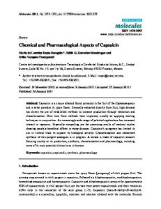
2. Unified treatment programme 2.1. During the catarrhal phase: calcium preparations with vitamin C parenterally, preparations calming the coughing and the pains, absolute bed rest. 2.2. During the subsiding phase: calcium preparations and vitamin C ampoules applied together venally every 4 hours; glucocorticoids - at average treatment doses. 2.3. During the pulmo-toxic phase: 2.3.1. Reanimation - respiratory, oxygen inhalations; - cardiovascular - Strophanthin, digital injections, antishoock (Urbason etc.), high-molecular solutions applied venally in drops under control of the haemodynamic indications. 2.3.2. Antidotes: 3% sodium bicarbonate solution, Becotide ampoules, aerosol mixture - fractionated inhalation; Acetylcystein. 2.3.3. Pulmo-protective treatment: inhalation of aerosol mixture (see above), diuretics (Furanthril, Mannitol etc. parenterally, calcium preparations and vitamin C - ampoules). 2.3.4. Antihystamin drugs: Fenistil, Sandosten-calcium, Allergosan and other applied parenterally. 3. Clinical model: Male. Name initials: Z.D.D. 42 years old. Patient: 5905/75 Data from the anamnesis: After wreckage and spill of a significant quantity of concentrated nitric acid, the patient inhaled the evaporations in the air containing nitric oxide, he felt ill, with difficult breathing and pains behind the chest bone. The objective status: General status - very severe, shock state, dispnoea, severe lung oedema, cyanoses, heavy respiratory insufficiency. After treatment with reanimation preparations, pulmo-protective therapy and detoxic drugs, the patient recovered. 3.1. Information. Male. Name initials: Z.D.D. 42 years old. Patient: 5905/75
34
C/Pp
21.3 18.7 16.0 13.3 10.7 8.0 5.33 2.67 0
Dg: Intoxicatio per Inhalationem gravis cum nitrogenio oxydato Z.D.D. 42 years old (Patient 5905/75)
Legend:
Treatment: 1). Pulmoprotection 2). Respiratory reanimation 3). Antishock treatment 4). Antidotes – by inhalations 5). Diuretics
Arterial pressure c/Pp
19.04
14.7
12.0
beats/min
Central venal pressure c/Pp
13.3
16.0 10.7
Diuresis
1.18
120 100 95 90 85 80
Shock
Pulse
60
Toxic pulmopathy: Severe pulmonary oedema, cyanosis, acute respiratory insufficiency
650 3.04. 2 p.m. 4 p.m.
3100 ml
Checked out healthy
9 p.m. 9.30 p.m. 10 p.m. 10.30 p.m. 4.04. 6.04. 22.04.
2.1.3. Poisonings by sulphur oxide The injuries are caused by the sulphur acid, which is formed in the respiratory tract from sulphur oxide and water of the bronchi secretion. Clinical picture. It is characterized by catarrhal phenomena in the respiratory tract, rarely acute tracheobronchitis, mild toxic oedema and bronchopneumonia may occur. Treatment. The same treatment is applied as for chlorine poisonings. 2.1.4. Acute poisonings with pulmotoxic effect in specific conditions purposefully created by terrorist or similar activities Main agents: phosgene, chlorpicrin, and others. Phosgene poisoning will 35
be taken for a model for this kind of poisonings. Acute phosgene poisonings. This gas has been used during World War I as a chemical weapon. Even today it is included in the chemical weapons' arsenal of many countries. In peaceful conditions, this gas is formed as an intermediary product in a number of today's industries producing chloroform, tetrachlormethane etc.). Gas mixtures containing chlorine and carbon oxide under high temperatures, produce phosgene ex tempore and at the same time increase the damaging effect of the mixture. Toxic mechanisms. The toxic dose is 0,003-0,015 mg/l air. Above 0,10 mg/l - a lethal outcome is expected within minutes. Irritative pulmotoxic mechanism: It injures the respiratory tract mucosa provoking catarrhal and necrotic changes depending on the dose. Enzyme toxic mechanism: It suppresses the function of a number of enzymes in the parenchyma organs' cells, which ensure oxidation and protein synthesis. Clinical picture. It is manifested after a latent period lasting 2 - 6 - 10 hours in average, by the following symptoms and syndromes: – catarrhal phenomena in the respiratory tract with coughing and pains behind the chest bone; – toxic pulmonary oedema; – dispnea, cyanosis and acute respiratory insufficiency; – vomiting and abdominal pain, Meniere's and conscience disorders observed when large doses of the poison have been taken in. A severe bronchial spasm is also observed. These severe forms may have lethal outcome, before pulmonary oedema appears. Superposed pulmonary infections and severe miocard damages are established in the process of the severe forms of intoxication. Laboratory tests. They show data for haemoconcentration with high haematocritis values, polyglobulism and multiple thromboembolic incidents. Paraclinical data. The roentgenogram shows a number of spot shades of different size in both lung sides, determining the toxic oedema and the bronchpneumonia; the electrocardiogram points to miocardiac lesion. Diagnosis is determined on the basis of anamnesis about gas contact with specific smell (in gas mixtures it may not be established), about the combination of a latent period of specific duration after catarrhal phenomena with pulmonary oedema. Treatment. The following antidotes are prescribed: 36
1. Glucocorticoids (Urbason etc.) ampoules of average daily dose 120 - 160 mg. 2. Calcium gluconicum ampoules 10% 10 ml and vitamin C ampoules 5,0 ml mixed, applied venally 3 - 4 times daily every 24 hours before occurrence of the shock. 3. Pulmoprotective medication - sodium bicarbonate 3% solution with broncholitics and glucocorticoids in aerosol mixture, inhaled fractionated; diuretics (Furanthril, Mannitol solution applied venally); for the pulmonary oedema - Acetylcystein - administered by inhalation in the traditional way. 4. Wide spectrum antibiotics applied parenterally upon occurrence of bronchopneumonia and other inflammatory processes. 5. Reanimation: respiratory methods; cardiovascular - Isolanid or Strophanthin ampoules injected muscularly or venally; digitalis preparations - ampoules, antiarrhythmia drugs. 6. Organoprotective and symptomatic medication - upon indications. 2.1.5. Acute poisonings by chlorpicrin (trichlornitromethane) Toxic effect. Gas with strong odour. Local effects - irritation, metabolite disorders and haemotoxic processes. Clinical picture. Catarrhal phenomena in the respiratory tract, pulmonary oedema in the severe forms. – somnolence, stupor; – cyanosis and methaemoglobinaemia; – shock state and acute respiratory insufficiency – in the extremely severe forms. Diagnosis is determined on the basis of data about contact with the poison, about existing combination of cerebral with pulmotoxic and methaemoglobuline syndrome in the clinical picture. Treatment. The same treatment is applied as with poisonings by phosgene gases. 2.2. Pulmocerebral type of monotoxic poisonings This type of injury is characterised by the data presented hereafter on its main representatives [7]. 2.2.1. Poisonings by sulphur hydrogen gases Damaging mechanisms. Injuries from the above mentioned gases occur as a result of two main harmful mechanisms: a) the strong chemical activity of the poison provokes local irritative or necrotic lesions on parts of the respiratory tract or the conjunctiva mucosa; b) the 37
blocking of the enzyme systems, providing intracellular respiration in the latter (cytochromoxidase etc.) provokes severe intracellular hypoxia of brain cells. As a result of the mentioned mechanisms, severe tracheo-bronchitis with toxic oedema affect the respiratory tract, as well as bronchospasm and toxic bronchopneumonia; the toxic effect in the severe cases and the prolonged exposure to the poison damage the central nervous system by forming encephalomalatic foci and multiple blood haemorrhages Clinical picture. The following main syndromes are characteristic: a) pulmotoxic syndrome - manifested by severe tracheobronchitis respiration disorders and conjunctivitis; the mild cases show catarrhal changes in the respiratory tract, the severe forms - strongly expressed tracheo-bronchial-broncholitic symptoms, toxic pulmonary oedema, bronchospasm, early multiple bronchopneumonic foci, severe acute respiratory insufficiency, high temperature; b) cerebro-toxic syndrome - the mild cases suffer of headache, somnolence, occasionally convulsion equivalents, the severe cases - coma, convulsions, bulbar paralysis, red conjunctiva, cardio-vascular syndrome. The general condition is severely affected. Paraclinical data show: at roengenography of the lungs - diffuse hilus images and spot-like equal shades in both pulmonary halves; at electrocardiography - strongly expressed depression zones; laboratory tests indicate leucocytosis, accelerated reaction of the sedimentation of erythrocytes (RSE), acido-alkalizing state (AAS), indications for acidosis at different degrees. Treatment. It is carried out by the following means and methods: 1. Oxygen therapy or respiratory reanimation, in mild forms - oxygen mixture is inhaled with oxygen mask, in severe forms - intubation and assisted or controlled respiration and hyperbar camera are applied; in mass incidents the equipment for reanimation, including the barocameras are taken at the site of the incident and these manipulations are carried out at the begin-ning of the treatment. 2. Antihypoxia complex antidote, a combination of Nootropil (Piramem), vitamin B6, Centrophenoxin and small doses of Diazepam (upon convulsions) in 250 ml glucose serum are applied by drops venally every 6 hours in different doses depending on the indications. 3. The aerosol mixture of 3% sodium bicarbonate, broncholitics (Alupent, Novphyllin), Becodite drops is applied by inhalations, every 3-4 hours throughout twenty four hours. 4. Antibiotics - included in the first hours of the intoxication. 5. Diuretics - Furanthril or Mannitol solution - about 250 ml in drops venally. 38
6. Glucocorticoids - Urbason average dose of 80 - 160 mg daily and cardiotonics; 7. Infusion therapy by drops venally according to the haemodynamic indications (glucose and levulose solutions, Alvesin, alkaline solutions etc.) 8. Cardiotonics - Strophanthin or digitalis preparations in ampoule form - parenterally. 9. Symptomatic medication - upon indications. Clinical model for acute poisonings by hydrogen sulphide - diagnostic antidote treatment method 1. Clinical characteristics 1.1. Catarrhal syndrome: irritation in the upper respiratory tract and the eyes, coughing, strangulation in the chest. 1.2. Pulmo-toxic syndrome: toxic lung oedema, bronchial spasm, early bronchopneumonia, acute respiratory insufficiency, apnoea. 1.3. Cerebral toxic syndrome: excitement, drowsiness, convulsions, coma. 1.4. Cardio-circulatory syndrome: shock state, acute lung heart (at a later stage!). 1.5. Metabolite syndrome: severe acidosis of mixed type. 1.6. Paraclinical tests: roentgenogramme: spot shadows on both sides of the lungs; electrocardiogramme: dysrepolarization changes, arrhythmia, Ppulmonale. 2. Unified treatment programme 2.1. Respiratory reanimation: assisted or directed respiration after intratracheal intubation, oxygen inhalation. 2.2. Cardiovascular reanimation: cardiotonics, antishock and antiarrhythmic drugs. 2.3. Pulmoprotective drugs: broncholitics, diuretics, antibiotics. 2.4. Antidotes: 3% sodium bicarbonate solution, Becotid - aerosol mixture; antihypoxia drugs Nootropil (Pyramem or Urbason doses) or by means of antihypoxic methods. 2.5. Symptomatic treatment.
39
3. Clinical model Information: The patient T.G.I. aged 42, during wreckage at a plant, together with three other workers, inhaled hydrogen sulphide. He felt catarrhal manifestations in the eyes and the throat and pain in the chest. He lost consciousness. Objective state: coma, toxic-clonical convulsions. Also established were: conjunctivitis, acute lung oedema, electrocardiographic data for hypoxia of the myocardum and disorders in the atrioventricular conductivity. The roentgenogramme shows spot shadows on both-sides of the lungs. The whole group was treated after the described method, adapted to the condition of each patient, by a team of doctors under the leadership of Prof. Monov.
C/Pp
Dg: Intoxicatio per Inhalationem gravis cum sulfuro hydrato. Pulmopathia. Encephalopathia toxica. Shock. Insufitientia respiratoria gravis. T.G.I. 42 years old Treatment: 1). Respiratory reanimation 2). Antishock therapy 3). Pulmo-protection 4). Antidotes
19.0 15.0 10.7 6.0 0
Legend: Blood pressure Clinical picture Coma, convulsions, shock, conjunctivitis, toxic pulmopathy
Checked out healthy 7.10.1975
40
9.10.
15.10.
29.10.1975
2.2.3. Poisonings by cyan compounds Mass forms of this type of intoxication can be provoked most often by preparations, which in specific conditions release the highly toxic hydrocyanic gases and methylizocyanate. 2.2.4. Poisonings by hydrocyanic gases They are caused by hydrocyan, which is formed in the industrial premises after failures in operations producing synthetic material or other modern chemical preparations. Damaging mechanisms. It penetrates into the blood and the cells through the respiratory tract. There, it blocks the cytochromoxidase enzyme groups ensuring the intracellular oxydation. Quick intracellular hypoxia occurs, especially manifested in the brain cells. Clinical picture. Mild and medium-severe cases are characterised by constrictions in the chest, air insufficiency, breathing disorders, dizziness, accompanied sometimes by convulsions. The severe forms are characterised by unconsciousness, reaching deep coma, quick respiratory disorder, convulsions, red face, sometimes - mild mydriasis and shortly after lethal outcome. Catarrhal changes in the respiratory tract are observed, provoked by the irrita-tive effect of the inhaled gas. 2.2.5. Methylizocyanite-, chlorcyanite- and other organic-cyan poisonings Etiology. These are provoked by organic compounds of hydrocyanite. Very often some of today's mass intoxications are due to chlorcyanite and methylizocyanite. Methylizocyanite is obtained in dozens of chemical operations in modern industries such as methylation of unorganic chlorcyanite, pyrolysis reactions, industrial decomposition of the compounds of acetic acid (acetamid, acetylchloride etc.). The fact that methylizocyanites can be obtained in the abovedescribed conditions from such substances as dymethyluren derivatives and diphenylcarbonates, which are comparatively harmless for men, is of great practical importance. Damaging mechanisms work mainly in two directions, namely: a) local affection of mucous membranes and skin in the contact areas, provoked by the new irritative-destructive effect products formed by the interaction between the poison and the water component of the mucous secretions (for methylizocyanite etc.); b) blocking of the enzyme groups in brain and other specialised cells, which conduct the intracellular oxidation (cytochromoxidase) etc. Clinical picture. It is characterised by severe general condition, often com41
bined with coma and the following main syndromes: a) catarrhalnecrotic – expressed by catarrhal phenomena in the eye and the upper respiratory tract mucosa, resulting often in severe destructive changes in the affected areas; b) pulmotoxic – it is often observed in methylizocyanite and other organic cyanite compounds intoxication and is manifested by severe tracheobronchial bronchiolitis and toxic pulmonary oedema. The latter can be observed at the beginning of the poisoning or after a latent period and is often complicated by inflammatory foci, severe destruction of the respiratory tract and acute respiratory insufficiency; c) respiratory – fast malfunction and ceasing of breathing, established in cases when free chlorcyanite has penetrated the blood and from there into the cells; d) cardiovascular – rhythm and myocardial disorders, shock states. Sometimes liver and kidney affections are established (organic chlorcyanite compounds). Treatment. The following medication is used: a) antidotes: 1-3% sodium nitrosum ampoule venally, after which through the same needle 10% sodium hyposulphurosum of average 30-50 ml venally is introduced or 4 dimethyl-aminophenol of average dose 4 mg/kg is applied after the standard methods; b) respiratory reanimation - applied at the beginning of the treatment for severe forms of affection and combined upon indications with apparatus respiration; c) glucocorticoids - Urbason 3x60mg at average venally or betamethazone preparations, which have a very good effect to overcome the toxicity, applied 4x4mg at average daily; d) other pulmoprotective means: aerosol intake of bronchiolitics (allopent, Novphyllin (Aminophyllinum) etc.) every 2-4 hours during the first day, glucocorticoids (Becotide), 3% sodium bicarbonate solution, Acetylcystein etc.; e) antishock therapy by vasopressor and cardiac medication (Effortil, Noradrenalin, Strophanthin or Isolanid - parenteral application in doses upon indication); f) antibiotics - in big doses againts the quickly developing bronchopneumonia; g) antihypoxia intracellular preparations - Pyramem, etc.); h) barocamera - hyperbaric oxygenation against the severe hypoxia lesions of the central nervous system. 2.2.6. Acute cyan poisonings with general toxic effect Main representatives: chlorcyan and other cyan gas compounds. In gas media it smells like bitter almonds. Toxic mechanisms. The mentioned poisons provoke the following: – local irritative damage of the respiratory tract mucosa; – enzyme toxic disorders, blocking the cytochromoxidase and other enzymes of the cell redoxenzyme system of different organs and re42
sulting in a very fast general intracellular hypoxia; – temporary transformation of haemoglobin into cyanhaemoglobin. Clinical picture. Catarrhal skin symptoms: eye irritation and watering; coughing and pain behind the chest bone (short lasting), red face and neck areas. Pulmotoxic: dispnea, bronchospasm, painful coughing, combined with stenocardia. Tonic clonic spasms. Comatose states, bulbar paralysis. Very often the severe cases are accompanied by a strong cry, convulsions, coma and shortly after - respiratory paralysis with lethal outcome. The diagnosis is put on the basis of existing information on the presence of gas in the air (bitter almond smell) combined with manifested catarrhal convulsions and pulmotoxic phenomena in the clinical picture. Treatment. Antidote preparations: Natrium nitrosum 1-3% ampoule, 5-10 ml at average with 20 % glucose solution 10-20 ml venally, followed by Natrium hyposulphurosum 10%, 40-60 ml at average venally through the same needle; Amilium nitrosum ampoule - a few drops are spread over a tissue and it is inhaled and the breath is kept; 4 DMAPh dimethylaminophenol) ampoule 250 mg i.v. followed by 10% sodium thiosulphate, 60-80 mg at average through the same needle. Nitrosum groups of the mentioned compounds quickly form methemoglobin, which connects immediately to the cyan groups of the poison due to its affinity, thus forming cyanmethemoglobin; the latter forms sulphocyan by connecting to the sulphur groups in the sodium thiosulphate and is thrown away in the urine; the antidote treatment should be started immediately at the beginning of the therapy, together with the other treatment methods. Respiratory reanimation: intratracheal intubation and artificial respiration; oxygen therapy in different forms. Anticonvulsion medication: Diazepam ampoule 10mg, muscularly. Hemotransfusion - 250-500 ml of blood according to hemotransfusion rules; partial exchanged blood transfusion - in high methemoglobinaemia cases. Infusion treatment: 20% glucose solutions 500-1000 ml, aminoacid and saltwater solutions attributed venally in drops of 2000-4000 ml average daily dose; 10% calcium gluconate of 10 ml daily dose about 3 times daily; with vitamin C ampoule. Antihypoxia medications: Pyramem (Nootropil) ampoule, vitamins of the B-group parenterally. Symptomatic drugs and methods for removal of poison from the entry area.
43
2.3. Mass poisonings by rhenal cerebral type of toxic agents These poisonings are mostly caused by metalalkaline coumpounds, formed by harmless final products in modern industrial specific technologies (e.g. microelectronic equipment production and others) after unexpected occurrence of the above described mechanisms. The aggressive character of these substances is defined by easy spread in the air and high chemical activity. The main representatives of this type of agent causing heavy intoxication are dymethyl-mercury compounds. 2.3.1. Poisonings by dymethyl-mercury compounds They penetrate the human organism through the skin, the mucosa, the respiratory tract and the mouth. Strongly expressed renal-tropic and cerebro-tropic features are manifested. They fix to the sulphohydril groups, which represent the enzyme receptors of the mentioned organ's cells and provoke severe damage of these organ's metabolism. Clinical picture. It is manifested by the following syndromes: a) irritative - catarrhal expressions on the contact mucosa; b) renotoxic - by oligo- or anuria, pathological deviations in urine indications and manifestation of kidney insufficiency; c) cerebro-toxic - manifested by convulsions, delirious fits, sometimes by consciousness derangement, gradual fall into neurotoxic degradation and dementia events. Treatment. It is carried out by the following methods: a) Unithiol ampoule - 1 ampoule injected subcutaneously or muscularly every 6 hours; b) antihypoxia anditote complex consisting of Pyramem, vitamin B6, Centrophenoxin at average dose of 1 ampoule diluted in 250 ml glucose solution every 8 hours applied in drops venally; c) early dialysis, specialised with Unithiol; d) symptomatic and organ-protective medication. 2.4. Acute mass poisonings with prevailing cerebral psychotoxic effect The acute mass poisonings with prevailing cerebral psychotoxic effect include intoxications, which occur in specific conditions, listed hereafter by groups: 2.4.1. Acute poisonings by incapacitating agents with psychic effect Analysing their chemical features, some of these poisons resemble the main mediators in the organism and are classified in three groups. 1. Similar to serotonin: amides of the lysergic acid, psilocibin, etc. 2. Similar to adrenaline: main representative - mescalin etc. 3. Similar to acetylcholine: ditran, benactysin etc., which are mainly esters of the glycol and benzile acids. 44
4. Another group includes phencyclidine (sernyl), BZ, marihuana etc. (Krastanov, L., J-P. Frejaville, Rapport de l'OMS, Geneve, 1970) Basic damaging mechanisms and effects. The pointed incapacitating agents have a specific mechanism for affecting the central nervous system, depending on their chemical characteristics and toxikinetics. The following basic mechanisms have been established: – disorder of the mediators' metabolism in the central nervous system through inhibition of enzyme groups, like preparations of the lysergic acid which block the monoaminooxidase and other enzymes, resulting in accumulation of serotonin and catecholamines metabolites in the cerebrum; – blocking of the mediators' chemioreceptors, mainly of the serotonin, adrenaline and acetylcholine, resulting in accumulation of these biochemical substances in the cerebral structures; – direct effect on the cerebrum neurones and synapses and especially on the reticular formation. By means of the pointed mechanisms the incapacitating agents of psychic effect accomplish temporary mental disorders of hallucinogenic depressive or excitement character, which in specific conditions may be accompanied by damages to other organs and systems. The toxic dose and the illness period are different for the separate representatives of these mass poisons and depend on their chemical characteristics. The main representatives of the different groups are given in Table 2 Table 2 Main representatives Type of poison Amides of the lysergic acid (LSD) Mescalin Sernyl BZ (bizet)
Toxic dose 0,001 mg/kg 5-10 mg/kg 0,03-1,0 mg/kg -
Period of the effect 5-10 hours 10-12 hours 40-48 hours 3-4 hours
Clinical features. They are manifested after different latent periods depending on the specific poison and also have different duration (Krastanov, L., S. Andreeva, J.-P. Frejaville, etc.). 2.4.2. Poisonings by preparations of the lysergic acid (LSD) The latent period is 30-40 minutes. Main syndromes: – Psychogenic syndrome. It is expressed by the following phenomena: 45
hallucinogenic visions of coloured objects, distorted geometric figures and fantastic panorama, auditory hallucinations; mental disorders - slow thinking, psychosis - euphoria or depression, depersonalisation, etc. – Neurogenic syndrome. Somnolence, dysarthria, tremor of the eyelids and hands, dizziness, ataxia. – Neurovegetative syndrome. Tachycardia, mydriasis, face erythema, salivation, voluminous sweating, vomiting. 2.4.3. Poisonings by mescalin preparations Latent period - 1-2 hours. Symptoms and syndromes: – Psychogenic hallucinations - mainly visionary, less so auditory; euphoria, lack of orientation for time and space. – Neurogenic: headache, parestesia and pains in the extremities. Chills and photophobia. 2.4.4. Poisonings by sernyl Latent period - 1 hour at average. Symptoms and syndromes: – Sleepiness and adynamia. – Depersonalisation and mental disorders. – Derealisation of the surroundings. 2.4.5. Poisonings by BZ Latent period - 2-3 hours at average. Expresses a central cholinolitic effect - blocks the Acetylcholine chemioreceptors. Symptoms and syndromes: – Cholinolitic: mydriasis, tachycardia, dry skin and mucosa. – Vomiting. – Neurogenic: mental disorders, stupor. – Psychogenic: hallucinations - visionary and auditory. Diagnosis and differential diagnosis of psychotoxic poisonings. The diagnosis is put on the basis of the psychotoxic syndrome, combining hallucination and psychic phenomena. The differential diagnosis considerations include reactive psychosis, schizophrenia, that are excluded according to anamnesis data and lack of other specific features for these diseases. Treatment. The following treatment medication is included: For all poisonings: patients are taken outside of the gased environment and receive the following antidote cocktail: – Pyramem 1,0 (Nootropil) 1-2 ampoules at average; 46
– Centrophenoxin 100-250 mg at average; – Vitamin B6 - 50-100 mg at average; – 5% Glucose serum - 250 ml. The above cocktail is applied in drops venally at every 8 hours till the patient is out of the poisoned state. Beside the described mixture, the following treatment is applied for the different types of intoxication: Poisonings by lysergous preparations: – benzodiazepin preparations (Diazepam amp. 10 mg) or phenothiazin preparations (Chlorazin or Largactil amp. 25 mg) muscularly 2 times at average; – nicotine acid and its derivatives - amp. 1 ampoule 2-3 times daily at average; – Atarax and glutamine preparations - in the pharmacological doses. For mescalin poisonings, treatment includes: – benzodiazepin preparations (Diazepam amp. 10 mg) muscularly 2 times daily at average; – respiratory and cardiovascular reanimation; – symptomatic means. Phenothiazin, barbiturates and reserpin drugs are contraindicated. For sernyl poisonings treatment is similar to the lysergic preparations poisonings. For BZ poisonings: – cholinomimetics and cholinesterase reversible inhibitors. Syntostigmin amp. 0,5 mg 1ml; Eserin etc. – benzodiazepine preparations: Diazepam, Valium amp. 10 mg with vitamin B6 ampoule applied muscularly upon indication 2 times daily; – respiratory reanimation - for the severe cases. For all the intoxications of the mentioned psychogenic poisons' origin, substitute and correc-tive therapy is applied using venal drops infusion of waterelectrolyte glucose, aminoacid and other solutions according to indications. 2.5. Mass poisonings with polyorganic effect The mass poisonings with polyorganic effect are described below including their most often met with representatives [2, 3, 4, 5, 6]. 2.5.1. Poisonings by carbon oxide These are amongst the most popular mass poisonings. 47
Damaging mechanisms. The poison penetrates the organism exceptionally through the respiratory tract. It manifests specific affinity to haemoglobin iron and forms the stable compound carboxihaemoglobin. With the mioglobin of the scarred muscularity it forms carboximioglobin, further it blocks the free iron in the plasma, due to its increased affinity towards it. For the same reasons, it sticks to the enzyme systems in the cerebrum neural cells and breaks their function. Severe hypoxia of intracellular type and haematogene-transport mechanism occurs in the organism. Clinical forms. The following clinical forms have been established by the author: a) foudroyant - the patient dies in a few minutes, due to inhaling air with high carbon oxide concentration; b) severe (comatose) form - the patient is in coma, often has pathological reflexes, noisy snory breathing, red face and neck, sometimes - nidus symptoms; electrocardiography shows evidence of hypoxia changes or infarction-like indications; blood in the vein is bright red; c) mediumsevere forms - the poisoned patient is for a short time in a state of disordered consciousness - sopor, of which he is taken out quickly; headache, dizziness, vomiting are observed; d) mild form - carboxide encephalopathy is manifested characterised by headache, skin paleness, nistagm, nausea and vomiting, faintness and adynamia. Laboratory tests show different increased values of carboxihemoglobin in the severe cases, immediately after the contact with the poison, electrocardiography of many of the patients with different degrees of poisonings, shows evidence of dispolarizing changes in the severe forms of infarction-like images. Treatment. It is carried out by the following methods: a) oxygen therapy: for the mild forms - inhalation of oxygen mixture, for the severe forms - intratracheal intubation and artificial respiration, hyperbar oxygenation with barocamera; b) antihypoxia medication - antihypoxia cocktail (Pyramem, Centrophenoxin, vitamin B6, 250ml glucose serum) or only Pyramem (Nootropil) ampoule in the established treatment dose; c) symptomatic treatment and antibiotics - in complications and bronchopneumonia. 2.5.2. Poisonings by hydroxile benzol preparations (phenol etc.) These poisonings occur in the operation technologies of some modern fields of the chemical industry. The agent penetrates the organism through the respiratory tract, skin and mucosa and the gastrointestinal tract, the first two entries being significant for mass poisoning results. Damaging processes. This type of poisonous steam affects the organism with local irritative and necrotic lesions, with cell damages - erythrocytes (hemoly48
sis), other organs' specialised cells (central nervous system, liver, kidneys), with enzyme inhibition and with strongly expressed and quickly developing metabolite disorders. Clinical manifestation. It includes several syndromes: a) pulmotoxic and irritative syndrome - expressed by catarrhal changes on the contact mucosa and respiratory tract, toxic pulmonary oedema; b) hemotoxic syndrome - expressed by severe hemolysis of different degrees, in the severe forms - hemolytic shock and anaemia; c) hepatal syndrome - characterised by subicterus or icterus, increased liver and bilirubinaemia; d) renal syndrome - by oliguria or anuria, pathological deviations in the urine and acute kidney insufficiency. In the extremely severe forms consciousness is disordered. Laboratory blood and urine chemical tests show evidence of phenol metabolites, data for blood damage (increased values of free hemoglobin, reduced number of erythrocytes), positive liver tests etc. Treatment. It is carried out by the following drugs and methods: a) respiratory and antishock therapy as a beginning for the severe degrees of affection; b) antidotes - calcium gluconate ampoule, applied venally 1 amouple at the beginning and after 2 hours, then every 4 hours, by accounting for the indications and contraindications; at the beginning 2-4 ampoules are accepted in water perorally; calcium fenolate is formed which is thrown away; c) hemotransfusion and partial exchange blood transfusion, as well as detoxic dialysis depuration - in the first hours of the intoxication; d) hepato- and renoprotective means and glucocorticoid preparations against hemolysis and shock state; e) antibiotics - against the secondary inflammatory processes in the respiratory tract; f) symptomatic medication. 2.5.3. Poisonings by amino- and nitrobenzol derivatives (anilin and other preparations) These, easily evaporating liquid poisons are used in many fields of modern chemical industries, in laboratories and in rockets' production, in everyday life. After failures or carelessness, they penetrate the organism through the respiratory tract, the skin and mucosa and the gastrointestinal tract. Mass intoxication occurs mainly after penetration through the first two entries [1, 5, 6]. Damaging processes. The organism is injured by the hemotoxic processes - hemoglobinaemia and severe hemolysis with shock fits, neural, liver and kidney cells' metabolism disorders occur. A mild irritative effect on the contact mucosa is also observed. Clinical manifestation. The following characteristics appear: a) general status - affected in different degrees, in the extremely severe forms - by coma; b) 49
hemotoxic syndrome characterised by cyanosis, hemiglobinaemia and hemolysis, acute anaemia; c) hepatotoxic or hepatorenal syndrome expressed by icterus, hepatomegalia, sometimes by oliguanuria and liver or kidney insufficiency. Laboratory tests prove chemical toxic substances and their metabolites in biological samples of the patients. Toxic steam penetrated by inhalation complicates the patient status by pulmonary lesions of the toxic pulmonite and acute respiratory insufficiency type. Treatment. It is carried out as follows: a) respiratory and cardiovascular reanimation - in comatose states, pulmonary and cardiovascular affections; b) glucocorticoids (Urbason) ampoule 60-120 mg venally daily and partial exchange blood transfusion; c) antidotes - blue methylene solution 1% (chromosmon) ampoule 20 ml (average dose 5-10 ml) in 250 ml glucose serum applied in drops venally, ascorbic acid (vitamin C) ampoule 2000-3000 mg average dose per day (as antidote); d) symptomatic treatment and pulmoprotective medication by aerosol drugs combinations. 2.5.4. Poisonings by methyl-benzol (toluol) preparations They are produced during operation technologies in different fields of the chemical industry, and exist as ready-made products as organic solvents. They penetrate the organism mainly through the respiratory tract, when they provoke mass poisonings, less so - through the gastrointestinal tract and the skin. Damaging mechanisms. Due to their physical and chemical properties, toluol molecules cause local irritative hyperemic lesions on the contact mucosa of the respiratory tract, dystrophic damage of the cerebrum and liver cells mainly by inhibition of enzyme groups and intracellular hypoxia. Clinical manifestation. It includes the following syndromes: a) irritativepulmotoxic syndrome - with evidence of catarrhal tracheobronchitis, combined with toxic oedema in the severe cases; b) cerebral toxic syndrome characterised by ataxia, Menier's syndrome, the severe forms manifest disordered consciousness; c) hepatotoxic syndrome - observed in the extremely severe cases of poisoning, manifested as icterus, hepotomegalia and increased blood bilirubin and transaminase values; sometimes it is manifested as hepatorenal syndrome; d) hemotoxic syndrome - rarely met in acute intoxication by methemoglobinaemia, hemolysis, leukopoenia. Treatment. The following medication and methods are used: reanimation procedures, pulmoprotective drug combinations, early dialysis detoxic depuration, partial exchange blood transfusion and others upon indication.
50
2.5.5. Poisonings by polychlordibenzodioxin and dibenzofuran gases Physicochemical properties. This gas poison is formed in modern chemical industries' failures in the process of synthetic matter molecules transformation. Thus upon the interaction with odorous chlororganic compounds combined with thermal effect, polychlordibenzo-dioxin and lesser quantities of polychlordibenzofuran are released. In specific conditions they can be used for toxicochemical terrorist acts. Dioxin and difuran are the cause of some of the severest mass poisonings of our days (Ludvigshafen in 1953, Sevezo in 1976). Mass intoxication occurs when the poison penetrates the organism by the respiratory tract and the skin, less often these poisons reach the organism as liquid substances through the gastrointestinal tract. Damaging mechanisms. Dioxin and difuran injure the patients by provoking local irritative and ulcerous processes on the skin and mucosa contact surfaces, as well as severe disorders of the substances' metabolism in the brain, myocardum and liver cells on the basis of enzyme failure and suppressed RNA and amino-acid groups' interaction. Clinical manifestation. It is characterised by the following events: a) general condition - severely damaged; b) cerebrotoxic syndrome - consciousness disorders reaching coma; c) pulmotoxic syndrome expressed by toxic catarrh and respiratory tract oedema, acute respiratory insufficiency; d) epicutaneous syndrome which includes erytheme and ulcerous damages in different areas of the body and the eyes; e) hepatic and hepatorenal syndrome manifested by icterus and data for liver and kidney disorders; f) teratogenic syndrome expressed by malformation in the progeny. Treatment. It is carried out by means of the following means and methods: a) respiratory and cardiovascular reanimation and pulmoprotective treatment by parenteral or aerosol forms medications; b) detoxic dialysis depuration of the blood; c) organoprotective treatment combinations (referring to liver, kidney etc.); d) symptomatic treatment. Clinical models in mass polytoxic and combined poisonings - diagnostic antidote treatment combined method (after Al. Monov) 1. Clinical characteristics 1.1. General state - very severe. 1.2. Syndromes of combined respiratory insufficiency: 1.2.1. Neurogenic-regulatory type: toxic damage of the brain centres and peripheral nerves. 51
1.2.2. Respiratory-pulmonary type: damage of the lungs. 1.2.3. Haemo-oxygen-transport type: toxic affection of the blood. 1.2.4. Enzyme-cellular type: toxic damage of the oxidising cell enzymes. 1.3. Syndromes of acute polyorgan humoral insufficiency. 1.3.1. Pulmo-cardiac insufficiency. 1.3.1.1. Pulmo-toxic syndrome: dispnea, orthopnea, tachipnea, apnea, cyanosis; obtrusional phenomena: broncho-vesicular-capillary block (multiple dendrite obtrusions in the bronchi, multiple atelectatic bronchopneumonia niduses) and "moist lungs" (toxic lung oedema). 1.3.1.2. Cardiovascular syndrome: acute left-ventricular insufficiency, severe “lung heart”, acute hypertonia in the pulmonary arteria, shock state. 1.3.2. Cerebral insufficiency: 1.3.2.1. Cerebral depressive syndrome: consciousness disorders: somnolence, stupor, coma. 1.3.2.2. Convulsive syndrome 1.3.2.3. Delirious syndrome 1.3.3. Toxo-hepatic insufficiency 1.3.3.1. Icteric syndrome 1.3.3.2. Hepatalgic syndrome 1.3.4. Renal insufficiency 1.3.4.1. Oliguria syndrome 1.3.4.2. Uremia syndrome 1.3.5. Haematogenic insufficiency 1.3.5.1. Syndrome of haemoglobin deficiency: carbo-haemoglobinaemia, cyan-haemoglobinaemia, methaemoglobinaemia, haemiglobinaemia, haemolysis, etc. 1.3.5.2. Dyshaemostatic syndrome: haemorrhagic diathesis. 1.3.6. Metabolite insufficiency 1.3.6.1. Acid-alkaline syndrome: metabolite acidosis etc. 1.3.6.2. Hypoproteinaemic syndrome 1.3.6.3. Water-electrolyte syndrome: dehyrdatation, electrolyte disorders, hydraemia. 1.3.7. Immune insufficiency
52
2. Unified treatment programme 2.1. Reanimation 2.1.1. Oxygen inhalation, assisted or commanded respiration (after intratracheal intubation). 2.1.2. Hyperbarric oxygenation (barocamera) after restoration of respiratory tract passage and spontanea respiration. 2.1.3. Cardiovascular reanimation 2.1.3.1. Cardiotonics: Strophanthin, digital injections; enzyme preparations, phosphorus-donators, vitamins of the B group etc. 2.1.3.2. Phlebotomy and/or blood transfusion - according to indications. 2.1.3.3. Anti-shock therapy: Urbason big pharmacological doses - infusion, adequate to the haemodynamic indications. 2.2. Pulmoprotection: removing of the brocho-vesicular-capillary block and the lung oedema: inhalation of aerosol mixtures: sodium bicarbonate solution. Becotide, Novphyllin, Acethylcystein - fractionated several times during the day, combined with oxygenation; diuretics, proteolytic medication against the obtrusive stoppers in the respiratory tract. 2.3. Antidotes 2.3.1. Antihypoxia antidotes: Nootropil (Pyramem), Orocetam, Centrophenoxin, vitamines of the B group etc. 2.3.2. Aerosol type poisons - see above. 2.4. Organ-protection: lungs - see above: heart, cerebrum, kidneys and others; antibiotics; immune protection (stimulating and substituting). 2.5. Symptomatic treatment. 3. Clinical models (author's studies). 3.1. Patient with severe acute inhalation poisoning by gas mixture: severe toxic pulmopathy with strongly expressed pulmo-cardiac insufficiency. (Fig. 1, 2, 3, 4).
53
Before Treatment
Fig. 1
Fig. 2 After Treatment
Fig. 3
54
Fig. 4
3.2.
Clinical models (the patient was treated under the leadership of Prof. Monov) 3.2.1. Patient: M.S.K.
Dg: Intoxicatio per inhalationem gravis cum fumo toxico. Pulmopathia toxica. Insufitientia respiratoria gravis. Shock. M.S.K.
1) 2) 3) 4) 5) 6)
General state – very severe Toxic pulmopathy – strongly expressed Lung oedema – very severe Dispnea – strongly expressed Cyanosis – severe Shock state – strongly d
Anti-shock therapy
Treatment: 1). Reanimation: respiratory (O2 masque), anti-shock therapy 2). Pulmo-protection 3). Antidotes
Moved to an internal clinic in improved state for general rehabilitation
Acute pulmo-cardiac insufficiency
CHOB = 5% AAS Ph = 7,44 PO2 = 51,5 PCO2 = 33 BE = -0,8
20-21.03.
7,384 110,3 57,3 +7,2
22.03.
7,405 130,3 50,6 +5,4
7,488 79,5 52,2 +13,5
23.03.
7,532 66,9 46,8 +14,8
29.03.
55
3.2.2. Roentgenography of the lungs of patient M.S.K.
During treatment Fig. 5
During treatment Fig. 6
At the end of treatment Fig. 7
56
3.2.3. Patient: M.I.N. Dg: Intoxicatio per inhalationem gravis cum fumo toxico. Pulmopathia toxica gravis. Shock. M.I.N.
Tracheostomy
Treatment: 1). Reanimation: respiratory, anti-shock 2). Pulmo-protection 3). Antidotes
General state – very severe Toxic pulmopathy – very strongly expressed Lung oedema – very severe Severe dispnea Cyanosis Shock state
Pulmo-cardiac insufficiency
CHOB AAS Ph PO2 PCO2 BE
20-21.03.
Respiratory insufficiency was overcome; Moved to internal clinic with pulmonary and cardiac damages
= 13% = 7,256 = 47,2 = 39,3 = -9,3
7,438 91,8 23,4 -7,4
22.03.
7,48 22,3 56,3 -6,0
7,404 84,8 33,8 -2,8
28.03.
7,488 76,3 38,8 +5,5
2.04.
57
3.2.4. Roentgenography of the lungs of patient M.I.N.
Before treatment Fig. 8
During treatment Fig. 9
At the end of treatment Fig. 10
58
Information: The presented clinical models illustrate the effect of the described model. The patients are affected by mass polytoxic combined poisoning by smoke gases after burning of plastic material. The treatment was carried out under the direction of Prof. Monov and his doctrine. 2.6. Poisonings by incapacitating agents with different effects These include poisons with irritative-reflectory effect and with sensomotive and other effects. They enter in contact with the human organism through the mucosa and the skin as well as by inhalation, very seldom by per oral way. 2.6.1. Poisonings by incapacitating agents with irritative-reflectory effect The listed below intoxications belong to this group (R. Ludowig, K. Lohs, etc.) Poisonings by tear-provoking poisonous compounds. Here are included the following poisons: Brombenzylcyanide (C6H5CHBrCN) - a colourless crystal body with bitter almond smell, toxic dose - 0,0003 mg/l. Chloracetophenon (C6H5COCH2CCl) - a colourless crystal body with wild violets' smell, toxic dose - 0,0003 mg/l. CS substance - a colourless solid crystal body, toxic dose - 0,00010,005 mg/l. All of the enumerated are weakly soluble in water and well soluble in organic solvents. Damaging mechanisms. They strongly irritate the neural receptors of the eye mucosa tropical nerve thus by reflex provoke a strong tear-provoking effect and contraction of the eyelids. In a lesser degree, they provoke excitement effect on the nose and the upper respiratory tract mucosa and the skin. Clinical manifestations: The following syndromes are observed: – Ophthalmotoxic syndrome: burning in the eyes at the beginning, lacrimation and often blinking; if the contact with the toxic environment is prolonged appear very strong lacrimation, blepharospasm, photophobia, strongly inflamed conjunctivas; sometimes pain in the eyes and headache are felt. – Catarrh of the upper respiratory tract characterised by coughing, retrosternal pain and sneezing - for the severe forms; – Nausea, vomiting are observed in the severe forms
59
2.6.2. Poisonings by CS The clinical picture includes the following events: – Ophthalmologic syndrome - very strongly expressed; – Respiratory syndrome - strongly expressed with acute dispnea; toxic pulmonary oedema; – Skin syndrome - erythema-bullous dermatitis; Additionally, copious vomiting, adinaemia, dehydration are observed. Treatment. It is carried out by means of the following: – The patients should be immediately taken away from the poisonous environment. – Copious washing of the eyes with water and 2% sodium bicarbonate solution; application of 1% ophthalmoanalgetics 1-2 drops in each eye. – Cortison preparations - ampoule, parenterally (Urbason, Celestone, etc.). – Pulmo-protective medication - calcium preparations and vitamin C ampoule combined applied venally, codein preparations combined with analgin and expectorating syrups perorally, inhalation of solution containing sodium bicarbonate, cortison preparations, Novphyllin etc. - upon indication. – Respiratory reanimation - for the severe forms upon indications – Skin-protective cremes containing cortison and other drugs; Linen, olive oil etc. – Symptomatic treatment - upon indication. – Dark glasses and wet compresses on the eyes till the catarrhal events subside. 2.6.3. Poisonings by sneezing toxic compounds Their main representatives are: – Adamsit - crystal substance of yellow colour, weakly soluble in water, better soluble in organic solvents; average toxic concentration 0,00038 mg/l. – Dyphenylchlorarcin - colourless crystal substance with the same characteristics; average toxic concentration - 0,00046 mg/l. Damaging mechanisms: a) irritative effect on the nose and the upper respiratory tract organs mucosa and irritation on the following nerves' extremities: olphactorius, laringealis supperior and trigeminus. 60
b) toxic effect on the cerebrum, muscular, liver and other cells with oxidation processes and metabolism disorders. Clinical manifestations. They are expressed by: – burning in the nose and the pharynx and sneezing, which is strong and prolonged in the gased environment; – lacrimation accompanied by burning in the eyes, saliva and voluminous watery nose secretions; – erytheme phenomena, vesicular changes, sometimes after contact of the skin with greater poison concentrations, ulcerous areas appear on the skin; – persistent vomiting; in peroral intake of the poison - abdominal pain with diarrhoea; – generalised manifestations: adynamia, pain in the joints, ataxia, bradipnoea, bradicardia etc. occurring after prolonged exposition of the human organism to the poison. The diagnosis is established on the basis of anamnesis showing strongly expressed presence in the air of sneezing gas, the leading sneezing syndrome combined with disorders of other organs and systems. Treatment: – taking out the patients from the gased environment; – lavage of the nose and nose-swallowing area with 2% solution of sodium bicarbonate; washing out of the mouth cavity with 3% tanin acid solution; – application of skin-protective unguents containing Ol. Olivarum, Ol. Lini and vitamin A, glucocorticoids, lanolin etc., and antibiotics - for ulcerous and secondary inflamed areas; – infusion of water-salt and monosaccharide solutions - 5 and 20% glucose solution at average of 2 x 500ml. Ringer solution 2 x 500ml and other - upon indication. – Symptomatic medication - analgetic and cardiology drugs, oxygen therapy etc. - upon indication. 2.6.4. Poisonings by incapacitating agents with combined effect This group includes compounds, which cause mass disorders of the motile, sense and other functions of the organism. Their affection in many cases is transient, but may very quickly render inactive great number of the population and put at risk the life of some of them. Here belong:
61
2.6.4.1. Poisonings affecting the sensory, motility and other functions They are: A.Poisonings with mio-relaxing effect. Main agents are: glyketal, miastenin, miocain, etc. Toxic mechanism. The function of the synapses between the intermediary neurones of the spinal morrow is blocked and no impact on the neuromuscular structures is exercised. Clinical manifestations. – paresis or paralysis of the different muscular groups - curare-like type; – disordered function of the lower extremities, spreading ascending towards the body muscles and the upper extremities; preserved respiratory function; – nausea, vomiting and abdominal pain - in some more damaged organisms; Treatment: - oxygentherapy, including respiratory reanimation if needed; strichnine injections - 1-2 mg at average muscularly; - 10% Calcium gluconicum amp. - 10 ml at average applied 2-3 times daily 1 ampoule venally; - infusion therapy with glucose and water-salt solutions - 2000-3000 ml at average in drops venally per day. B. Poisonings with cerebral effect. Main agents: tremorin, indoclon etc. Agents of chemical character are: hexafluorindi-ethylether and others. Toxic mechanism: Fluorin derivative substances (indoclon etc.) in contact with the organism release hydrofluorin. Below are described the main damaging mechanisms: – local - on the contact mucosa and skin, often provoking ulcerous lesions; – metabolite - they combine with the blood calcium and provoke strong decrease of its level; – enzyme metabolite - they suppress a number of cell enzyme systems (phosphatase etc.), provoke disorder of the lemon acid cycle in brain cells and damage the carbohydrate cell metabolism. Clinical manifestation. After 30 min and during the following hours, symptoms and syndromes appear, as follows: – Parkinsonlike syndrome with strong tremor (for intoxication with tremorine preparations); 62
– catarrhal syndrome: pulmotoxic or gastrointestinal - depending on the entry area: dispnoea, coughing, pulmonary oedema, pain behind the chest bone; nausea, vomiting, diarrhoea; severe abdominal and chest pain; – skin-ulcer syndrome - in contact with the skin surfaces; – tonic-clonical convulsions and consciousness disorders - related to the acute hypocalcinaemia and the complex intracellular damage; – abortive syndrome - provokes abortion in pregnant women; – cardiotoxic syndrome - arrhythmia, shock state, asystolia. Diagnosis. It is determined on the basis of the Parkinson-like and convulsion syndromes in the clinical picture, combined catarrhal and ulcerous lesions and strong pain in the affected areas. Treatment: - inhaling of aerosol mixtures with sodium bicarbonate - 3% solution and Novphyllin; - stomach washing and intestine cleaning - in peroral intake of the poison; - 10% Calcium gluconicum amp. 10 ml venally 3-4 times daily; - benzodiazepine preparations - Diazepam ampoule 10 mg muscularly for convulsion incidents; - oxygentherapy, respiratory reanimation and antishock preparations - applied by the established methods. 2.7. Poisonings by highly toxic compounds with delayed effect 2.7.1. Acute poisonings by predominantly skin-eruption-provoking compounds Main provoking agents: Yperites (sulphur and nitric compound), Lewisite (chlorine vynil di-chlorine arsinic). Toxic effect. These poisons are transparent or yellow-brownish oil-like liquids. Their evaporations are about 5 times heavier than air. They penetrate the organism through the skin, the respiratory tract, the gastrointestinal tract and the surface mucosa. They manifest lypotropic characteristics, dissolve in the underskin fat depots and penetrate mainly in the lipid rich cells of the brain, liver etc. Damaging mechanisms. Severe local and general toxic damages are provoked in the organism, by suppressing the function of a number of enzyme systems in some vital organs cells, such as central nervous system, skin and mucosa, respiratory tract, myocardum, liver etc. More specifically, latest investigations show that yperite's molecules metabolised in the cells. Their metabolites (thyodiglycol, dichlorinediethylsulphoxide etc.) break the colloid osmotic balance and provoke changes in the protein structures. The toxic products suppress a number of enzyme systems in the cells, such as the catalase, the hexochinase etc. Phosphorilation of carbohydrates is broken, nucleic acid damage provokes pro63
tein synthesis disorders, mitotic processes are also affected. The lipid structures in the rich-in-lipids' cells is disrupted. Thus the yperites act as a polycell poison, which affects multilaterally the metabolism of the different specialised cells. Clinical manifestations. The following symptoms and syndromes are observed: – skin syndrome: it is characterised by polymorphic lesions of the skin, namely erytheme, vesica and ulcerus areas with slow healthing tendency; – pulmotoxic syndrome: manifested by catarrhal bronchial changes and pulmonary oedema; – gastrointestinal syndrome: including salivation, nausea, vomiting, abdomen pains, diar-rhoea; sometimes the vomited matter and the faeces are bloody; – breathing and blood circulation disorders, observed in most of the patients; – cerebral syndrome: headache, somnolence, adynamia, excitement are felt; – ophthalmologic syndrome: appears with lacrimation, red eyes, blepharospasm; – cardiovascular syndrome: characterised by bradycardia, hypotonia and in the extremely severe forms - shock states; – hepatalgic syndrome (established by A. Monov et al.): occurs later (about 1-2 weeks after the beginning of the intoxication) and is manifested by haepatomegalia, sometimes by icterus, increased transaminase and other enzyme samples values, and sometimes bilirubin's. The pointed syndromes appear at different time periods after contact with the poison, after a hidden period of approximately 3 to 10 hours. Thermal-regulation disorders with high temperature are observed (caused by the noxa at the beginning of the intoxication and later provoked by secondary infections). Diagnosis. It is determined on the basis of contact with the poison, combined with the clinical picture including skin-eruption and cerebral syndromes, a continuous latent period and slow healing of the skin lesions leaving cicatrices' marks. Treatment: - interrupting contact with the poison and cleaning on the entry area; caution measures for the hands of assistance personnel (gloves etc.); - reanimation methods: respiratory reanimation (inhalation of oxygen and oxygen mixture, commanded breathing for the extremely severe cases), stimulation of the 64
respiratory centre (Micoren ampoule, Coramin ampoule subcutaneously or venally if the medicament in ampoule is adapted for this application); - cardiovascular reanimation; anti-shock medication - Urbason ampoule 80 - 120 mg venally several times daily or other glucocorticoids; cardiotonic preparations - Isolanid ampoule once or twice daily one ampoule muscularly or Strophanthin ampoule applied slowly venally with glucose solution; - pulmoprotective medication: for the antidote medication are used: 3% sodium bicarbonate solution for washing the mouth and for inhalation; inhalation of aerosol mixture solution including Becotide and Alupent; N-Acetylcystein solution for inhalations, pain relieving drugs including codein and Amidophen, expectorating sirups at a later stage; Urbason ampoule venally and diuretics (Furanthril ampoule, 10% Calcium gluconicum solution against the pulmonary oedema); antibiotics against inflammatory pulmonary complications: - haepatal-protective treatment with antidote effect: 1020% levulose solutions twice daily of 500 ml venally in drops; - skin protective manipulations and means; surgical aseptic processing of vesica and necrotic areas, prolonged application of epithelium tonic and antibiotic gels and paraffin applications, physical therapeutic procedures: - symptomatic means: drug combinations against ophthalmic and gastrointestinal affections are applied. 2.8. Mass poisonings by organophosphorus compounds in specific conditions These are among the severest up-to-date intoxications and are mainly caused by two big groups of organophosphorus preparations: organophosphorus pesticides and organophosphorus compounds applied in specific conditions. Toxic effect. It is caused by highly toxic organophosphorus compounds and their cyanite, fluorin and other derivatives, manifesting a very quick effect. Due to their strongly affecting characteristics, they are produced as chemical weapons and are used for mass terrorist acts in some countries. Further on, the specific damaging mechanisms, the clinical manifestations and treatment of some of the representatives in this group will be presented. 2.8.1. Acute poisonings by neuroparalytic organophosphorus compounds Acute poisonings by alkyl phosphorus: Trilon etc. It is a colourless, easily evaporating liquid. Toxic effect. Suppression of the cholinesterase and accumulation of acethylcholine in the choline-reactive systems, inhibition of a number of enzyme systems of the neural and other cells. It penetrates through the respiratory system, the skin, the mucosa and the gastrointestinal tract. Clinical manifestation. The following damages are observed: 65
– local irritative phenomena of the contact areas: skin, eyes, etc.; – M-cholinomimetic phenomena (myosis, salivation, perspiration, pulmonary oedema, etc); – cholinolytic phenomena (nausea, vomiting, abdominal pain, diarrhoea, bronchial spasm); – multiple severe neuroparalytic manifestations, occurring shortly after contact with the poison. Treatment: The same treatment is applied as with the fluorin-phosphorus propil ester compounds (on page 68). Acute poisonings by cyan alkyl-phosphates: Tabun (Gelan), etc. Physico-chemical characteristics: It is a brownish liquid with repulsive odour, easily evaporated. Penetrates the organism through all entry areas. Toxic affect. The same effect is manifested as with the alkyl-phosphorus compounds, but a much stronger one in what concerns the inhibition of cholinesterase and enzyme groups in brain and other specialised cells (Ludowig, R., K. Lohs, Rapport de l'OMS, Geneve, 1970). Clinical picture. The following phenomena are observed: – cholinomimetic: miosis, perspiration, lacrimation, watery nose secretion at the beginning of intoxication; – cardiovascular symptoms: retrosternal pain and heaviness behind the chest bone; – pulmotoxic syndrome: catarrhal symptoms, pulmonary oedema, bronchial spasm, severe pulmonary insufficiency; – cerebral manifestations: headache, especially in the forehead area, photophobia and cilia spasm, convulsions, coma (especially in the severe forms); – gastrointestinal affection: abdominal pain, vomiting, diarrhoea. Diagnosis is made on the basis of the combination between M-cholinolytic with cerebral and neuroparalytic phenomena. Laboratory data show indications similar to organophosphorus pesticides intoxication. The lethal outcome occurs quickly due to the severe bronchial spasm and the neuroparalytic damages. Treatment: It is the same as for the fluorin-phosphorus propil ester compounds (see below). Acute poisonings by fluorin-phosphorus propil ester compounds. Main agents: Sarin (Trilon 46, T46). 66
These are three times more toxic than Tabun. Very small quantities penetrated through a healthy skin, beside other entry areas, cause severe poisonings. The lethal doses are 0,02-0,05 mg/l in the air after exposition for 2-3 minutes at average, 10-15mg/kg - affecting intact skin. Soman (Trilon preparations) is several times more poisonous than the other representatives in this group. DR (di iso propil iso phosphate); CPF (dicyclo hexil fluorin phosphate), fluorin phosphoryl choline and triphosphorylcholine compounds (tameline gases and amitones). Lethal dose: 0,001 mg/l after 1-2 min exposition, through the skin - 0,0050,08 kg b.w. (Krastanov, L. et al.). Toxic mechanisms: These are manifested by: – inhibition by blocking the cholinesterase function and rapid accumulation of acetylcholine to the choline-reactive structures; – direct blocking of a number of enzymes in the brain, liver and other types of cells and acute disorder of their metabolism and intercellular acidity; – direct irritative or necrotic damage on the mucosa contact zones. Clinical manifestation. The following phenomena are observed: – M-cholinomimetic symptoms (miosis, salivation, perspiration, toxic pulmonary oedema); – gastrointestinal symptoms: nausea, vomiting, diarrhoea; – cerebral neural symptoms: convulsions, neuropsychic disorders; – multiple paralytic events in the bulbar centres and the different motility nerves; – cadiovascular symptoms: expressed by rhythm disorders, atrioventricular block, shock state, preceded sometimes by a short hypertonia; – pulmotoxic symptoms: dispnoea, tightness in the chest, coughing with watery expectoration; physical data for toxic oedema and bronchial spasm: sometimes - apnoea and ventilation functions disorder due to paralysis of the interribs area and the diaphragm. Clinical forms: a) Foudroyant: expressed by a quick lethal issue due to paralysis of respiration, shock and asystolia; b) Cerebral paralytic (extremely severe): characterised by paralysis of bulbar centres, apnoea, paralysis of interbone and other nerves, conscience disorders, coma; c) polyorgan respiratory coma (severe): acute respiratory and cardiovascular insufficiency are observed expressed by and combined with the symptoms described in the clinical picture. Laboratory and paraclinical tests. The following intoxication specific 67
data are established: strong decrease of cholinesterase in the serum reaching zero value, worsened haemostatic, acid-alkaline and immune indications; electrocardiographic data for myocardial and rhythm disorders; biochemical changes pointing to liver damages; positive tests for presence of organophosphorus compounds in blood and urine. Diagnosis. It is established on the basis of anamnesis for contacts with the pointed poisons, of a clinical picture with existing combination between M-cholinomimetic with cerebral-paralytic phenomena and changes in the cholinesterase activity of the blood. Treatment. It is carried out by means of the following medications: – Antidotes, among which Toxogonin ampoule of 500 mg applied 2-3 times daily muscularly or a different cholinesterase-reactivator; atropine sulphate during the first 1-2 days - ampoules of 0,5 mg or 1 mg, 1-3 ampoules applied every 15-20 min, upon estimation of the clinical and paraclinical indications and their dynamics, till the final disappearance of the M-cholinomimetic phenomena or appearance of atropinisation symptoms; cholinolitic antidote complex A (after Al. Monov): Atropinum sulphuricum 0,5 mg ampoule - 1-3 ampoules applied every 30 min at average upon indications: cholinesterasereactivator - toxogonin 0,500 mg ampoule - 2 ampoules per day at average applied muscularly or venally for the first 2 days; Nootropil (Pyramem) 1,0 g ampoule - 1 ampoule applied venally every 6 hours at average; vitamin B6 4% 2 ml ampoule - 1 ampoule applied muscularly at average every 6-8 hours; calcium gluconicum 10% 10 ml ampoule - applied venally upon indications of hypocalciaemia. – Reanimation: respiratory, cardiovaclular and substitutive-corrective - it is implemented at the beginning of the poisoning concurrently to the antidote treatment. Hyperbar oxygenation - if not contraindicated. – Detoxic depuration: it is applied by dialysis methods and partial exchange blood transfusion taking into consideration indications and occurred contraindications. – Organ protective means: pulmonary, cerebral and hepatoprotective drug combinations according to manifested indications. – Symptomatic means - anticonvulsion, analgetics and other medications according to existing indications.
68
3. Mass polytoxic industrial poisonings They are caused by a complex etiological factor, in which several types of toxic compounds are simultaneously effective. Investigations show that they considerably diversify the consistence of the complex toxicological agent and increase its harmful effect. The polytoxic gas mixtures formed after thermal-acid destruction of organic compounds and after explosion are highly aggressive. 3.1. Poisonings by thermally-produced gas combinations from plastic compounds They are produced during thermal impact on a number of plastics, widely spread in everyday life of modern humanity. The basic compounds of the oftenproduced poisonous gas complexes, provoking toxic aggression on the injured, are as follows: carbon oxide, toluol evaporation, phormaldehydes, phenol, isocyanates, hydrocyan, ammonia compounds, chlorine gases etc. Injuring mechanisms. The enumerated poisons in the combined gas agent, penetrating the organism during mass intoxication mainly through the respiratory system and the skin and mucosa, mutually potentiate the injuring effect. This leads to the multiplication of the pathological processes in the patient's cells and tissues, equally concerning degree and rapidity of effect. The injuring mechanisms can be local - with destructive effect mainly on the respiratory tract and the contact mucosa and general - enzyme blocking, dysmetabolite and dystrophic. They affect almost all organs and systems, but before all - respiratory tract, CNS, blood, cardiovascular system and others. Clinical manifestation. It is characterised by a great variety of syndromes in the different affected organs on the background of a severe general condition. Most often observed are the following phenomena: a) pulmotoxic syndrome: severely laboured breathing with acute respiratory insufficiency, combined with chemical and bacterial pulmonitis, toxic oedema, bronchial spasm and atelectatic areas; b) cerebral syndrome, expressed by disordered consciousness, reaching coma, often convulsions and vision and auditory analysers' function disorders; c) cardiovascular syndrome - characterised mainly by shock states and rhythm disorders; d) haemotoxic syndrome - expressed mostly by haemolysis, metahaemoglobinaemia or carboxyhaemoglobinaemia; e) parenchymal-toxic syndrome - characterised by hepatotoxic and renal-toxic phenomena, mostly present in the extremely severe forms of affection. Treatment. It is carried out by means of the following medication: 1. Interrupting the contact with the gas mixture and taking the patient out of the polluted 69
environment. 2. Respiratory and cardiovascular reanimation and oxygen therapy. 3. Antidote preparations - non-specific anti-hypoxia complex, directed mainly at the brain damages etc., depending on the leading noxa of the gas mixture: Nootropyl - average dose - 1-2 ampoules, Centrophenoxin - average dose 100-250 mg, vitamin B6 - average dose 50 - 100 mg, 5% glucose serum of 250 ml, The mentioned cocktail is applied in drops venally at average every 8 hours. 4. Pulmo-protective and other organ-protective drug combinations and symptomatic medication. 5. Exchanged blood transfusion and other detoxic depuration methods upon indication. 3.2. Poisonings by explosion gases Such poisonings happen during explosion incidents mostly with compounds containing ammonium nitrate and nitrate derivatives from the aliphat group in insufficient quantities of oxygen in the mentioned mixtures. Their main components are carbon oxide (highest percentage), nitric and sulphur oxides etc. Toxic effect. Penetrating the organism, mostly through the respiratory tract, smaller quantities - through the eye mucosa, they cause severe local damages on the contact mucosa (by the nitric and sulphur oxides), cerebral-haemo- and cardiotoxic effects provoked by the inhibition of the enzyme systems in cerebrum, myocardum and in the cells of other organs and the blocking of haemoglobin (mainly by the carbon oxide). Clinical picture. It is characterised by the following syndromes: a) ophthalmic-toxic syndrome expressed by catarrhal or catarrhal-ulcerous effects on the eye mucosa accompanied by pains, lacrimation and blepharospasm, oedema and reddening of the conjunctiva and the eye-lids; b) pulmo-toxic (main) syndrome - by catarrhal larynx-tracheal-bronchitis, toxic oedema (occurring in the hours after the intoxication), larynx- and bronchial spasm and oedema of the bronchial mucosa, multiple atelectatic sites, bronchopneumonia, acute respiratory insufficiency with difficult breathing; c) cerebral-toxic (main) syndrome - by consciousness disorders in different degrees, reaching coma, with pathological reflexes; d) haemo-toxic syndrome - by carboxy- and methaemoglobinaemia, sometimes haemolysis; e) cardiovascular syndrome - by hypoxia manifestations of the myocardum, in the severe cases - by shock state, rhythm disorders and toxic myocardum infarction. Treatment: It is carried out by means of the following means and methods: 1. Oxygen mixture inhalation, in the severe cases - respiratory reanimation and hyperbaric oxygenation. 2. Pulmo-protective drug combinations with bronchiolitics aerosol, anti-hypoxia, anti-toxic and anti-oedema combinations etc. 3. Haemo70
transfusion and partial exchange blood transfusion - for the severe cases; 4. Cardiovascular reanimation, antibiotics and symptomatic drugs; 5. Antidote means (anti-hypoxia antidote complexes, Acetylcystein etc.). 4. Combined mass industrial and other poisonings (after the doctrine of Al. Monov) Etiology. These poisonings are provoked by an etiologic agent, combining toxic-chemical and thermal, mechanical and baro-factors. The latter appear during wreckage incidents, after fires or fire and explosion on the sites of which the victims happened to be present. Damaging mechanisms. They are grouped as follows: 1. Toxic-chemical damages provoked directly by the poisonous chemical compounds, released by the aggression. 2. Thermal injuries of the respiratory tract, skin and exterior mucosa - by the heated mixture of chemical compounds and ashes. 3. Blocking of the bronchi and the bronchioles - by the penetrated ashes with the toxic gases and other hard particles and occurrence of bronchiole-alveolar block, which obstructs air and oxygen penetration in the alveoli and blood capillaries. 4. Microruptures of the respiratory tract mucosa and cerebrum concussions with violation of the membrane and intracellular structure, as well as the intracellular metabolism, due to the explosion wave and the abruptly risen pressure on the compounds of the inhaled mixture. Clinical picture. It is quite varied due to the different aspects of the aggression, but the clinical manifestations are grouped in the following directions: a) general state - severe; b) respiratory tract - severely affected, with strongly disturbed ventilation, with dispnea, cyanosis, acidosis, multiple destructive and inflammatory changes in the lungs with toxic oedema and acute respiratory insufficiency; c) central nervous system - consciousness disorders, psychotic manifestations, convulsions, coma; d) cardiovascular system - shock state and myocardum damages. Severely affected other organs (liver, kidneys, etc.) and metabolite disorders, different degrees of skin burns are observed as well. The combined mass industrial poisonings provoke poly-organ pathology with effects of different character, among which leading are either the toxic or the thermal. Treatment. Treatment is carried out by a team, including toxicologists, reanimators, neuro- pathologists, etc. It includes a) Respiratory cardiovascular and corrective reanimation; parenteral forms of drugs and antibiotics application; anti-hypoxia preparations (nootropic group, Nootropil (Pyramem), anti-hypoxia 71
combinations, etc.); b) Other treatment - by symptomatic drugs; surgical intervention - anti-combustion and plastic procedures on the face, the skin, the extremities. Recommendations The mentioned therapeutic means and methods should be applied only in case of mass traumatism and toxochemical terrorism. The individuality of the patient, the degree of infliction, the phases' flow of the disease process, the existing contraindication and the main rules for the medical treatment process should be taken into consideration. Main References: 1. 2. 3. 4. 5. 6. 7. 8. 9. 10. 11. 12. 13.
72
Faivre, M. Armand, J. Faivre. Les methemoglobinemies toxiques. Paris, Masson & Cie, Editors, 1979; Frejaville, J-P et al. Toxicologie clinique et analytique. Paris, Flammarion Med. Sc. 1971; Gleason, M., R. Glosselin et al. Clinical Toxicology of Commercial Products. Baltimore, The Williams & Wilkins Co 1969; Ludewig, R., K. Lohs. Acute Vergiftungen, Jena, VEB. G. Fischer Verlag, 1875; Monov, Al. Clinical Toxicology. Vol. I, Publisher "Venel", Sofia, 1995, 306; Monov, Al. Clinical Toxicology. Vol. II, Publisher "Venel", Sofia, 1997, 368; Monov, Al. Diagnosis and Treatment of Unconscious States, Publisher "Enthropy", Sofia, 1992, 96; Monov, Al. Medical Strategy in Mass and Severe Damages. Medical University, Sofia, First Edition, 1997, 97; Second Edition; Monov Alexander. Shock states in acute toxic and allergic diseases. Medicine and physical culture, Sofia, 1982, 237; Monov, Al. Early Diagnosis and Treatment of Convulsion States, Medical University - Sofia, 2001; Monov, Al. Early Diagnosis and Treatment of Acute Poisonings, Medical University - Sofia, 2001; Monov, Al. "Humanism and Terrorism". Union of Scientists in Bulgaria Publi-shing House, Sofia, 2003; The 5th World Congress of World Federation of Clinical Toxicology Abstract Book, 1994.
2
Medical Issues of Chemical Terrorism
Boris Filatov CONTENS I. Medical response efforts under chemical contamination ..........................73 II. Planning for medical response to terrorist act .........................................75 III.Potentially dangerous chemical agents ..................................................77 IV. Non-armament facilities .........................................................................78 1. Ricin .................................................................................................78 2. T-2 Mycotoxin ...................................................................................80 V. Terrorist scenarios considered ................................................................80 VI. Chemical expertise of contaminated focal points ....................................81 VII. Personal protective equipment ...............................................................83 VIII.Informational technologies for mitigating chemical exposure consequences ........................................................................................84 1. Medical decision support tools ...........................................................86 2. Expert tools of syndrome-based diagnosing ........................................87 IX.Antidote properties of natural bioactive substances .................................87 X. Personnel training ...................................................................................88 I. MEDICAL RESPONSE EFFPRT UNDER CHEMICAL CONTAMINATION The 1995 Tokyo subway incident involved sarin release, which killed 12 and sent over 500 people to hospitals. This incident vividly illustrates how the world community and each individual nation continue to face the real threat of casualties from chemical warfare agents used for terrorist purposes. Since those critical events, however, the preparedness of health-care providers in developed countries towards best countering of chemical terrorist attacks has not significantly improved. 73
Disintegration processes on the territory of the former USSR were accompanied by great concern about the security of its nuclear weapon arsenals. Other “ weapons of mass destruction “, namely chemical and biological agents, attracted less attention, but the extent of the Soviet chemical stockpiles is a cause for concern about sales to or theft by terrorist groups or rogue states. The fact that some chemical agents and devices to deliver them efficiently are easily producible in or even legally available from simple laboratories is also a cause of anxiety. Small quantities of hazardous chemicals can cause massive numbers of casualties, and the attack may not have terrorist involvement immediately evident, with the exposure proceeding in a covert fashion. For instance, the incident in Tokyo subway that happened before the appointed time, because the perpetrators had strong suspicion that Japanese police were about to launch a suppression raid, employed a very crude delivery system; otherwise, the number of causalities might be far higher. Admittedly, there is no way to prepare in an optimal fashion for a terrorist attack. Traditional military approaches to battlefield detection of chemicals and the protection, and treatment of casualties are not necessarily suitable or easily adapted for use in a civilian setting where a chemical attack is to come about or has occurred. Very many tools that are potentially applicable for terrorist acts cause a rapid development of clinical manifestations and they are of small zone of toxic action. There is no guarantee that the terrorist will announce the attack. Without such an announcement, there will be no recognition that a chemical attack is occurring until enough cases, including a number of fatalities, are observed and reported to allow recognition of a chemical damage. Since exposed victims will almost certainly not seek medical care in the same facility, the problem of quick revealing the chemically exposed setting becomes compounded even more greatly. Detection and identification of the chemical agent at the scene of a terrorist incident is quite possible to carry out with joint efforts of chemists and healthcare providers, but it must not be accomplished at the expense of rapid and appropriate medical treatment of chemical casualties. It is precisely a delayed onset of the treatment that often results in death of exposed individuals and further complications in disease course of survivals. There are certainly many chemical agents for which there are no known treatments. We should not expect that terrorists would choose the agents for which we are prepared, and for which we have effective treatment, even if they are the easiest to create and disperse, such as sarin. The problem is likely to be approached by superimposing a response to a terror 74
attack upon the systems that are already in place to deal with nonterror events. It would be irresponsible to focus solely on research and development, while ignoring potentially simpler, faster, or less expensive mechanisms, such as organization, staff, training, and procurement. All health establishments must be surveyed and evaluated for the tasks of being prepared and responding adequately to potential terrorist violence. Not only should a special attention be paid to supplies of antidotes, drugs, ventilators, personal protective equipment and decontamination capacity, but the spotlight must also be fixed on an insufficient familiarity of medical stuff with the acute effects and treatment of chemical weapons. The tasks of being prepared and responding adequately appear at times to contain insurmountable obstacles. It is believed, however, that by utilizing the resources that are present, along with improvements in communications, monitoring capabilities, detection, and therapeutics, it will be possible to minimize the damage that a terror attack will cause. Many of the things that are needed to avert or mitigate consequences from a chemical terrorist event are routinely needed and regularly used to mitigate health outcomes from chemical household and industrial emergencies. The response of even the most well prepared medical facilities will be markedly improved by advance notice from the law enforcement community. The substantial impact of even very general information about possible incidents can scarcely be overestimated in facilitating a rapid and effective response by the medical community. Receipt of information concerning a possible mass-casualty event need not involve more than a few key individuals. These can review the organization’s emergency plan and begin to prepare to response and to think where and how to obtain needed antidotes and drugs, as well as how to make hospitals available on short notice. There needs to be a system capable to ensure that medical facilities receive information on actual, suspected, and potential terrorist activity. This function can be successfully performed by intelligence service provided that a mechanism for the distribution of clinical data to intelligence and medical communities after an actual event or exercise is created. II. PLANNING FOR MEDICAL RESPONSE TO TERRORIST ACT Even if individual first responders can be adequately equipped and prepared to safely handle the physical and emotional hazards of attending victims and their families following an act of chemical terrorism, it is crucial that the efforts of these individuals be administered and coordinated systematically and 75
objectively. Such direction is necessary for minimizing or eliminating additional exposures, averting needless pain and suffering, and preventing any amplification of serious adverse health effects in the population or among first responders. A clear understanding of how to quickly adjust to different environmental circumstances (e.g. human-behavior, infrastructure limitations and communication interruptions) plays an important role in promoting a mechanism of selfcoordination needed by the first-responder community to react rapidly, competently, collaboratively, and instinctively during and after a chemical terrorist event. Field exercises are one such mechanism. An alternative is offered by advances in computers and software, which make it possible to address the essential training and operational requirements more conveniently and cost-effectively. In fact, such computer-related tools could also help formulate responses to unintended releases of conventional chemicals and hazardous materials. Thus, if developed, such computer-related tools could permit first-responders to enhance and sustain their ability to assess and plan for variety of different situations. During any threatened or actual act of chemical terrorism the first-responder community faces the tasks of different complexity. The immediate reaction should aim to identify specific agent, determine the best methods for reaching and treating any exposed individuals, decide whether to evacuate any critically ill or other potentially susceptible members of the population, and consider the most appropriate ways to avert further exposures and casualties. These efforts will require an understanding of chemical and physical properties of the agent, its likely mechanism and location of release, its environmental transport and fate, and the acute and chronic health effects resulting from both low and high dose levels. This can involve individual or multiple exposure routes and translate into variety of symptoms, some which may not even require medical intervention. Once the agent is known, its toxicological and chemical properties will be of interest to those responding to the incident. Currently, this information can be obtained by verbal communication with local poison centers and/or experts in the federal government, but eventually the information and experts might be even more promptly available by computer network. Much of the currently available toxicological information is not comprehensive for the chemical substances under discussion, and in most cases documents only lethal dosage or acute effects by a specific exposure pathway. It is nevertheless reasonable to expect even such limited information will be used and extrapolated, if necessary, pending the development of more precise and relevant data. Developing more information to address 76
the toxicological behavior of such substances is necessary for understanding the full range of health effects likely to be seen during and after a release, especially those to occur from low-dose exposures and that might not require extensive medical intervention. What is more, such information is valuable for providing realistic instructions for using protective equipment and for authorizing reentry into contaminated areas and the decontamination of property. Scenarios for terrorist acts that involve the release of chemical substances into ambient air are considered among the most likely, as this is an easy way for a terrorist to achieve dispersion, affect a large population, and gain attention. Therefore, models currently available or that undergoing development primarily focus on identifying the consequences of a release into air. III. POTENTIALLY DANGEROUS CHEMICAL AGENTS The issues of chemical terrorism belong to especially vexing problems, because potential terrorist have a much longer list of agents from which to choose than does a list, which incorporates substances supported by actual system of opposing effects. Moreover, genetic engineering may eventually make the list of potential terror agents extremely long. In practice, the few chemical terrorist incidents that have occurred to date have involved only a few different agents, and these agents are well known from military weapons programs. However, it would be a great mistake to assume that terrorists will not be able and willing to take advantage of biotechnology to produce new agents. The pre-incidence intelligence about the specific agent suspected will always be important, for it is not possible to be prepared for all possible agents in all possible circumstances. As a practical matter, the analysis of the possible utility of chemical toxic agents can be limited to those referred to chemical warfare agents by the Convention on chemical weapons prohibition. These are nerve agents, cyanides, phosgene, ricin, T-2 mycotoxin and vesicants. The most realistic means of delivery of the agents in question are vapors and aerosols, the inhalation of which causes poisoning in living beings. However, it would be a mistake to assume that terrorists will not be able to use other agents, even novel ones, or other means of delivery, including contamination of foods or water supplies.
77
IV. NON-ARMAMENT FACILITIES 1. Ricin Ricin is a protein found in the bean of the castor plant, Ricinis communis, which has been widely cultivated for its oil since ancient times. Ricin remains in the castor meal after the oil is extracted, but is readily separated and concentrated. Although its lethal toxicity is about 1,000-fold less than that of the botulinum toxins, the worldwide availability of large quantities of castor beans makes ricin a potential biological weapon. At the cellular level, ricin kills through inhibition of protein synthesis. Clinical signs and symptoms appear 8 to 24 hours after exposure and vary with the route of exposure: respiratory distress and airway lesions after inhalation; vomiting, diarrhea, gastrointestinal, liver, and kidney necrosis after ingestion. Little human data exist on mortality rates after ricin poisoning by the aerosol route. The death rate in cases of castor bean ingestion accounts for less than 10 percent. Ricin acts rapidly and irreversibly, which makes treatment very difficult after signs and symptoms appear. Symptomatic care is the only intervention presently available to clinicians treating aerosol ricin poisoning. The finding that has enormously contributed to the understanding of ricin intoxication mechanism deals with the functional activity of its sub-units, i.e. the A-and B- chain fragments, and their ability to conserve specific individual activity, even once a basic structure of ricin is disintegrated. Determined currently are basic biotargets of each chain, down to those presented by amino acid composition of the ribosomal RNA site that is inactivated by the A-chain. It has been found that cell protection barrier composed of polysaccharides is the primary target of the B-chain, whereas binding of the chain to cell proceeds through glycoprotein receptors. It has been shown that the A- and B-chains isolated from ricin conserve their ability to penetrate into cells and inhibit ribosomal protein synthesis provided that they share a common route of administration into the body. In that event, inhibiting action of the A-chain on ribosomal protein synthesis is two orders of magnitude higher than in a whole ricin administration. There are several investigational anti-ricin strategies being pursued, including passive immunization through antibodies. The U.S. Department of Defense is pursuing the testing of the A-chain antigen as a possible vaccine. The investigators have been unable to demonstrate effective passive immunization however, and an in vitro screening program has examined over 150 compounds of a wide variety, but has not found a compound that provides any protection to 78
laboratory animals. Additional study of the toxin’s mechanism of action may provide useful leads for specific mediator blocking agents, but these research ventures are in a very early stage and raise fundamental questions about risk, benefit, and potential utility, in view of the exceptionally low potential for mass exposure. When presented together, the said features of ricin appear to be a major obstacle for creating a safe protective system against this dangerous poison. High rate at which the poison penetrates into cell is thought to be a main cause of low antibody potency. Although experimental evidence gained by immunochemists, immunologists and oncologists on optimal application conditions of specific antiricin sera is great, there is still no essential progress in this direction. A general evaluation of the available data leads to the following conclusion. The potency of specific antibodies depends on the poison’s route of entry into the body, its dose or concentration (as applied to experiments on isolated cells), and on the period that has passed between the onset of poisoning and the time of antibody introduction. Specific antibodies, when the LD50 of ricin is subcutaneously injected in rats and mice, continue to show their effect even within 8-12 hours after poisoning. When a similar dose is introduced parenterally and peritoneally, that interval is reduced to 2-3 hours (in rats). Intravenous injection of and inhalation exposure to ricin reduce the interval up to 40 minutes, even though specific antibodies have been injected I/V in rats. Should administered amounts of ricin are equal or greater than a minimal lethal dose, there is only room for extending the interval of animals loss, which is also very important, because the time necessary for effective employment of other medicines increases. It should be remembered, however, that efficiency of specific immune sera or pure anti-ricin antibodies decreases as the dose of ricin increases. This is associated not only with an error in optimal antigen-antibody ratio, but also with the aggravation of many pathological changes that are running in vitally important organs and systems. Of all the tools used for suppressing the inhibiting action of ricin on intracellular protein synthesis of ribosomes, something that should be noted is the positive result that emerges from the introduction of topical anesthetics, such as dibucaine and procaine. However, they also have failed to contribute to the rehabilitation of protein synthesis system in ricin-transformed cells. The Manasonana D has been shown to inhibit ricin toxicity in isolated cells. The authors have suggested that suppressed cytotoxicity of ricin is associated with a change in permeability of cell membranes. There is sufficient evidence that a change in cell permeability can enhance protective forces of cells against toxic action of ricin. Those results have been obtained by experimentation on isolated cell cultures 79
under conditions of low acidity and in the absence of Ca2+ that is considered to be favorable to penetration of toxin into cytosol. When transferred into normal media, those cells restored the permeability of their membranes to ricin. It has been determined that a solution of 0,25 M lactose in a volume of 3 ml per mouse, when introduced concurrently with a minimal lethal dose of ricin, inhibits the toxin binding to cell surface receptors. 2. T-2 mycotoxin Mycotoxins are by-products of fungal metabolism. A wide variety of fungi produce substances that have adverse health effects in animals and humans. However, mycotoxin production is most commonly associated with the terrestrial filamentous fungi. T-2 mycotoxin is a representative of a family of nearly 150 toxins that are produced by Fusarium and related fungi. These molds infect wheat and other grains which are integral parts of human food chain (T-2 mycotoxin-contaminated grain is thought to have been responsible for the death of 10 percent of the population of the Russian town Orenburg in the 1940s). These toxins are nonvolatile compounds with low molecular weight. They are insoluble in water and highly resistant to heat. Their primary toxic effects appear to be caused by inhibition of protein synthesis. Clinical effects of acute exposure, in addition to local effects specific to route of exposure, include vomiting and diarrhea, weakness, dizziness, ataxia and acute vascular effects leading to hypotension and shock. In the 1970s, the U.S. government accused the Soviet Union and its allies of using trichothecene mycotoxins as weapons in conflicts in Southeast Asia and Afganistan. Both no vaccine and no specific therapy are currently available for protection against any of the trichothecene mycotoxins and for poisoning by them. Skin decontamination with soap and water or the hypochlorite solution is capable of effective removing toxin up to six hours after exposure, although none of them neutralize the toxin. Treatment of respiratory, dermal and GI effects currently must be symptom based and supportive in nature. V. TERRORISM SCENARIOUS CONSIDERED Terrorist incidents can take a wide variety of forms. Evaluation of civilian medical and public health capabilities will be scenario dependent. Like the number of possible agents, the number of possible scenarios is also very large. Important variables include the extent of prior intelligence or warning about the time, place, or nature of the attack; the degree to which time and place of the attack 80
itself is obvious; and the number and location of individuals exposed. The American Committee on chemical and biological terrorism believes that medical community must be prepared to three general scenarios. The first is an overt attack leading to exposure of population to chemical or biological agents followed by an abrupt increase in morbidity and mortality. In most cases, the fact of exposure risk is realized, once its nature has been identified, and medical response actions, if they are undertaken immediately afterwards, are able to effectively mitigate the post-exposure health outcomes. A second, quite different scenario is a covert attack, wherein the agent effects will only become manifest after an incubation period particular to the material used. The danger of an identifiable event is that the victims might be widely dispersed, which would play a role in promoting a disease or disorder incidence well before the jobs on consequence management can be initiated by hospitals, medical laboratories, and public health officials. A third scenario involves the actions of a terrorist team that is under intelligence surveillance. This is the case when medical experts must serve as supervisors who trace medical indicators of suspected terrorist activities and report their findings to appropriate law enforcement authorities. Unusual diseases or disorders can be such indicators. Since medical community has never been a receiver of intelligence information concerning a possible mass-casualty event, this practice should be crucially changed in the light of present concerns about chemical and biological capacity of terrorist forces worldwide. Consequence management in these three general scenarios is obviously quite different, qualitatively and quantitatively. For chemical and biological agents and scenarios, a particularly threatening means of delivery is as vapors or aerosols designed to cause poisoning or infectious disease as a result of inhalation. Nevertheless, it would be a mistake to assume that terrorists will not be able to use other agents, even novel ones, or other means of delivery, including attacks on crops, contamination of food or water supplies. VI. CHEMICAL EXPERTISE OF CONTAMINATED FOCAL POINTS Since the threat of possible terrorist attack involving military nerve agents is no longer an event of little likelihood, the development of a chemical analytical expert system capable of disclosing chemically contaminated focal points is getting a kind of increasing urgency. A number of organizational and technical steps should be implemented. Of 81
special importance therefrom will be a prompt engagement of expert chemists. Having initial information about the contaminated hotbed (evidence, witness testimony, clinical symptoms, organoleptic characteristics, etc.), the chemists together with other subject matter experts will classify the exposed area and carry out competent sampling for subsequent express assays and quantitative determinations of detected agents. To identify trace quantities of organic substances in environmental objects is the most compounded procedure. Automated system for identification of toxic organic substances that is currently being devised would be able to correlate and generalize the data obtained by several independent analyses, such as GC-MS, Fourier Transform Infrared (FTIR) Spectrometry, CG with Atom Emission Detection, and gas chromatographic retention time parameters. It is expected that the quality and soundness of obtainable data on most agents can be greatly improved with this technology. The device will allow the user to receive directory information about physical and chemical properties of the interest agent, to provide the ways of its isolating from matrices of different composition, and to deal with substantiated options of chromatographic isolation and partition of analytical mixture components. The advances of greatest benefit will be rapid and sensitive assays for microcomponents of organic substances in different media, which will provide verification and justification for initiation of terrorist (subversive) events investigation. When the identification step is completed, the contaminant has to be normally determined in quantitative terms. This procedure is performable on the spot of identification using, specifically, gas chromatography combined with atom emission detector, or in laboratory setting of sanitary and epidemiological stations, and environmental departments, assuming that a specific analytical technique to quantify detected agents is available. Civilian specialized teams that are prepared for working in chemically contaminated focal points are usually provided with different types of chemical detectors and monitoring kits, which are only capable to point to the presence or absence of the suspected chemical substance or a class of chemical. The mostused detectors are designed for detection of organophosphate pesticides, chlorine and cyanide, but the detectors against classical CW agents are usually not available to civilian chemists. Because, currently, the anti-terrorist activities have spiralled upwards, much of todays’s military technology has been developed into commercially available equipment, however, the cost of detection equipment items 82
associated with their acquisition leads to insufficient provision of the civilian community with them. Laboratory assays indicating exposure to cyanide and anticholinesterase compounds, such as nerve toxic agents, are known and available at many clinical facilities. However, there is currently no clinical test for skin-blistering agents and, therefore, initial diagnosis and treatment of injured are likely to be guided by observation of vital signs and symptoms by health care professionals on the scene. The main conclusion that can be drawn here is that terrorist acts involving chemical agents appear to differ in significant ways from those committed with biological agents. Therefore, capabilities of medical communities, which are expected to match the requirements of responding in two these scenarios should obviously be quite different. What is more, there is huge gap between probable terrorism situations encountered by military and civilian medical aid services. In the first case, military medical community faces the situation where an enemy is known or suspected to have a stockpile of poisoning substances, and the community is generally prepared to rapid and confident actions at the scene. In the second, civilian medical community has to deal with prompt identification of terrorist act outcomes and after-exposure management of consequences, having no prior knowledge of the enemy, the agent, the time, and the place of attack. Additionally, there is frequently a lack of special toxicological knowledge of the effects that many of these chemicals may cause. Furthermore, most hospitals lack highly specific antidotes, therapeutic and pretreatment drugs, and decontamination tools. These characteristics must therefore shape the manner in which civilian medical community deals with the realistic case of a no warning chemical attack. VII. PERSONAL PROTECTIVE EQUIPMENT Personal protective equipment (PPE) refers to clothing and respiratory apparatus designed to shield an individual from chemical, biological, or physical hazards. The “universal precautions” (gloves, gown, mask, goggles, etc) employed by medical personnel to prevent infections will generally provide protection from some biological agents, but its is difficult to say with confidence which, if any, civilian workers and general population have suitable chemical PPE. In addition, work-related PPE available to occupational stuff to deal with chemical substances in chemical industrial and laboratory settings has not been tested for protection against military nerve agents. The subject matter specialists who have to work in unsafe zones of con83
taminated areas need to be provided with respirators and filter-type gas masks capable to protect against a wider spectrum of hazardous chemicals. All types of commercially available respirators and protective suits should better meet the requirements of less bulk, weight and heat stress level. Appropriate personal protective equipment that is intended for different child age groups should also be available from manufacturers. A great deal needs to be done towards the direction of better provision of hospitals with medical protective tools, such as antidotes. Bearing in mind that producing a stock in antidotes should be implemented through social funds and charity programs, a significant increase in their production is unlikely to become a realistic policy even in developed countries. In addition, all kinds of antidotes existing today are limited to the group of a few hazardous chemicals. Thus, the analysis of demands for personal protective equipment for general population groups shows that the former grow faster than the economy, presenting thus an almost insurmountable obstacle for achieving these goals. VIII. INFORMATION TECHNOLOGIES FOR MITIGATING CHEMICAL EXPOSURE CONSEQUENCES Current advances in information technologies may play a considerable role in optimizing decision-making at chemical disasters, including the events of chemical or biological terrorist attacks. The computer-related information technologies which have been designed by people at the Research Institute of Hygiene, Toxicology and Occupational Pathology (RIHTOP), Volgograd, Russia focus on solving applied tasks of health monitoring strategies for chemical demilitarization. These technologies include those aimed at: sustained health monitoring of chemical demilitarization workers and people living in buffer zones of appropriate facilities; medical and ecological systems in support of decision-making to oppose chemical accident consequences; and differential diagnostics as a tool to recognize human poisoning by hazardous chemicals. Specially designed methodology and health registers as a new dimension of activity for occupational health services that goes beyond their classical role, which has been limited to the control of occupational diseases and injuries, now cover a far more broad spectrum of issues. The most essential requirements of the chemical demilitarization program are safety of chemical agents and munitions demilitarization workers, citizens living in surrounding communities and protec84
tion of the environment. Considerable effort is being made at the RIHTOP to extend health registers as applied to the needs of the Russian Munitions Agency. The way in which health registers are developed allow to obtain initial information about health sate, both in the individual, working groups and in a population living in buffer zones as whole. Medical and ecological systems to support decision-making at the time of an accident or disaster within the boundaries of chemically dangerous industrial objects and adjacent buffer zones refer to innovative research program tools that successfully link information and decision-making. The systems, which are fully adapted to all probable scenarios of chemical accidents or disasters on the premises of 17 chemical facilities in the boundaries of Volgograd have been developed to order of local administration. Three more systems of the same type are available to and cater for the needs of chemical agents and munitions demilitarization facilities in the settlements of Gorny, Kambarka and Schutchy’e of Saratov, Udmurt and Kurgan regions, correspondingly. The methodology to recognize hazardous chemicals that are a cause of poisoning in humans settles one of the most compounded issues the physicians deal with routinely - differential diagnostics. Of special importance is diagnosing of a disease cause in a massive number of individuals exposed to unclear adverse factor, especially, if terrorist or subversive act is strongly suspected. A universal system capable of recognizing chemical substances by diseased manifestations of intoxication that has recently been developed is now of great help in revealing an etiological factor of poisonings accompanied by unclear clinical picture. The development of computer-related systems for syndrome-based diagnosing solves the problem of differentiating between signs of chemical-produced disorders and somatic or infectious diseases. The diagnosing of pathological conditions resulting from exposure to chemicals is based on expert estimates of frequency with which some signs and symptoms being an integral part of a certain syndrome actually occur. Thus, there are already available expert systems for differential diagnostics of many pathological conditions, such as diseases of red and white blood cells, disorders of blood clotting, toxic forms of hepatitis, and skin and mucous membrane lesions.
85
1. Medical Decision Support Tools Russia is well known to be the possessor of the largest CW stockpiles in the world (40,000 tones of toxic agents, 32,000 tones of which are highly toxic organophosphorus compounds -OPC). The greatest challenge for Russia is destruction of its stockpiles involving such life-threatening compounds as Vx, sarin, soman, phosgene, mustard gas, lewisite, and etc. There are currently no procedures capable of an estimated forecasting in terms of loss of lives and sanitary consequences that are likely to arise from a terrorist attack involving military OP nerve agents. However, some forecasts based on the knowledge of potential action of toxic agents allow to suggest that such an event will be incomparable with those expected, for example, at accidental releases of chlorine, ammonia and other hazardous chemicals. An early preparedness of appropriate medical facilities for incidents involving chemical weapons of mass destruction may appear to be an effective direction towards countering the threat from terrorists. A useful starting point is therefore to attempt to develop a decision support system to be employed in the regions where CW destruction plants are under construction. Different computer software systems provided with special program packages can assist in promoting new approaches that stem from the concept of decision-making support system, in order to create a computer-related forecasting technique in the form of soft-and hardware complexes. These will give the most effective use of information systems for safety/security purposes. Because of unpredictability shown by any one of a number of terrorist acts, and the amount of resources and action needed to prevent the occurrence of such events, the decisions to be made and emergency plans to be used for on- and off site contingencies must be realistic and sustainable. To arrive at a reasonable decision under stress of a great body of information and lack of time is very difficult or impossible without addressing to capabilities of information management system. The aim of the system in question is to provide information support for decision-making, once a terrorist act or industrial failure at CW destruction facility occurs. Installed at the facility control room, the computational system operates under a specific program that allows an individual user responsible for decision-making to respond to the events occurring at any moment during facility operation and to do this in prompt and correct fashion. The system is designed to assist medical services and local committees, which deal with various aspects of the problem of chemical accidents.
86
2. Expert tools of syndrome-based diagnosing Because most of the intoxications, especially, of light degree and thereby of unclear origin always give rise to recognition problems, improved diagnostic approaches are urgently needed. Computer-related expert tools appear to be the right approach to the problem for two reasons. The difficulty of unclear etiological factor or the determination of acting chemical agent can be overcome by additional reliability criteria, and the selection of a definite clinical syndrome allows to choose between other types of nosological entities and the one having characteristic clinical symptoms. The structure of such a tool normally involves a database, register of signs, prevalence of each of the signs, which is expressed in % for every nosological entity, and a knowledge base, that enables the system to upgrade. Several computer-related tools for syndrome-based diagnosing have been developed at the RIHTOP (Volgograd, Russia). These systems deal with differential diagnosis of blood and liver diseases, skin lesions and the nervous system involvement. For instance, the impairment of liver function arising from exposure to chemical compounds is mainly manifested by toxic hepatitis of any type of severity. The differential diagnostic criteria are expressed as signs of the pathological condition produced by a definite chemical compound in other body systems, such as the nervous system, airways, gastrointestinal tract, the kidneys, blood and etc. These signs that vary with a substance structure play a leading part in differentiating process. The database incorporates information about 54 nosological entities pertaining to chemical-produced acute intoxications, which give rise to hepatotoxic effects of any type of severity, with manifestations presented by 455 signs. The differential diagnosis within a definite disease class is predominantly based on clinical features of an individual disease, i.e. etiopathogenesis. The database of known types of jaundice includes 116 nosological forms, 41 of which refer to acute poisonings arising from different chemical compounds used in industry and the home, and being an integral part of pharmaceuticals. IX.
ANTIDOTE PROPERTIES OF NATURAL BIOACTIVE SUBSTANCES
Antidote properties of vitamins and food additives to mitigate health outcomes from acute poisonings by chemical compounds were the subject of investi87
gation carried out at the RIHTOP (Volgograd, Russia). From this investigation it was concluded that a search for effective antidotes among biological active substances with due regard for the mechanism of poison action is a worthwhile approach. In studying the mechanism of action of bisquaternary ammonium compounds upon mediator exchange in the vegetative nerve system, most attention has been concentrated on the part played by thiamine and panthothenic acid. The results obtained from these studies have shown that thiamine and calcium pantheonate as antidotes exert a certain positive effect in acute poisonings by one of the bisquaternary ammonium compounds, such as hexamethylen-bis-(4-nitrobenzene) chlorine ammonium. Calcium pantheonate has shown a high antidote activity. In experimentation with white rats, the efficiency of riboflavine in acute poisonings by 2,3,7,8-tetrachlorodibenzo-p-dioxin has also been determined. Some structural resemblance of riboflavin’s molecules to the interest substances and similar action that it has on Ah-receptor were decisive for its choice. The findings of morphological investigations and an increase in animal survival were evidence in favor of antidote effects of riboflavine in acute poisonings by dioxin. The efficiency of vitamins, medicinal plants and agricultural vegetable produce in acute and subacute poisonings from lewisite in white rats has been studied. Amaranth seeds, which are very rich in amino acids combinations, have demonstrated the highest efficiency. X. PERSONNEL TRAINING Overall, there is a general agreement that local preparedness of medical professionals for chemical terrorist incidences, as well as the levels of organization and systemic preparedness required and available are nominal. Although there are some highly trained personnel available and excellent capabilities in many consequence management organizations to respond to a domestic chemical disaster, chemical response personnel and equipment are everywhere limited compared to the potential threat, and there is still much room for improvement. Current hospital and professional response capabilities should be reviewed for current knowledge about chemical warfare agents so that these personnel will be better prepared to address not only health outcomes from exposure to toxic agents, but also emotional sequelae likely to follow an attack with such agents. This lack of knowledge further emphasizes the need for updating protocols and providing additional training of health providers to assure adequate mental health 88
support in existing disaster networks. Health providers need to be taught how to face problems of survivals’ fitness to life and work. It is essential that education programmes include attention to psychosocial stress resulting from terrorist events. Licensed psychologists, counselors, and psychiatrists are required to complete ongoing professional continuing education. Specific training programs awarding continuing education credits, and possible certification, for mental health crisis intervention after terrorist attacks could be developed and provided by the corresponding major professional organizations. Those who are responsible for providing medical response aid to civilian population will certainly have to deal with extraordinary problems of consequence management at the acute stage of the aftermath of a chemical terrorist attack. Depending on a specific nature of the event, the first-responders may have to implement protective measures, which cover the following points: 1. Emergency measures to be promptly taken at the place of incident for providing first aid for different types and levels of exposure. 2. Identification of the location, prospectively or retrospectively, type of a chemical substance and possible release patterns (i.e. stationary or mobile spray, an explosive device, etc.) and construction of a reasonable footprint for exposure (e.g. dispersion in air over space and time), and potential doses. 3. Assessment of hazard types and recommendations on practical intervention procedures (e.g. isolation, shielding, distribution of pharmaceuticals) to limit exposures and further amplification of adverse health effects. Clearly, for civilian medical and law enforcement first responders to address these acts of terrorism optimally and rapidly, their collective efforts will need some self-coordination, and they will have to react instinctively and collaboratively as they do in other emergency situations for which they have been adequately trained. Fortunately, medical and other first responders can acquire these essential instinctive and collaborative reactions for responding to an actual or threatened chemical terrorist act by enhancing their existing skills, knowledge and abilities for dealing with more conventional disasters. However, as unlikely a chemical terrorist act is in any given local, its potential impact makes it vital to the firstresponder community, especially the principle decision-makers, that such enhancement of existing capabilities also be sustained. Accordingly, there needs to be 89
dluoc taht noitamrofni stceffe-htlaeh tnenitrep dna sloot detaler-retupmoc tnaveler -arepo esu neve ro ylraluger niart ot srednopser tsrif rehto dna lacidem yb desu eb egral ni noitapicitrap tneuqerf rof deen eht esaerced osla lliw sloot esehT .yllanoit -orpnu dna ,evisnepxe ,detacilpmoc yllacitsigol ,evitpursid eb nac taht sesicrexe .evitcud
90
3
CW Terrorism: a Compara tive Analysis of the Cases of the Revolutionary Armed Forces of Colombia (FARCs) (low tech) and Aum Shinrikio (high tech)
Maria Espona, Ignacio Aladro CONTENTS I. Introduction ............................................................................................91 II. Methodology ..........................................................................................92 III.General considerations ..........................................................................92 IV. Main aspects to evaluate and compare both regions regarding the use of CW ...................................................................................................95 V. Conclusions ............................................................................................98 Acknowledgements ..................................................................................99 References .............................................................................................100 I. INTRODUCTION When we talk or read about the CW terrorism or the threat of use of toxic chemical substances or weapons, in most of the cases we assume a capacity to manage certain level of technology to make chemicals (or to purify them) and also the means to deliver them. In this paper we are going to consider the case of CW use by two different organizations: FARCs (in Colombia) and Aum (in Japan). 91
One issue that is relevant to point out here and to avoid misunderstandings in this study, is that we will considered the FARCs as a guerrilla organization that sometimes uses terrorist tactics to achieve its goals rather than a terrorist organization. Recently, The United States and the European Union, as well as the Colombian government, have modified their approach towards the threat posed by the FARCs, by classifying it as a terrorist organization. The neighboring countries, instead, have not modified their status of this group. Since they still consider that the FARC is a guerrilla group and that its activities are part of Colombia's domestic affairs, they take distance from the conflict by claming the non intervention principle. We will consider several aspects that relate both organizations - regardless of the formal international classification given to them - that are relevant to the analysis and to reach some conclusions about non conventional terrorism. The seizure of cyanide ammunitions, in spite of the economical consequences for the FARCs could be seen connected with the will of cause as psychological effects on the governmental troops. The ultimate goal of this paper is make a comparative analysis between both non state actors leading to find the differences their in the use of CW. II. METHODOLOGY We make our research based on the information that we can obtain in open sources. Also we take into account the veracity and credibility of each one of the sources consider to make the study. III. CENERAL CONSIDERATIONS FARCs The last annual "Patterns of Global Terrorism" report of the United States Department of State, described the origin of the group connected to the "growing out of the turmoil and fighting in the 1950s between liberal and conservative militias, FARC`s was established in 1964 by the Colombian Communist Party to defend what were then autonomous Communist-controlled rural areas", and it "is Latin America's oldest, largest, most capable, and best-equipped insurgency of Marxist origin. Although only nominally fighting in support of Marxist goals 92
today, the FARCs is governed by a general secretariat led by longtime leader Manuel Marulanda (a.k.a. "Tirofijo") and six others, including senior military commander Jorge Briceno (a.k.a. "Mono Jojoy"). It is organized along military lines and includes several units that operate mostly in key urban areas such as Bogota". The FARCs controls the south territory of Colombia, its base of support came from peasant farmers and has an organization with a modus operandi follow that of most guerrilla groups to do a successfully attacks against Colombian political, military, and economic targets. It is divided in different "fronts," which works as military-like units with his own operational roles such: urban warfare, kidnapping, drug trafficking (including cultivation and distribution), arms acquisitions, guerrilla campaigns against military targets, urban attacks, and security requirements. Current estimates of FARCs membership range from approximately 15,000 to 17,500 armed members and approximately US$ 250 million to US$ 600 million in annual income. The group has historically used improvised devices for its attacks, including gas canisters filled with explosives. However, FARCs also maintains a fairly sophisticated smuggling network of assault rifles that accesses illegal weapons supplies in both Central and South America. Finally, FARCs has also reportedly attempted to acquire man-portable surface-to-air missiles. If true, these weapons would help FARCs in its attacks on the Colombian military, especially against counternarcotics helicopters. The Colombian conflict occurs within a context of economic crisis, underdevelopment, unemployment, poverty, an increasing level of informal and illegal economic activities, internal migrations, and a strong state legitimacy crisis caused by the state impossibility to grant the rule of law and the weakness of political institutions. If this "spill over" of the Colombian conflict to the regional level finally does take place, it might encourage the emergence of a complementary cooperative strategy in the region that involves an integral vision, taking into consideration the political, economic and social aspect of the phenomenon. Aum Shinrikio [1] The Aum Shinrikyo is a cult centered in Japan. Their name is a combination of Aum which is a sacred Hindu syllable, and Shinri Kyo which means "supreme truth". It appears to be a syncretistic religion, founded in 1987, and com93
bining elements of Buddhism with Christianity. It has been rejected as a legitimate Buddhist faith group by Buddhist leaders in Japan. Asahara, leader of Aum, is regarded as Christ by his followers. His group reached a peak membership of about 20,000 worldwide. Many of them were drawn to the group because of a promise that they would develop supernatural powers; others were attracted by the group's rejection of the corruption and materialism which they saw throughout modern Japan. Many arbitrary, strict rules of behavior were enforced on the membership. They were explained as being part of an ancient tradition. Supreme Truth emphasized a siege mentality: that outside groups, including federal governments, were intent on destroying their organization. In July 1989, Asahara professed political action was necessary to save the world and hence the Shinrito ("Supreme Truth party") political party emerged (Reader: 44). Their purpose was to publicize Aum's teachings, offer salvation to a wider audience, and provide Aum with access to publicity. (Reader: 44). This period marked a major shift in Aum ideology. A group that initially sought to prevent an apocalypse now realized a new goal; they had to limit the number of deaths through religious activities and preparations (Mullins: 316). They could no longer save the world but needed to protect themselves (Reader: 46). Asahara announced the need for followers to prepare for the inevitable Armageddon, and they began construction on nuclear shelters and communes where they could escape worldly distractions (Reader: 46). This isolation strengthened the influential power of Aum's leadership and the hierarchic structure that was based on ascetic attainment (Reader: 49). Many failed attempts to improve the group's public image led to a stronger feeling of persecution among the Aum members and inner dependence (Reader: 54). Takahashi Masayo was one of four members accused in the 1995 Tokyo sarin gas attack, and he outlined a sequence of events in court. In this sequence, Takahashi indicated that in March 1993, Asahara gave orders to manufacture sarin gas, however it has not been ruled impossible that such plans were made as early as 1990 (Reader: 72). An Aum official named Murai Hideo (who was murdered in April 1995) is believed to have received Asahara's orders to develop chemical weapons (Reader: 73). Murai then placed Tsuchiya Masami, who has a Master's degree in organic chemistry, in charge of chemical weapons research (Reader: 73). Tsuchiya's team successfully made sarin in late 1993 (Reader: 77), and he now faces various charges in connection with the "Aum Affair." In the summer of 1994, Aum established its own "government" in opposi94
tion to the Japanese government (Reader: 81). Similar in organization to that of the Japanese nation, Aum's governmental structure promoted Asahara's personal "imperial aspirations" (Reader: 82). The cult's operations [2] were worldwide; cult membership around the world was likely 20,000 to 40,000. One cult leader estimated the cult's net worth in March of 1995 at about US$ 1.5 billion. The money was collected through donations, tithing, sales of religious paraphernalia, videotape and book sales, and other sources. The cult conducted seminars and hosted training courses for members, offering indoctrination in Aum's teachings, charging believers from hundreds to tens of thousands of dollars for attending these sessions. The cult manufactured illegal drugs and had a marketing agreement with the Japanese Mafia (the Yakuza). In 1996, the Yakuza would be found responsible for the assassination of the cult's lead scientist, Dr. Hideo Murai, in the days following the Tokyo subway attack. Concerned at his frequent televised appearances, the Yakuza silenced him for fear that he would betray the linkage between the two shadowy groups. Extortion, theft, and murder were also part of the cult's fund-raising activities. Another cult leader, Fumihiro Joyu, was a bright young engineer with the Japanese space program, specializing in artificial intelligence. He left that organization to go to work for Aum, where he very quickly rose through the ranks, ultimately to head the cult's operations in Russia. Joyu oversaw this important cult expansion, among other things "investing" as much as US$ 12 million in the form of payoffs to well-placed officials. The cult's investment paid off with expedited access to office buildings, dormitories, and other facilities throughout Russia. At the time of the Tokyo subway attack, the cult's principle venture in Russia was the Moscow-Japan University, with headquarters in offices across the street from the Bolshoi Ballet. Their senior Russian partner in the university was a man by the name of Oleg Lobov, at that time also chairman of Russia's National Security Council and a close confidant of Boris Yeltsin. IV. MAIN ASPECTS TO EVALUATE AND COMPERE BOTH REGIONS REGARDING THE USE OF CW CW used In the case of Colombia, on September 2nd of 2001 the FARCs used a toxic gas, identified later as a cyanide compound, and that could have also been cyanogen chlorine [3]. During this event four officials died and others survived with 95
permanent after effects. This event occurred in San Adolfo (Department of Huila), 370 Km. from Bogota [4]. The release of the compound was made using hand grenades with the cyanide compound inside plastic canisters plus explosives. Even when this was the only event of use of chemical weapons in a confrontation, during the last years the security forces (the ones who respond to the government) had captured ammunitions filled or smeared (or impregnated) with cyanide (or a cyanide compound), but was not communicated their use in confrontations. This pattern continues until today. In the case of Aum Shinrikio, they used the neurotoxic agent sarin in 1995, a well know event, in the subway system [5]. Even when the casualties where very low, 12, it is important to point out that it was not an isolated event: they made use in previous occasions of others CBW agents to support their ideology [6]. Delivery system As was mentioned before, the FARCs used hand grenades ammunition filled with a cyanide compound with the cyanide inside plastic canisters. In the case of the Aum's attack to the Tokio subway, they use nylon polyethylene double bags containing 600 grams of sarin, wrapped in newspaper, and to release the agent the Aum' members punctured the bags using umbrellas with sharpered tips. Then they left their trains [7], and the material was allowed to spill onto the floor of the subway car. As the liquid spread out and evaporated, vaporous agent spread throughout the car. Tokyo was experiencing a coordinated, simultaneous, multi-point assault. The attack was carried out at virtually the same moment at five different locations: five trains, many kilometers apart, all converging on the center of Tokyo. The resulting deaths and injuries were spread throughout central Tokyo [8]. Technology level The information regarding how the FARCs had access to CW primitive technology refers to the IRA [9]. Several press reports mentioned that groups of IRA's members made trips to Colombia with the objective of training FARCs troops, first in the use of gas cylinders and later in the use of cyanide in bombs or how to ammunition it in bullets. As a consequence, we are talking of a low level of knowledge and know how transfer by the members of the IRA [10]. But when we talk about Aum the scenario changes because in order to achieve their objectives, they try - most of the times successfully - to recruit scientific and professionals from different fields to produce CBW agents. This 96
"recruitment system", and of course the bibliography to support that, is well known in the CBW experts community. In summary, here we are talking about a organized group, with a well planned strategy to obtain goods, raw materials and know how, so we can consider them as a organization with high level of development of a non conventional weapons program. Regional and global impact Analyzing the national, regional and global impact of the abovementioned events, we can observe several differences between them. Taking into account the objectives of the paper, the main differences related to it are: – The results pursued by both groups: in the case of the FARCs, the intention was to cause a tactic effect, without the objective of producing an extreme damage; also they did not want to give publicity to this special attack abroad. In the case of Aum, the objective was to cause a great impact in the society, and even promote a political change. – Neither the FARCs nor the Colombian government made official statements about the use of cyanide ammunitions. Regarding the seizure of ammunitions with cyanide, the press impact was limited. Jointly analyzed those elements make us think that the parts involved have not the intention of known deeply the facts, creating an atmosphere of doubts and limiting the spread of information. As opposed, the Aum case made a paradigmatic change, because this was the first terrorist use of CW in large scale. This event provoked an increase on the risk perception in the international community, as well as in the governmental sphere. Also it was a trigger factor to develop response capabilities for non conventional attacks. It is important to point out that over the last years the risk perception increased and as well as the budgets linked to be prepared to face this kind of threat. – The Colombian governmental response could be consider as null, even more taking into account that the event was clarified almost a year after its happen. Meanwhile on the Japanese event, where the sarin used was impure, together with the inappropriate delivery system the impact that Aum was looking for was not reached, specially because the Japanese response system acted very efficiently. 97
– After the attack, in the Colombian case, the IRA's members were judged (not connecting with the knowledge transfer related to non conventional weapons but with their visa status) and none FARCs members were accused for the use of CW. On the Aum's case their members were judged and punished. – The events and the results did not affect the FARCs, linked with his name, structure or organization. In the Aum case, was the opposite, considering that now they named their organization as "Aleph", even when they conserve some of their activities and without refused his leaders that are in prison. V. CONCLUSIONS The attack made by Aum Shinrikio, was the first CW terrorist attack in a large scale. This event had a big impact in the international community: it was a change of paradigm. Also was a wake-up call to the governments and also the academic arena to study deeply this kind of phenomena and take measures to prevent and mitigate it. After all the facts presented in this paper, we can said that a technological level reached by Aum was made only with high budget, organized structure, scientific and technological capacitated personnel and a production infrastructure well installed; all of this is possible in a urban environment. When we compare this situation with the FARCs' one, we noted some differences, even when they have a well organized structure and budget, they do not have scientific and technological high trained personnel. The chemical weapons were manufactured in an isolated environment, the fronts move for not to be detected fact that means that it is impossible to have a stationary (not mobile) facility. In this context, Colombia's position in the Amazon River basin has great strategic importance, mainly because the neighbors Brazil, Ecuador, Panama, Peru and Venezuela; its territory is part of the Amazon basin; and finally its access to the Pacific Ocean the Caribbean Sea. Other point that we must take in account, as mentioned in the Introduction, it is the change from guerrilla to terrorist status. This fact has encouraged the Uribe Administration to try implementing a military security initiative, known as "Plan Patriota", with the initial purpose of expelling the insurgent armed groups from the territories in the southern region of the country, where they hold their strongest positions. This plan will not be implemented without bringing about some collateral 98
effects. If it is successful in forcing the FARC to retreat, this will probably increase its presence in the rural areas near the Pacific coast and the Venezuelan border. It could also cause a raise of violent attacks in urban areas both to increase the sensation of insecurity and vulnerability among the population. Another collateral effect could be that the FARCs use again CW. Regarding AUM, and considering that even when the new leaders recognize the older ones, they do not have the obsession that Asahara has with the use of weapons of mass destruction. Together with this, they still have problems with the production of viable delivery systems and weapons. In summary, it is more probable that they use (if they consider it appropriate to their goals) conventional weapons rather that non conventional. This table summarizes, as a final conclusions, all the differences we found between both groups. FARCs
Aum Shinrikio
CW used
Cyanide compound
Sarin
Delivery System
Plastic canisters inside hand
Nylon polyethylene double
grenades
bags.
Technological level
Low
High
Impact of the CW use
Tactical
Strategic
Publicity (Mass Media
Local media and scare
Global
impact)
repercussions abroad the country
Impact on the group
None
Change of name and some activities.
Governmental response
None
Appropriate
Prosecution of the
None
Yes
groups for the attack
ACKNOWLEDGEMENTS We would like to thank to Christophor Dishovsky for give us the opportunity to participate in the second part of the scientific series "Medical Aspects of Chemical and Biological Terrorism", related to "Chemical terrorism and trauma99
tism". Also we thank to Javier Fernandes for his critical review of the manuscripts and to Agustin Mendioroz for his support all the time. REFERENCES 1.
http://religiousmovements.lib.virginia.edu, chapter over Aun Shinrikio. In the original is included the following references: Mullins, Mark R. 1997. "Aum Shinrikyo as an Apocalyptic Movement." in Millennium, Messiahs, and Mayhem: Contemporary Apocalyptic Movements. Thomas Robbins and Susan J. Palmer, eds. New York, NY: Routledge. 313-324. Reader, Ian. 1997. A Poisonous Cocktail? Aum Shinrikyo's Path to Violence. Copenhagen, Denmark: NIAS Publications. Reader, Ian. 2000. Religious Violence in Contemporary Japan: The Case of Aum Shinrikyo. Honolulu: University of Hawaii Press. 2. Kyle B. Olson, Aum Shinrikio: Once and Future Threat, Emerging Infectious Diseases Journal, Special Issue, Vol.5 No.4, July - August, 1999. 3. The quantity of chlorine in this agent could justify the pulmonary edema, but not in itself the effects of the hydrogen cyanide. 4. ASA Newsletter 03-05 October 31, 2003, number 98. Chemical and Biological terrorism in Latin America: the Revolutionary Armed Forces of Colombia, Mariano C. Bartolome and Maria Jose Espona. 5. It is well known in the international community and the bibliography is broad, about the previous events in which the cult uses CBW agents to reach their objectives or test to evaluate the properties of the agents. 6. "Cult Unleashed Germ Attacks," New York times Service, 1998-MAY-25 7. http://religiousmovements.lib.virginia.edu 8. Aum Shinrikyo: Once and Future Threat?, Kyle B. Olson; Research Planning, Inc., Arlington, Virginia, USA in Emerging Infectious Diseases, Special Issue Vol. 5 Number 4, 1999, CDC, Atlanta, USA. 9. El Espectador, September 16th, 2001, Bogota, Colombia. 10. "Colombian/N.Ireland: more links drawn Between IRA and FARCs". Emergency Response Research Institute (ERRRI), January 8th, 2002.
100
4
AUM SHINRIKYO AND TERRORIST USE OF NERVE AGENTS IN JAPAN
Milos Stojiljkovic, Milan Jokanovic CONTENS I. Introduction ...........................................................................................101 II. Shoko Asahara and the AUM Shinrikyo cult ...........................................102 III.Chronology of events .............................................................................103 IV. Discussion ............................................................................................111 Acknowledgements ................................................................................113 Reference .............................................................................................113 I. INTRODUCTION This article is aimed at comprehending the circumstances and consequences of the use of organophosphorus cholinesterase inhibitors by the terrorists in Japan in 1994 and 1995. The importance of these incidents was best described by Frederick R. Sidell: "On March 20, 1995 the world terrorism changed. For the first time, terrorists used a chemical warfare agent against a civilian population. The nerve agent sarin (GB) was released in the Tokyo subway system causing over 5,500 people to seek medical attention. Although terrorists had released sarin previously outside an apartment building in the city of Matsumoto in June 1994, this was felt to be directed at a few people living in the building and not attack on the general population" [1]. There are a lot of examples of fatal accidental massive poisonings, such as the Bhopal catastrophe caused by a leak of 4,000 tons of methylisocyanate, used in manufacturing of the insecticide carbaryl, from the Union Carbide facility in 101
Bhopal, India, during the night of December 2/ 3, 1984, with 100,000 intoxicated and at least 2,000 dead [2, 3] or the massive poisoning with ammonia which leaked from the cistern in Lahore, Pakistan on January 8, 1997, when more than 900 people were poisoned, 25 of whom died [4]. Terrorist attacks with many casualties, performed mainly with various explosive devices, are also frequent, such as the World Trade Centre bombing in New York, which killed 6 and injured about 1,000 people, performed by Ramzi Ahmed Yousef and his radical Islamic group in 1993 [5], or the Oklahoma City Federal Building "Alfred P. Murray" bombing organised by American right-wing extremists Timothy McWay and Terry Nichols on April 19, 1995, in which 168 people were killed and more than 500 wounded [6]. The unique dimension of this Tokyo incident consisted in the fact that the causative agent of this terrorist-induced poisoning was sarin, one of the four still actual organophosphorus chemical warfare agent. These four agents - tabun, sarin, soman and VX - are also known as nerve agents, owing to their tropism towards central and peripheral nervous system. This new dimension of the gas attack in Tokyo subway was stressed in the 1995 U.S. Department of State Report on Patterns of Global Terrorism: "Lethal acts of international terrorism and the number of deaths declined in 1995, but a gas attack in Japan raised the spectre of mass casualties by chemical terrorism. … One of the most chilling terrorist acts of the year was the gas attack on the Tokyo subway by the Aum Shinrikyo cult, indicating that terrorism involving materials of mass destruction is now a reality" [7]. U.S. Department of State Co-ordinator for Counterterrorism Ambassador Philip Wilcox gave a similar comment on this matter: "The poison gas attacks on the Tokyo subway last year are a clear warning that terrorists also may use materials of mass destruction. This is a new and ominous dimension of terrorism" [8]. II. SHOKO ASAHARA AND THE AUM SHINRIKIO CULT The AUM Shinrikyo (in Japanese, "the supreme truth") cult or sect is accused of all the nerve gas attacks which happened in Japan in 1994 and 1995. This cult was founded in 1987 by Shoko Asahara (real name Chizuo Matsumoto), who was born in 1955. AUM Shinrikyo is the world's largest militant Buddhist sect. It inspired its guru Shoko Asahara to his most notorious doctrine - Final War ("saishu senso"), the imminent Armageddon which will annihilate the Christian West. It is a doctrine that Asahara learned from Soka Gakkai, a Buddhist sect 102
which claims a world-wide membership of 15 million and with political influence of controlling Japan's Shinshito Party and which extends to key agencies of the United Nations. Two Asahara's brothers were active members of Soka Gakkai before he decided to become a guru in 1984. Members of AUM Shinrikyo call themselves Buddhists. According to Buddhist philosophy, time is not a straight line (like Christian time) but unfolds in a circular movement like the hands of a clock. According to the proportion of good and bad actions performed by a person during the time they are alive, the soul is born again as a god, person, or insect, which dies, and the soul is reborn again. Taking another life is looked down upon for the reason that flies or cockroaches might be reincarnations of one's dead ancestors. For Buddhists, the ideal is to achieve eternal peace of mind and be released from this circular movement through "satori", spiritual awakening. This is redemption [9]. Asahara has called himself the last redeemer. When Asahara founded AUM Shinrikyo in 1987 as a school of yoga, he synthesised an amalgam of Buddhist and Hindu theology around the practice of yoga. Devotion to his teachings, according to his own writings, could lead adherents not only to a state of enlightenment, but also to superhuman feats like levitation. On August 25, 1989 AUM Shinrikyo was given a religious corporation license by the Tokyo Metropolitan Government. With the passage of time his vision grew darker. He spoke more frequently about an imminent apocalypse. In his book entitled Disaster Approaches the Land of the Rising Sun, published in Japan in 1995, Armageddon arrives in a gas cloud from the U.S. which is said to be ruled by Freemasons and Jews. The end of the world, placed previously in 1997, 1999 and 2000, would leave behind enlightened followers of AUM Shinrikyo and 10% of everyone else. III. CHRONOLOGY OF EVENTS The chain of events that ended with the arrest of Shoko Asahara and his followers in May, 1995 is summarised in Table 1. One of the first widely known criminal acts ascribed to AUM Shinrikyo was the abduction and murder of anti-AUM lawyer Tsutsumi Sakamoto and his wife and son on November 4, 1989. Later in 1991 the sect, whose property was estimated at $ 400,000,000 ($ 8,000,000 in cash) tried to purchase a Mil-17 military helicopter and a variety of heavy weaponry from Russia.
103
Table 1. EVENTS ASSOCIATED WITH AUM SHINRIKYO ACTIVITIES AND NERVE GAS POISONINGS Date April 11, 1989 June 27, 1994 January 4, 1995 January 16, 1995 March 20, 1995 March 22, 1995 March 30, 1995 April 19, 1995 May 5, 1995 May 1995
Event [reference number] Abduction and murder of anti-AUM attorney Tsustumi Sakamoto, his wife and son Sarin poisoning in the apartment area of Matsumoto, Nagano prefecture, 7 dead [10-14] Three former AUM members sprayed with VX in Tokyo, no fatalities [15, 16] Earthquake in Kobe with 6,000 casualties, heralding the world's end, according to AUM Sarin poisoning in Tokyo subway with 12 dead [17-33] Insecticide poisoning in a business building in Seoul, South Korea [34] Murder of chief of Tokyo Metropolitan Police Takaji Kunimatsu Ammonia poisoning in Yokohama subway [35] Unsuccessful HCN poisoning in Tokyo subway [36] Arrest of Shoko Asahara and his main followers
One of the first widely known criminal acts ascribed to AUM Shinrikyo was the abduction and murder of anti-AUM lawyer Tsutsumi Sakamoto and his wife and son on November 4, 1989. Later in 1991 the sect, whose property was estimated at $ 400,000,000 ($ 8,000,000 in cash) tried to purchase a Mil-17 military helicopter and a variety of heavy weaponry from Russia. On June 27, 1994 around 9 p.m. people in the residential area near the centre of the city Matsumoto, Nagano Prefecture, located on one of the four main Japanese islands Honshu, began to sneeze or have rhinorrhoea. At 11.09 p.m. the first request for an ambulance was made, by a man whose wife had lost consciousness and whose dog had died in the garden. The man was nauseated and his vision was clouded. From that time to around 2 a.m. on the next day, 3 people were found dead, 4 died on the way to hospital, 56 were admitted to hospitals, and 253 consulted doctors. Neither of them died, but 8 out of 53 rescuers and one doctor had mild signs of poisoning [10]. Signs and symptoms observed in intoxicated people (miosis, bronchoconstriction, short breath, hypersalivation) as well as decreased activity of red blood cell acetylcholinesterase indicated that the causative agent could be either carbamate or organophosphorus cholinesterase inhibitor. Since the route of poisoning was obviously inhalation, a nerve agent poisoning was suspected. Finally, on July 4, 1994 the local government and police reported that the cause of death and illnesses was poisoning with sarin (isopropyl 104
methylphosphonofluyoridate), which was detected by gas chromatography-mass spectrometry in specimens taken from a pond [11]. In another analysis of 18 out of 56 cases of people poisoned with sarin vapours and complaining of darkness of vision, ocular pain, vomiting, dyspnoea and headache, were admitted in Matsumoto Kyoritsu Hospital on June 27 and 28, 1994. Four of them were in critical state, but all survived. Seventeen patients were discharged from the hospital fully recovered. The eighteenth patient was still suffering from akinetic mutism two years later. In the acute phase of intoxication, six biochemical features could be seen: (1) decreased plasma cholinesterase activity in 94%; (2) hypoxaemia (partial pressure of arterial blood O2 9,000 leukocytes/mm3 in 72%); (4) hypotriglyceridaemia in 67%; (5) hypocapnia in 29%; and (6) hypokalaemia in 22% [12]. Due to the lack of gas masks and other protective clothes and gear, the total number of victims poisoned with sarin in Matsumoto who needed professional help (600) included not only residents, but also members of the rescue team and health care professionals. Seven out of 58 residents admitted to the hospitals died. A physician from the duty ambulance vehicle and seven rescuers out of the 95 engaged, had mild symptoms of organophosphate intoxication [13]. By application of fluoride anion to butyrylcholinesterases obtained from the blood of Matsumoto intoxication victims, it was later confirmed that the causative agent in the first terrorist poisoning was sarin [14]. On January 4, 1995 three cases of sudden loss of consciousness were admitted to Tokyo hospitals, one of which was described in a case report [15]. This particular patient first became aware of impaired vision just about noon and then experienced seizures and loss of consciousness. Upon admittance at 2.25 p.m. he was semicomatose (Glasgow scale E2M4V2), frothing at the mouth, with marked sweating and cyanosis. Blood pressure was elevated (177/83 mm Hg), and heart rate increased (120 beats per minute). Due to impaired lung ventilation induced by skeletal muscle fasciculation, the patient was intubated at 2.30 p.m. There was no sign of miosis and pathological reflexes at that time, but convulsions persisted and fasciculation spread during the next hour from the upper extremities and the trunk to almost all muscles. Computed tomography disclosed haematoma or infarction of the brain. At 3.27 p.m. bilateral miosis, bradycardia (43/min) and hypotension (80/44 mm Hg) developed, and dopamine and isoprenaline were unsuccessfully administered. At 3.32 p.m. atropine 1 mg iv was administered, which resulted with prompt increase of blood pressure to 117/71 m Hg and heart rate to 125/min within 2 minutes. Metabolic acidosis was treated with bicarbonates. 105
However, despite the use of phenytoin, the number of convulsive attacks gradually increased and ceased only after diazepam 10 mg iv was administered at 7.25 p.m. The next day decreased serum cholinesterase was found (14 IU/l; normal 245-470 IU/l), a clue for the doctors to suspect intoxication with unidentified organophosphate compound. The patient was maintained on continuous atropine 3 mg iv per day and mechanical ventilation. On day 7 his pupil size became normal, and on day 9 he became alert and was extubated. On day 15, the patient was discharged with amnesia and right brachial plexus neuropathy. Serum cholinesterase activity increased gradually, returning to normal values (258 IU/l) on day 20. After 6 months he recovered from neuropathy, but still had both anterograde and retrograde amnesia. It was not until June, 1995 (i.e. after the Tokyo subway poisoning) that these authors learned from the press that some AUM Shinrikyo members confessed that these three cases were deliberately induced by spraying the victims' back or neck with nerve gas O-ethyl-S-[(diisopropylamino) ethyl] methylphosphonothioate (VX) [16]. On January 16, 1995 a catastrophic earthquake destroyed the Japanese city Kobe, killing more than 6,000 people. This event was used by Shoko Asahara as the final proof that the doomsday was rapidly approaching. He decided to take even more radical actions aimed at fulfilling his own prophecy. On March 20, 1995 at 8.05 a.m. the nerve gas sarin was used in a terrorist attack on crowded commuter subway trains in Tokyo, killing 12 and injuring over 5,000 people [17-19]. Although it was first announced by the media that the causative agent was cyanide, it was subsequently definitively confirmed that the nerve gas sarin was used [20]. Out of some 5,000 poisoned men and women, 1188 were reported in medical literature, mainly published in Lancet. A short review of these figures is given in Table 2. In the first article reviewed, 58 patients brought to Tokyo University Hospital were reported [21]. Patients with severe poisoning showed greatly reduced consciousness levels, miosis, marked fasciculation, flushing, tachycardia, raised blood pressure, respiratory distress and flaccid paralysis. Patients with mild poisoning complained of headache, dizziness, nausea, chest discomfort, abdominal cramps and showed marked miosis. Patients with severe poisoning requiring hospitalisation had reduced mean cholinesterase values (174.5 IU/l) compared with those with mild poisoning (492.0 IU/l). None of the patients showed abnormal bradycardia or excess secretion, which are common manifestations of orga106
nophosphate poisoning. Contrastingly, tachycardia and hypertension were often seen. It is concluded that sarin gas produces mainly nicotinic responses (e.g. tachycardia, raised blood pressure and fasciculation). Severe cases of sarin intoxication were successfully treated with atropine and pralidoxime iodide iv, while in mild cases of sarin intoxication only atropine eye drops were used to treat miosis, while iv atropine was avoided due to lack of hypersalivation and bradycardia. Table 2. SURVEY OF THE HOSPITAL DATA ON VICTIMS OF TOKYO SUBWAY SARIN POISONING
Hospital
Attended Admitted
Died
Reference number
58
58
0
[21, 25]
Kelo University hospital
113
15
1
[22]
Tokyo Teishin Hospital
71
43
0
[23]
Taranomon Hospital
213
166
0
[24]
St. Luke's International Hospital
641
111
2
[26, 27]
91
91
0
[28]
1
1
0
[29]
1188
485
3
[21-29]
Tokyo University Hospital
Japanese Red Cross Medical Centre Tokyo Metropolitan Bokuto Hospital Total
Authors from the Kelo University Hospital described 113 patients, one of which was dead on arrival and 15 of which were admitted. The article contains a detailed report on the most severe case hospitalised [22]. This 29-year-old patient was admitted one hour after poisoning, i.e. at 9.05 a.m. in a coma (Glasgow coma scale E1M1V1), frothing at his mouth, sweating and with marked cyanosis. His blood pressure and heart rate were 150/80 mm Hg and 155/min, respectively. He had a respiratory arrest due to strong fasciculation of respiratory muscles. His trachea was immediately intubated. His body temperature was 36.1°C, he had pinpoint pupils and convulsions. Since a carbamate or organophosphate poison107
ing was suspected, atropine sulphate 0.5 mg, diazepam 5 mg and pralidoxime iodide 1,000 mg were given iv. Mechanical ventilation and iv bicarbonates significantly improved the results of gas analyses and slightly reduced the heart rate to 140-146/min. During the next 20 minutes additional atropine sulphate 4 mg and diazepam 20 mg were administered iv. The results of biochemical tests became available about 90 minutes after admittance (i.e. at 10.30 a.m.). Plasma cholinesterase was decreased to 16 IU/l (normal range 245-470 IU/l). Number of leukocytes (15.8x10⁹/l), concentration of inorganic phosphates (2.3 mmol/l) and ammonia (407 mmol/l) were increased. In the blood sample drawn at 12.00 noon plasma cholinesterase activity was significantly increased to 151 IU/l. The patient gradually regained consciousness and was extubated at 1.30 p.m. For the next two days he was treated iv with atropine sulphate (total dose 15 mg) and was discharged on March 27, 1995 without any sequelae. According to the report from Tokyo Teishin Hospital, on March 20, 1995, 71 patients sought help in this hospital and 43 were admitted [23]. Of these, 39 were secondarily exposed emergency medical staff, 25 of whom were treated as inpatients. All of them had local symptoms, such as eye pain, cough, tightness in the throat and nausea. Miosis was found in 41 and ataxia in 3 patients. Headache was also frequent. Neither of these patients had respiratory or circulatory abnormalities and they were discharged within four days. Out of 66 patients investigated, decreased erythrocyte acetylcholinesterase and plasma butyrylcholinesterase activity was found in 34 and 12 cases, respectively. Test of red blood cell acetylcholinesterase activity was thus proved to be more sensitive and strongly correlated with the occurrence of miosis. Report from Taranomon Hospital refers to 213 patients presented to this hospital with sarin poisoning [24]. Their clinical profiles can be summarised as follows: coughing, nasal discharge and pupillary constriction. Out of total of 166 patients with symptoms associated with cholinesterase inhibition, the most frequent one was miosis, which occurred in 89% cases. Abnormal laboratory findings included: elevated creatine kinase activity in 15.1%, hypokalaemia in 11%, and decreased serum cholinesterase activity in only 7.2% of patients. Only one out of 20 patients with elevated creatine kinase activity had at the same time decreased serum cholinesterase activity and the authors concluded that the latter finding is not sensitive enough as a sign of poisoning with cholinesterase inhibitors. On the contrary, miosis persisted for up to two weeks. These authors treated their patients with atropine, while one especially severe case of sarin intoxication was additionally and successfully treated with aggressive haemoperfusion. 108
In another letter to Lancet, doctors from Kelo University Hospital confirmed the somewhat unexpected domination of nicotinic signs and symptoms over the muscarinic ones (only one patient had mild bradycardia, 50 beats per minute) and questioned usefulness of treatment of mild cases of sarin intoxication with atropine sulphate eye drops, because of atropine-induced photophobia and poor focusing [25]. The physicians from St. Luke's International Hospital in Tokyo reported 641 victims of the subway sarin poisoning seen at the hospital, with 111 of them hospitalised [26, 27]. This number included five patients admitted with cardiopulmonary or respiratory arrest with marked miosis and extremely low serum cholinesterase values. Two of them died, while the rest of them recovered completely. Another 106 patients (including four pregnant women) had symptoms of mild to moderate exposure. The rest of victims (530) had only mild symptoms and were released after 6 hours of observation. Major signs and symptoms were: miosis, headache, dyspnoea, nausea, ocular pain, blurred vision, vomiting, coughing, muscle weakness and agitation. Almost all of them had miosis, headache, blurred vision and visual darkness. Although all these symptoms disappeared within two weeks, psychological problems associated with post-traumatic stress disorder persisted longer. Besides, 20% of the medical staff showed some sort of physical abnormality due to secondary contamination. The ophthalmologists from the Japanese Red Cross Medical Centre treated 96 people intoxicated with sarin gas on March 20, 1995 [28]. Twenty-one of them were passengers from the subway train cars which contained the source of sarin gas generation, while the rest of them were passengers from different cars of the same train, or from different trains. The duration of their exposure to vapours of sarin ranged from several to 20 minutes. The most frequent pathological findings were conjunctival injection (91.7%) and miosis, associated with a sensation of darkness (81.3%). Miosis was particularly severe and accompanied with complete loss of accommodation in patients who were in the same car with the source of nerve gas generation. Despite iv or im treatment with atropine sulphate 0.5-3.0 mg, recovery of normal pupillary diameter required 3-21 days. Ocular pain was the third most frequently (58.3%) found symptom and it was successfully treated with 0.5% tropicamide eye drops. About 37.5% of these victims complained of subjective accommodation impairment. Intraocular pressure was 11.6 mm Hg 2 hours after exposure to toxic vapours, but it increased to 14.6 mm Hg after the resolution of miosis. Only five patients had a decreased plasma cholinesterase activity. 109
Personnel from the Tokyo Metropolitan Bokuto Hospital reported a case of one patient who developed amnesia after the sarin Tokyo subway attack [29]. Seven minutes after exposure to sarin vapours he had clonic-tonic generalised convulsions and episodes of dyspnoea, during which he needed artificial ventilation. On admittance, the patient was comatose, mildly cyanotic and had strong miosis and oral and nasal secretions, accompanied by profuse sweating and vomiting. He was treated with atropine sulphate and pralidoxime iodide iv. Eight and 54 hours after the exposure he regained consciousness and full mobility, respectively. Plasma cholinesterase activity was only 6% of the normal values, but normalised fully after 3 weeks, while erythrocyte acetylcholinesterase regained its control values after 3 months. Neuropsychological tests after 6 months revealed no global intellectual impairment. All his errors were on the Mini Mental State and were related to recall and temporal orientation, suggesting a defect in his ability to consolidate new learning and memory. Besides, he showed a retrograde amnesia that extended to 70 days before the exposure to sarin. The authors ascribed this impairment to cerebral hypoxia. Although the police investigation collected enough evidence to prove that sarin was used by AUM Shinrikyo terrorists in Tokyo, the final scientific proof came this summer, when two laboratories independently determined sarin metabolites in blood and urine samples drawn from Tokyo subway attack victims. The Holland group of investigators liberated them from plasma butyrylcholinesterases [14], while the Japanese group used urine instead [30]. Several other articles stressed the importance of proper organisation and co-operation between various parts of the rescue/medical system in massive chemical incidents, such as the Tokyo subway catastrophe [31-33]. Two days later, on March 22, 1995, a mysterious poisoning with an unidentified insecticide happened in a 19-floor office building in Seoul, South Korea. The people working on the 15th, 18th and 19th floor were attacked through the ventilation system with a gas smelling like an insecticide. The attack was preceded by an anonymous telephone warning to the American insurance company from the 18th floor. The building was promptly evacuated without casualties. However, 19 cases of severe contamination were hospitalised. They recovered uneventfully after oxygenotherapy. Local authorities accused South Korean AUM Shinrikyo branch for this diversion [34]. In response to massive arrests of AUM members in Japan, Takaji Kunimatsu, chief of Tokyo Metropolitan Police, was shot dead on the street outside his flat on March 30, 1995. 110
On April 20, 1995 at 1.00 p.m. local time AUM Shinrikyo organised another attack on civilian population. Ammonia gas was used simultaneously in the train and in the premises of the main subway station in Yokohama, Japan. All 261 passengers had the same symptoms: heavy breathing and ocular and chest pain. Upon emergency evacuation, only 12 out of 261 victims were hospitalised. This attack was in a way heralded by AUM Shinrikyo, whose prophesy depicted April 16, 1995 as the date "when worse catastrophe than Kobe earthquake will happen". Asahara four days later obviously wanted to keep his own promise. It is logical to suppose that this particular date and hour of attack was chosen by AUM leaders, because at that same time the House of Commons of the Japanese Parliament was voting for the government's proposal of the law which bans use, manufacture, import and possession of sarin and other toxic gases[35]. On May 5, 1995 this fanatic sect attempted to attack passengers in the Tokyo subway with hydrocyanic gas. In fact, a primitive device was found in a men's lavatory at the Shinyuku subway station in Tokyo. One of these bags contained several pounds of sodium cyanide, while the other contained sulphuric acid. They were connected with a slow-burning fuse involving two condoms filled with sodium chlorate. The device had been ignited and if the sodium had burned through the condoms it would have caused the cyanide and sulphuric acid to explode and release hydrocyanic gas. The Japanese police prevented this crime, saving thus from almost immediate death an estimated number of 10,000 people[36]. After all these terrorist actions, all AUM members known to police, including Shoko Asahara, were arrested and prosecuted. IV. DISCUSSION The data presented above could be analysed from security and medical points of view. Security aspect. This review of AUM Shinrikyo's terrorist activity reveals a new threat to stability of democratic societies - chemical terrorism. Especially terrifying dimension of the problem is the fact that members of this fanatic cult used nerve gases three times against civilian population - sarin in Matsumoto and Tokyo in June, 1994 and in March, 1995, respectively, and VX in Tokyo in January, 1995. The two sarin attacks were especially dangerous, because they threatened the lives of hundreds of citizens. Nerve gases were synthesised in Nazi Germany by Gerhard Schrader and 111
his team from IG Farbenindustrie before and during the World War II - tabun in 1936, sarin in 1937 and soman in 1944; the only exception being VX, which was synthesised in UK by Tammelin and colleagues in 1957 [37]. However, the first proved use of nerve gases happened in the Iraq-Iran war, where Saddam's army used tabun in combination with sulphur mustard against Iranian soldiers in 1984 [38, 39]. It means that the events in Japan described previously not only represent the use of nerve gases for the second time, but also the first known use of nerve gases against civilian population. Further, it was for the first time that a chemical warfare agent was used by a non-military group, in this case a terrorist religious sect. It means that not only developing countries with politically radical leaderships, but also the terrorist groups could come into possession of weapons of mass destruction, such as the chemical ones. Medical aspect. These data stress the need for good organisation of both the rescuers and the medical staff engaged in the management of massive exposure to nerve gases. Early recognition of parasympathetic muscarinic and nicotinic signs and symptoms of poisoning is crucial for timely administration of antidotes. Although mildly intoxicated victims could be effectively treated with atropine sulphate iv or im alone, proper treatment of any intoxication with cholinesterase-inhibiting agent (including nerve gases) is triple, and consists of parenteral administration of atropine, oxime and diazepam [40]. Since the causative agents in Japan were sarin and VX, pralidoxime was quite an efficacious reactivator of inhibited cholinesterase. In the case of tabun and soman intoxication, better choices would be obidoxime or trimedoxime and HI-6, respectively [41]. Getting to the right diagnosis was somewhat harder because in some cases of sarin poisoning muscarinic parasympathetic signs were almost absent, while the nicotinic ones were dominant [21, 25] A special issue from this Japanese experience are ocular effects of poisoning with sarin and their treatment. Some authors unsuccessfully treated strong miosis and consequential visual darkness with systemic atropine [15, 29]. Others used 0.25% or 1.0% atropine sulphate eye drops, but these patients complained of atropine-induced photophobia and poor focusing [25]. Our suggestion for optimal treatment of ocular manifestations of intoxication with organophosphorus cholinesterase inhibitors is topical use of pralidoxime chloride eye drops instead of atropine [42]. Ocular pain should be treated with tropicamide 0.5% [28]. It could be concluded that the experience with AUM Shinrikyo's chemical terrorism, although terrifying and bitter, is of great value for more in-depth knowl112
edge of management of massive nerve gas intoxication, especially in civilian population [43]. ACKNOWLEDGEMENT The authors are deeply indebted to Mr. Hiroshi Yamasaki-Vukelic, Asahi Shinbun Belgrade correspondent, for translating the first published report on the terrorist use of sarin in Matsumoto from Japanese into Serbian[11]. REFERENCES 1. 2.
3.
4. 5. 6. 7. 8. 9. 10.
11. 12. 13.
Sidell FR. Chemical agent terrorism. Ann Emerg Med 1996 Aug;28(2):223-4. Kamat SR, Mahashur AA, Tiwari AKB, Potdar PV, Gaur M, Kolhatkar VP, Vaidya P, Parmar D, Rupwate R, Chatterjee TS, Jain K, Kelkar MD, Kinare SG. Early observations on pulmonary changes and clinical morbidity due to the isocyanate gas leak at Bhopal. J Postgrad Med 1985;31:63-72. Menzel DB, Amdur MO. Toxic responses of the respiratory system. In: Klaassen CD, Amdur MO, Doull J, editors. Casarett and Doull's Toxicology. The basic science of poisons. 3rd edition. New York: Macmillan Publishing Company, 1986: 330-58. Reuters. U pakistanskom gradu Lahoreu: Od otrovnog gasa umrlo 26 lica. Politika 1997 Jan 10; 93 (29890) : 1 (col 4-5). Ribnikar D. Islamski ekstremisti se utvrdili sirom Amerike: Strah od crnih hronika. Politika 1997 May 27;94(30025):3(col 3-5). Croft A. Pripreme za spektakularno sudenje bombasu iz Oklahoma Sitija: Sacuvati hladnu glavu. Politika 1997 Apr 5;94(29975):5(col 1-3). Department of State Report on Patterns of Global Terrorism. Washington, DC: Department of State, 1995. Wilcox P. Remarks by Department of State Coordinator for Counterterrorism [daily press briefing]. 1996 April 30. Washington, DC: Department of State, 1996. Tesic D. Na razmedi dva milenijuma: Hriscanstvo na proveri. Politika 1997 Apr 20; 94 (29990): (col 2-5). Morita H, Yanagisawa N, Nakajima T, Shimizu M, Hirabayashi H, Okudera H, Nohara M, Midorikawa Y, Mimura S. Sarin poisoning in Matsumoto, Japan. Lancet 1995 Jul 29;346 (8970):290-3. Yoshida T. Sarin poisoning in Matsumoto, Nagano prefecture. J Toxicol Sci 1994;19: Suppl 85-8. Suzuki J, Kohno T, Tsugakosi M, Furuhata T, Yamazaki K. Eighteen cases exposed to sarin in Matsumoto, Japan. Intern Med 1997 Jul; 36(7): 466-70. Okudera H, Morita H, Iwashita T, Shibata T, Otagiri T, Kobayashi S, Yanagisawa N. Unexpected nerve gas exposure in the city of Matsumoto: report of rescue activity in the first sarin gas terrorism. Am J Emerg Med 1997 Sep; 15 (5): 527-8.
113
14. Polhijs M, Langenberg JP, Benschop HP. New method for retrospective detection of exposure to organophosphorus anticholinesterases: application to alleged sarin victims of Japanese terrorists. Toxicol Appl Pharmacol 1997 Sep;146(1): 136-61. 15. Nozaki H, Aikawa N, Fujishima S, Suzuki M, Shinozawa Y, Hori S, Nogawa S. A case of VX poisoning and the difference from sarin. Lancet 1995 Sep 9;346(8976): 698-9. 16. Nagaoka sanwo XV de shuugeki. Asahi Shinbun (evening edition) 1995 Jun 20:17 (col 1). 17. Lijima M. Nesreca u tokijskoj podzemnoj zeleznici: Trovanje gasom u metrou. Politika 1995 Mar 21;92(29246):1(col 4-5), 2 (col 4-5). 18. Murder on the metro. Nature 1995 Mar 30; 374 (6521):392(col 2). 19. Tokuda Y. Teaching ethics in Japan. Lancet 1995 Jun 17; 345(8964):1574. 20. Nagao M, Takatori T, Matsuda Y, Nakajima M, Iwase H, Iwadate K. Definitive evidence for the acute sarin poisoning diagnosis in the Tokyo subway. Toxicol Appl Pharmacol 1997 May; 144(1):198-203. 21. Suzuki T, Morita H, Ono K, Maekawa K, Nagai R, Yazaki Y. Sarin poisoning in Tokyo subway. Lancet 1995 Apr 15; 345 (8955): 980. 22. Nozaki H, Aikawa N, Shinozawa Y, Hori S, Fujishima S, Takuma K, Sagoh M. Sarin poisoning in Tokyo subway. Lancet 1995 Apr 15; 345(8955):980-1. 23. Masuda N, Takatsu M, Morinari H, Ozawa T. Sarin poisoning in Tokyo subway. Lancet 1995 Jun 3;345(8962):1446. 24. Yokoyama K, Yamada A, Mimura N. Clinical profiles of patients with sarin poisoning after the Tokyo subway attack. Am J Med 1996 May; 100(5):586. 25. Nozaki H, Aikawa N. Sarin poisoning in Tokyo subway. Lancet 1995 Jun 3; 345 (8962): 1446-7. 26. Okamura T, Takasu N, Ishimatsu S, Miyanoki S, Mitsuhashi A, Kumada K, Tanaka K, Hinohara S. Report on 640 victims of the Tokyo subway sarin attack. Ann Emerg Med 1996 Aug;28(2): 129-35. 27. Ohbu S, Yamashina A, Takasu N, Yamaguchi T, Murai T, Nakano K, Matsui Y, Mikami R, Sakurai K, Hinohara S. Sarin poisoning on Tokyo subway. South Med J 1997 Jun;90(6):58793. 28. Minami M, Hui DM, Katsumata M, Inagaki H, Boulet CA. Method for the analysis of the methylphosphonic acid metabolites of sarin and its ethanol-substituted analogue in urine as applied to the victims of the Tokyo sarin disaster. J Chromatogr Sect B Biomed Sci Appl 1997 Aug 1;695(2):237-44. 29. Kato T, Hamanaka T. Ocular signs and symptoms caused by exposure to sarin gas. Am J Ophthalmol 1996 Feb;121(2):209-10. 30. Hatta K, Miura Y, Asukai N, Hamabe Y. Amnesia from sarin poisoning. Lancet 1996 May 11; 347(9011):1343. 31. Volans AP. Sarin: guidelines on the management of victims of a nerve gas attack. J Accid Emerg Med 1996 May; 13(3):202-6. 32. Kulling P, Persson SA. Terroristattacken med nervgas i Tokyos tunnelbana 1995. Samverkun nodvandig i raddningsarbete. Lakartidningen 1997 Jun 18; 94(25):2395-8. 33. Woodall J. Tokyo subway gas attack. Lancet 1997 Jul 26. 350(9073):269. 34. Lim CW. Trovanje gasom i u Seulu. Politika 1997 Mar 23; 92(29248):3(col 4-5). 35. Reuters. U japanskom gradu Jokohami: Trovanje gasom u gradskoj zeleznici. Politika 1997 Apr 20; 92(29276):1(col 3-5).
114
36. Reuters. Japanska policija osujetila novi napad gasom u podzemnoj zeleznici. Politika 1995 May 7; 92(29290):3(col 4-5). 37. Stojiljkovic MP. Profilaksa trovanja somanom. Beograd: Zaduzbina Andrejevic, 1997: 15-18. 38. Marshall E. Iraq's chemical warfare: case proved. Science 1984 Apr 13;224:130-2. 39. Andersson G. Analysis of two chemical weapons samples from the Iran-Iraq war. NBC Def Technol Int 1986 Apr;1(1):62-5. 40. Gall D. The use of therapeutic mixture in the treatment of cholinesterase inhibition. Fundam Appl Toxicol 1981;1:214-6. 41. Dunn MA, Sidell FR. Progress in medical defense against nerve agents. J Am Med Assoc 1989 Aug 4;262(5):649-52. 42. Dobric S, Aleksic P, Dragojevic-Simic V, Jovanovic D, Milovanovic SR. Double-blind safety study of 2.5% eye drops of oxime PAM-2 in healthy volunteers. Arch Toxicol Kinet Xenobiot Metab 1994; 2(3): 579-90. 43. Buckley NA, Roberts D, Eddleston M. Overcoming apathy in research on organophosphate poisoning. Br Med J 2004;329:1231-3.
115
116
5
Toxicological Aspects of Investigation of Act of Chemical Terrorism in Matsumoto (Japan)
Victor Shulga, Evgeny Fokin, Sergey Shokin Application of well-known chemical agents in terrorist acts as complex compositions with other compounds may significantly to hamper their identification in the air and on the ground with use of usual analytical methods. In this case we can expect that clinical course of intoxication of victims will be atypical and curing effect of known antidotes and other medications will be brought to nothing. Practical confirmation of such application of chemical agent in terrorist purposes with use of sarin took place in Japan (Matsumoto, 1994; Tokyo Subway, 1995). For successful investigation of such acts of chemical terrorism we have developed and approved the method of clinical-and-toxicological analysis. This method is based on: (1) Identification of characteristics of injurious actions of chemical agents depending on the method of their criminal application; (2) Establishment of the regularities of the beginning, course, and follow?up outcome of the intoxication. In November of 1994 Russian Academy of Science received the letter from the public organizations and journalists of Japan with the request to render assistance to the experts in investigation of inexplicable incident in Matsumoto (Japan). The letter informed that on 27 June of 1994 at 22:40 the escape of poisonous substance occurred. The first complaint on the health came at 23:09 ("Wife is in pain"). The second complaint came at 23:48 ("It makes me sick"). And the third complaint came at 00:06 ("Friend became unconscious").After that about 50 people went to hospital. That day the weather conditions were as follows: south-west of 5 m/sec, 117
mist (fog), drizzle, humidity was 95 %, air temperature was 29 Celsius degree. There were revealed in the atmosphere poison particles. Chemical analysis had indicated that they might be assigned to sarin series substance. Within a radius of 100 m from the leakage epicenter about 200 people were injured at different severity degree; within a radius of 50 m from the leakage epicenter 7 people had lethal outcome. Among all injured people there was observed pupil contraction, decrease in cholinesterase activity 10 times less against the norm. Among people exposed weakly there was observed a headache, nausea, vomiting, dizziness, eye socket pain. At sever exposure along with clinical signs mentioned above there were observed auditory and visual hallucinations, loss of consciousness (Table1). In case of lethal outcome there were observed both fast (quick) death and delayed death after convulsions lasted up to 2 hours. The following information as an additional one was presented in the letter. Among 60 - 80 % of injured people had windows opened or ventilation turned on. The grass and foliage in the supposed leakage epicenter were faded, and eyewitnesses had felt odor of hypochlorite. Besides, the authors of the letter emphasized that the leakage of poison gas (possibly sarin) took place not from chemical plant but from parking lot in usual residential area. In conclusion of the letter the authors put questions they could not find the answers: 1. If it was a leakage of sarin why all lethal outcomes had occurred at the level of the second floor. In this case due to additional heating aerosol (vapor) contained sarin went up and formed lethal concentrations at the certain height above the ground (beginning from the level of the second floor of resident houses). Dispersion of chemical substances by exhausts of the car resulted in formation of the system "agent - carrier" (combustion products of fuel were as a carrier). Inhalation exposure with toxic agent placed on carrier gives atypical clinical finding of intoxication, namely long eclipse period. Such method allows generating low and stable concentrations of agent for a long time that will complicate subsequent chemical and analytical examination during investigative actions. Therefore, to accumulate the necessary dose of poison there required considerable period of time. It was proved by slow development of clinical signs of intoxication. Injured people as we can see from the letter had a time to feel ill and to go to a hospital. 118
Table 1. Ditribution of clinical signs of intoxication among injured people Clinical sign
Number of injured people
Intoxicated deadly – 7 persons Salivation, vomiting
6
Pupil contraction, decrease in cholinesterase activity
7
Involuntary urination, encopresis
7
Autopsy revealed internal hemorrhage
7
Serious intoxication – 6 persons Temporary cardiac arrest
2
Total loss of consciousness
2
Partial loss of consciousness, speechlessness
2
Involuntary urination, encopresis
1
Involuntary urination
3
Pupil contraction, rhinitis, vomiting, heart disturbance, headache, convulsions, trembling in extremities, auditory and visual hallucinations, inhibition of creatine kinase, blood was revealed in urine CONTINUOUS
6
Intoxication of light severity rate – 30 persons Pupil contraction, rhinitis, heart disturbance, nausea, headache, trembling in extremities, decrease in cholinesterase activity by 50 – 70 %
30
Vomiting
50 %
Breathing difficulty
50 %
Eyesocket pain
50 %
People feel indisposition – 155 persons Pupil contraction, decrease in cholinesterase activity (by 20 – 30 %), rhinitis
all injured
Blackout
50 %
Headache
50 %
Nausea
50 %
Breathing difficulty
50 %
Sore throat
50 %
Eyesocket pain
50 %
119
2. If it was a consequence of malicious hooliganism what possible motives were and what methods of application of chemical substance criminals used. 3. It was unclear if the chemical agent was synthesized at the incident place or was delivered ready to use. 4. If it is assumed that sarin was applied in act of terrorism, it is unclear why women were more sensitive to it in a comparison with men. The incident described in the letter most likely was a result of applying in terrorist purposes of highly toxic substance (similar to chemical warfare agents inhibited cholinesterase), and by physicochemical properties similar to sarin. Exposure of people occurred by penetration of chemical substance via respiratory apparatus as no setting fine aerosol or vapour. Aerosol may be obtained using specially equipped tailpipe of a car as an aerosol generator. The presented method of clinical-and-toxicological analysis is especially important in those situations when at the moment of crime it is impossible to identify the nature of the act of terrorism using known methods of chemical analysis as it was occurred in conditions of tragedies in Japan. The noted coincidence of victims as the result of application of sarin on solid aerosol-carrier possibly proves the elaboration of median lethal inhalation doses, thoroughly prepared composition in conditions of ground application (Matsumoto) and closed space (Tokyo Subway). Especially it is necessary to emphasize that the mentioned elaboration was performed at one go what from the point of view of inhalation toxicology was impossible. If additionally to note that the exposure was characterized by atypical clinical intoxication picture in a comparison with sarin, and gas alarms with sensitivity at the level of maximum allowable concentration for organophosphorous compounds indicated nothing at lethal concentrations, it becomes clear why leading experts in the world could not come to know the particulars of these acts of chemical terrorism. Thus, using clinical-and-toxicological method we succeeded in implementation of distance examination of the most sophisticated acts of terrorism with use of chemical agents in Matsumoto and Tokyo. This method was applied successfully as well for investigation a number of the other crimes with use of chemical agents. In this connection the use of clinical-and-toxicological method along with chemical and biochemical investigations should be fundamental and mandatory requirement for investigation of acts of terrorism and their long-term effects.
120
6
Low-Level Nerve Agent Exposure: Objectives of Future Research for Military and Civilian Populations
David H. Moor CONTENS I. Introduction ........................................................................................121 II. Background ..........................................................................................122 III.Future Research Needs .........................................................................124 IV. Conclusion ...........................................................................................126 References .............................................................................................126 I. INTRODUCTION Prior to the initiation of destruction of chemical warfare agent (CWA) stockpiles under the Chemical Weapons Convention and preceding the sarin terrorist attacks in Matsumoto City and Tokyo, Japan, the development of defensive measures against CWA was primarily focused on the military use of these chemicals. However, military and civilian defense planners face very different situations when considering today's potential chemical threats. For example, the value of a chemical detection and alarm system and the use of highly specific medical countermeasures diminish considerably in the most probable civilian terrorism situation, in which the agent, the time, and the place of attack are unknown. Another significant difference between military and civilian response planning for a CWA incident is that the populations to be protected are fundamentally different. 121
In the case of the military, the population of interest is primarily one of healthy young males, while the civilian community includes an equal mix of genders, the very young, the very old, and the sick. In addition, acceptable levels of exposure for military personnel operating in a CWA environment differ significantly from those for the general civilian population that may be involved in a terrorist CWA incident. Furthermore, in most instances some level of physical protection would be available to a prepared military force, while civilian populations would be unprotected. Following an extensive review of available toxicological data on CWA, the U.S. Department of Health and Human Services published final recommendations in the Federal Register on March 15, 1988. The recommendations stated: "Questions related to the nerve agents proved relatively easy to resolve. The information bases are fairly complete, and there appears to be little risk either of adverse health effects from long-term exposure to low doses or of delayed health effects from acute exposures." Up until the appearance of unexplained illnesses in returning Gulf War veterans in 1991 and following the first terrorist use of sarin in Matsumoto City, Japan in 1994, there was little additional debate about these findings. The question of adverse health effects following low-level exposure to nerve agents was examined closely by the Presidential Advisory Committee on Gulf War Veterans' Illnesses (1). Their conclusions related to low-level nerve agent exposure were as follows: 1. available scientific evidence does not indicate that long-term, subtle neuropsychological and neurophysiological effects occur in humans following asymptomatic exposure to nerve agents, 2. there is minimal human or animal research data on low-level exposures to nerve agents, and 3. the Department of Defense (DOD) should support additional research on the long-term health effects of low-level exposures to nerve agents. In 1998, the DOD research program was critically reviewed by the U.S. Government Accounting Office (2), and it was found that the program lacked a focused strategy for low-level CWA research. Since this time, the DOD has developed a research plan and objectives and has begun to execute research outlined in that plan.
122
II. BACKGROUND Chemical warfare nerve agents were designed to kill or incapacitate enemy forces, disrupt military operations, and deny terrain to the adversary (3, 4, 5). However, in terrorist's hands the nerve agent sarin was utilized as an effective weapon against a civilian population (6). Toxicity of the nerve agents is both concentration and time dependent; with acute effects of nerve agents being elicited at very low vapor concentration-durations that can cause mild symptoms such as miosis, rhinorrhea, and bronchospasm (5, 7, 8). Exposure of skin to small to moderate amounts of liquid nerve agent causes localized sweating, muscle fasciculation, nausea, vomiting, and lethargy (5, 7, 8). Large doses of vapor or liquid cause convulsions, loss of consciousness, apnea, paralysis, and death (5, 7, 8). Additionally, after both vapor and liquid agent exposure, there are CNS effects that vary in intensity and duration. After mild to moderate exposure to nerve agent there may be transient signs such as forgetfulness, inability to concentrate, insomnia, impaired judgment, nightmares, irritability and depression (3, 7, 8). All of the above effects would be elicited by exposures that exceed what could be reasonably considered a low-level exposure. The majority of CWA toxicological research efforts during the past two decades have focused on developing new antidotal interventions, pretreatments, and preventive measures for nerve agent exposures (9). Several recent comprehensive reviews describing the pharmacology of and general treatment principles for the major nerve agents have been prepared by Sidell (3) and Spenser et al (10). Recent comprehensive reviews of the health effects of low-level exposure to nerve agents are provided by Sidell (11), Romano et al. (12), Brown and Brix (13), Ray (14), and Moore (15). There are clear ethical constraints that prevent human research that could definitively answer the questions of concern regarding the military operational and civilian health risks of exposure to low-levels of chemical warfare nerve agents. Only three sources of relevant human data are available for analysis. These data are from either past human volunteer studies, reports based on accidental exposures, or reports of the consequences of malicious releases of the agents. While these sources are valuable, the data have some limitations for deriving dose-response relationships because of inferior analytical and clinical methods or the lack of precise estimates of exposure. There have been uncontrolled exposures of human populations to airborne sarin that lasted several hours (16). In 1994, sarin was intentionally released in a 123
residential area of the city of Matsumoto, Japan. About 600 residents and responders to the attack were affected; 58 were admitted to hospitals and 7 died (17). Symptoms of exposure included ocular pain, darkness and narrowing of visual fields, nausea, vomiting, headache, rhinorrhea, sore throat, fatigue, and dyspnea (18). Abnormal electroencephalograms were recorded in two patients (19). Red blood cell acetylcholinesterase inhibition was documented and recovery within 3 months demonstrated for all examined subjects. However, very mild miosis and peripheral neuropathy were present in some individuals up to 30 days after exposure. A three-year follow-up study of the exposed population revealed persistence of symptoms among those with lower acetylcholinesterase activity (20). There is additional information on possible persistent effects following symptomatic exposure to sarin from studies of victims of the 1995 Tokyo, Japan subway attack. Eighteen victims were examined by computerized posturography 6-8 months after the poisoning. It was suggested that a delayed effect on the vestibulocerebellar system was induced by acute sarin poisoning (21). Another follow-up study found visual evoked potential latencies to be significantly prolonged in sarin cases compared with the matched controls (22). One subject developed neuropathy with pathological evidence of nerve fiber degeneration at death 15 months after sarin exposure (23). Unfortunately, all these studies were accomplished on patients that received symptomatic exposures to the agents. There are no reliable follow-up studies on people who were exposed to the agent to the level that they experienced no effects or only mild symptoms such as miosis. III.
FUTURE RESEARCH NEEDS
For an exposed military population, the dose of a CWA considered to be "low-level" is the lowest dose that results in either an immediate observable adverse health effect or which causes operationally relevant performance decrements. The most sensitive marker of an observable health effect and the purported cause of early significant performance degradation is nerve agent-induced miosis. While various exposure durations can be considered in the planning of future research, a one-time exposure or continuous exposures lasting from minutes to several hours should be the primary target duration of exposure. Chemical warfare agent research relevant to the military must address the effects of low-level agent exposure on operational performance of military personnel at the time of the exposure as well as the potential delayed adverse health effects caused by the exposure. Paramount in this effort will be arriving at best 124
estimates of the dose response curve for humans so that the operational and health risks of CWA exposure can be reliably compared to other deployment or battlefield threats. Another consideration for the military to pursue investigations of low-level CWA exposures is based on the military's need to insure that current doctrine, equipment and training are adequate to protect forces from effects of exposure to low-levels of nerve agents (2). The research required for this military requirement must address the development of best estimates of concentrationduration of nerve agents causing mild human incapacitation. Research needed includes reliable and reproducible experimental systems to deliver and quantify very low levels of nerve agents in laboratory animals. If determining the lowest dose causing a significant performance decrement in humans is the objective, then studies conducted in non-human primates where accurate nerve agent inhalation dosimetry is combined with biochemical and physiological measurements in addition to operant and behavioral testing will be of particular benefit. Only under these conditions can dose response relationships be assessed and used to develop best estimates in humans for use in developing operational courses of action. Little of this research can be applied to practical operational application for the military without the development of better detection technology (24). A practical consideration must be to insure that greater sensitivity will not involve a higher incidence of false positive measurements. False alarms themselves distract soldiers from combat-related tasks and may initiate the requirement for wearing personal protective gear, which could result in a significant decrease of combat effectiveness. In other words, the presence of measurable CWA is not the only significant health or military threat. Thus, emphasis on the development of highly sensitive and reliable field detection devices must go on in parallel to any toxicological studies of low-level effects. It is critical that operational doctrine does not require implementation of maximum physical protective measures at exposure levels that are significantly below those likely to produce casualties or long-term disabilities. In order to do this, human toxicity must be estimated as accurately as possible, and appropriate toxicological data are required to minimize the uncertainty around these values. In a civilian setting, such as the terrorist nerve agent attacks in Japan, a single exposure lasting from minutes to hours is the most realistic scenario. Consequently, for both civilian populations and the military, research on potential acute and delayed adverse health effects from low-level exposure to nerve agents should be of the highest priority-human performance should not be an important factor when considering civilian populations. A source of potentially valuable 125
perspective is epidemiological studies of pesticide-exposed workers, particularly those in under-developed countries where personal protection and exposure controls are less sophisticated. While recognizing that those agents do not share all of the specific toxicological properties of the nerve agents, there is sufficient similarity of toxicological mechanisms that such studies would provide numerous clinical observations that could usefully serve as a means to validate the relevance of animal findings. Likewise, follow-up assessments of patients surviving self-inflicted exposures would provide additional case-related information regarding delayed-onset and persistent adverse clinical effects. The use of sensitive neurochemical and immunohistochemical methods in the examination of tissues from exposed animals is an important component of any new animal study to be conducted. Similarly, modern molecular techniques should be implemented when new studies are designed. IV. CONCLUSION There is a requirement for accurate and reliable estimates of the effects of low-level CWA exposures on human performance as well as a need for more precise data for acute, long-term, and delayed health effects. Some estimates can be derived from the universe of existing data on studies utilizing animals and human research subjects; however, well designed toxicological and behavioral studies conducted in non-human primates may be required to validate the estimates. Additionally, basic research designed to measure sensitive markers of nerve agent exposure should be expanded in an effort to assure that low-level exposures are not associated with long-term or delayed health effects. REFERENCES 1. 2.
3.
4. 5.
126
Presidential Advisory Committee on Gulf war Veterans' Illnesses: Final Report Washington, DC, U.S. Government Printing Office, December 1996. U.S. Government Accounting Office Report to Congressional Requestors, Chemical Weapons: DOD Does Not Have a Strategy to Address Low-Level Exposures, GAO/NSIAD-98-228, September, 1998. Sidell, F., R., Textbook of Military Medicine, Medical Aspects of Chemical and Biological Warfare, Nerve Agents, ed. Sidell, F., R., Takafuji, E., T. and Franz, D., R., Office of the Surgeon General, U.S.. Army, p.131, 1997. Somani, S., M., Chemical Warfare Agents, Academic Press, San Diego, pp. 156-194, 1992. Chemical Casualty Care Office, Medical Management of Chemical Casualties Handbook, Medical Research Institute of Chemical Defense, Aberdeen Proving Ground, MD, p. 17, 1995.
6.
7. 8. 9. 10.
11.
12.
13. 14. 15. 16. 17. 18. 19. 20. 21.
22.
23. 24.
Yokoyama, K., Araki, S., Kaysuyutci, M., et al, Chronic neurobehavioral effects of Tokyo subway sarin poisoning in relation to post-traumatic stress disorders, Arch. Environ. Health, pp. 53, 245, 1998. Sidell, F., Soman and Sarin: Clinical manifestations and treatment of accidental poisoning by organophosphates, Clin. Toxicol. 7, 1, 1974. Sidell, F., Clinical Considerations in Nerve Agent Intoxication, In Somani, S., (Ed.) Chemical Warfare Agents, New York: Academic Press, 1992. McDonough, J., H., and Shih, T., M., Neuropharmacological mechanisms of nerve agentinduced neuropathology, Neurosci. Behav. Rev., 21, 559, 1997. Spencer, P., S., Wilson, B., W., and Albuquerque E., X., Sarin other "nerve agents," and their antidotes, In Spencer, P., S., Schaumburg H., H., eds, Experimental and Clinical Neurotoxicolog , 2nd ed., Oxford, New York, pp. 1073-1093, 2000. Sidell, F., R. and Hurst, C., G., Textbook of Military Medicine, Medical Aspects of Chemical and Biological Warfare, The Long-Term Health Effects of Nerve Agents and Mustard, ed. Sidell, F., R., Takafuji, E., T., and Franz, D., R., Office of the Surgeon General, U.S. Army, 1997. Romano, J., McDonough, J., Sheridan, R., and Sidell, F., Health effects of Low-Level Exposure to Nerve Agents, In Chemical Warfare Agents: Toxicity at Low Levels, Somani, S. and Romano J., Editors, 2001. Brown, M. and Brix, K. Review of health consequences from high-, intermediate- and lowlevel exposure to organophosphorus nerve agents, J. Appl. Toxicol., pp. 393-408, 1998. Ray, D., E., Chronic effects of low-level exposure to anticholinesterases - a mechanistic review, Toxicology Letters, 527-533, 1998. Moore, D., Health effects of Exposure to Low-Doses of Nerve Agent - A Review of Present Knowledge, Drug and Chemical Toxicol. 21(SUPPL. 1) 123-130, 1998. Nakajima, T., Ohta, S., Morita, H., Midorikawa, Y., Mimura, S., and Yanagisawa, N., Epidemiological study of sarin poisoning in Matsumoto City, Japan, J. Epidemiol., pp. 8, 33, 1998. Morita, H., Yanagisawa, N., Nakajima, T., Shimizu, M., et al, Sarin poisoning in Matsumoto, Japan, Lancet 346:290-3, 1995 . Nakajima, T., Sato, S., Morita, H., and Yanagisawa, N., Sarin poisoning of a rescue team in the Matsumoto sarin incident in Japan. Occup Environ Med., 54:697-701, 1997. Okudera, H., Clinical features on nerve gas terrorism in Matsumoto, J. Clin. Neuroscience, 9:17-21, 2002. Nakajima, T., Ohta, S., Fukushima, Y., and Yanagisawa, N., Sequelae of sarin toxicity at one and three years after exposure in Matsumoto, Japan., J. Epidemiol., 9:337-43, 1999. Yokoyama, K., Araki, S., Murata, K., et al, A preliminary study on delayed vestibulo-cerebellar effects of Tokyo Subway Sarin Poisoning in relation to gender difference: frequency analysis of postural sway, J Occup Environ Med., 40:17-21, 1998. Murata, K., Araki, S., Yokoyama, K., et al, Asymptomatic sequelae to acute sarin poisoning in the central and autonomic nervous system 6 months after the Tokyo subway attack, J Neurol., 244:601-6, 1997. Himuro, K., Murayama, S., Nishiyama, K., et al, Distal sensory axonopathy after sarin intoxication. Neurology., 51:1195-7, 1998. O'Hern, M., R., Dashiell, T., R., and Tracy, M., F., Textbook of Military Medicine, Medical Aspects of Chemical and Biological Warfare, Chemical Defense Equipment, ed. Sidell, F., R., Takafuji, E., T. and Franz, D., R., Office of the Surgeon General, U.S. Army, p.377-382, 1997.
127
128
7
How to Confront Chemical Terrorism (Medical Management of Nerve Agent Casualties)
Mostafa Ghanei, Shahriar Khateri CONTENS I. Introduction ..........................................................................................129 II. Nerve Agent ............................................................................................130 III.Triage system ........................................................................................131 IV. Medical response System .......................................................................132 V. Antidote (Nerve agent therapy) ...............................................................133 VI. Assisstex-1 ............................................................................................135 VII. Conclusion .........................................................................................140 References .............................................................................................141 Appendixes ...........................................................................................142 I. INTRODUCTION Terrorists have used chemical warfare agents and may use them again. These agents range from those that cause death quickly, such as the nerve agents and cyanide, to those with effects beginning hours after exposure, such as mustard and the pulmonary agents. Although prevention of such an attack would be the best strategy, this may not be possible. Medical personnel must be prepared to diagnose, manage, and triage casualties. To do this, they must have equipment and knowledge. Medical care to a large number of critical patients during a mass casualty situation can also be a challenge in terms of patients, providers, and priorities. The large number of patients often exceeds the medical community's 129
ability to provide timely treatment. An overwhelming number of patients needing medical care can force providers to alter or abandon traditional treatment. Because of the specific nature of chemical warfare agents in terms of their lethal or disabling effects and overwhelming number of casualties needing medical treatment in chemical attacks or terroristic use of chemical warfare agents, an effective medical system for management of chemical casualties can save lives of so many patients. II. NERVE AGENT Nerve agents are organophosphates and therefore cause the characteristic cholinergic toxic syndrome. Organophosphates inhibit acetylcholinesterase, which results in an excess accumulation of acetylcholine at muscarinic and nicotinic receptors. Clinically, these agents cause increased bodily secretions (sludge; salivation, lacrimation, urination, defecation, emesis), miosis, bronchospasm, increased airway secretions, sudden loss of consciousness, seizures, apnea, muscle fasciculations that progress to flaccid paralysis, and death. The primary cause of death in patients exposed to nerve agents is respiratory failure caused by respiratory muscle weakness, bronchoconstriction, and increased respiratory secretions. Maintain a high index of suspicion of organophosphate and/or nerve agent poisoning whenever presented with a patient who has pinpoint pupils with seizures, muscle fasciculations, or flaccid paralysis. Exposure to nerve agents occurs from either vapor or liquid forms. A patient's vapor or liquid exposure can be classified as mild, moderate, and severe based on clinical criteria. What can we as medical responders do about a terrorist attack? The first rule for medical personnel must be that they protect themselves. Failure to use appropriate procedures or protective equipment places the individual, health care workers and the medical treatment facility at risk. During a mass casualty incident, many people require medical care; neither health care personnel nor health care facilities can be compromised. The responder must have knowledge of the agent, its effects, and countermeasures and knowledge of how to protect self and others, which includes decontamination of self and others. Equipment includes material for countermeasures, such as antidotes if known, and material for protection, such as protective clothing and masks. 130
III. TRIAGE SYSTEM One of the most important parts of such a system is triage which is crucial to establish an effective mass casualties management system. The first task is to triage casualties before decontamination and as they enter the hospital. The purpose of triage is to sort the injured by priority and determine the best use of available resources (eg, personnel, equipment, medications, ambulances, hospital beds). The emphasis is on saving as many people as possible. Triage must occur at each site due to changes in patient status. In a chemical incident, victims are triaged for 3 purposes at the incident site: decontamination, treatment, and evacuation. Triage at the hospital primarily is for decontamination and treatment. Evacuation to other medical facilities may be necessary to provide specialty care or to distribute the patients throughout the regional health care system. Effective triage requires the presence of a triage officer who is trained to identify the type of casualties that will be sorted. For instance, a person contaminated with a liquid nerve agent who arrives as an ambulatory casualty may deteriorate rapidly once the agent is absorbed. Triage officers are in a key position, and the individual must be capable of making quick and frequently difficult decisions. Also, triage officers must be very familiar with the medical staff and the hospital's capabilities and limitations. According to the experience of Iranian medics and paramedics confronting the mass casualties management in large scale chemical attacks during the war with Iraq (1980-1988) , casualties can be divided into four groups: Immediate, Delayed, Minimal and Expectant and each group are recognized by a special tag with a special color as following: (Fig. 1)
– – – – – – –
Immediate (Red tag): Require lifesaving care within a short time (the care is available and of short duration). Relief an airway obstruction, administering antidotes, acute lifesaving surgery Delayed (Yellow tag): Casualties with severe injuries who are in need of major or prolonged surgery or other care those who will require hospitalization Delay of this care will not adversely affect the outcome Minimal (Blue tag): Casualties who have minor injuries Can be helped by non physician medical personnel 131
– Will not require hospitalization Expectant (Gray tag): – Casualties with severe life-threatening injuries who will not survive with optimal medical care – Whose injuries are so severe that their chance of survival does not justify expenditure of limited resources. – Can be reexamined and possibly be re-triaged to a higher category Figure 1. The principles of triage in the EMT point and Field hospital for nerve agent casualties which used in the ASSISTEX-I
IV. MEDICAL RESPONSE SYSTEM Confronting a chemical terrorist attack needs a well organized medical system, the main elements of such an effective response system are: z Emergency medical treatment point EMT z Field hospital z Transportation system z Command system z Medical personnel z Equipment The other step to manage the casualties of a chemical terrorist attack is to 132
determine the level of contamination to chemical agent in order to use the most proper treatment protocol based on the severity of exposure. Below is an effective method for rapid assessment of patients with nerve agent contamination. In this protocol the casualties are divided into three groups according to the level of exposure to nerve agent: Mild, Moderate and Severe, and based on this categorization the patients received appropriate medical treatment (Antidote, Oxime, and Diazepam). Table 1. The criteria for determining the level of exposure according to sign and symptoms of patients
GRADE
SYMPTOMS
SIGNS
R f.o.a R runny nose R sweating R salivation R coughing R lacrimation R Pallor R myosis R Restlessness R R f.o.c R tachycardia R confusion MODERATE R hypertension R dyspnea R muscle twitching R disorientation abdominal pain R fasciculation R of f.o.a = failure accommodation f.o.c = failure of concentration l.o.c = loss of consciousness R res.depression R diarrhea R bronchorrhea R l.o.c V. ANTIDOTE (NERVE AGENT THERAPY)R bronchospasm Convulsion Antidote recommendations following exposure toRnerve agents: R res.failure RSEVERE After determining the level of contamination or the severity edema of exposure the R pulmonary flaccid paralysis R most important step is to use the most effective treatment protocol including antiR involuntary micturition/defecation Rcyanosis 133 R deep coma RMILD
R Dizziness R anxiety R headache R nausea R weakness R tightness of breath
dotes and the other medications. Depending on the patient's clinical status, treatment with atropine and pralidoxime may be feasible. Atropine dries secretions and relaxes smooth muscle via antagonism of acetylcholine at its muscarinic receptors. Clinically, this results in decreased sludge and improved ventilation by decreasing bronchoconstriction and bronchorrhea. Pralidoxime improves the patient's muscle strength by removing the nerve agent from acetylcholinesterase. The acetylcholinesterase, when freed from the nerve agent, then resumes breakdown of acetylcholine. Pralidoxime's effectiveness decreases with time, because the enzyme/nerve agent complex can age and form an irreversible bond. The time course for this depends on the specific agent and can vary from minutes to hours. Tables 2 and 3 show the standard protocol for treatment of nerve agent casualties which includes administration of atropine as the gold standard antidote for nerve agent therapy and oxime as a complementary treatment which has a significant role in treatment of patients with nerve agent intoxication, as well as diazepam. Table 2. Nerve Agent Therapy (Emergency Department Management) Mild / Mod Atropine* Child
Oxime 2-PAM Cl
Atropine Adult
Oxime 2-PAM Cl
134
0.05 mg/kg IV in 1-2 min if not atropinized repeat
Severe
0.1mg/kg IV in 1-2 min if not atropinized 0.2 - 0.4 mg/kg IM or IV in 5 min repeat until atropinization
25 mg/kg IVin 15-30 min then 10- 20 mg/kg/h in 5% dextrose 2-4 mg IV in 2-3 min if not atropinized repeat
4 mg IV in 1-2 min (test dose) if not atropinized then 5mg/min IV until atropinization
30 mg/kg IV in 30 min then 8 mg/kg/h in 5% dextrose
Administration of atropine after early atropinization: – 1-2 mg PO or IM - 6h to 24-48 hour – If severe bradycardia occurred; reatropinization with lower doses is necessary. – If only myosis exists (no more symptoms): 0.5 mg Atropine PO - 6h to 24 h – If only mild dyspnea remains: use β2 agonists (Salbutamol) – In the case of severe Poisoning, increased airway resistance should be treated with positive pressure O2 * Atropine should be given intravenously in doses to produce mild to moderate Atropinisation ( tongue, oropharyngeal and bronchial tree dryness, returning airway resistance to near normal , tachycardia, mydriasis and flushing) as soon as possible. *Atropine should be given I.V. to a hypoxic patient with caution and under v/s monitoring. * If the patient is hypotensive, atropine can be given through an endotracheal tube or intratracheally for more rapid absorption through the peribronchial vessels. Atropine is contra indicated in the following disease: glaucoma, chronic lung disease, unstable cardiac rhythm, reflux esophagitis (relative). Infants < 5 years 0.2-0.5 mg IM or IV Children > 5 years 1 mg IM orIV Adults 5 -10 mg IM or IV z
Table 3. Diazepam dose Infants < 5 years Children > 5 years Adults
0.2-0.5 mg IM or IV 1 mg IM orIV 5 -10 mg IM or IV
VI. ASSISSTEX I - as the first international exercise to deal with a chemoterrorist attack: The First International exercise on Delivery of Assistance (ASSISTEX-1) was a good opportunity to assess the effectiveness of a medical plan for mass casualty management in case of a chemo-terroristic attack. ASSISTEX-1 was organized by the OPCW - Organization for the Prohibi135
tion of Chemical Weapons - , it took place on September 2002. The exercise was intended to deal with a "terrorist action" scenario as the one of the most dangerous events for unprotected civilians. Medical scenario for exercise proposed by Iranian experts and was focused on terrorism usage of VX agent. In terrorism events like this one during the exercise, huge number of victims suffer from high poisoned nerve chemical warfare agents. The most important of this is VX. In the liquid form of VX, it is absorbed through the eyes or the skin of the victim. It takes an hour or two to take effect and its effects result in death so it is very important to keep the non-symptom patients under-observe in a safe area close to the medical facilities for any possible late effects of chemical agent. A secondary or even tertiary triage is also important to assess the clinical situation of those patients who may have been exposed with liquid chemical agent and may have late intoxication. The medical management plan for the exercise was first treatment for casualties in the Emergency Medical Treatment (EMT) point as well as in the field hospital according to the special protocol for mass casualties management containing: z Triage System z EMT Establishment z Field Hospital Establishment z Circulation system in the field hospital z Medical treatment protocol (type and dose of antidotes and other medications) for casualties with various level of exposure z Follow up system Here we have described the details of medical management operation done by Iranian medical team in collaboration with Croatian medics and paramedics during the ASSISTEX 1, as well as lessons learned form this first international cooperation experience for confromting a chemo-terroristic attack. Objectives of the Exercise: 1. Assess the overall validity of current Standard Operating Procedures (SOPs), Working Instructions (WIs) and other documents dealing with the Delivery of Assistance (DoA). 2. Assess management and control procedures 3. Assess communications management and systems 4. Assess notification and activation procedures 136
5. Assess capability of field operations: z Examine co-ordination among emergency response elements z Determine resources and response requirements z Determine staffing requirements The main tasks of medical team during the exercise Operation was: z Setting up the EMT Point for initial Triage and Emergency Treatment z Setting up a Field Hospital in Collaboration with Croatian Medical Units z Providing standard protocol for treatment of casualties z Supervision on management of casualties during 2 Chemo-terroristic attacks According to the medical scenario for the exercise, it was focused on terroristic usage of VX agent and according to the medical operation procedure, the main medical operations was focused on management of VX casualties in two points: 1. EMT - Emergency Medical Treatment- point. 2. Field Hospital EMT Point was: – Close to Decontamination Point which was established in the scene of main attack by the Swedish Chemical Support Team – There was One Triage Officer (Expert in Mass Casualties Management) at the entry point of EMT for primary triage of casualties – There were two medical doctors (Expert in Treatment Of C.W Casualties) in the EMT point for administration of antidote and other prehospital medical managements. Field Hospital was: – 10 km from Main Place of Attack – Entering Point for Re-triage of Casualties – ICU for Casualties with Severe Exposure – 2 Tents for Delayed & minimal Casualties – 1 Tent for Under-Observe (no- symptom) Persons There was a Triage officer at the entry point of Field hospital (Expert in Mass Casualties Management) who was responsible to re-triage the patient as well as two other medical doctors one of them expert in Intensive care to supervise the treatment of patients with severe exposure or traumatic injuries in the ICU and another one to supervise the treatment of casualties in other wards of 137
field hospital. In the field hospital the patients were treated with antidotal therapy (Atropine sulfate), oxime therapy (HI-6 or Pralidoxim), anticonvulsive therapy (Diazepam) and other supportive treatment such as ventilatory support. Victim with serious symptoms like unconsciousness, fasciculation, miosis, uncontrolled urination and defecation, severe dyspnea, convulsions and flaccid paralysis were immediately lifted in Zadar hospital. There was also a special ward in the field hospital separated from the main part of hospital for under-observe (non-symptom) individuals. The hospital also was supported with four Croatian medical teams (doctor and two nurses) and equipped with proposed and sufficient medical equipment. One part of field hospital was formed from Ministry of Health of Croatia and this part had emergency life saving department and specific section of emergency room with enough equipment, antidotal and symptomatic therapy. This part of hospital was supported with two or three medical teams. The second part of field hospital was formed from Croatian Army Health Service. This part of hospital was equipped with sufficient equipment and supported with two medical teams (doctor and two nurses). An important activity in the medical operation of the exercise was Triage of chemical casualties, the Iranian Medical team used an standard procedure for triage (primary and secondary) in the EMT point and field hospital as well. Lessons learned and Recommendations for Real Assistance Operation: The ASSISTEX-I as the first OPCW international exercise on delivery of assistance in case of a chemo-terroristic attack was a unique experience in such a large scale multinational anti chemo-terroristic operation and an invaluable experience for all participating teams to assess their capabilities in terms of deployment to a requesting state party in a short time, working in an multi-national multi-organizational assistance system , evaluating the strengths and weaknesses of each team. From the medical point of view, ASSISTEX-I showed that: – Each medical unit must develop a standard operating procedure (SOP) that defines how each task will be accomplished. The unit's SOP must be simple but all-inclusive; each task must be defined. Cue cards or checklists to address specific tasks or the sequence of tasks can be prepared as appendices to the SOP. The cue cards, distributed at the outset of an event, prevent essential tasks from being overlooked. The SOPs must be consolidated and reviewed 138
closely to identify shortcomings and to ensure a coordinated response. Staff members must practice frequently to maintain their competency. Standard Operation Procedure (SOP) for setting up field emergency unit and field hospital is also crucial in terms of direction of tents or containers, circulation of patients and ambulances and staff. – A medical command post for medical operation control is also seems to be necessary for on-site coordination and control of medical teams, ambulances, coordination with other rescue teams, providing logistic needs such as trustable communication measures, rapid transportation and safety measures for medical personnel. – At least one safety officer who is knowledgeable in the operations being implemented should be at the medical treatment facility. The safety officer has specific responsibility for identifying and evaluating hazards and provides direction with respect to the safety of operations. The safety officer's primary function is contamination control to ensure the safety and welfare of the hospital personnel and prevent the contamination of the facility. To avoid contaminating treatment areas, constant vigilance is required to ensure that contaminated casualties, staff, and equipment are not permitted to enter the hospital area prior to decontamination (during the ASSISTEX-I main exercise some outpatient casualties evacuated to the field hospital without decontamination and contaminated the medical facilities and staff). Besides of the aforementioned medical issues, following are some recommendations for facing chemo-terroristic attacks as the lessons learned from the ASSISTEX-I: – Improving national capabilities countries to be handling a chemo-terroristic attack in the first 48 hours with their own resources and capabilities (before arrival of international assistance). – Organizing regional assistance networks; in the regional model, delivery of assistance is more available and rapid than the present international model. – Facilitating administrative issues for entering assisting teams & equipments to requesting state parties -RSP- (needs some special rules in all state parties or bilateral agreement).
139
VII. CONCLUSIONS There have always been fears that terrorists might be tempted to acquire and use chemical weapons particularly nerve agents. The world received a shocking reminder of the potential impact of terrorist use of chemical weapons when the Aum-Shinrikyo sect used the nerve gas Sarin to attack civilian targets in Japan during 1994 and 1995. There is much that local, national, and international authorities can, and should, be doing to counter the threat of chemical terrorism. Adequate planning and regular training are the key to preparedness for terrorism-related events. Healthcare providers should be alert to illness patterns and reports of chemical exposure that might signal an act of terrorism. The following clinical, epidemiological and circumstantial clues may suggest a possible chemical terrorist event: z An unusual increase in the number of people seeking care, especially with respiratory, neurological or gastrointestinal symptoms. z Any clustering of symptoms or unusual age distribution (e.g., chemical exposure in children). z Location of release not consistent with a chemical's use. z Simultaneous impact to human, animal and plant populations. z Any unusual clustering of patients in time or location (e.g., persons who attended the same public event). Psychological aspects: Emergency planners should be aware that the release of any CW agent in a chemoteroristic attack is likely to induce a psychological reaction on the part of a largely unprotected civilian population, and those problems with crowd control, rioting, and other opportunistic crime could be anticipated. The primary counter to these effects must involve an effective "psyops" operation to include extensive participation by public information/affairs officers and the media. Extensive attempts must be made to prevent a "panic reaction" among those that might potentially be exposed to a warfare agent. It is anticipated that early interventions/statements by technical experts and political leaders can help to defuse public feelings of confusion and lead citizens to appropriate behaviors. In mass casualties events like a chemical terrorist attack , very important group of victims are acute stress-affected individuals and those who are suffering from a panic attack, the large number of these patients often exceeds the medical community's ability to provide timely treatment. So, the role of clinical psycholo140
gists and psychiatrists in the mass casualties management system is also very significant. REFERENCES 1. Robert M Gum,John D Hoyle.CBRNE - Chemical Warfare Mass Casualty Management, 9 Jan 2003 2. Khan A, Levitt A, Sage M: Biological and chemical terrorism: strategic plan for preparedness and response. Recommendations of the CDC Strategic Planning Workgroup. MMWR Morb Mortal Wkly Rep 2000 Apr 21; 49 (RR-4): 1-14 3. Young F, Roberts B: Terrorism With Chemical and Biological Weapons: Calibrating Risks and Responses. 1997: 113-120.
141
Appendix I: RECOGNIZING AND DIAGNOSING HEALTH EFFECTS OF CHEMICAL TERRORISM Agent Type Nerve
Agent Names - Cyclohexyl sarin (GF) - Sarin (GB) - Soman (GD) - Tabun (GA) - VX
Asphyxiant/Blood - Arsine - Cyanogen chloride - Hydrogen cyanide
Any Unique Characteristics
Initial Effects
- Miosis (pinpoint pupils) - Copious secretions - Muscle twitching/ fasciculations
- Miosis (pinpoint pupils) - Blurred/dim vision - Headache - Nausea, vomiting, diarrhea - Copious secretions/ sweating - Muscle twitching/ fasciculations - Breathing difficulty - Seizures
- Possible cherry red skin - Possible cyanosis - Possible frostbite*
- Confusion - Nausea - Patients may gasp for air, similar to asphyxiation but more abrupt onset - Seizures prior to death
Choking/ Pulmonarydamaging
- Chlorine - Hydrogen chloride - Nitrogen oxides - Phosgene
- Chlorine is a greenishyellow gas with pungent odor - Phosgene gas smells like newly-mown hay or grass - Possible frostbite*
- Eye and skin irritation - Airway irritation - Dyspnea, cough - Sore throat - Chest tightness
Blistering/ Vesicant
- Mustard/Sulfur mustard (HD, H) - Mustard gas (H) - Nitrogen mustard (HN-1, HN-2,HN-3) - Lewisite (L) - Phosgene oxime (CX)
- Mustard (HD) has an odor like burning garlic or horseradish - Lewisite (L) has an odor like penetrating geranium - Phosgene oxime (CX) has a pepperish or pungent odor
- Severe irritation - Redness and blisters of the skin - Tearing, conjunctivitis, corneal damage - Mild respiratory distress to marked airway damage - May cause death
Incapacitating/ Behavior-altering
- Agent 15/BZ
- May appear as mass drug intoxication with erratic behaviors, shared realistic and distinct hallucinations, disrobing and confusion - Hyperthermia - Mydriasis (dilated pupils)
- Dry mouth and skin - Initial tachycardia - Altered consciousness, delusions, denial of illness, belligerence - Hyperthermia - Ataxia (lack of coordination) - Hallucinations - Mydriasis (dilated pupils)
*Frostbite may occur from skin contact with liquid arsine, cyanogen chloride or phosgene.
142
8
Effects of Mustard Gas Exposure in Pediatric Patients: Long-term Health Status of Mustard-exposed Children, 14 Years After Chemical Bombardment of Sardasht
Shahriar Khateri, Mustafa Ghanei, Mohammad Soroush, David Haines CONTENS I. Introduction ...........................................................................................143 II. Methods ................................................................................................144 III.Results .................................................................................................144 IV. Discussion ...........................................................................................145 References ............................................................................................148 I. INTRODUCTION On 28 June 1987 the Iraqi Air Force attacked Sardasht, a town in the northwest of Iran close to the Iraqi border, with chemical warfare agents. The attack involved detonation of 4 bombs containing sulfur mustard in densely populated residential areas of the central part of the town, causing at least 4500 casualties [1-5]. 3000 of these persons sustained mild exposure and were treated for acute effects on an outpatient basis; whereas 1500 of the casualties exposed to higher 143
mustard concentrations developed moderate to severe medical complications and required hospitalization [6]. Since Sardasht was not a military target the population was both unprotected and unprepared for a chemical weapons assault and most individuals in the vicinity of the munitions impact sites developed severe acute effects [7]. Infants and children tended to exhibit the most profound medical complications due to their heightened sensitivity to toxic challenge relative to adults; and at the time of this writing (more than 14 years after the attack) most of the mustard-exposed children who survived into adulthood are experiencing some form of chronic health problems. II.METHODS In the present study we investigated the occurrence of late medical complications among the 50 individuals (20 female, 30 male) confirmed to have been within the toxic plumes generated by the 4 impacts on the day of the attack. Validation of this exposure is provided by medical records of each individual dating from the time of exposure [8]. Each member of this cohort was under 10 years of age at the time of the attack (Fig. 1). Since the chronic disorders among this population most commonly include persistent lesions of the eye, skin and respiratory system, we here report the distribution of these pathologies among the mustard-exposed population according to the severity of the lesions. III. RESULTS We observed that among Sardasht children exposed to mustard agent 84% exhibited mild lesions, 8% moderate lesions and 8% severe lesions of the lung; 82% suffered from mild, 16% moderate and 0% severe skin lesions; 82% exhibited mild, 4% moderate and 0% severe eye lesions and 14% with no eye lesions. Lesions were categorized according to severity based on a standardized scale used by Janbazan Organization [9]. Examination of medical records dating from the time of the attack revealed that when the occurrence of lesions in the acute phase of mustard exposure was compared with chronic phase lesions, children's symptoms were considerably more severe than that of adults given equal levels of mustard exposure. Conversely, we have observed that the chronic effects tended to be significantly more pronounced in adults than in children (as shown in Table 1).
144
IV. DISCUSSION Military use of chemical weapons: Chemical warfare is recognized as a deeply abhorrent addition to the repertoire of combat strategies. The capacity of chemical weapons to cause casualties extends beyond the battlefield, with potential for enormous death and suffering of noncombatants and soldiers alike. Accordingly, efforts have been made to establish conventions for military operations in which employment of poisons would be prohibited. In modern times these efforts have met with mixed success. An agreement not to use such weapons was approved by the Hague Convention of 1899 [10]; but failed to prevent their widespread use during the First World War, a conflict in which chemicals caused approximately one million casualties, of which more than 90,000 were fatal [11]. In 1925 the Geneva Protocol, which banned chemical agents from military use, was established [12] but ignored by the Italian government during their 1935 invasion of Ethiopia; and by Japan during its war with China 1937-1945. The principles of the Geneva Protocol were nevertheless generally adhered to by combatants in World War II and military use of toxins remained contrary to the policy of most nations for 35 years thereafter. Nevertheless, chemical weapons development continued during this period; and by the late 1970s, the Soviet Union and its client states possessed sophisticated technologies for both defense against chemical attack and military employment of chemical warfare agents. These capabilities were put to use by Iraq during its war with Iran from 1980-88. During that period, UN fact-finding teams confirmed the use of chemical weapons on massive scale by the Iraqis. In 1984, 1986 and 1987 UN inspectors verified through field inspections, clinical examination of casualties and laboratory analysis of chemical ammunition that the Iraqis had used aerial bomb-delivered chemical weapons; and that the main type of chemical agent used was mustard, with occasional use of the nerve agent Tabun [13]. This experience has inflicted an enormous toll on Iran. As a society we have the world's largest population of chemical attack survivors, a significant proportion of who are chronically ill. Medical impact of chemical weapons: Exposure to mustards is associated with development of chronic health problems including chronic neurophathic pain [14]; increased susceptibility to cancers [15-21]; possible defective spermatogenesis [22]; ocular injury [23-29]; skin lesions [30-34]; and respiratory disease [35-45]. During our war with Iraq, reports from Iranian combat aid stations, field hospitals in battle zones and reports by civil authorities, where non-combatants had endured chemical warfare (CW) exposure, more then 100,000 military and civilian personnel had received treatment for acute effects of CW agents [46]. 145
These reports, which included both inpatient and outpatient populations were compiled primarily in frontline military medical facilities; and hospitals in population centers close to the front lines (which were also subjected to chemical attack). Today, more than 13 years postwar, approximately 34,000 Iranians, both military veterans and civilians, are afflicted with medical problems arising from exposure to Iraqi chemical weapons [46]. In the present investigation we have documented occurrence and severity of the three most common health problems experienced by children of Sardasht exposed to mustard 14 years prior to physical evaluation for this study. Problems experienced by these patients underscores a particularly insidious aspect to use of chemical weapons: beyond the horrific effects produced by initial exposure to these agents, the chemistry of interaction with essential cellular components so as to create enormous potential for latent disease years after exposure. For example, the capacity of mustards to add alkyl groups to nucleotide bases confers substantial mutagenicity on these agents. Typically such changes cause chromosomal loss or single base mutations, both of which may result in malignant transformation. Hematopoeitic cells of the bone marrow are most susceptible, with myelogenous leukemia being the most common medical consequence [47]. Mustards also react directly with peptide residues of proteins, thus altering the structure and function of critical macromolecules in cells of exposed persons [48]. The higher prevalence and greater severity of chronic effects in mustardexposed adults versus children may be due to a number of factors including hormonal constitution, diet, stress levels and other variables affecting response to toxic burden. Nevertheless, we hypothesize that since mechanisms of cellular repair are more dynamic than in adults, this may account for lower overall susceptibility to chronic health problems by mustard-exposed children. Currently we are conducting a comprehensive survey of health disorders among residents of Sardasht. Results of this investigation should provide insight into specific mechanisms by which mustard-induced damage is repaired. The results of this study underscore the long-term cost of chemical warfare. Historical precedent has demonstrated that although use of these weapons may enable achievement of temporary tactical advantage, well-protected troops can easily remain mission-capable in a chemically contaminated environment. Moreover once the effects of a chemical agent fire plan become widespread through an area of military operations, their use as a combat multiplier is substantially reduced - as both opposing forces are forced into similar mission-oriented protective postures. By contrast, their effects on an unprotected civil population are devastating. If the objective in use of such agents 146
is to terrorize residents of a target area, their employment is very effective. The health effects sustained by residents of Sardasht developed as a consequence of the unique chemistry of sulfur mustard interaction with bodies of the victims. Mustards are blister causing alkylating agents with substantial capacity to affect hematopoietic tissues. These and other tissues containing a large fraction of rapidly dividing cells are profoundly affected by the mutagenicity of mustards, which are capable of causing ethyl additions to cellular DNA (as well as protein components). This results in substantial cell death and deterioration of tissue. Surviving mutated cells exhibit higher susceptibility to oncogenic transformation due to altered function of tumor suppressors such as P53 and other elements acting to regulate cell division and apoptosis. The effect of exposure to mustards in acute phase is particularly severe in children under 10 since many of their tissues experience higher rates of cell division than adults as they are growing and developing physically. Hence the serious chronic illness observed in the population we describe in this study is expected, based on the high mutagenicity mustards should induce in cases of pediatric exposure. Ironically, the tragic circumstances inflicted on the residents of Sardasht, may ultimately help to save lives of thousands of people in coming years. This Kurdish community has one of the largest pools of mustard-exposed individuals in the world. Moreover since the attack was indiscriminate, exposed persons include a representative crossection of age and gender. Hence by carefully monitoring the health of this population and defining their illnesses according to both clinical and molecular biological parameters, it will be possible to obtain substantial insight into the long-term effects of mustard exposure. Regrettably the world is presently spiraling toward a broad conflict since chemical weapons are likely major contributors to this conflict, the lessons of Sardasht may become a mainstay of medical management of chemical casualties. Thus despite their suffering, the children who became victims of a chemical attack in 1987 may provide a key to treatment of one of the most horrible weapons ever developed. Acknowledgment We express our sincere appreciation to Dr. David Haines from University of Connecticut for his help in preparation of the manuscript.
147
REFERENCES 1. 2. 3. 4. 5. 6. 7. 8. 9.
10.
11. 12. 13. 14. 15. 16.
17. 18. 19.
20.
21. 22. 23.
148
UN document S/18953 Jun.29.1987 UN document S/18956 Jun.30.1987 UN document S/18966 Jul.6.1987 UN document S/18967 Jul.7.1987 UN document S/18953 Jun.29.1987 Conference of Disarmament CD/827 Apr.12 .1988 UN document S/19006 Jul.30.1987 Society for medical care of chemical war victims Crime Against Humanity Nov.1987 TehranIran S. Khateri, Statistical Views on Late Complications of Chemical Weapons in Iranian Chemical Warfare Victims ASA NEWSLETTER (Applied Science and Analysis, Inc.) Issue Number 85. 01-4 Chapter I: The Introduction of Gas Warfare in World War I., pg.3. Leavenworth Papers; Paper #10, C.E. Heller, USAR, Combat Studies Institute, US Army Command and General Staff College, (1984) Chemical disarmament, basic facts (1999) - organization for the prohibition of the chemical weapons chapter I Hu H, Fine J, Epstein P, Kelsey K, Reynolds P, Walker B.Tear gas--harassing agent or toxic chemical weapon? JAMA 1989 Aug 4;262(5):660-3 United Nations official reports: S/16433 (1984); S/ 17911 (1986); and S/18852 (1987) Thomsen AB, Eriksen J, Smidt-Nielsen K. Chronic neuropathic symptoms after exposure to mustard gas: a long-term investigation. J Am Acad Dermatol. 1998 Aug;39(2 Pt 1):187-90 Norman J. Lung cancer mortality in World War I veterans with mustard-gas injury: 1919-1965. J Natl Cancer Inst 1975 Feb;54(2):311-7); Yamakido M, Ishioka S, Hiyama K, Maeda A. Former poison gas workers and cancer: incidence and inhibition of tumor formation by treatment with biological response modifier NCWS.Environ Health Perspect. 1996 May;104 Suppl 3:485-8 Easton DF, Peto J, Doll R. Cancers of the respiratory tract in mustard gas workers. Br J Ind Med. 1988 Oct;45(10):652-9 Nishimoto Y, Yamakido M, Ishioka S, Shigenobu T, Yukutake M. Epidemiological studies of lung cancer in Japanese mustard gas workers. Princess Takamatsu Symp. 1987;18:95-101 Tokuoka S, Hayashi Y, Inai K, Egawa H, Aoki Y, Akamizu H, Eto R, Nishida T, Ohe K, Kobuke T. Early cancer and related lesions in the bronchial epithelium in former workers of mustard gas factory. Acta Pathol Jpn. 1986 Apr;36(4):533-42 Nishimoto Y, Yamakido M, Shigenobu T, Onari K, Yukutake M. Long-term observation of poison gas workers with special reference to respiratory cancers. J UOEH. 1983 Mar 20;5 Suppl:89-94 Manning KP, Skegg DC, Stell PM, Doll R. Cancer of the larynx and other occupational hazards of mustard gas workers. Clin Otolaryngol. 1981 Jun;6(3):165-70 Azizi F, Keshavarz A, Roshanzamir F, Nafarabadi M. Reproductive function in men following exposure to chemical warfare with sulphur mustard. Med War 1995 Jan-Mar;11(1):34-44); Safarinejad MR, Moosavi SA, Montazeri B. Ocular injuries caused by mustard gas: diagnosis, treatment, and medical defense. Mil Med 2001 Jan;166(1):67-70)
24. Pleyer U, Sherif Z, Baatz H, Hartmann C. Delayed mustard gas keratopathy: clinical findings and confocal microscopy. Am J Ophthalmol 1999 Oct;128(4):506-7) 25. Dahl H, Gluud B, Vangsted P, Norn M. Eye lesions induced by mustard gas. Acta Ophthalmol Suppl. 1985;173:30-1) 26. Balali M. Clinical and laboratory findings in Iranian fighters with chemical gas poisoning. Arch Belg. 1984;Suppl:254-9) 27. Asboe S, Fledelius H. Mustard gas: a medical-ecological problem. Eye and skin injuries in 3 Ostersjo fishermen. Ugeskr Laeger. 1978 Aug 21; 140(34):2048-50) 28. Geeraets WJ, Abedi S, Blanke RV. Acute corneal injury by mustard gas. South Med J. 1977 Mar;70(3):348-50) 29. Blodi FC. Mustard gas keratopathy. Int Ophthalmol Clin. 1971 Fall;11(3):1-13.); 30. Momeni AZ, Aminjavaheri M. Skin manifestations of mustard gas in a group of 14 children and teenagers: a clinical study. Int J Dermatol. 1994 Mar;33(3):184-7) 31. Tim Bullman and Han Kang A Fifty Year Mortality Follow-up Study of Veterans Exposed to Low Level Chemical Warfare Agent, Mustard Gas Annals of Epidemiology, Volume 10, Issue 5, July 2000, Pages 333-338) 32. Olajos EJ, Olson CT, Salem H, Singer AW, Hayes TL, Menton RG, Miller TL, Rosso T, MacIver B. Evaluation of neutralized chemical agent identification sets (CAIS) for skin injury with an overview of the vesicant potential of agent degradation products.J Appl Toxicol. 1998 NovDec;18(6):409-20) 33. Petrali JP, Oglesby-Megee S. Toxicity of mustard gas skin lesions. Microsc Res Tech 1997 May 1;37(3):221-8) 34. Smith KJ, Hurst CG, Moeller RB, Skelton HG, Sidell FR. Sulfur mustard: its continuing threat as a chemical warfare agent, the cutaneous lesions induced, progress in understanding its mechanism of action, its long-term health effects, and new developments for protection and therapy. J Am Acad Dermatol. 1995 May;32(5 Pt 1):765-76. Review); 35. Ghanei, M. Late Pulmonary Complications of Mustard Gas Inhalation. Abstract B2, World Congress on Chemical and Biological Terrorism Dubrovnic- Croatia-23-27 April 2001) 36. Aslani J, Ghanei M. A case of unilateral lung collapse in a mustard gas victim. Military Medicine Journal. 1998;1(1&2):49-50) 37. Emad A, Rezaian GR. The diversity of the effects of sulfur mustard gas inhalation on respiratory system 10 years after a single, heavy exposure: analysis of 197 cases. Chest. 1997 Sep;112(3):734-8) 38. Assennato G, Ambrosi F, Sivo D.Possible long-term effects on the respiratory system of exposure to yperite of fishermen. Med Lav. 1997 Mar-Apr;88(2):148-54) 39. Andrew DJ, Lindsay CD.Protection of human upper respiratory tract cell lines against sulphur mustard toxicity by hexamethylenetetramine (HMT). Hum Exp Toxicol. 1998 Jul;17(7):3739) 40. Anderson DR, Byers SL, Vesely KR. Treatment of sulfur mustard (HD)-induced lung injury. J Appl Toxicol. 2000 Dec;20 Suppl 1:S129-32) 41. Rappeneau S, Baeza-Squiban A, Marano F, Calvet J. Efficient protection of human bronchial epithelial cells against sulfur and nitrogen mustard cytotoxicity using drug combinations. Toxicol Sci. 2000 Nov;58(1):153-60) 42. Emad A, Rezaian GR. Immunoglobulins and cellular constituents of the BAL fluid of patients with sulfur mustard gas-induced pulmonary fibrosis. Chest. 1999 May;115(5):1346-51)
149
43. Calvet JH, Planus E, Rouet P, Pezet S, Levame M, Lafuma C, Harf A, D'Ortho MP. Matrix metalloproteinase gelatinases in sulfur mustard-induced acute airway injury in guinea pigs. Am J Physiol. 1999 May;276(5 Pt 1):L754-62) 44. Calvet JH, Gascard JP, Delamanche S, Brink C. Airway epithelial damage and release of inflammatory mediators in human lung parenchyma after sulfur mustard exposure. Hum Exp Toxicol. 1999 Feb;18(2):77-81) 45. Langford AM, Hobbs MJ, Upshall DG, Blain PG, Williams FM. The effect of sulphur mustard on glutathione levels in rat lung slices and the influence of treatment with arylthiols and cysteine esters. Hum Exp Toxicol. 1996 Aug;15(8):619-24) 46. Statistic annals booklet of the Janbazan organization (2000) - clinical status of chemical warfare victims - Janbazan organization, health and treatment department 47. Shulman LN. The biology of alkylating-agent cellular injury. Hematol Oncol Clin North Am 1993 Apr;7(2):325-35 48. Dacre JC, Goldman M. Toxicology and pharmacology of the chemical warfare agent sulfur mustard. Pharmacol Rev. 1996 Jun;48(2):289-326)
Table 1. Comparative frequency of long term health effects
Lung Lesions
Eye Lesions
Skin Lesions
Children
Adults
Children
Adults
Children
Adults
Normal
0%
0%
14%
4%
2%
2%
Mild
84%
74%
82%
76%
82%
74%
Moderate
8%
14%
4%
16%
16%
24%
Severe
8%
12%
0%
6%
0%
0%
150
Fig 1. Two cases of this study and their photos of then (left) & now (right)
151
152
9
Cholinesterase Blockers as Potential Agents for Chemical Terrorism and Contemporary Approaches to Therapy of Acute Poisonings Induced by Anti-Cholinesterase Neuroparalytic Substances
N. V. Kokshareva, N. G. Prodanchuk, P. G. Zhminko, V. E. Krivenchuk At present time, mass terrorism acquires a progressively growing scale all over the world. As a result of acts of terrorism, a lot of people perish, and extensive damage is caused to the ecosystems. Scales of terrorism threat can be evidenced by the known cases in Matsumoto (1994), Tokyo (1995), Amman (1997), New York (2001), Moscow (1997, 2002, 2003), etc. During the last decades, mass terrorism has developed into international terrorism. Known are transnational terrorist groupings, particularly "Aum Shinrikyo", "al Qaida" and others, which are able to use any methods of mass terrorism to demolish people, animals, vegetation, stocks of materials and capital equipment [1]. As means for terrorism, chemical, biological, physical, technical and other means can be used. Their usage makes up world threat for the development of eco-toxicological catastrophes. Chemical substances hold an important position 153
among the terrorist means. Particularly, it can be military chemical agents (MCA), which can fall into terrorists' hands by force of either taking or damaging the places of their storage as a result of armed attacks. Besides, the usage of chemical substances of other destination (such as industrial chemicals, pesticides, etc.) cannot be excluded because of their greater accessibility. The usage of modern knowledge, training, hardware and financial resources by a subject appeared for a terrorist does not exclude the synthesis of toxic substances by underground laboratories [1, 2]. Among chemical substances that are potential substances for terrorist actions, a special place belongs to cholinesterase (ChE) blockers, particularly to organophosphorous compounds (OPC), which are either in arsenal of many countries (sarin, soman, tabun, V-gases, etc.) or are used as plant protectants against diseases and pests. The usage of some of them even in small amounts can affect a large contingent of the population, cause death or invalidization, and do damage to the environment as well. According to the data by certain authors [3, 4], regional climatic/geographic conditions (temperature, humidity, dust), vaccination against infectious organisms, usage of pesticides to control carriers of diseases and venomous insects, combustion materials of petroleum and diesel oil, stress are of great importance in the formation of mass poisonings with chemical substances. It was suggested that, along with the chemical factor, environmental factors and individual susceptibility to chemical substances of various destination played a significant role in the formation of "Sverdlovsk Region syndrome", "Persian Gulf syndrome", "Balkan syndrome". The prolonged exposure to small doses of xenobiotics was shown to be extremely dangerous and unpredictable. Among chemical substances capable of inducing fast acute poisoning with lethal outcome, cholinesterase blockers are of great importance. Referred to the substances possessing the anti-cholinesterase effects are substances of very different chemical structures, which have rather diverse biological properties. Most of anti-cholinesterase substances can be divided into 4 main groups depending upon a functional chemical group that determines the anti-cholinesterase effects, namely quaternary ammonium compounds, carbamic acid esters (urethane and carbamates), organophosphorus compounds, and the rest. Other substances, e.g. narcotics, strychnine, substances with curare-like and local irritant effects, nitrous yperites, etc., can also possess pronounced anti-cholinesterase effects. However, they only manifest such effects at comparatively high concentrations, thereby capability to inhibit ChE activity does not play a determining role in the mecha154
nism of their biologic effects [5-10]. Based on features of their interaction with ChE, anti-cholinesterase substances are clas-sified as reversible and irreversible inhibitors. Referred to the reversible anti-cholinesterase substances are quaternary ammonium compounds and aminoformic acid esters; OPC are referred to the irreversible anti-cholinesterase substances. At the same time, a great amount of anti-cholinesterase substances, carbamates in particular, though induce transitional reversible inhibition of ChE, are irreversible reagents. They are destroyed on the enzyme surface. The mechanism of interaction between such compounds and cholinesterase has been studied in detail. Detailed descriptions of such a mechanism could be found in monographs [5-8, 11-15]. OPC manifest toxic effects as a result of certain structural similarity with natural ChE substrate, acetylcholine (ACh), by both stereochemical features and reactivity. The interaction between OPC and cholinesterase represents a reaction of phosphorylation, which can be sketched out as follows: EH + (RO)2P(O)X → (RO)2P(O)E + XH, where EH = active cholinesterase. A difference between ChE interaction with ACh and OPC lays in the fact that, in the first case, an acetylated enzyme is formed, which is a rather unstable compound undergoing to fast hydrolysis. As a result, ChE active centers become free for new reactions with ACh. During the interaction between OPC and ChE, an esterase center binds firmly with the residue of phosphoric acid, which leads to the formation of an extremely hydrolysis-resistant phosphorylated enzyme incapable of reacting with ACh molecules and thereby losing its main catalytic function. The ChE blockade with OPC occurs in two phases. The first phase begins immediately following the contact between the inhibitor and enzyme. During the first phase, inhibition of the enzyme is reversible. It is only after a certain time period that the second phase follows. The transition from reversible to irreversible inhibition occurs gradually and depends upon temperature, structure and concentration of the inhibitor. Derivatives of aminoformic acid react with ChE similarly to OPC, i.e. in two stages with establishing a covalent bond. A degree of the anti-cholinesterase action of carbamates depends upon strength of the complexes formed. Evidently, the phosphorylated ChE becomes disabled for a more prolonged period, as compared with the carbamylated ChE. Rapidity of the initial activity restoration of the enzyme inhibited by carbamates is determined, respectively, by the carbamylated enzyme hydrolysis speed, which depends upon the inhibitor structure. Carbamy155
lated esterases are hydrolyzed considerably sooner than phosphorylated ones. However, in both the cases the initial molecule of inhibitor is restored no longer, such that the reaction is irreversible [8, 9, 12]. Because of the fact that ChE and cholinoreceptors (ChR) have in their structures much in common, their interaction not only with the enzyme, but with ChR as well can be of certain importance in the mechanism of anti-cholinesterase compound action. At that, some OPC (phosphacol, DFP, parathyon, armin, etc.) can manifest both the stimulating and blocking actions on ChR [16-18]. The blocking action of such substances as diisopropylfluorophosphate, armin and phosphacol on ChR seems to be connected with their interaction with an esterophilic zone of ChR [7]. The effect on N-cholinoreactive systems is mainly manifested in case of the large-dosed administration of such substances. Organophosphorus substances are the most pronounced blockers of cholinesterase among all the anti-cholinesterase substances; therefore, poisoning pathogenesis and antidote therapy will be considered in this review by the example of this group of substances. It has been shown that pronouncedness of the OPC anti-cholinesterase effect depends upon a dose and a route of their entering the organism. The intravenous route is the most dangerous one as long as a chemical finds itself in blood immediately and reaches a target of its toxic effects fast. The inhalation, peroral and dermal routes of entering the organism are most probable during terrorist acts. In most cases, toxicity is higher for the inhalation OPC entry into the organism than for the administration of the same dose perorally. However, despite quantitative differences between effects of the inhalation and peroral entry, one can observe not infrequently their qualitative unidirectionality manifested in similarity of changes in organs and biochemical parameters of test animals [13, 15, 19]. In case of the dermal and peroral routes of OPC entering, the maximum reduction in ChE activity is noted during the first day. However, the enzyme inhibition grows up more slowly, and the enzyme activity begins be restored somewhat later than in case of the peroral entry. More prolonged changing of ChE activity observed for the dermal route of entry is connected with depositing the substance in lipids of the skin and its gradual release from such a depot [13, 15, 20]. Dose-effect dependence is shared by many OPC in both the acute and chronic tests. 156
With a rise in dose of the substance entered, effect grows up irrespective of route of its entry into the organism. The higher dose of an anti-cholinesterase substance, the higher degree of both acetyl cholinesterase (AChE) inhibition in neural tissue and intoxication evidence. At high levels of exposure, any doseeffect dependence can be described by an exponential curve. The dynamics of efficient doses of lower level shows different variations, which however always come to either S-like or exponential curves [5, 8, 13]. The inhibition of erythrocytic AChE can differ significantly from the inhibition of neural tissue AChE with exposure to the same dose of substance. Effects on ChE of plasma and internal organs (liver, kidneys, spleen, heart, muscles) also depend upon dose. However, a disproportion, sometimes significant, exists between degrees of cholinesterase activity inhibition in different biosubstrates. For certain substances, plasma ChE is more susceptible to inhibition than erythrocytic AChE; however, inverse dependence is observed more frequently. A degree of the plasma ChE activity inhibition not always corresponds to severity of intoxication. The typical cholinergic intoxication only occurs with significant inhibition of neural tissue AChE. The signs of OPC intoxication can develop either immediately or several hours later after the exposure. The intoxication symptoms can develop slowly and sustain for several days in case of more lipophilic compounds that require metabolic activation. The clinical picture of acute OPC intoxication includes muscarine-like and nicotine-like disorders, changes on side of the central nervous system and respiration. Depending upon the substance structure, metabolism rate and direction as well as the evidence of one or another disorder on side of the central nervous system can change. In most cases, the appearance of first cholinergic signs and symptoms is observed at the time when AChE activity in blood is reduced down to 50%. It is generally recognized that the inhibition of AChE and ChE activities in human blood by 75% is an indicator of danger and requires taking immediate measures to discontinue the exposure. The inhibition of AChE activity by 25 to 30% is the threshold effect, which means that no adverse consequences for health are observed as yet. Blood ChE activity is restored slowly and depends upon the respective substance dose and a route of its entry. A degree of the inhibition evidence with the administration of the same dose depends upon specific susceptibility of animals. Substances with an in vitro high anti-cholinesterase effect manifest their toxic actions during the first hours following the administration. As for substances with less pronounced in vitro anti157
cholinesterase properties as well as substances requir-ing preliminary activation (thionophosphates), their toxic actions and anti-cholinesterase effects manifest themselves at more late term [5, 8, 13]. In case of the subchronic and chronic OPC exposures, dependence between a degree of the blood ChE activity inhibition and intoxication severity may not be preserved. In certain cases of the repeated exposure to OPC, erythrocytic AChE activity can be inhibited by almost 100% with neither the appearance of intoxication signs nor any connection with the existing symptoms, which develop following the first exposure. One cause for such a reaction of erythrocytic AChE to the repeated exposure to a cholinesterase inhibitor lays in low rapidity of its activity restoration [21]. Since OPC block ChE selectively in every cholinergic structure (M- and Ncholinoreceptive systems), practically all physiological systems and organs can be involved in a pathological process. At that, changes in activity of the central and peripheral nervous systems as well as the resulting impairments in respiratory and cardiac activities have a critical influence on an outcome of the poisoning. In this connection, it seems necessary to consider in brief effects of the anticholinesterase substances on the principal, vitally important organs and systems of the organism. In the clinical picture of an OPC poisoning, symptoms of CNS impairments (mentality modifications, tremor, periodic clonic/tonic convulsions, etc.) play a leading role. Functional impairments of different CNS parts (encephalon and spinal cord) under influence of OPC are associated with the cerebral ChE activity inhibition and the accumulation of ACh mediator in central synapses. It should be taken into consideration that permeability of the hematoencephalic barrier (HEB) differs in various CNS parts; thereby the same substance inhibits ChE in a different degree in various cerebral structures. Along with the factor of selective permeability of various HEB areas and not identical ChE distribution within the brain, one cannot leave out of account selectivity in OPC effects of their own; OPC can exhibit higher activity towards a certain cerebral structure. The most pronounced differences in degree of OPC penetration from blood to brain were revealed between tertiary and quaternary compounds. Such substances, as DFP, sarin and soman, which dissolve well in lipids and penetrate readily into the brain, exert pronounced central effects. In case of the intravenous DFP administration in a dose of 1 mg/kg, AChE is inhibited in various cerebral parts not identically: by 70% in mesencephalon, 35% in medulla oblongata, and 21% in cortex. Octamethyl pyrophosphate having a low coefficient of distribu158
tion in the system oil-water does not penetrate practically into CNS and does not inhibit cerebral ChE. In the clinical picture of poisoning with such OPC, peripheral muscarine-like and nicotine-like symptoms of intoxication are predominant [5, 7, 8]. Based on the comprehensive studying of effects that different anti-cholinesterase substances (OPC inclusive) have on cerebral electrical activity, three phases of changes in electro-encephalogram (EEG) were established: preconvulsive phase, which is registered on EEG as a reaction of activation; phase of generalized convulsive discharges; and phase of decay in cerebral electrical activity. Practically all OPC, being administered in small doses, induce changes in cerebral bioelectrical activity by the awakening reaction type, which is characterized by low-amplitude fast activity. The usage of toxic doses is accompanied by more significant and qualitatively different changes. For such highly toxic OPC, as sarin, DFP and parathion, the appearance of convulsive discharges is the most characteristic feature [5, 7]. In case of rat poisoning with DDVP in a dose of 40 mg/kg (DL50), registered in EEG are the generalized reaction of desynchronization, appearance of convulsive discharges, and then acute inhibition of biopotentials, which confirms central effects of the chemical. Similar changes in bioelectrical activity have been described for the exposure to carbophos, trichlorfon, phosalon, phosphacol, triorthocresyl phosphate (TOCP) in toxic doses. The appearance of pathological α-waves in EEG is observed in chronic poisoning with mercaptophos [5, 7, 22-24]. Since cerebral ChE is hypersensitive to OPC effects, changes in higher nervous activity could be one of the early signs of these poisons influence upon the organism. As a rule, OPC possess two-phase dose-dependent action on higher nervous activity. In small doses (0.01 to 0.1 DL50), they amplify the differentiation and extinction inhibition, while in high doses (0.3 DL50 and over) they inhibit conditioned reflexes by the narcotic phase type [25]. Changes in conditioned reflexes are connected with immediate OPC influence on the brain. Thus, chemicals with poor penetration into CNS (octamethyl) are only able to change conditioned reflexes when administered in toxic doses shortly before animal death [5]. Poisons, such as sarin, soman, dichlorvos, parathion and others, being administered even in small doses, 0.1 to 0.3 LD50, exert influence on regularity and reproducibility of the congenital responses (perception of auditory and visual stimuli, consumption of water and food) [25]. In case of acute poisoning with OPC (phosphacol, DPP, sarin, tabun, parathion, DDVP, hostaquick, etc.), the clonic/tonic character of convulsions indi159
cates the spinal cord involvement in the pathological process. The mechanism of OPC action on reflexes of the spinal cord have been associated with their influence on the cholinergic synapse located between a collateral branch of the motor axon and the Renshow inhibitory inserted neuron, as well as on the reticular formation that regulates functional level of the spinal reflexes. It has been shown that ACh and anti-cholinesterase substances induce stimulation in a synapse of Renshow cells. At the same time, no direct dependence is observed between a degree of ChE inhibition and the corresponding effect pronouncedness [6]. Along with an influence on CNS, an important role in the pathogenesis of OPC poisoning belongs to effects of these substances on neuromuscular synapses. The blocking OPC effects on neuromuscular synapses include three principal points: 1 - the anti-cholinesterase mechanism disturbing the conduction of highfrequency pulses in synapse; 2 - direct impact on CR disturbing the conduction of single pulses (with preserved muscular responses to direct irritation); 3 - influence on contractility of a muscle itself [26]. The existence of the three mechanisms indicated in the development of neuromuscular blockade has been confirmed experimentally, by means of the administration of a number of highly toxic OPC to rats [27]. Along with blocking neuromuscular transmission, a number of organophosphorus pesticides (OPP), such as DDVP, aktellic, metaphos, in toxic doses induce a sharp (by 40 to 50%) reduction in velocity of stimulation propagation along a peripheral nerve, which is associated with disturbance of the membrane processes and degeneration of motor axons [28, 29]. A number of OPC are capable of rendering a delayed neurotoxic effect (DNE). This effect becomes apparent gradually, after a certain latent period (usually 14 to 21 days, sometimes 1 to 5 years after the acute poisoning survived) and is characterized clinically by the development of ataxia, muscular weakness, paresis and paralysis of the extremities. Morphologically, it is characterized by fiber demyelinization of spinal pathways and peripheral nerves. Till present time, near 40,000 cases have been described, when paresis and paralysis developed in human beings as a result of their exposure to OPC (TOCP, mipaphox, chloropyrophos, trichlorfon, etc.) [30-34]. The DNE mechanism for OPC has not been elucidated finally. Neither in experiment, nor in clinical material, a direct dependence has been established between their anti-cholinesterase and neuroparalytic effects. The phosphorylation of a protein, which belongs to carboxyesterases and is named neurotoxic (or neuropathy) esterase (NTE), is considered to be an important pathogenetic compo160
nent of injury [30, 35]. Delayed neuropathies develop only in the case of exposure to such OPC, which inhibit NTE by 70 to 80%, with the DNE development being connected not only with NTE inhibition, but also with its subsequent aging. The aging of NTE that was inhibited by OPC (TOCP, leptophos, mipaphox, aphos, etc.) occurs extremely rapidly, as early as 1 to 24 hours following a single exposure [36, 37]. One of early manifestations of neuropathy consists in a sharp (by 35 to 40%) reduction in conduction velocity of stimulation along peripheral nerves and a decrease in action potential amplitude of nerve, which are noted prior to the development of other clinical signs (ataxia, paresis, paralysis). The changes observed indicate the loss of excitability firstly by the A-á thick fast-conducting nerve fibers [34, 36]. In the pathogenesis of DNE induced by OPC, there take place an immunopathological component (the development of autoimmune process) [38, 39], intensification in lipid peroxidation in neural tissue, labilization of lysosomal membranes, and activation of acid hydrolases [40, 41]. DNE is characteristic of OPC with different structures - phosphates, phosphonates, amidophosphates. In vitro analysis of OPC structure and NTE inhibition showed that OPC with hydrophilic and heterocyclic substitutions as well as carbamates are the least dangerous in respect of the development of neuropathy [31]. A number of cases have been described, when mental disorders appeared in persons who survived either acute poisoning or chronic intoxication with OPP, such as mercaptophos, trichlorfon, carbophos, thiophos, etc. In victims there were observed derangements of memory, speech alteration, sleep disturbance, anxiety, fright, aggressiveness or depression, schizophrenic psychosis, asthenic syndrome, etc. Sometimes neuropsychic disorders aroused a prolonged time following either the contact with OPP or survived acute poisoning (3 months, 1-3 years). It is supposed that the inhibition of cerebral ChE underlies psyche alteration [5, 42]. Respiratory disorders play a leading part in the pathogenesis of OPC poisonings. Three principal mechanisms underlying respiratory disorders induced by anti-cholinesterase substances can be distinguished: 1 - direct and reflex influence on the respiratory center; 2 - bronchospasm and amplified secretion by the bronchial glands; 3 - paralysis of the respiratory musculature. The experimental studies indicate that the OPC, which penetrate into CNS, can induce direct depression of the respiratory center. Sometimes, respiratory depression is preceded by the excitation phase. Its presence can be connected with both immediate OPC effects on the respiratory center and excitation of N161
cholinoreactive systems of the carotid body, which was confirmed in the tests with passing thiophos via isolated carotid sinus of the cat [21, 43]. Bronchospasm results in impairing pulmonary ventilation, makes difficult respiration, and plays an important pathogenetic role in the development of intoxication. Respiratory disorders are aggravated even in a greater degree because the bronchospasm is associated with an amplification of bronchial secretion. The accumulation of secretion within the respiratory tract serves as a mechanical barrier for air entry. Paralysis of the respiratory musculature develops as a result of impairing neuromuscular conduction and is mainly observed with the exposure to high doses of OPC. At that, along with the anti-cholinesterase mechanism of blockade development, direct influence of chemicals on CR plays a significant role as well as a rise in sensitivity of the transversal striated muscles to ACh. In case of acute OPC intoxication, a functional disorder of the cardiovascular system develops in parallel with respiration depression. At that, arterial hypotension, bradycardia, lowered force of myocardial contraction and coronary spasm have an extremely unfavorable impact on the poisoning course and aggravate the respiratory failure picture considerably. These effects are induced by OPC influence on central and ganglionic synapses as well as ACh stabilization in the peripheral cholinoreactive systems. Changes in blood pressure induced by OPC (sarin, tabun, DFP, thiophos, octamethyl, carbophos, mercaptophos, trichlorfon, DDVP) depend significantly upon a dose administered. High (lethal) doses induce a persistent fall in blood pressure, which is usually preceded by short-term hypertension. On the contrary, an effect of low doses can be accompanied by a rise in blood pressure [5, 6]. The changes associated with OPC effects on the M-cholinoreactive system are of no small importance in the development of poisoning. OPC induce contraction of the orbicular muscle of eye, which results in pupil narrowing (miosis) and accommodation spasm, bronchial musculature (bronchospasm), musculature of the gastro-intestinal tract, bladder, uterine as well as amplify secretory function of the stomach and intestine. All these effects are determined by the anti-cholinesterase action of OPC on the peripheral M-cholinoreactive systems. OPC with the pronounced anti-cholinesterase effect (I50 = 1⋅10-7 - 1⋅10-9 M) are strong miotics. Miosis may serve as both a diagnostic criterion and a criterion of severity of patient's condition. In case of severe poisonings, dotty pupils retain for a long time, reaction for light is absent; nystagmus is vertical and horizontal [22, 43]. 162
In high doses, anti-cholinesterase substances are able to cause intestinal paralysis, change contractive activity of the uterine. OPC are stimulators of secretion of the digestive glands, pancreas; they amplify secretion of sudoriferous, lachrymal and other glands. The amplification of secretion of the saliva glands is one of the first signs of OPC poisoning. OPC effects on musculature of the urinary bladder, which has cholinergic innervation, cause dysuric phenomena [5, 8, 13, 42]. Referred to non-cholinergic mechanisms of OPC action is their capability to change the peripheral blood picture, functional activity of the immune system, and affect the liver, kidneys, proteolytic enzymes, etc. In persons with OPC poisonings (trichlorfon, carbophos, DDVP, mercaptophos, methylmercaptophos), changes in peripheral blood were characterized by leukocytosis with blurred neutrophilosis, shift of leukocytic formula to the left, lymphopenia, eosinopenia, and elevated hemoglobin content. These changes were in direct dependence on severity degree of intoxication and did not correlate with inhibition of ChE activity. The exposure to certain OPC is accompanied with anemic effect and impaired haemopoiesis [5, 13, 44]. The OPC influence on the immune system is characterized by immunosuppression, predominantly by T-type, lowered antibody genesis, increased susceptibility to infectious diseases; allergic states can be observed. Changes in immune reactivity of the organism appear more often afterwards specific signs of intoxication and retain longer than anti-cholinesterase effect [45, 46]. It is considered [47, 48] that OPC exert indirect influence upon the immune system, with their immunosuppressive effect being associated with elevated production of corticosteroids. Since lymphocytes have on their membranes both receptors to acetylcholine and AChE enzyme, ACh expressed from nerve cells to microenvironment of lymphoid cells can serve as a factor of lymphocytic activity regulation, while AChE enzyme can serve as a factor that confines the immunomodulating effect of a neuromediator. As a result of this, one of mechanisms for OPC immunosuppressive action can lie in impaired neuromediator function of lymphoid cells [49]. In addition, as a result of OPC cytotoxic effect on T-cells, a reduction in interleukin production is observed as well as a decrease in ability of T-lymphocytes to proliferate [50, 51]. Taking into account the role of T-cell growth factor (IL-2) in the regulation of proliferation and activation of the immunocompetent cells, one may conclude that disordered production of IL-2 can also cause immunosuppressive condition [51]. In severe OPC poisonings (trichlorfon, phosphamide, carbophos, etc.), char163
acteristic changes in the liver consist in impairments of the coagulation and anticoagulation systems of blood; protein-synthesizing, carbohydrate, antitoxic and excretory functions of the liver; disorders of regional hemodynamics with subsequent development of phenomena of protein dystrophy and cholestasis. Non-specific OPC effects can be manifested by changes in renal function and altered activity of a number of enzymes in various organs and tissues. Non-cholinergic mechanisms become apparent mainly during repeated entries in the organism of small doses of the substances incapable of inducing pronounced cholinergic reactions; usually, they play an important role in effects of less toxic OPC, to which referred are numerous pesticides [8, 13, 15]. Antidotic agents presently used in poisoning with organophosphorus substances are aimed at blocking the cholinergic effects resulted from a rise in ACh level as well as lowering its concentration in synapses. A principle of the therapy for acute poisonings with anti-cholinesterase compounds (OPC, carbamates) lies in the complex performance of specific antidotic therapy including methods for poison excretion and intensive resuscitation measures. The specific therapy consists in concurrent use of two antidotes differing by mechanism of action: cholinolytics eliminating anti-cholinesterase effects on CNS, and cholinesterase reactivators (ChR) ensuring the restoration of inhibited enzymatic activity. A reduction of the mediator concentration in synapses can be also achieved by the AChE activation [52, 53], inhibition of mediator release into synaptic slit, slowing-down of re-verse choline intake, as well as by using AChE compounds [54]. The usage of cholinolytics becomes a basis for antidotic therapy of poisonings induced OPC and carbamates. Atropine sulfate has received the most wide spread occurrence among all the cholinolytics suggested for this purpose. Atropine sulfate eliminates muscarine-like effects (bronchospasm in particular), reduces glandular secretion and salivation. For the purpose of therapy, atropine is used in high doses and repeatedly, since its action ceases much sooner than OPC effects. To eliminate nicotine-like effects, such cholinolytics are used, as tropacine, diphacil, benactyzine, aprophen, etc. At the same time, with the accumulation of both experimental and clinical data, it becomes evident that atropine and other cholinolytic compounds only exert a pronounced therapeutic effect in poisonings of mild and moderate severity, 164
and are less efficacious in severe intoxications. As disadvantages of atropine, one may consider the following: the absence of antagonism toward effects of acetylcholinesterase compounds on neuromuscular transmission; danger of the development of complications after survived acute poisoning in form of psychic states; possibility of atropine intoxication, when high doses are used repeatedly; impossibility to use with prophylactic aim [42]. Therefore, researches pay special attention to selection cholinolytics for reducing symptoms of intoxication, modification of their structure, and widening spectrum of pharmacological activity [56, 57]. The essence of ChR action consists in dephosphorylation of the enzyme inhibited, which becomes apparent as restoration of its activity, i.e. ability to perform enzymatic hydrolysis. Therapeutic efficacy of ChR is associated with their capability to eliminate toxic effects of anti-cholinesterases on nicotine receptors. Based on studying intimate components of the OPC and ChR mechanisms of action, it was established that summary antidotic effect of oxime, along with reactivation of phosphorylated AChE, can consist of the following pharmacological effects: deblockade of neuromuscular conduction (ability to destroy the OPC+ cholinoreceptor complex), ganglioblocking action, M- and N-cholinolytic activity, AChE protection against irreversible OPC inhibition [55], inhibition of the AChE aging reaction, direct chemical interaction with ACh in blood. Ability of oximes to interact directly with some OPC [58] and also with phosphorylated enzyme [59] is considered as an additional protective mechanism of their action. A value of the reaction indicated for the organism depends upon subsequent fate of the phosphorylated oxime. The formation of less toxic products in case of fast degradation of the phosphorylated oxime is evaluated as the process of OPC detoxication [60], while amplification of the poisoning enzyme toxicity is accompanied with the formation of strong phosphorylated oxime [61]. That is the reason why prophylactic use of oximes in the last case can result in the reverse effect. A significant role in ChR mechanism of action belongs to their ability to restore activity not only AChE, but also other serine-containing enzymes (for example, ATP-ses) inhibited with OPC [62]. The analysis of literature allows several principal directions to be singled out for the development of efficacious ChR [63]. Referred to ChR of the first generation (the development started in the middle 60-ies of the last century) are pralidoxime (2-PAM-iodide and chloride), 165
dipiroxime (TMB-4), toxogonine (obidoxime, LuH-6), P2S, isonitrosine. They are means of medical protection in cases of OPC poisoning and are implemented as antidotes in medical practice. The first generation ChR are well studied, possess high specific activity in the animal treatment for affection with sarin and substances of VX type [64, 65]. At the same time, being quaternary compounds, they penetrate poorly biological membranes and so have predominantly peripheral type of action. According to available data [42], the administration of dipiroxime and toxogonine to 725 patients with poisonings induced by organophosphorus pesticides (OPP) ensured neither normalization of EEG, nor relieving clinical symptoms of CNS affection. A significant disadvantage of these compounds lies in their relatively high toxicity (DL50 = 100-220 mg/kg, i.m., mice); some of them (dipiroxime) are able to form difficult-to-hydrolyze esters with OPC, whose toxicity is higher than that of the initial compounds [66, 67]. Severe hepatic lesions, unusual for OPC toxic effects, were described, when dipiroxime was used for a long time [42]. Another disadvantage of the first generation reactivators lies in their lacking of antidotic effect in respect to affections induced by soman, which causes fast aging of the phosphorylated enzyme [67]. The end 70-ies was noted by the development of universal efficacious agents for the therapy of OPC affection, H-oximes or reactivators of the second generation, by means of structural modification of the first generation reactivators. The most efficacious compounds were synthesized in Germany under the direction of I. Hagedorn [68]; they are considered to be rather perspective for the development of antidotic agents [69, 70]. Among H-oximes, high activity is demonstrated by such compounds, as HI-6, HS-6, HGG-12, HGG-42 and their analogues under "BDB" cipher, that were produced later on in Yugoslavia [71]. One of the last compounds synthesized by I. Hagedorn laboratory was a perspective Hlo-7 compound [69] containing two oxime groups, in contrast to other H-oximes, which bear one oxime grouping at 2 or 4 positions in one of pyridinium rings. High efficacy of H-oximes can be explained by the presence in their structure of carbonyl, ester and ammonium groupings, which are positioned at distances close to those in cholinergic compounds (cholinolytics, cholinomimetics and certain inhibitors of cholinacetyltransferase) [71]. HI-6 is the most efficacious antidote in poisonings with soman. This agent has less toxicity as compared with HS, H6-6, toxogonine and 2-PAM. Because of their weak lipophily, H-oximes overcome poorly the 166
hematoencephalic barrier. Their antidotic action is based on reactivation of peripheral AChE (in blood and respiratory musculature) [72]. In the experiments with carbon-labeled HI-6, it was established that only its small amounts penetrate CNS, however sufficient to induce reactivating effect in the brain of animals poisoned with various OPC, soman inclusive [73]. A positive property of the most active reactivators of the second generation lies in their ability to reduce 2 to 2.5 times aging rate of AChE inhibited with soman [74]. To restore AChE following an exposure to soman (in vitro tests), it is necessary to create very high concentrations of ChR (1·10-4 to 1·10-3) [75], which is practically impossible. However, certain oximes (HI-6, HLo-7) owing to high rate of reactivation have time to restore activity of the soman-inhibited enzyme before it becomes aged [76]. The literature contains data about certain species susceptibility to ChR. Thus, H-oximes are more efficacious in poisoning of rodents, than primates and the human being (rodents' AChE aging rate is 10 to 20 times lower). The absence of functional antagonism between oximes and OPC can be compensated by the usage of cholinolytics. Efficacy of oximes grows up, when they are combined with atropine sulfate. HI-6 reserves for itself superiority over other H-oximes. H-oximes used in combination with atropine sulfate against a background of the intramuscular or inhalation administration of soman are able to protect animals (dogs, monkeys) from 4-5 DL50 of this poison [77]. Efficacy of the combined usage of two or more ChR depends upon OPC type. The prophylactic usage of atropine, HI-6 and toxogonine is indicated in animals poisoned with tabun; at the same time, symptoms of soman intoxication are eliminated without use of toxogonine [78]. Gender differences are observed in the antidotic-and-therapeutic action of oximes. The single administration of an oxime concurrently with atropine protects males from 4.2 DL50 of soman and 2.5 DL50 of tabun, with respective values for females being 10.5 and 4.3 DL50 [79]. The pharmacokinetics of oximes was studied using the chromatographic, electrophoretic, fluorimetric and polarographic methods [80]. It was established that HI-6 possesses the same bioavailability, whether administered intravenously or intramuscularly [81]. A significant disadvantage of the 1st and 2nd generation ChR created on the basis of pyridine aldoximes is their own, relatively high toxicity (DL50 amounts to 167
146-250 mg/kg, i.m.) as well as low stability in aqueous solutions. The introduction into a molecule of bispyridine monoximes HI-6 and HS-6 of a carbamyl group (especially in 4 position) lowers their toxicity 2.5 to 5 times as compared with the basic compound HS-14 [82]. Medium-lethal doses (DL50) of the most active H-oximes differ insignificantly and range from 495 to 653 mg/ kg. HI-6 of Bulgarian production has proved its low toxicity and high efficacy. The experimental studies have shown that the administration of this oxime does not induce any significant changes in functional state of the cardiovascular and respiratory systems. The pathomorphological studies did not reveal changes neither in organs nor in the site of injection. The pharmacokinetic studies showed that HI-6 was eliminated fast from the organism [83]. Stability of H-oximes was studied most deeply by the example of HI-6 [34]. It was shown that this compound (similarly to other H-oximes) was very labile, which became one of the reasons hampering its usage. Hydrolysis rates of HI-6 and HS-6 are an order of magnitude higher than that for toxogonine [84, 85]. Quaternary ChR, which possess lipophily at the expense of long allyl radicals present in their molecule, seem to be of certain interest. A representative of this type compounds is alloxime (an analogue of 2-PAM) synthesized and studied at Institute of Pharmacology and Toxicology (Kyiv, Ukraine) [86, 87]. By its chemical structure, alloxime is referred to monoquaternary ammonium salts and represents -N-allyl-2-pyridinaldoxime bromide:
The compound gave good account of oneself in clinic in the therapy for acute OPP poisonings. The clinical studies on alloxime demonstrated its high efficacy in the treatment of patients with acute OPC poisonings. In cases of poisonings with trichlorfon and carbophos, alloxime surpasses dipiroxime in therapeutic effect. A cumulative daily dose of alloxime amounts to 0.3 g (0.9 to 1.8 g 168
for a course of the treatment). In contrast to dipiroxime, alloxime in therapeutic doses demonstrates a pronounced therapeutic effect in poisonings induced both by sarin and soman (rats and dogs) and by high toxic carbamates (carbofuran, ellocron, pirimor). Advantages of alloxime over ChR, such as dipiroxime and isonitrosine, consist in more pronounced antidotic-and-therapeutic power at the expense of its ability to penetrate the hematoencephalic barrier and restore activity of cerebral ChE. In addition, it reactivates well activity of blood ChE. The central effect of alloxime is characterized by fast restoration of patients' consciousness and improvement in bioelectrical cerebral activity. Its peripheral effects consist in restoration of neuromuscular conduction, normalization of cytochrome P-450 content in the liver and kidneys. Alloxime, similarly to all quaternary oximes of the first generation, is a sufficiently toxic compound. DL50 for the intravenous administration to rats and cats amounts to 135 mg/kg and 126 mg/kg, respectively. In case of the combined use of alloxime and atropine sulfate, potentiation of their antidotic effect is observed. Under the conditions of acute poisoning with trichlorfon, the index of protection (IP) amounts to 3.23 in rats. Alloxime is produced in form of lyophilized powder in 0.0075-g ampoules. In different years, Russian toxicologists implemented into practice antidotic agents for medical aid to those affected with high toxic organophosphorus poisonous substance (PS), such as aphin, dipiroxime, budaxim. The studies have been completed on development and implementation into industrial production of an antidote for self-care and mutual aid, pelixime, as well as a ChR, carboxime (1-4-methyl-5-[21-(benzyl dimethyl ammonium) ethyl]) carbamoilpyridinium-2aldoxime dichloride [88, 89]. The antidotic complex of atropine (0.1% solution of atropine sulfate in 1.0ml ampoules) and carboxime (15% injection solution in 1.0-ml ampoules) is proposed as a basic antidotic means aimed at the usage for the OPC destruction. High therapeutic effect was received with the usage of atropine (10 mg/kg, i.m.) and carboxime (30 mg/kg, i.m.) in animals poisoned with sarin, soman and VX. It was shown that carboxime by its ability to reactivate ChE (in cerebral tissue inclusive) inhibited with OPC surpassed TMB-4 at least an order of magnitude and was superior to HI-6. Consistently, the indices of protection (IP) obtained for the combined usage of a cholinolytic and carboxyme surpassed significantly those for the comparable compounds. Being not inferior to HI-6 in efficacy, carboxime is more convenient, since it is proposed as a ready-for-use solution for injection [90]. In case of the intramuscular administration to cats, albino 169
mice and rats, carboxime toxicity (DL50) amounts to 92 mg/kg, 118 mg/kg and 250 mg/kg, respectively. The usage of atropine in combination with carboxime and caventon not only reduces severity and spreading of structural changes in sensorimotor region of the cortex, but also prevents the development of vascular disorders (normalizes state of cerebral circulation) in animals [91]. Convulsions are among the most severe symptoms of intoxication with high toxic OPC. The reduction of central convulsive syndrome exerts positive effect on both the course of OPC intoxication and the delayed consequences. For this, antidotic formulas are supplemented with anticonvulsants of different chemical structures (diazepam, clonazepam, phenazepam, etc.). Anticonvulsants facilitate normalization of metabolism of biogenic amines and CNS uptake of glucose, whose consumption grows up steeply in poisonings with soman [92]. Efficacy of the anticonvulsant usage in antidotic formulas depends considerably on OPC type. Thus, index of protection (IP) for the composition of ChR (HI-6 inclusive), cholinolytics and diazepam is much higher for the treatment of intoxication with soman and tabun, than in poisonings with sarin and VX [93]. During the 70-80-ies of the last century, a new direction appeared in the development of ChR on the basis of previously unknown thiohydroxime esters, which are close structurally to acetylcholine [94-98]. It seems that this type of oximes may be referred to cholinesterase reactivators of the third generation. This group of compounds lacks the disadvantages inherent to reactivators of the 1st and 2nd generations. Thus, thiohydroxime esters are low toxic and stable in aqueous solutions. Being the substances having a tertiary nitrogen atom in their structure, they penetrate the hematoencephalic barrier and enter CNS readily. When working out on this direction, diethyxime: S-[2-(Diethylamino) ethyl]4-Bromobenzothiohydroximate has been developed, studied in detail and implemented into medical practice:
This is a low toxic ChE reactivator (DL50 = 950 mg/kg, rats, i.m.) of central action. During the clinical studies, it was established that, in severe OPC poisonings, diethyxime could be administered intramuscularly in a dose of up to 170
120 ml of 10% solution a day (12 g of dry substance). At that, no side effects were revealed. Therefore, diethyxime solves the problem of a low toxic ChR. Diethyxime penetrates readily the central nervous system and, unlike dipiroxime, normalizes bioelectrical activity (EEG), functional state motoneurons of the spinal cord (Fig. 1), prevents the development of deep disorders in ultrastructure of rats' spinal nerve fibers induced by OPC [99]. The diethyxime absorption and distribution was studied in animals. As can be seen from Fig. 2, its level in the brain is twice as high as that in blood serum. In acute poisoning with DDVP or dioxycarb, percent of AChE reactivation in different parts of the brain (medulla oblongata, tectum of mesencephalon, hippocampus, nucleus caudatus) amounts to 60-80% (Fig. 3). By the cholinolytic effect, dietyxime in 12 to 20 times surpasses dipiroxime. The compound possesses a pronounced antidotic action in acute poisoning with organophosphorus and carbamate pesticides of different chemical structures; it restores function of the respiratory and cardiovascular systems. The mechanism of diethyxime therapeutic action on the neuromuscular
Fig. 1. Monosynaptic potential amplitude in spinal cord of rats poisoned DDVF (DL50) and treated with TMB-4 (3mg/kg) and Diethyxime (20 mg/kg) 171
Fig. 2. Absorption and distribution of diethyxime in tissues of rats after intramusculare injection of 100 mg/kg
conduction in poisonings with anticholinesterase compounds is associated both with reactivation of synaptic AChE and a deblockaded postsynaptic membrane cholinoreceptor and with normalization of mediator release by the postsynaptic membrane. In case of the combined usage of diethyxime with TMB-4 and atropine sulfate (each in a dose of 10 mg/kg, i.m.), the effect of their antidotic action potentiation is observed. A value of IP amounts to 15 in acute poisoning of rats with DDVP, which was confirmed by Indian scientists. This effect of potentiation allows a reduction to be reached in ChR therapeutic doses down to 1/130 DL50 for dipiroxime (1.5 mg/kg) and 1/180 DL50 for 172
Fig. 3. Reactivation of acetylcholinesterase in different parts of brain rabbits after poisoning DDVF (LD50) and treatment with Diethixime (20 mg/kg) and TMB-4 (3 mg/kg)
diethyxime (5 mg/kg), which creates wide margins of therapeutic action [100102]. Unfortunately, the therapeutic efficacy of diethyxime has been studied insufficiently on the model of intoxication with sarin and VX. A subsequent development of this direction became the purposeful designing of reativator-cholinolytics. The chemical structures of ACh and its antagonists (cholinolytics) have many in common. However, all antagonists without exception have high molecular weights. On this basis, a supposition appeared that ACh antagonists could be obtained by means of "charging" its molecule. Indeed, among synthetic atropinelike substances, there is a numerous group of amino esters of diphenylacetic and diphenylthioacetic acids, for example spasmolytin, thiophene, diprophen, arpenal, amysil (benactyzine), etc. These principles of developing synthetic atropine-like substances can be used in full measure for the synthesis of reativator-cholinolytics in the rank of thiohydroxime esters [97, 102]. This can be represented by the example of amysil.
173
amisil
thiohydroxime ester (LA-240)
As can be seen from the above formulas, a thiohydroxime analogue of amysil under a cipher of "LA-240" preserves all elements of amysil structure and has to preserve the cholinolytic properties inherent to it. The change of a =C=O group of amysil for a =C-NOH- group of its thiohydroxime analogue (LA-240) allows the property of ChR to be added to amysil. V.Ye. Kryvenchuk has synthesized over 30 compounds of similar structure, including thiohydroxime analogues of cholinolytics (thiophene, dipraphene, arpenal) [97]. The cholinolytic properties of reactivator-cholinolytics were characterized in in vitro tests on the model of contracture suppression in rat's isolated intestine, induced by ACh. Antidotic-and-therapeutic effects of the compounds were studied in the model of rats' intoxication with dichlorvos. Therefore, it was for the first time that the synthesis of ChR-cholinolytics, as new antidotes of OPC, was realized in the rank of thiohydroxime esters. Most of OPC undergo different transformations in the organism with involvement of the endoplasmatic reticulum enzymes. These enzymes can be activated or inhibited with various therapeutic agents. It was shown that the organism resistance to effects of OPC (TEPP, parathion, DDVP, afugan, dioxycarb, carbofuran, etc.) could be amplified by means of preliminary activation of hepatic microsomal enzymes using phenobarbital and benzonal. The prophylactic administration of phenobarbital (14 mg/kg, i.m.) or benzonal (20 mg/kg) induces significant increases in amplitude of ESR-signal (by 105-140%), activities of carboxylesterase (by 42%) and orylesterase, level of lipid peroxidation in the liver as well as reduces hexonal sleep, normalizes neuromuscular transmission and spontaneous activity of the myoneural synapse. At that, DL50 of anti-cholinesterase compounds reduces 1.4 to 2.2 times [103, 104]. The introduction of inductors into the known combination of specific therapeutic agents (diethyxime ChR + atropine sulfate) results in its roused antidotic activity. In acute poisoning of rats with DDVP, IP increases from 5.5 to 7.0; in poisoning with furadane from 8.7 to 11.0. The regulation of OPC metabolism using hepatic monooxigenase system (MOGS) inductors seems to be most perspective in subacute poisonings, when augmenting of toxic effects develops slowly. At that, it is necessary to take into 174
account OPC metabolism, since in a number of cases the formation of more toxic products (lethal synthesis) can results in the reverse effect. When working out methods for prophylaxis and treatment of the postintoxication immunodeficiency states accompanied with different infectious complications, it is important to know the character of immune response modulation by antidotic means used for therapy of acute poisonings with OPC. There were studied effects of atropine and dipiroxym on the basic humoral and cellular immune reactions in acute intoxication with sarin [105]. In the experiments in rats with acute sarin poisoning (DL50), antidotes were administered intraperitoneally as follows: dipiroxym 15 mg/kg, atropine 20 mg/ kg (2 times a day, every 12 hours, for 2 days). The first dose of antidote was administered to the animals 10 minutes after exposure to the poison. It was established that, in acute intoxication with sarin, the usage of atropine amplified its immunotoxic effect. Dipiroxyme, on the contrary, reduced manifestations of the postintoxication immunodeficiency state. A number of immunomodulators were used to eliminate immunosuppression induced by sarin, against a background of pretreatment usage of atropine. Their activities grew up in the following order: immunophan (5 mcg/kg), T-activin (10 mcg/kg), thymogen-myelopid (5 mcg/kg) and immunophan-myelopid-T-activinthymogen. AChE of high purity and peptide toxins isolated from the venom of cobra Naja naja oxiana are efficacious as prophylactic means in poisonings with organophosphorus inhibitors of ChE [106]. Antidotic activity of AChE out of the cobra venum and toxins was compared with action of horse serum butyrilcholinesterase and α-bunharotoxin. It was established that the AChE preparations of high purity demonstrated pronounced prophylactic activity. The enzyme administered in dose of 2000 U/mouse was able to protect animals against several DL50 of the GD-42 substance (index of protection 2.9), specific high-toxic inhibitor of AChE. However, AChE occurred to be inefficacious in poisonings with diisopropyl fluorophosphate (DFP), whose toxicity was 160-fold lower than that of GD-42. In contrary to AChE, T-1 neurotoxin (especially in the combination with atropine) protects more efficiently against toxic effects of DFP, than against intoxication induced by GD-42 (index of protection 3.2). It was concluded that the prophylaxis of OPC poisonings by means of the preliminary administration of exogenous ChE was only efficient in cases of poisonings with high toxic substances, whose effective doses were very low. Prophy175
lactic effect of the combination of T-1 neurotoxin and atropine seemed to be associated with their synergistic blockade of muscarine and nicotine cholinoreceptors. Reversible ChE inhibitors of the pyridostigmine type are used for prophylaxis of OPC poisonings. Good effect was gained in the combined use of pyridostigmine and diazepam [107]. The development of efficient means for prevention of neurotoxic action of OPC (TOCP, mipaphox, dichlorvos, DFP, leptophos, aphos, etc.) of the delayed type (DNE) remains to be relevant. The usage of specific therapeutic means (quaternary ChE reactivators, cholinolytics) with this purpose prevents only the development of cholinergic symptoms of intoxication, but does not exert influence on the development of paresis and paralysis in the remote period. The retardation of development DNE was noted in cats and chickens poisoned with DPF, when preliminary they were administered corticosterone, methyl prednisolone or phenilacetate. To ensure specific prophylaxis to intoxications, the diet of persons engaged in the production of neuroparalytic OPC was supplemented with B6 and E vitamins as well as phytin and sulfur-containing amino acids [108]. In the experiment (rats and chickens), we showed that central ChR (diethyxime and other synthesized thiohydroxime esters - LA-81) were capable of reactivating activity of NTE in the brain of chickens and rats (by 56-71%) poisoned with certain OPC (triorthocresylphosphate, leptophos), thereby attenuating the development of neuropathies in the remote period. One cannot also exclude a possibility of direct interaction between ChR and OPC resulting in neutralization of the latter. The best effect was obtained in case of the prophylactic-and-treatment usage of oximes (20 mg/kg, i.m.) and an inductor of the hepatic monooxygenase system, e.g. phenobarbital (20 mg/kg, intragastrally). The electrophysiological and morphological studies confirmed positive effects of the therapeutic agents indicated on neural structures and skeletal musculature [108, 109]. It appears that phenobarbital facilitates accelerated detoxication of either TOKP and leptophos or their intermediate products, and reduces accumulation of OPC in target tissues. Since the neurotoxic effect develops usually 2-3 weeks following a contact with OPC, reactivation of phosphorylated proteins in the nervous system and amplification of processes of their neutralization at the expense of enzyme induction can be used efficiently in this period. The data obtained indicate expediency of the usage of central ChR and hepatic MOGS in the development of complex therapy for poisonings with neuroparalytic OPC. 176
The experiment in chickens demonstrated that the immune system played a significant role in the pathogenesis of DNE. Changes in immune reactivity are observed prior to the development of paresis and paralysis. Aggravation of autoimmune disorders in chicken was accompanied by progression of pathological impairments in the nervous system [110, 111]. It was shown that the usage of an immunosuppressor, cyclophosphan, with prophylactic-and-treatment purpose, in dependence upon the administered dose of neurotoxicants (TOCP or aphos), prevented the development of pareses and paralyses. At that, in chickens poisoned with TOKP and dosed with cyclophosphan, impairments in activation propagation speed along the nerve (n. fibularis) were less pronounced, than in untreated animals. At Day 28 of the observations, this parameter in treated chickens restored to the control level, paralyses did not develop. The usage of hemocarboperfusion at Day 7-10 after poisoning with aphos facilitated stabilization of the immune system state (elevated metabolic activity of neutrophils, absolute number of T- and B-lymphocytes, restored function of Tlymphocytes, normalized a proportion between T-lymphocyte subpopulations, the number of NK-cells, lowered a level of pathogenic circulating immune complexes in blood serum), thereby preventing the development of pareses and paralyses [112]. These experimental data indicate that immunosuppressors and hemoperfusion can be an efficient means for prophylaxis of severe complications of DNE. The analysis of literature indicates that the search of antidotes for the treatment of OPC intoxications during the second half of twentieth century was performed preferably among H-oximes and thiohydroxime esters. A great number of active compounds were synthesized and studied (2-PAM, TMB-4, HI-6, HGG12, HGG-42, HLo-7, alloxime, aphine, carboxime, di-ethyxime, etc.). However, oximes may be only considered as one of the basic components of formulas. The usage of any of them as monotherapy does not ensure a high antidotic effect. At present, professional toxicologists should determine more clearly a range of neurotoxicants, which can be used with maximal probability in terrorist acts, and concentrate their efforts on the development of combined antidotic means composed of ChR and cholinolytics (or agents possessing concurrently reactivating and cholinolytic activities), anticonvulsants and reversible AChE inhibitors. At the same time, while working out such combinations, one should solve a number of problems - compatibility, solubility, and stability of various ingredients in a combination. In view of possible use in terrorist acts of neuroparalytic OPC, the development of antidotic-and-therapeutic means is an extreme relevant problem. Strongly 177
corroborative in this respect is the known terrorist act in the Tokyo underground performed with the use of sarin. REFERENCES 1. 2. 3. 4. 5. 6. 7. 8. 9. 10. 11. 12. 13. 14. 15. 16. 17. 18.
178
(Shumeyko V.M.) Øóìåéêî Â. Ì. Åêîëîã³÷íà òîêñèêîëîã³ÿ ³ òåðîðèçì. Á³îòîêñèêàíòè.Ê.- ÝÊÎÐÅòÎ-ÅÒÕI.- 2002.- Ñ.4-27. Raymond S. Weinstein, Kenneth Alibek Biological and Chemical Terrorism.- Thieme.- New York-Stuttgart.- 2003.- P.114-147. (Loshadkin N.A., Goldenkov V.A., Dikiy V.V. et al.) Ëîøàäêèí Í.À., Ãîëäåíêîâ Â.À., Äèêèé Â.Â. è äð. // Ðîññèéñêèé õèìè÷åñêèé æóðíàë.- 2002.- ò.ÕLVI, ¹6.- Ñ.46-57. (Goldenkov V.A., Dikiy V.V., Lizunova G.V.) Ãîëäåíêîâ Â.À., Äèêèé Â.Â., Ëèçóíîâà Ã.Â. // Ðîññèéñêèé õèìè÷åñêèé æóðíàë.- 2002.- ò.ÕLVI, ¹6.- Ñ.39-45. (Kagan Y.S.) Êàãàí Þ.Ñ. Òîêñèêîëîãèÿ ôîñôîðîðãàíè÷åñêèõ ïåñòè-öèäîâ.- Ì.: Ìåäèöèíà, 1977.- Ñ. 48-73. (Golikov S.N., Rosengard V.I.) Ãîëèêîâ Ñ.Í., Ðîçåíãàðä Â.È. Õîëèíýñòå-ðàçû è àíòèõîëèíýñòåðàçíûå âåùåñòâà.-Ë.: Ìåäèöèíà, 1964. - 140 ñ. Ðóêîâîäñòâî ïî òîêñèêîëîãèè îòðàâëÿþùèõ âåùåñòâ / Ïîä ðåä. Ñ.Í. Ãîëèêîâà. - Ì.: Ìåäèöèíà, 1972. - 470 ñ. (Kagan Y.S., Kokshareva N.V., Zhminko P.G.) Êàãàí Þ.Ñ., Êîêøàðåâà Í.Â., Æìèíüêî Ï.Ã. Áëîêàòîðû õîëèíýñòåðàçû // Îáùàÿ òîêñèêîëîãèÿ / Ïîä ðåä. ïðîô. Á.À.Êóðëÿíäñêîãî, ïðîô. Â.À.Ôèëîâà.-Ì.: Ìåäèöèíà.- 2002.- Ñ.176-236. Êàðáàìàòíûå ïåñòèöèäû: îáùåå ââåäåíèå // Ãèãèåíè÷åñêèå êðèòåðèè ñîñòîÿíèÿ îêðóæàþùåé ñðåäû 64/Ìàòåðèàëû ÂÎÇ.- Æåíåâà, 1991. -127 ñ. Êëèíè÷åñêàÿ òîêñèêîëîãèÿ äåòåé è ïîäðîñòêîâ / Ïîä ðåä. Ìàðêîâîé È.Â., Àôàíàñüºâà Â.Â., Öûáóëüêèíà Ý.Ê., Íåæåíöåâà Ì.Â.- Ñàíêò-Ïåòåðáóðã, Èíòåðìåäèêà, 1998.- 304 ñ. (Rosengard V.I., Sherstobitov O.E.) Ðîçåíãàðä Â.È., Øåðñòîáèòîâ Î.Å. Èçáèðàòåëüíàÿ òîêñè÷íîñòü ôîñôîðîðãàíè÷åñêèõ èíñåêòîàêàðèöèäîâ. Ñðàâíèòåëüíî-áèîõèìè÷åñêèå àñïåêòû / Ïîä ðåä. À.Ï.Áðåñòêèíà..- Ë.: Íàóêà, Ëåíèíãðàäñêîå îòä.- 1978.- 173 ñ. Clinical and Experimental Toxicology of Organophosphates and Carbamates / Eds. B. Ballantyne and T.C. Marrs, Foreword by W.N.Aldridge - Oxford: Butterword-Heinemann Ltd.- 1992.641p. (Kagan Y.S.) Êàãàí Þ.Ñ. Òîêñèêîëîãèÿ ôîñôîðîðãàíè÷åñêèõ ïåñòèöè-äîâ//Ãèãèåíà ïðèìåíåíèÿ ïåñòèöèäîâ, ñáîðíèê ó÷åáíî-ìåòîäè÷åñêèõ ìàòåðèàëîâ ïîä ðåä. Êóíäèåâà Þ. È. - Ì.: Öåíòð ìåæäóíàðîäíûõ ïðîåêòîâ Ãîñêîìïðèðîäû ÑÑÑÐ, 1991. - Ñ. 153-167. (Î"Brien R.D.) Î"Áðàéí Ð.Ä. Òîêñè÷íûå ýôèðû êèñëîò ôîñôîðà. Ïåðåâîä ñ àíãëèéñêîãî ïðîô. Â.È. Ðîçåíãàðòà / Ïîä ðåä. àêàä. È.Ë.Êíóíÿíöà.-Ì.- Ìèð.- 1964.- 631 ñ. Ôîñôîðîðãàíè÷åñêèå èíñåêòèöèäû: îáùåå ââåäåíèå. Ãèãèåíè÷åñêèå êðèòåðèè ñîñòîÿíèÿ îêðóæàþùåé ñðåäû 63 .- Æåíåâà: ÂÎÇ, 1990.- 168 ñ. (Golikov S.N., Kuznetsov S.G.) Ãîëèêîâ Ñ.Í., Êóçíåöîâ Ñ.Ã. //Âåñòíèê ÀÌÍ ÑÑÑÐ. -1970. -¹ 2. - Ñ. 67-85. (Anitchkov S.V) Àíè÷êîâ Ñ.Â. Èçáèðàòåëüíîå äåéñòâèå ìåäèàòîðíûõ ñðåäñòâ. - Ë.: Ìåäèöèíà, 1974. - 295 ñ. (Prozorovskiy V.B., Savateev N.V.) Ïðîçîðîâñêèé Â.Á., Ñàâàòååâ Í.Â. Íåàíòèõîëèíýñòåðàçíûå ìåõàíèçìû äåéñòâèÿ àíòèõîëèíýñòåðàçíûõ âåùåñòâ. - Ì.: Ìåäèöèíà, 1976. - 160 ñ.
19. Hayes W.J. Pesticides studied in man. - Baltimor; London: Williams and Wilkins, 1982. - 672 p. 20. (Kundiev Y.I.) Êóíäèåâ Þ.È. Âñàñûâàíèå ïåñòèöèäîâ ÷åðåç êîæó è ïðîôèëàêòèêà îòðàâëåíèé. Êèåâ.- Çäîðîâüå.- 1975.- 200 ñ. 21. (Kagan Y.S.) Êàãàí Þ.Ñ. Òîêñèêîëîãèÿ ôîñôîðîðãàíè÷åñêèõ èíñåêòèöè-äîâ è ãèãèåíà òðóäà ïðè èõ ïðèìåíåíèè. Ì.- Ãîñóäàðñòâåííîå èçäàòåëü-ñòâî ìåäèöèíñêîé ëèòåðàòóðû.1963.- 326 c. 22. (Luzhnikov E.A., Kostomarova L.G.) Ëóæíèêîâ Å.À., Êîñòîìàðîâà Ë.Ã. Îñòðûå îòðàâëåíèÿ. Ðóêîâîäñòâî äëÿ âðà÷åé. Ì.: Ìåäèöèíà, 1989. - 432 ñ. 23. (Ershova L.K., Kokshareva N.V.) Åðøîâà Ë.Ê., Êîêøàðåâà Í.Â. //Ôèçèîëîãè÷åñ-êèé æóðíàë. - 1985. - ò. 31. - ¹ 4. - Ñ. 439-444. 24. (Tremasov M.I., Zhukov Y.A.) Òðåìàñîâ Ì.ß., Æóêîâ Þ.À. //Ðàçðàáîòêà ýôôåêòèâíûõ ìåòîäîâ ïðîôèëàêòèêè è ëå÷åíèÿ æèâîòíûõ ïðè èíôåêöèîííûõ çàáîëåâàíèÿõ. Êàçàíü, 1982. - Ñ. 118-120. 25. D'Mello D.G. Neurobehavioural toxicology of anticholinesterases// Clinical and experimental toxicology of organophosphates and carbamates/Eds. B. Ballantyne and T.C. Marrs. Foreword by W.N.Aldridge - Oxford:Butterworth-Heinemann Ltd, 1992. - P. 61-75. 26. (Danilov A.F., Rozhkova E.K.) Äàíèëîâ À.Ô., Ðîæêîâà Å.Ê. // Ãèãèåíà è òîêñèêîëîãèÿ ïåñòèöèäîâ è êëèíèêà îòðàâëåíèé. - Êèåâ: Çäîðîâüå, 1965. - Ñ. 284-291. 27. (Kokshareva N.V., Kovtun S.D.) Êîêøàðåâà Í.Â., Êîâòóí Ñ.Ä. //Äîêëàäû àêàäåìèè íàóê Óêðàèíñêîé ÑÑÐ, ñåðèÿ Á: "Ãåîëîãè÷åñêèå, õèìè÷åñêèå è áèîëîãè÷åñêèå íàóêè", Êèåâ, 1981. - ¹ 2. - Ñ. 78-81. 28. (Kagan Y.S., Voytenko G.A. et al.) Êàãàí Þ.Ñ., Âîéòåíêî Ã.À. è ñîàâò. // Ãèã. è ñàí. - 1983.¹ 6. -Ñ. 32-36. 29. (Kovtun S.D., Kokshareva N.V.) Êîâòóí Ñ.Ä., Êîêøàðåâà Í.Â. //Ôèçèîëîãè÷åñ-êèé æóðíàë. - 1980.- Ò. ÕÕVI, ¹ 4. - Ñ. 541-545. 30. Johnson M.K. //Arch. Toxicol. - 1977. -N 37. - P. 113-115. 31. (Makhaeva G.F., Malygin V.V. Martynov I. V.) Ìàõàåâà Ã. Ô., Ìàëûãèí Â.Â., Ìàðòûíîâ È.Â. // Àãðîõèìèÿ. - 1987.- ¹ 12. - Ñ. 103-124. 32. Lotti M., Becker C.E., Aminoff M.Y. //Neurology.- 1984. - N 34.- P. 658. 33. Tkachenko I.I., Kokshareva N.V., Kagan Yu.S. et al. //Fresenius Env. Bul. - 1992. - N 1. - P. 571-576. 34. Tkachenko I.I., Kokshareva N.V., Kagan Yu.S. et al. //Fresenius Env. Bul. - 1993. - N 2. - P. 131-136. 35. Êagan Y. S., Kokshareva N.V., Tkachenko I.I. et al. // The 6th Intern. congress of pesticides chemistry IUPAC, Aug. ,1986.- Ottawa, Canada. - P. 3-A-34. 36. (Êagan Y. S., Kokshareva N.V., Tkachenko I.I.) Êàãàí Þ.Ñ., Êîêøàðåâà Í.Â., Òêà÷åíêî È.È. // Áþë. ýêñï. áèîë. è ìåä. - 1986.- ¹ 9. - Ñ. 310-312. 37. Hayes W.J. Pesticides studied in man. - Baltimor; London: Williams and Wilkins, 1982. - 672 p. 38. Zhminko P.G., Lysenko Ye.A., Yankevich M.V. // Abstr. of 35-th European congr. of toxicology - EUROTOX'96 - Alicante, Spain. September 22-25 1996.- Ð.22. 39. (Zhminko P.G.) Æìèíüêî Ï.Ã. // Ñîâðåìåííûå ïðîáëåìû òîêñèêîëîãèè.- 1999.- ¹4.- Ñ.1824. 40. (Tkachenko I.I., Kokshareva N.V., Leonenko O.B., Kagan Y.S.) Òêà÷åíêî È.È., Êîêøàðåâà Í.Â., Ëåîíåíêî Î.Á., Êàãàí Þ.Ñ. //Ãèãèåíà ïðèìåíå-íèÿ, òîêñèêîëîãèÿ ïåñòèöèäîâ è ïîëèìåðíûõ ìàòåðèàëîâ: Ñá. Íàó÷í. Òðóäîâ / ÂÍÈÈÃÈÍÒÎÊÑ.- Ê.- 1986.- Âûï. 16.- Ñ. 66-68. 41. (Kuzminskaya U.A., Bersan L.V., Veremenko L.M.) Êóçüìèíñêàÿ Ó.À., Áåðñàí Ë.Â., Âåðåìåíêî Ë.Ì., // Ãèãèåíà è ñàíèòàðèÿ.- 1987.- ¹10.- Ñ. 73-75.
179
42. (Luzhnikov E.A.) Ëóæíèêîâ Å.À. Êëèíè÷åñêàÿ òîêñèêîëîãèÿ. -Ì.: Ìåäèöèíà, 1982. - 368 ñ. 43. (Cymbal F.A., Subbotina S.N., Svetlova N.M.) Öèìáàë Ô.À., Ñóááîòèíà Ñ.Í., Ñâåòëîâà Í.Ì. // Ìåäèêî-áèîëîãè÷åñêèå ïðîáëåìû ïðîòèâîëó-÷åâîé è ïðîòèâîõèìè÷åñêîé çàùèòû.Ñàíêò-Ïåòåðáóðã.-ÎÎÎ "Èçäàòåëü-ñòâî Ôîëèàíò".- 2004.- Ñ.211-212. 44. Shulyak V.G., Zhminko P.G., Nedopytanska N.M. // Toxicology Official Yournal of the British Toxicology Society. Special Issue. Abstracts of the Ixth International Congress of Toxicology. 8-12 Yuly 2001 - Brisbane, Australia, P. 85. 45. Repetto R., Baliga S.S. Pesticides and the immune system: The Public Health Risks.- World Resources Institute.- 1996.- P. 8-58. 46. (Zhminko P.G.) Æìèíüêî Ï.Ã. // Ñîâðåìåííûå ïðîáëåìû òîêñèêîëîãèè.- 1998.- ¹2.- Ñ.5358. 47. Tiefenbach B., Lange P. // Arch. Toxicol.- 1980.- Vol. 45.- ¹ 4.- P. 167-170. 48. Tiefenbach B., Hennighausen G., Lange P. Zum // Zbl. Pharm., Pharmakother und Laboratoriumstiagn.- 1983.- Vol. 122.- ¹ 2.- P. 22. 49. (Gushin N.V., Haydarova D.S., Kugusheva L.I. et al.) Ãóùèí Í.Â., Õàéäàðîâà Ä.Ñ., Êóãóøåâà Ë.È., è äð. // Áþëë. ýêñïåðèì. áèîë. è ìåä.- 1991.- ò.ÑÕI, ¹2.- Ñ.122-145. 50. (Arilova T.U., Medzhidov A.V., Alibekova M.G.) Àðèëîâà Ò.Ó., Ìåäæèäîâ À.Â., Àëèáåêîâà Ì.Ã. // Èììóíîëîãèÿ.- 1991.- ¹ 2.- Ñ. 67-68. 51. (Kondratenko I.V., Jarilin A.A., Khokhalin L.N.) Êîíäðàòåíêî È.Â., ßðèëèí À.À., Õîõàëèí Ë.Í. // Èììóíîëîãèÿ.- 1992.- ¹ 1.- Ñ. 6-10. 52. (Tuchek S.) Òó÷åê Ñ. Ñèíòåç àöåòèëõîëèíà â íåéðîíàõ. - Ì.: Ìèð, 1981. - 288Ñ. 53. Daris F.F. //Gov. Rep. Ann. Index. - 1988. -V. 88, N19. - P. 165. 54. Ricordel L, Meunier J. // Ann. Pharm. Fr. - 2000. - V. 58, N1. -P. 5-12. 55. Kassa J., Fusek J. // Acta, Medica (Hradec Kralive) - 2002. - V. 45, N1. -22 P. 19-27. 56. Hamilton Murray G., Lundy Paul //Arch. Toxicol. - 1989. -V. 63,1-P. 144 -149 57. Brezenoff H.E. //Gov. Rep Ann. Index.- 1988.- V.88, N7.-P. 182 58. (Tikhonenko V.M.) Òèõîíåíêî Â.Ì. // Ôàðìàêîë. è òîêñèêîë. - 1982. - N1. - Ñ. 26-29. 59. Wall T.J., Doebler J.A., Anthony A. // Fed. Proc. - 1984. - V. 43, N3. - P. 1642. 60. Smith A.P., Wolthuis O.L. // J. Pharm. Pharmacol. - 1983.- V. 35, N3. - P. 157-160. 61. (Trinus F.P., Braver-Chernobulskaya B.S., Luyk A.I.) Òðèíóñ Ô.Ï., Áðàâåð-×åðíîáóëüñêàÿ B.C., Ëóéê À.È. // Áþëëåò. ýêñïåðèì. áèîëîãèè è ìåäèöèíû - 1982, N6. - Ñ. 66-68. 62. Baskin S.J., Wilkerson G. // Fed. Proc. - 1985. - V. 44, N5.- P. 7233. 63. (Mokhort N.A., Pritula T.P.) Ìîõîðò Í.À., Ïðèòóëà Ò.Ï.// Ñîâðåìåííûå ïðîáëåìû òîêñèêîëîãèè.- 2003.- ¹2.- Ñ.18-26 64. Krummer S., Thiermann H., Worek F. et al. // Arch. Toxicol.- 2002. - V. 76, N10. -P. 589-595. 65. Sudakin D.L., Mullins M.E., Horowitz B.Z. et al. // J.Toxicol. Clin. Toxicol. - 2000. - V. 38, N1.-P. 47-50. 66. Eksanov K. //Chem. Stosow., - 1982. - V. 26, N2. - P. 205-210. 67. Wong L.,Radio Z.,Brugemann R.J. et al // Biochemistry - 2000. - V. 39, N19. - P. 5750-5757. 68. Hagedorn I., Stark I., Lorenz H.P. // Angew. Chem. -1972.-V. 11, N4. - P. 307-309. 69. Binenfeld Z, Deljac V, B. Kamenar, I. Vickovic //Acta pharm.Jugosl. - 1984. - V. 34, N4. - P. 195-199. 70. Arbogast H. // Arch. Pharmacol. - 1987. - 28. - P. 335. Suppl. 7. 71. Mager P.P. Quantitative structure-activity relation ships of reactivators of phosphorilated acetylcholinesterase. Part. 3 // Pharmazie. - 1982. - Bd. 37, N11. - S. 800-801 72. Claiã P., Wiberg K., Granelli I. et al. // Eur.J. Pharm.Sci.- 2000.- V.9,- N3. - P. 259-263. 73. Simons K.J., Briggs C.J. //J. Pharmac. Pharmacol. - 1985. l. 37, N5. - P. 367-369.
180
74. Boskovic B., Kovacevic V., Jovanovic D. //Fundam. Appl. Toxicol.-1984.-V.4.-N 2 (Pt.2).-P. 106-115. 75. Artusson E., Puu G. //FDA Report.-1986.- P. 1-10. 76. Kovacevic V., Maksimovic M., Deljac V., Binenfeld Z. // Acta pharm. Jugosl. - 1989. - V. 39, N2. - P. 167-170. 77. Clement I.W., Lockwood P.A. // Toxicol. Appl. Pharmacol. - 1982. - V. 64, N1. -P. 140-146. 78. K. Schoene, D.Hochrainer, H.Oldiges et al. //Fundem. Appl.Toxicol.-1985.-V.5, N 6.- P.8488. 79. Wilson B.W., Henderson J.D., Coatney E.M. //Drag. Chem. Toxicol.-2002.-V.25,- N 2.-P. 131139. 80. Stemler F. W. et al. // Fund. Appl. Toxicol. - 1991. - V. 40, N1.- P. 119-120. 81. Simons K.J., Briggs C.J. //Biopharmaceutics and Drug Disposition. - 1983. - V. 4. - P. 376-388. 82. Reiner R. // Arzneim.-Forsch. -1971 - Bd.21, N12.-S. 1032-2033. 83. Dishovsky Ch. // Abstract of the 1-st Congress of Ukrainian Toxicologists - 2001.-P.-49-50. 84. Clement I.W., Lockwood P.A., Thompson A.G. //Arch. Toxicol.-1988.-V.62, N 2-3.- P.220223. 85. Eyer P., Ladstetter Â., Schaffer W., Sonnenbichler I. // Arch. Toxicol. - 1989. - V. 63, N1. - P. 59-67. 86. (Loboda Y.I.) Ëîáîäà Þ.È.// Ôàðìàêîë. è òîêñèêîë..- 1990.-ò. 53.-¹1.- Ñ. 24-28. 87. (Kokshareva N.V.) Êîêøàðåâà Í.Â. - Ôàðìàêîë. è òîêñèêîë..- 1992.-ò. 55.-¹6.- Ñ.51-53. 88. (Petrov A.N., Netchiporenko S.N.) Ïåòðîâ À.Í. Íå÷èïîðåíêî Ñ.Ï.//  êí.: 2-îé ñúåçä òîêñèêîëîãîâ Ðîññèè.- Ìîñêâà.-2003- Ñ. 20-21. 89. (Petrov A.N., Netchiporenko S.N.) Ïåòðîâ À.Í. Íå÷èïîðåíêî Ñ.Ï. //  êí.: Ìåäèêîáèîëîãè÷åñêèå ïðîáëåìû ïðîòèâîëó÷åâîé è ïðîòèâîõè-ìè÷åñêîé çàùèòû.- ÑàíêòÏåòåðáóðã.- "Èçäàòåëüñòâî Ôîëèàíò".- 2004. - Ñ.367-368. 90. (Neñhiporenko S.N., Zatsepin E.P., Korolev S.M.) Íå÷èïîðåíêî Ñ.Ï., Çàöåïèí Ý.Ï., Êîðîëåâ Ñ.Ì. //  êí.: Ìåäèêî-áèîëîãè÷åñêèå ïðîáëåìû ïðîòèâîëó÷åâîé è ïðîòèâîõèìè÷åñêîé çàùèòû.- Ñàíêò-Ïåòåðáóðã.- "Èçäàòåëüñòâî Ôîëèàíò".- 2004. - Ñ.364-365. 91. (Maslov S.K.) Ìàñëîâ Ñ.Ê. //  êí.: Ìåäèêî-áèîëîãè÷åñêèå ïðîáëåìû ïðîòèâîëó÷åâîé è ïðîòèâîõèìè÷åñêîé çàùèòû.- Ñàíêò-Ïåòåðáóðã.- "Èçäàòåëü-ñòâî Ôîëèàíò".- 2004. - Ñ.362363. 92. Arndt H., Arbogast H., Sprengard M., Schults-Herbruggen Ò., Daniel P. et al. // NaunynSchmiedebergs Archiv. pharmacol. - 1988.- 338. - S. 188. 93. Maksimovic M., Pantelic D., Kovacevic V. //Acta Pharmacol. Jugosl.- 1987.-V.37 - N3.-P.227229 94. (Krivencuk V.E., Petrunkin V.E. ) Êðèâåí÷óê Â.Å., Ïåòðóíüêèí Â.Å.//À.ñ. 287931(ÑÑÑÐ).Á.È. - 1970.- ¹36.-Ñ.27. 95. (Krivencuk V.E. Êðèâåí÷óê Â.Å. .//À.c.683744(ÑÑÑÐ).- Á.È. - 1979.- ¹33.- Ñ.23 96. (Krivencuk V.E., Petrunkin V.E. ) Êðèâåí÷óê Â.Å , Ïåòðóíüêèí Â.Å. // À.c.419523 (ÑÑÑÐ). -Á.È.-1974.-¹10. 97. (Krivencuk V.E., Kokshareva N. V. ) Êðèâåí÷óê Â.Å., Êîêøàðåâà Í.Â.//À.ñ. 579761 (ÑÑÑÐ) (íå ïóáëèêóåòñÿ) . 98. (Krivencuk V.E., Bakhishev G.N. ) Êðèâåí÷óê Â.Å., Áàõèøåâ Ã.Í. //À.ñ. 892876 (ÑÑÑÐ) (íå ïóáëèêóåòñÿ). 99. (Kokshareva N.V.) Êîêøàðåâà Í.Â.// Ñîâðåìåííûå ïðîáëåìû òîêñèêî-ëîãèè.- 1999.-¹4.Ñ.13-18. 100. (Kagan Y.S., Kokshareva N.V., Zhminko P.G.) Êàãàí Þ.Ñ., Êîêøàðåâà Í.Â., Æìèíüêî Ï.Ã. // êí.: Îáùàÿ òîêñèêîëîãèÿ.- Ìîñêâà, "Ìåäèöèíà".- 2002.- C.180- 200.
181
-îêèäåÌ :.íê. //.Å. êó÷íåâèðÊ ,.Â.Í àâåðàøêîÊ ).E.V kuhcnevirK ,.V.N averahskoK( -òêíàÑ -.ûòèùàç éîêñ å÷èì-èõîâèòîðï è éîâå÷óëîâèòîðï ûìåëáîðï åèêñ å÷èãîëîèá .453-353.Ñ - .4002 -."òíàèëîÔ îâòñüëåòàäçÈ" -.ãðóáðåòåÏ -îêèäåÌ :.íê. // .Â.Í àâåðàøêîÊ,.Å. êó÷íåâèðÊ ).V.N averahskoK ,.E.V kuhcnevirK( -òêíàÑ -.ûòèùàç éîêñ å÷èì-èõîâèòîðï è éîâå÷óëîâèòîðï ûìåëáîðï åèêñ å÷èãîëîèá .853-753.Ñ - .4002 -."òíàèëîÔ îâòñüëåòàäçÈ" -.ãðóáðåòåÏ ,.Í.Ì âå÷þíèË ,.Â.Í âååòàâàÑ ).V.N averahskoK ,.S.Y nagaK .N.M vehctuniL ,.V.N veetavaS( 401-001.Ñ -.2¹ -.5891 -.ëîêèñêîò è .ëîêàìðàÔ // .Â.Í àâåðàøêîÊ ,.Ñ.Þ íàãàÊ -.seirtnuoc naklaB fo pohskrow yticixot dna msilobatem citoibonex ht4 // .V.N averahskoÊ .321.P-.0002 -/ yekruT ,ailatnA -îêèäåÌ .íê Â// .Ô.Ï éèêñäîðáàÇ ,.Ã. êó÷íàìðåà ).F.P yiksdorbaZ ,.G.V kuhcnamereG( -òêíàÑ -.ûòèùàç éîêñ å÷èì-èõîâèòîðï è éîâå÷óëîâèòîðï ûìåëáîðï åèêñ å÷èãîëîèá .633-533 .Ñ - .4002 -."òíàèëîÔ îâòñüëåòàäçÈ" -.ãðóáðåòåÏ -îêèäåÌ .íê Â//.Ñ. éèêñíÿðáîÄ ,.À.Ê îêíåèêèíÀ ).S.V yiksnairboD ,.A.K okneikinA( -òêíàÑ -.ûòèùàç éîêñ å÷-èìèõîâèòîðï è éîâå÷óëîâèòîðï ûìåëáîðï åèêñ å÷èãîëîèá .772.Ñ - .4002 -."òíàèëîÔ îâòñüëåòàäçÈ" -.ãðóáðåòåÏ .224-114-.P-.2 N- .07.V-.3891-.locamrahP .lppA .locixoN // ,.T rekaB ,.A seditnokarD îêíå÷àêÒ ,.Ñ.Þ íàãàÊ , .Â.Í àâåðàøêîÊ ).I.I oknehcakT ,.S.Y nagaK ,.V.N averahskoK( .76-26.Ñ -.2 ¹-.0991-.ÿèðàòèíàñ è àíåèãèÃ// .È.È -.69-XOTORUE(-.sretteL ygolocixoT// .E.V kuhcnevirK ,.I.I oknehcakT ,.V.N averahskoK .22.P-.6991-,niapS ,etnacilA ûìåëáîðï åûííåìåðâîÑ// .È.Þ àäîáîË ,.Ã.Ï îêüíèìÆ ).I.Y adoboL ,.G.P oknimhZ( .37-27.Ñ -.2¹ -.3002 -.èèãîëîêèñêîò ,.Ì. üëîðîÊ ,.Ã.Ï îêüíèìÆ ).G.M kuhcnadorP ,.G.V oknedailoK ,.M.V loroK ,.G.P oknimhZ( .ð 2002.50.51 ä³â 67883¹ ä³õaíèâ àí )àí¿àðêÓ( òíåòàÏ // .Ã.M êó÷íàäîðÏ ,.Ã. îêíåäÿëîÊ XOTORUE fo tcartsbA ,sretteL ygolocyxoT // .N.V loroK ,.G.M kuhcnadorP ,.G.P oknimZ .55 .P-.1002 -.321 1.lppuS ,yekruT ,lubmatsI ,muesuM yratiliM - 1002 rebmetpeS 61-31 ,1002
182
.101 .201 .301 .401 .501 .601 .701 .801 .901 .011 .111 .211
10
Organophosphate Poisoning: Possibilities of Prophylaxis
Jiri Bajgar
CONTENT I. Introduction ..........................................................................................183 II. Protection of AChE against inhibition ...................................................185 III.Detoxification .......................................................................................186 IV. Simulation of treatment .........................................................................187 V. Combinations of different drugs ............................................................188 VI. Conclusion ............................................................................................188 References .............................................................................................189 I. INTRODUCTION The nerve agents from the group of organophosphates (OP) including nerve agents, can be considered as the most dangerous group of chemical warfare agents and chemicals misused by terrorists. They are mainly represented by G-compounds - sarin (O-isopropyl methylphosphonofluoridate), soman (O-pinacolyl methylphosphonofluoridate), cyclosarin (O-cyclohexyl methylphosphono-fluoridate), tabun (O-ethyl N,N-dimethyl phosphoroamidocyanidate) and V- compounds, i.e. VX (O-ethyl S-2-diisopropylaminoethyl methyl phosphonothiolate) and others, e.g. Russian VX etc. (1-3). Sarin was terroristically used in Japan in 1994 and 1995 (1,4) Prophylactic measures against effect of these compounds is of importance especially in cases of their increased risk exposure. Prophylaxis against poisoning with different chemicals can be understood as preparing of the organism to be resistant or less sensitive to health disturbance/ 183
death caused by chemicals. However, the term prophylaxis used in this article is limited to medical countermeasures applied relatively shortly before penetration of a toxic agent into the organism. From practical point of view, it is obvious that when the drug is administered prior intoxication with the aim to protect the organism against toxic drug, the exposure to these agents is expected and, therefore, postexposure therapy can be very probably used, i.e. pretreatment could be used. For simplicity reasons, the term prophylaxis is used in this article for both types. Basic mechanism of action of OP is known: it is based on irreversible acetylcholinesterase (AChE, EC 3.1.1.7) inhibition at cholinergic synapses. The resulting accumulation of acetylcholine at synaptic junctions overstimulates cholinergic pathways and subsequently desentisizes cholinegic receptor sites. The evidence supporting AChE as the primary site of OP agents action has been based on a number of observations such as correlation between AChE inhibition in vivo and in vitro, good therapeutic efficacy of anticholinergics and cholinesterase reactivators and on the possibility to prevent intoxication (and cholinesterase inhibition) using reversible cholinesterase inhibitors, e.g. carbamates and others. The basic reaction of the OP in the organism is its interaction with cholinesterases, first in the blood stream in accordance with the principle "first come, first served" (5) and then in the target tissues - peripheral and central nervous system (1-3,5,6). From this basic mechanism (enzyme inhibition) it appears that prophylaxis will be focused to protection of AChE against the inhibition. Diminishing the level of OP using enzymes hydrolysing these agents or enzymes binding the agents (to specific proteins or to antibodies) and thus reducing OP level (and inhibition of cholinesterases) in the organism can be considered as detoxification. Another approach to prophylaxis is based on using of present antidotes. The standard treatment for OP contains combined administration of anticholinergics (preferably atropine) and reactivators. Anticholinergics block the effect of accumulated acetylcholine while reactivators repair the inhibition of AChE via dephosphorylation and thus obtaining normal enzyme. For the treatment of convulsions, benzodiazepines (diazepam) are used. The administration of present antidotes (anticholinergics, reactivators and others) to prevent effects of OP seems to be described as simulation of treatment or treatment in advance. The problem of this approach is to achieve of sufficient levels of antidotes for relatively long time. Combinations of these approaches are also possible (Fig. 1).
184
DECONTAMINATION
DETOXIFICATION (hydrolysis, binding /“scavengers“/) METABOLISATION
OP
PENETRATION
TRANSPORT TOXIC EFFECTS
DIFFERENT DRUGS
protection of AChE against inhibition (simulation of) treatment in advance other drugs, combinations
Fig. 1. Schematic representation of the fate of OP in the organism and possible target sites of prophylaxis. II. PROTECTION OF ACHE AGAINST INHIBITION The keeping of intact AChE is a basic requirement for effective prophylaxis, i.e. to change the enzyme in the way to be resistant to OP. It can be reached by using of reversible inhibitors which are able to inhibit reversibly AChE and after spontaneous recovery of the activity, normal AChE serves as a source of the active enzyme. The ability of some carbamates to protect organism poisoned with OP is known many years (7,8). Physostigmine and neostigmine were used to protect animals against DFP. The number of OP studied for protection was enlarged as well as the number of carbamates studied. These studies were performed both in vitro and in vivo. The results are very dependent on experimental conditions, nevertheless, protective effect of physostigmine, aminostigmine, pyridostigmine and others against AChE inhibition caused by different OP (mostly soman) was demonstrated (1,2,9,10). There were numerous studies demonstrated effectivity of carbamate pretreatment/prophylaxis against intoxication with OP. From the results published (and unpublished) it appeared that pyridostigmine was the most perspective prophylactic drug especially against soman poisoning (1,2,11-18). On the basis of these results, pyridostigmine was introduced into some armies as prophylactics against nerve agents. Its prophylactic effect (like effects of other carbamates) is limited by its dose. In higher dose, higher efficacy was observed but side effects were more expressed, too. This problem can be solved by adding of pyridostigmine antagonizing drugs - anticholinergics. Many anticholinergics were tested to protect organism against intoxication with soman (and other nerve 185
agents) and on the base of this research, prophylactic combination of pyridostigmine with trihexyphenidyle and benactyzine (1,13,14,19-24) was introduced into the Czech Army. The presence of two anticholinergics allowed to increase pyridostigmine dose and to increase its prophylactic efficacy. This combination (and following therapy) is not limited to soman, sarin and VX poisoning but it was observed its high efficacy against tabun (22), GV (20) and cyclosarin (23) intoxications. The mentioned nerve agents are known to be also resistant to common antidotal treatment. The prophylactic antidote called PANPAL has no side effects as it was demonstrated on volunteers: no statistically different changes in actual psychic state as well as no negative changes in dysfunction time were observed. An improvement of hits following PANPAL administration was demonstrated. A decrease in heart frequency 60 min following PANPAL administration lasting 480 min and returning to normal values within 24 hours was demonstrated (12). On the base of results with prophylactic efficacy of different carbamates, aminostigmine was chosen as the most effective (10). Other carbamates have also good prophylactic efficacy, especially physostigmine (due to its central effect on the contrary to pyridostigmine) (25-28). Human study with transdermal physostigmine suggests serious interest on prophylactic use of this drug (29,30). Mobam and decarboxyfuran were also experimentally considered as potential candidates for prophylaxis. From other inhibitors, aminophenols and OP were tested but their effects were lower in comparison with pyridostigmine (1,2). Structurally different inhibitors from carbamate and OP groups were also studied. From these compounds (preferably binding to AChE anionic site), tacrine, methoxytacrine a huperzine A were considered and experimentally studied with respect to prophylaxis in vitro and in vivo (31-36). The most interesting results were obtained with huperzine A. Huperzine A was tested as potential candidate against OP for its long-lasting efficacy and relatively low toxicity (37). However, the results obtained do not support replacement of pyridostigmine by these drugs. III. DETOXIFICATION This principle can be used in two different ways: administration of the enzymes splitting hydrolysing OP or evaluating specific enzymes (cholinesterases). OP is bound to the exogenously administered enzyme and thus the OP level in the organism is decreased (it acts as "scavenger"). Enzymes hydrolysing OP are under research (38,39). On the other hand, many studies have been made with cholinesterases as scavengers. Butyrylcholinesterase (EC 3.1.1.8, BuChE) and AChE 186
were observed to be very effective in protection against OP intoxication (1,2,16,4046). The administration of enzymes as scavengers seems to be very perspective: the enzyme is acting at very beginning of toxic action, without interaction with target tissues and without side effects (40,42,43). All these features are of great interest and they are reaching to practical results. Moreover, BuChE pretratment also showed protective effects on AChE inhibiton in the brain parts following low level sarin inhalation exposure (47).According to better knowledge in bioengineering and biotechnology, connection between two enzymes will be possible with the aim to obtain modified enzyme splitting OP and simultaneously reacting with AChE as scavenger (48). Antibodies against OP are in the stadium of research and they are focused more to detection of OP. IV. SIMULATION OF TREATMENT It appears from therapeutic approaches to OP poisoning that currently used antidotes can be considered and tested as prophylactics. Standard antidotes were studied in this respect i.e. anticholinergics, reactivators, anticonvulsants and others (1,2,17,49). The problem of their use is timing and duration and achievement of sufficient levels of these antidotes after administration. However, the prophylactic efficacy is good as it is demonstrated in studies with treatment - administration of antidotes is mostly very shortly (minutes) after the intoxication. The prolongation of the duration of antidote effect by achievement of their sufficient level in the blood by oral administration is not possible (especially reactivators) and therefore is excluded. It was a reason for searching of other routes of administration. Transdermal administration of one of the most effective reactivators (HI-6) was shown as the most realistic (50). The final result was new prophylactic transdermal antidote TRANSANT. This preparation was clinically tested (including dermal sensitivity) without any harmful effects and field testing was also successful. Therefore, TRANSANT was introduced as prophylactic and therapeutic antidote into the Czech Army. Prophylactic efficacy of other drugs was studied. As anticonvulsant drugs, benodiazepines (diazepam, midazolam, alprazolam, triazolam, clonazepam) were studied but isolated prophylactic administration has had not very good effects (1,2,51). Calcium antagonists (nimodipine), neuromuscular blockers (tubocurarine), adamantanes (memantine), opiate antagonist meptazinol (1,2,52,53) were also tested with different results but they were not very useful for practical use. On the other hand, positive prophylactic effect has been demonstrated with procyclidine (antimuscarinic, 187
antinicoticnic and anti-NMDA receptor drug) (54). Special importance can be focused to suramine (protease inhibitor). Administration of this compound prior soman intoxication (and followed by administration of atropine) showed good prophylactic effect (55). However, all these studies are experimental studies and did not reach to practical output. V. COMBINATIONS These combinations can be of very different character. They can be used simultaneously (combination of different drugs) or as pre-treatment and following treatment with different antidotes. Administration of pyridostigmine (or other inhibitors) prior intoxication and treatment with different drugs is a typical example (1,1120,21,25-27). There are other combinations such as the administration of triesterase (18), procyclidine (25,26), clonidine (56), sustained release of physostigmine and scopolamine (57). The results are very dependent on experimental conditions but this approach - administration of different drugs - has some good results though they are up to now on experimental level. The only three prophylactic antidotes were introduced into the armies - PYRIDOSTIGMINE BROMIDE, PANPAL composed from pyridostigmine, benactyzine and trihexyphenidyle, and TRANSANT (HI-6 for transdermal administration). These and many other studies were performed to improve prophylaxis of OP poisoning. They contribute to elucidation of OP action on the nervous system. This approach could lead to improvement of our knowledge of mechanisms of action of OP and other inhibitors and of the poisoning caused by these chemicals and their treament. Simultaneously, it could contribute to better understanding of cholinergic nerve transmission and thus to biochemistry, pharmacology and neuropharmacology in general. VI. CONCLUSIONS – Without postexposure treatment, simple prophylaxis against OP is not satisfactory sufficient – Pyridostigmine administered alone has an importance as prophylactic drug especially when postexposure antidotal treatment is followed – Combination of different drugs is of special importance for uprophylaxis against OP – It is necessary to search new drugs for further development of effec188
tive prophylaxis – New routes of administration of prophylactics are of interest for further evaluation – Preparations of cholinesterases are of special importance for development of more effective prophylactics – The only three prophylactics (Pyridostigmine, PANPAL and TRANSANT) are in regular equipment of the military medical service. REFERENCES 1. 2. 3. 4. 5. 6. 7. 8. 9. 10. 11. 12. 13. 14. 15.
Bajgar, J., Organophosphates/nerve agent poisoning: mechanism of action, diagnosis, prophylaxis, and treatment, Adv. Clin. Chem., 38, 151-216, 2004. Marrs, T.C., Maynard, R.L., Sidell, F.R. (1996) Chemical warfare agents. Toxicology and treatment. J. Wiley and Sons. Chicester, New York, Brisbane, Toronto, Singapore, 1-243, 1996. Lotti, M., Organophosphorus compounds, In Experimental and Clinical Neurotoxicology, Spencer, P.S. and Schaumburg, H.H., Eds., New York, Oxford University Press, 898-925, 2000. Ohtomi, S., Takase, S., and Kumagai, F., Sarin poisoning in Japan. A clinical experience in Japan Self Defense Force (JSDF) Central Hospital, Int. Rev. Arm. Ser., 69, 97-102, 1996. Benschop, H.P., De Jong, L.P.A., Toxicokinetics of nerve agents. In Chemical Warfare Agents: Toxicity at Low Levels, Somani, S.M. and Romano, J.A., Eds., Chapter 2. CRC Press, Boca Raton, Florida, USA, 2001. Bardin, P.G., van Eeden, S.F., Moolman, J.A., Foden, A.P., and Joubert, J.R., Organophosphate and carbamate poisoning. Arch. Intern. Med., 154, 1433-1441, 1994. Koelle, G.B., Protection of cholinesterase against irreversible inactivation by DFP in vitro, J. Pharmacol. Exp. Therap.. 88, 323-327, 1946. Koster, R., Synergisms and antagonisms between physostigmine and diisopropyl fluorophosphate in cats, J. Pharmacol. Exp. Therap.. 88, 39-46, 1946. Patocka, J., Effect of pyridostigmine and syntostigmine pretreatment on the inhibition of acetylcholinesterase by O-pinacolyl-methylphosphonofluoridate. In vitro experiments with rat tissues, Biomed. Biochim. Acta, 48, 715-720, 1989. Tonkopii, V., Structure and efficiency of carbamates as drugs for prophylaxis against OP poisoning, Symposium Proceedings, NBC 2003, Laihia, K., Ed., Res. Rep. No. 98, Jyvaskyla, 140-145, 2003. Anderson, D.R., Harris, L.W., Woodard, C.L., and Lennox, W.I., The effect of pyridostigmine pretreatment on oxime efficacy against intoxication by soman and VX in rats, Drug Chem. Toxicol., 15, 285-294, 1992.. Fusek, J., Bajgar, J., and Vachek, J., The prophylactic antidote against nerve paralytic agents PANPAL, Working Group TG 003, 11-13 September 2000, The Hague, The Netherlands, 2000. Kassa, J., and Fusek, J., The positive influence of a cholinergic-anticholinergic pretreatment and antidotal treatment on rats poisoned with supralethal doses of soman, Toxicology, 128, 1-7, 1998. Kassa, J., and Fusek, J., The influence of of anticholinergic drug selection on the efficacy of antidotal treatment of soman poisoned rats, Toxicology, 154, 67-73, 2000. Koplovitz, I., Harris, L.W., Anderson, D.R., Lennox, W.J., and Stewart, J.R., Reduction by pyridostigmine pretreatment of the efficacy of atropine and 2-PAM treatment of sarin and VX posoning in rodents, Fundam. Appl. Toxicol., 18, 192-106, 1992.
189
16. Maxwell, D.M., Brecht, K.M., Doctor, B.P., and Wolfe, A.D., Comparison of antidote protection against soman by pyridostigmine, HI-6 and acetylcholinesterase, J. Pharmacol. Exp. Therap., 264, 1085-1089, 1993. 17. Patocka, J., Jakl, A., Bajgar, J., and Fusek, J., Efficacy of various pretreatment and therapy regimens against soman lethality in mice, Sbor. Ved. Praci LFUK Hradec Kralove, 34, 243-247, 1991. 18. Tuovinen, K., Comparison of pyridostigmine, physostigmine, heptylphysostig-mine and phosphotriesterase treatments in sarin intoxication, Suppl. Proceedings from the 6th CBW Protection Symposium, Stockholm, May 10-15 1998, p. 254, 1998. 19. Bajgar, J., Fusek, J., and Vachek, J., Treatment and prophylaxis against nerve agent poisoning, ASA Newsletter, 94-4, 10-11, 1994. 20. Bajgar, J., Fusek, J., Vachek, J., Kassa, J., and Patocka, J., Organophosphate poisoning: improvement of prophylaxis, Proceedings of the 2nd CBMTS, 7-12 July 1996, Spiez, Switzerland, p.201204, 1996. 21. Bajgar, J., The influence of inhibitors and other factors on cholinesterases, Sb. Ved. Pr. Lek Fak UK (Hradec Kralove), 34, 3 - 75, 1991. 22. Kassa, J., and Vachek, J., A comparison of the efficacy of pyridostigmine alone and in the combination of pyridostigmine with anticholinergic drugs as pharmacological pretreatment of tabunpoisoned rats and mice, Toxicology, 177, 179-185, 2002. 23. Kassa, J., and Cabal, J., A comparison of the efficacy of acetylcholinesterase reactivators against cyclohexyl methylphosphonofluoridate (GF agent) by in vitro and in vivo methods, Pharmacol. Toxicol., 84, 41-45. 1999. 24. Wolthuis, O.L., Van Helden, H.P.M., Melchers, B.P.C., Busker, R.W., and Degroot, D.M.G., Search for a therapy against soman-intoxication, Neurosci. Neurobehav. Rev., 18, 469-486, 1994. 25. Kim, Y.B., Cheon, K.C., Hur, G.H., Phi, T.S., and Yeon, G.B., Prophylactic effect of physostigmine and procyclidine against nerve-agent poisoning, Seventh International Symposium on Protection against CBWA. Stockholm, Sweden, 15-19 June 2001, Abstracts , p.158, 2001. 26. Kim, Y.B., Cheon, K.C., Hur, G.H., Phi, T.S., Choi, S.J., Hong, D., and Kang, J.K., Effects of combinational prophylactics composed of physostigmine and procyclidine on soman induced lethality, seizures and brain injuries, Env. Toxicol. Pharmacol., 11, 15-21, 2002. 27. Tuovinen, K., and Hanninen, O., Protection of mice against soman by pretreatment with eptastigmine and physostigmine, Toxicology, 139, 233-241, 1999. 28. Sket, D., Efficacy of antidotes against soman poisoning in female physostigmine-protected rats, Pharmacol. Toxicol., 72, 25-30, 1993. 29. Walter, K., Muller, M., Barkworth, M.F., Niciecki, A.V., and Stanislaus, F., Pharmakokinetics of physostigmine in man following a single application of a transdermal sytem, Brit. J. Clin. Pharmacol., 39, 59-63, 1995. 30. Levy, A., Brandeis, R., Meshulam, Y., Shapira, S., and Levy, D., Human studies with transdermal physostigmine, Proc. 4th Int. Symp. Protection Against Chemical Warfare Agents, Stockholm, 812 June 1992, p.277-284, 1992. 31. Ashani, Y., Peggins, I.I.I., J.O., and Doctor, B.P., Mechanism of inhibition of cholinesterases by huperzine A, Biochem. Biphys. Res. Commun., 184, 719-726, 1992. 32. Bajgar, J., Fusek, J., Patocka, J., and Hrdina, V., Protective effect of 9-amino-7-methoxy-1,2,3,4tetrahydroacridine against O-ethyl S-(2-dimethylamino-ethyl) methylphosphonothioate in vivo, Arch. Toxicol., 54, 163-166, 1983. 33. Freeman, S.E., and Dawson, R.M., Tacrine: a pharmacological review, Prog. Neurobiol., 36, 257-277, 1991. 34. Fusek, J., Tacrin a and its analogues, antidotes against psychotomimetics with anticholinergic effect (in Czech), Voj. Zdrav. Listy, 46, 21-27, 1977.
190
35. Lallement, G., Baille, V., Baubichon, D., Carpentier, P., Collombet, J.M., Filliat, P., Foquin, A., Four, E., Masqueliez, C., Testylier, G., Tonduli, L., and Dorandeu, F., Review of the value of huperzine as pretreatment of organophosphate poisoning, Neurotoxicology, 23, 1-5, 2002. 36. Patocka, J., and Kassa, J., Huperzine A - prospective prophylactic antidote against organophosphate warfare agent poisoning, ASA Newslett., 99-2, 16-19, 1999. 37. Grunwald, J., Raveh, L., Doctor, B.P., and Ashani, Y., Huperzine A as a pretreatment candidate drug against nerve agent toxicity, Life Sci., 54, 991-997, 1994. 38. Li, W.F., Furlong, C.E., and Costa, L.G., Paraoxonase protects against chlorpyrifos toxicity in mice, Toxicol. Lett., 76, 219-226, 1995. 39. Raveh, L., Segall, Y., Leader, H., Rothschild, N., Levanon, D., Henis, Y., and Ashani, Y., Protection against tabun toxicity in mice by prophylaxis with an enzyme hydrolyzing organophosphate esters, Biochem. Pharmacol., 44, 397-400, 1992. 40. Clark, M.G., Saxena, A., Anderson, S.M., Sun, W., Bansal, R., Myers, T.M., and Doctor, B.P., Behavioral toxicity of purified human serum butyrylcholi-nesterase in mice, The 4th International CBMTS, 28 April-3 May 2002, Spiez, Switzerland, Abstract No 19, 2002. 41. Doctor, B.P., Raveh, L., Wolfe, A.D., Maxwell, D.M., and Ashani, Y., Enzymes as pretreatment drugs for organophosphate toxicity, Neurosci. Behav. Rev., 15, 123-128, 1991. 42. Doctor, B.P., Maxwell, D.M., and Saxena, A., Preparation and characterization of bioscavengers for possible use against organophosphate toxicitym, Mini CB Medical Treatment Symposium, 26-30 May 1997, Hradec Kralove, Czech Republic, Abstracts p. 17-18, 1997. 43. Doctor, B.P., Saxena, A., Clark, M.G., Bansal, R., Luo, C., Rosenberg, Y., Lenz, D., and Ashani, Y., Scavenger protection against organophosphates by human serum butyrylcholinesterase, The 4th International CBMTS, 28 April-3 May 2002, Spiez, Switzerland, Abstract No 24, 2002. 44. Saxena, A., Maxwell, D.M., Quinn, D.M., Radic, Z., Taylor, P., and Doctor, B.P., Mutant acetylcholinesterases as potential detoxification agents for organophosphate poisoning, Biochem. Pharmacol., 54, 269-274, 1997. 45. Moore, D.H. (1996) Bioscavengers as antidotes for organophosphorus (OP) agents, Proceedings of the 2nd CBMTS, 7-12 July 1996, Spiez, Switzerland, p. 330-349, 1996. 46. Saxena, A., Qian, N., Kovach, I.M., Kozikowski, A.P., Pang, Y.P., Vellom, D.C., Radic, Z., Quinn, D., Taylor, P., and Doctor, B.P., Identification of aminoacid residues involved in the binding of huperzine A to cholinesterases, Protein Sci., 3, 1770-1778, 1994. 47. Sevelova-Bartosova, L., Bajgar, J., Fusek, J., Saxena, A,., and Doctor, B.P., Protective effect of equine butyrylcholinesterase in inhalation intoxication in rats with sarin: determination of blood and brain cholinesterase activities, Inhal. Toxicol.,16, 531-536, 2003. 48. Broomfield C.A., Lockridge, O., Millard C.B., and Lenz, D.E., Design and construction of butyrylcholinesterase mutants that have organophosphorus acid anhydride hydrolase activity, Mini CB Medical Treatment Symposium, Hradec Kralove, Czech Republic, 26-39 May 1997, Abstracts p. 13-14, 1997. 49. Samnaliev, I., Draganov, D., and Dishovski, C., Cholinesterase reactivators as prophylactics against OP intoxication, Mini CB Medical Treatment Symposium, 26-30 May 1997, Hradec Kralove, Czech Republic, Abstract p. 27-28, 1997. 50. Dolezal, P., Vachek, J., and Hrabalek, A., In vitro transdermal permeation of a cholinesterase reactivator HI-6, In Perspectives in percutaneous penetration, R.K. Brain and K.A. Walters, Eds., Vol. 6A, STS Publishing, Cardiff, p. 84, 1988. 51. Herink, J., Experimental study of some functional and pharmacological properties of cholinoreactive structures of the septum (in Czech), Thesis, Doctor?s dissertation work, Hradec Kralove, Military Medical Academy, 251 p., 1992.
191
52. Stojiljkovic, M.P., Maksimovic, M., Bokonjic, D., Kilibarda, V., Tadic, V., and Boskovic, B., Adamantanes versus carbamatres as prophylactic agents in soman-poisoned rats, Proceedings from the 6th CBW Protection Symposium, Stockholm, May 10-15 1998, p. 197-202, 1998. 53. Karlsson, B.M., Koch, M., and Koskinen, L.O.D., Effects of soman in animals pretreated with nimodipine, Proceedings from 6th CBW Protection Symposium, Stockholm, May 19-15 1998, pp. 181-184, 1998. 54. Cowan, F.M., Shih, T.M., Lenz, D.E., Madsen, J.M., and Broomfield, C.A., Hypothesis for synergistic toxicity of organophosphorus poisoning-induced cholinergic crisis and anaphylactoid reactions, J. Appl. Toxicol., 16, 25-33, 1996. 55. Myhrer, T., Enger, S., and Aas, P., Soman-induced seizures in rats: possible treatment and prophylaxis, Symposium Proceedings, NBC 2003, Laihia, K., Ed., Jyvaskyla, 2003, p. 136-137, 2003. 56. Loke, W.K., Chua, E., Loo, H.P., Tan, S.H., and Teo, C., Anticonvulsant effects of post-intoxication administered atropine-clonidine drug combination in soman poisoned rats. Seventh International Symposium on Protection against CBWA. Stockholm, 15-20 June 2001, Abstracts p. 160, 2001. 57. Meshulam, Y., Cohen, G., Chapman, S., Alkalai, D., and Levy, A., Prophylaxis against organophosphate poisoning by sustained release of scopolamine and physostigmine, J. Appl. Toxicol., 21, Suppl. 1, S75-S78, 2001.
192
11
The Role of Oximes in the Antidotal Treatment of Chemical Casualties Exposed to Nerve Agents
Jiri Kassa
CONTENT I. Introduction ...........................................................................................193 II. Basic characterization .........................................................................194 III.In vitro and in vivo efficacy ....................................................................196 IV. Clinical usage ........................................................................................203 V. Conclusion ............................................................................................205 References ............................................................................................206 I. INTRODUCTION The organophosphorus compounds (OPC) widely used in agriculture as insecticides and ascaricides, in industry and technology as softening agents and additives to lubricants, and in military technology as chemical warfare agents called nerve agents (1) are extremely potent irreversible inhibitors of the enzyme acetylcholinesterase (AChE, EC 3.1.1.7) that is responsible for the termination of the action of acetylcholine (ACh) at cholinergic synapses (2). The irreversible inhibition of AChE leads to the accumulation of the neurotransmitter ACh in synapses of the central and peripheral nervous systems and overstimulation of postsynaptic cholinergic receptors (3,4). Especially nerve agents are highly toxic for mammals and, therefore, they are considered to be the most dangerous chemical warfare agents. Despite of the entry into force in April 1997 of the Chemical 193
Weapons Convention forbidding the development, production, stockpiling and the use of chemical warfare agents, the world has seen a rapid proliferation of such agents. They pose potential neurotoxic threat to both military and civilian populations as evidenced in recent terroristic attacks (5) as well as occupational hazard to individuals exposed to organophosphorus insecticides (OPI). As we are familiar with the basic mechanism of toxic effects of nerve agents, the medical countermeasures of poisonings with nerve agents include the administration of the special medicaments called antidotes that are able to counteract their main toxic effects. The current standard antidotal treatment of poisoning with nerve agents usually includes a muscarinic ACh receptor antagonist to block ACh-induced overstimulation of cholinergic receptors, and an oxime to reactivate nerve agent-inhibited AChE (4,6,7). II. BASIC CHARACTERIZATION Chemical structure and properties The compounds with oxime anion that is bound on the pyridinium ring are considered as the compounds able to reactivate OPC-inhibited AChE by dephosphorylating the enzyme molecule and restore its activity. Their reactivating activity is based on the nucleophilic activity of the oxime group (8). Several oximes are known to be used for the antidotal treatment of poisonings with nerve agents. Their basic structures are very similar. They differ from each other by the number of the pyridinium rings (monopyridinium and bispyridinium oximes), by the position of the oxime group on the pyridinium ring and by the chemical structure of the bridge connecting the pyridinium rings (Figure 1). The most important oximes are: – Pralidoxime (2-pyridiniumaldoxime-N-methyl chloride) – Obidoxime (bis-/4-pyridiniumaldoxime-N-methyl/ether dichloride) – Methoxime (N,N`-methylen/4-pyridinium aldoxime/ dichloride) – HI-6 (4-aminocarbonyl-pyridinium-1-methyleneoxy-2`-/hydroxyiminomethyl/-1`-me- tylpyridinium dichloride monohydrate) – HLo-7 (1-[[[4-(aminocarbonyl)pyridinio]methoxy]methyl]-2,4bis[(hydroxyimino) methyl] pyridinium dichloride) The stability of oximes in aqueous solutions belongs to the most important properties because of the necessity to store the oximes for possible clinical usage in the case of exposure of people to OPC. While pralidoxime, obidoxime and methoxime are relatively stable in aqueous solutions and therefore they can be 194
HON
NOH HON 2Cl-
+ + N N CH2OCH2 2Cl
Pralidoxime
Obidoxime H2N
HON
NOH
N CH2 N
NOH + N CH3 Cl
Methoxime
O
+ + N N 2ClCH2OCH2
HI-6
HON
HON
H2N
O
+ + N N 2ClCH2OCH2
Hlö-7
Figure 1. Chemical structure of oximes stored as solutions (6), the oximes HI-6 and HLö-7 are unstable in aqueous solutions and they must be stored as lyophilized substances for longer period (9,10). They are quickly decomposed under physiological conditions (pH 7.4, 37°C). The half-time of the decay of a 1mM solution was found to be about 12 h for both H oximes (11,12). Pharmacokinetics According to the experimental data, a plasma concentration of 4 µg/ml is needed for oximes to counteract the toxic effects of nerve agents such as neuromuscular block, bradycardia, hypotension and respiratory failure (13). This concentration has been assumed since then to be the minimum concentration of any oximes (regardless of identity or molecular weight) required to counter nerve agent intoxication in man (14). If pralidoxime is administered at a dose of 10 mg/kg, it produced a plasma concentration of > 4 µg/ml in 5-10 min and maintained a concentration of > 4 µg/ml for a further 50-55 min in humans (15). For the oxime HI-6, it was found that plasma concentrations of > 4 µg/ml were reached in 4-6 min for 250 or 500 mg of HI-6 and were maintained for 125 min (250 mg dose) or 200 min (500 mg dose) (16). Obidoxime has the similar profile. It produced a plasma concentration of > 4 µg/ml from 5 min to 2-3 h after administration at the dose of 5 mg/kg (17). The pharmacokinetics of HLö-7 is similar to the oxime HI6. The data presented by Eyer and his co-workers showed that the mean absorp195
tion half-time was about 14 min, maximal plasma concentration was found at some 30 min after injection and the elimination fitted the kinetics of first-order with t1/2 about 45 min (10). However, there are publications demonstrating that some oximes are not considered to reach a plasma concentration of > 4 µg/ml to counteract the toxic effects of OPC. Shiloff and Clement reported that a plasma concentration of HI6 of only 0.72 µg/ml and plasma concentration of pralidoxime of only 2.56 µg/ml were required to protect 50% of rats against a subsequent dose of three times the LD50 of sarin when followed immediately by atropine. On the other hand, obidoxime has to reach the plasma concentration of 9.05 µg/ml to protect 50% of rats poisoned with sarin at a dose of 3xLD50 (18). Toxicity The data describing acute lethality of common oximes (6,19,20) are summarized in the Table 1. The data show that currently used oximes, pralidoxime and obidoxime, are more toxic than methoxime as well as H oximes (HI-6 and HLö-7) regardless of the animals species and the route of exposure. Respiratory paralysis is considered to be a major factor in toxicity of pyridine aldoximes, chemicals related to the common oximes (21). Therefore, death due to respiratory paralysis is probable in oxime toxicity. III. REACTIVATING EFFICACY Reactivating Efficacy in Vitro Generally, the reactivating efficacy of oximes depends on their reactivity and affinity for OPC-inhibited enzyme. Their reactivity is derived from the nucleic activity of oxime anion that is bound on the pyridinium ring (8). Oximes differ from each other by the position of the oxime group on the pyridinium ring only. The reactivity of all available oximes is almost the same because their basic structure is very similar (8). The affinity of oximes for intact enzyme, characterized by dissociation constant of enzyme-reactivator complex (Kdis), and for nerve agentinhibited enzyme, characterized by dissociation constant of inhibited enzymereactivator complex (KR), is determined by various physicochemical factors such as steric compatibility, electrostatic effects, hydrophobic interactions and by the shape and the size of the whole molecule as well as functional groups (22). The ability of the oximes to reactivate nerve agent-inhibited AChE in vitro 196
Table 1. Summary of acute lethality of oximes Oxime (umol/kg) Pralidoxime Obidoxime
Species
Route
Mouse
i.v.
520-665
Mouse
i.m.
1040-1220
6 6
6
Mouse
i.p.
695-1270
Mouse
p.o.
23750
Rat
i.v.
555-585
6
6
Rat
i.m.
870-1200
Rat
i.p.
1230
Guinea pig
i.m.
Rabbit
i.v.
975-1070 6 550
6
HI-6 470
6
405-480
1950
20
6
6
1200-1230
1074
19
1702
20
590-1920
6 6
280-410
6
480-625
1949
20
2280
6
6
860
6
400-1460
280-625 6
HLo-7
6
6
285-535 10900
6
Methoxime
6
195-310
6 6
220-280 6 280
6, 19, 20 - numbers of References
is characterized by various kinetic parameters, summarized in Table 2 (20,23,24,25). The oximes could be divided into two groups according to their affinity to intact and phosphonylated AChE, as characterized by the dissociation constants (Kdis, KR) and their ability to reactivate nerve agent-inhibited AChE, as characterized by the first-order rate constants (kR) and the second-order rate con-
Table 2. Kinetic parameters of the reactivation of nerve agent-inhibited AChE in rat brain homogenate in vitro Kdis (uM)
OXIME soman
sarin
KR (uM)
cyclosarin
tabun
soman
Pralidoxime
210
210
210
210
Obidoxime
280
280
280
280
Methoxime
1200
sarin
cyclosarin
354
12000
781
2000
2000
2000
2000
7200
619
15
24
24
24
24
64
9
12
Hlo-7
5
5
5
5
19
6
76
-1
-1
kR (min ) soman
sarin
575 3,2
HI-6
OXIME
tabun
6,3
-1
kr (M min )
cyclosarin
Pralidoxime
0,14
0,04
Obidoxime
0,38
Methoxime
0,25
0,24
HI-6
0,21
0,35
Hlo-7
0,36
0,25
tabun
soman
sarin
cyclosarin
0,006
403
3,1
0,02
486 404
160000
0,007
22000
29000
65600
3200
tabun 0,4 87,5 3,8
197
stants (kr) of reactivation of OPC-inhibited AChE. While currently available oximes (pralidoxime, obidoxime) have low affinity for intact as well as phosphonylated AChE, the affinity of H oximes (HI-6, HLö-7) for AChE and its phosphonylated derivative is relatively high (20,23,24,25). This fact corresponds to the potency of the oximes in reactivating nerve agent-inhibited AChE in vitro. While currently used oximes (pralidoxime, obidoxime) seem to be generally poor reactivators of nerve agent-inhibited AChE, H oximes are relatively good reactivators (20,23,24) with the exception of tabun-inhibited AChE (25). The difference in the reactivating efficacy in vitro is the most pronounced in the case of the exposure to soman. It was even described that obidoxime makes AChE inhibition by soman worse (20). Methoxime is characterized by low affinity for sarin or soman-inhibited AChE and, therefore, it seems to be a poor reactivator of soman or sarin-inhibited AChE in vitro (20,23). On the other hand, its affinity for cyclosarin-inhibited AChE is relatively high and, therefore, it is considered to be a very good reactivator of cyclosarin-inhibited AChE (24). The values of the kinetic parameters of oximes for the reactivation of tabun-inhibited AChE are lower compared to the kinetic parameters describing the reactivation of sarin, soman or cyclosarin-inhibited AChE by the same oximes (20, 23-25). Therefore, all currently available oximes should be considered to be relatively weak reactivators of tabun-inhibited AChE. Especially the oxime HI-6, so efficacious against fluorophosphonates and some other phosphates, has very low potency to reactivate tabun-inhibited AChE because of its disadvantageous chemical structure (ether bridge connecting the pyridinium rings and carbamide group instead of the oxime group on the second pyridinium ring) (25). Above mentioned data demonstrate that the strength of reactivator binding to AChE is usually decreased because of a reduction of a space in the cavity of the AChE molecule following enzyme phosphonylation. Generally, nerve agents reduce the strength of binding of oximes to AChE and make their nucleophilic attack less effective (22). In spite of the reduction of the strength of binding to nerve agent-inhibited AChE, H oximes seem to be relatively good reactivators of nerve agent-inhibited AChE in vitro (with the exception of tabun) because their affinity for nerve agent-inhibited AChE is relatively high. On the other hand, currently used oximes, especially pralidoxime and obidoxime, have low affinity for OPC-inhibited AChE and thus their reactivating potency, found in vitro, is commonly lower in comparison with H oximes. Contrary to other oximes, cyclosarin significantly increases the strength of methoxime binding to cyclosarin-inhibited AChE because of an improvement of the 198
Table. 3. Diaphragm Soman
Treatment
Sarin
Brain
Cyclosarin
Tabun
Soman
Sarin
Cyclosarin
Tabun
Atropine
1,13
6,9
5,35
11,93
29,6
93
114
82,8
Atropine + pralidoxime
2,35
7,1
5,95
22,82
44,6
108
141,4
86,4
a
(% reactivation )
5,3*
1,1
3,2
18,6*
5,7*
7,2
14,9*
1,9
Atropine + obidoxime
1,85
7,8
8,33
27,36
40,5
125
149,3
126,5
25,6*
20,7*
(% reactivation)
3,2
5,5
16*
Atropine + methoxime
3,53
12,9
16,69
4,1*
16,1*
19,4*
47,8
143
194,2
(% reactivation)
10,5*
35,4*
60,8*
Atropine + HI-6
6,48
23,8
23,88
24,98
6,9*
24,8*
44,5*
69,8
191
271,3
(% reactivation)
23,4*
99,9*
99,4*
21,6*
104,8
15,3*
49,1*
87,7*
Atropine + Hlo-7
14,64
21,4
10,5
23,91
84,8
202
269,4
(% reactivation)
59,1*
85*
99,5*
20,9*
54,5*
86,6*
a
% reactivation was determined using the AChE activity values: 100 [((oxime)-(saline))/((atropine control)*(saline))]x100; significantly different from the atropine group at a level of P < 0,05 (as determined by the Student`s t-test).
methoxime orientation in the AChE cavity. That is why the reactivating efficacy of methoxime in vitro is higher than that of other oximes tested in the case of cyclosarininhibited AChE. Cyclosarin-induced increase in the strength of HI-6 binding to AChE was also demonstrated but this increase was not significant (24). Reactivating Efficacy in Vivo The reactivating efficacy of oximes, found in vitro, usually corresponds to their reactivating efficacy in vivo as shown in the Table 3 (20,23,24,25). While the ability of pralidoxime, obidoxime and methoxime to reactivate soman-inhibited AChE in the peripheral (diaphragm) as well as central (brain) compartment is very low, H oximes (HI-6, HLö-7) are significantly more efficacious in reactivation of soman-inhibited AChE although the percentage of H oxime-induced reactivation in brain is not satisfactory due to the difficulties with the penetration of H oximes across the blood-brain barrier. Nevertheless, previously published data demonstrate that H oximes penetrate the blood-brain barrier in a concentration sufficient to produce biochemical and physiological actions in soman poisoning (10,26,27). Presently used pralidoxime and obidoxime are also very poor reactivators of cyclosarin-inhibited AChE in vivo (28,29). On the other hand, H oximes seem to be very good reactivators of cyclosarin-inhibited AChE in peripheral and central compartments (30). The efficacy of methoxime to reactivate cyclosarin-in199
hibited AChE in vivo is not as high as its ability to reactivate cyclosarin-inhibited AChE in vitro. It means that not only the affinity of oxime for nerve agent-inhibited AChE but other factors such as their pharmacokinetics can play an important role in their therapeutic efficacy (31). The ability of pralidoxime, obidoxime and methoxime to reactivate sarininhibited AChE in rat diaphragm and brain is relatively low although methoxime seems to be better reactivator of sarin-inhibited AChE in vivo than expected on the basis of its in vitro reactivation potency (23). H oximes (HI-6, HLö-7) are very efficacious reactivators of sarin-inhibited AChE especially in diaphragm (23). However, they also seem to be good reactivators of sarin-inhibited AChE in the central compartment in spite of their quaternary structure that limits their penetration across the blood-brain barrier. Generally, the ability of currently available oximes including H oximes to reactivate tabun-inhibited AChE in peripheral as well as central compartment is relatively low. On the contrary of other nerve agents, obidoxime has a higher potency to reactivate tabun-inhibited AChE than HI-6 especially in the central compartment (25). Therapeutic Efficacy of Oximes The therapeutic efficacy of oximes is usually focused on the evaluation of the protective ratio (PR), which is the ratio of the LD50 value of nerve agents for therapeutically protected animals to the LD50 value of nerve agents for unprotected animals. The authors usually publish data obtained from experiments where a combination of atropine and an oxime is used as an antidotal treatment because this possibility is much more relevant to anticipated military use than atropine or oxime alone. The results of published experiments are listed in Table 4 (10, 3247). Low oxime-induced protection was found against tabun in all species. Obidoxime seems to be superior to pralidoxime and HI-6 in mice, but not in rats. Data appear to be lacking for obidoxime in guinea pigs, where no difference between the efficacy of pralidoxime and HI-6 was found. Published data confirm that currently used oximes (pralidoxime and obidoxime) as well as H oximes including HI-6 in combination with anticholinergic drugs are not considered to be sufficiently effective in decreasing the toxicity of tabun (48-51). The deleterious effects of tabun are extraordinarily difficult to counteract because of the existence of a free electron pair located on amidic nitrogen that makes the nucleophilic attack of oximes almost impossible (6, 50, 52, 53). 200
The efficacy of oximes against sarin depends on the species of the experimental animals. While most experiments show pralidoxime and obidoxime to be of comparable, moderate effectiveness in mice and rats, high effectiveness of both oximes was observed in guinea pigs. The oxime HI-6 seems to be more efficacious than currently used oximes (pralidoxime and obidoxime) in mice and rats but its efficacy does not reach the effectiveness of pralidoxime and obidoxime in guinea pigs (Table 4). The oxime HI-6 with atropine is reasonably effective against soman regardless of the choice of experimental animals while currently used oximes (pralidoxime and obidoxime) seem to be practically uneffective to protect mammals poisoned with supralethal dose of soman (Table 4). Presented data confirm that soman appears to be one of the most resistant nerve agent to the antidotal treatment because of the rapid aging of soman-phosphonylated AChE and the existence of a soman depot in the poisoned organisms (31, 54, 55). The somanAChE complexes age very quickly and this fact prevents the oxime-induced reacTable 4. Therapy of nerve agent poisoning with oxime and atropine NERVE Route of AGENT admin. i.m. s.c. s.c. s.c. TABUN s.c. s.c. s.c. s.c. i.m. s.c. s.c. s.c. SARIN i.m. s.c. s.c. s.c. s.c. i.m. s.c. s.c. SOMAN s.c. s.c. s.c. s.c. i.m. s.c. s.c. VX i.v. s.c. s.c. s.c.
OXIME PAM-Cl Obidoxime HI-6 PAM-Cl Obidoxime HI-6 PAM-Cl HI-6 PAM-Cl Obidoxime HI-6 PAM-Cl Obidoxime PAM-Cl Obidoxime HI-6 PAM-Cl Obidoxime HI-6 PAM-Cl Obidoxime HI-6 PAM-Cl HI-6 PAM-Cl Obidoxime HI-6 PAM-Cl Obidoxime HI-6 PAM-Cl
Dose Route of (umol/kg) admin. 145 i.m. 25 i.m. 244 i.m. 100 i.m. 50 i.m. 100 i.m. 145 i.m. 722 i.m. 145 i.m. 140 i.m. 244 i.m. 43 i.m. 111 i.m. 43 i.m. 21 i.m. 722 i.m. 289 i.m. 89 i.m. 214 i.m. 250 i.m. 50 i.m. 265 i.m. 290 i.m. 382 i.m. 145 i.m. 30 i.p. 30 i.p. 100 i.m. 84 i.m. 265 i.p. 145 i.m.
Atropine Route of dose admin. 32 i.m. 29 i.m. 29 i.m. 14 i.m. 10 i.m. 14 i.m. 92 i.m. 58 i.m. 32 i.m. 10 i.m. 29 i.m. 46 i.m. 86 i.m. 46 i.m. 46 i.m. 58 i.m. 32 i.m. 242 i.m. 32 i.m. 46 i.m. 10 i.m. 29 i.p. 46 i.m. 46 i.m. 32 i.m. 29 i.p. 29 i.p. 46 i.m. 29 i.p. 29 i.p. 92 i.m.
Species Mouse Mouse Mouse Rat Rat Rat Guinea pig Guinea pig Mouse Mouse Mouse Rat Rat Guinea pig Guinea pig Guinea pig Mouse Mouse Mouse Rat Rat Rat Guinea pig Guinea pig Mouse Mouse Mouse Rat Rat Rat Guinea pig
Protective Reference ratio 1,3 32 3 33 2,9 10 1,6 34 1,5 35 2 34 4,4 32 3,8 10 2,1 32 1,9 36 7,8 10 2,5 37 6,8 38 19 37 14 37 6,8 10 1,5 39 1,5 40 8,8 39 1,3 41 1,2 35 5,2 42 2 43 6,3 39 7,8 32 23 44 40 44 2,5 45 32 46 95 47 59 32
201
tivation of soman-phosphonylated AChE (6, 8, 42, 56, 57). VX agent is not resistant to the therapeutic efficacy of oximes regardless of the choice of oxime. Pralidoxime, obidoxime as well as the oxime HI-6, with atropine, are each highly effective against VX in rats. Good protection was also found for all tested oximes in mice. In addition, pralidoxime is also highly effective against VX in guinea pigs. The relatively low efficacy of pralidoxime against VX agent described by Anderson (45) can be explained by the route of administration of the nerve agent - intravenous in the case of Anderson`s publication and subcutaneous for the other cases (Table 4). The therapeutic potency of oximes can be also evaluated by other methods. One of the most frequently used method is the evaluation of the ED50 values of oximes able to protect animals from the death following the poisoning with supralethal dose of tested OPC including nerve agents. Published data (20, 23, 24, 25), summarized in Table 5, clearly demonstrate that pralidoxime is not able to protect rats from supralethal poisoning with all tested nerve agents when it is administered at therapeutical doses. On the other hand, obidoxime and methoxime are able to protect nerve agent-poisoned rats in the case of sarin or cyclosarin poisoning. In addition, obidoxime is able to protect tabun-poisoned rats when it is administered at therapeutical doses. However, soman-poisoned rats do not survive in the case of the antidotal treatment consisting of atropine and obidoxime or methoxime at therapeutical doses. Obidoxime is efficacious at doses that are significantly higher than those suggested for humans (about 2% of LD50). The oximes HI-6 and HLö-7 are really efficacious to protect rats poisoned with supralethal doses of nerve agents with the exception of tabun. The efficacious doses of HI-6 and HLö-7 against soman, sarin and cyclosarin are low, relevant to human therapeutical doses. On the other hand, the oxime HI-6 is able to counteract acute lethal toxic effects of tabun at doses that are significantly higher than those suggested for humans as it is shown in Table 5. Presented data demonstrate that the therapeutical effectiveness of oximes depends on many factors. The most important factor seems to be the type of nerve agent because some nerve agents (especially soman and tabun) are very difficult to treat while other nerve agents (especially VX agent) are not resistant to common antidotal treatment. Other very important factors are the species of the experimental aminal (especially guinea pigs and primates are considered to be a good model for humans in the treatment of nerve agent poisoning - 58), the route of administration of nerve agents as well as antidotes and the timing of poisoning and therapy. Generally, the H oximes seem to be very perspective efficacious 202
Table 5. The antidotal potency and safety ratio of oximes in nerve agentpoisoned rats ED50 (mg/kg)a
OXIME
SR (LD50/ED50)b
soman
sarin
cyclosarin
tabun
soman
sarin
cyclosarin
tabun
Pralidoxime
> 100
> 100
> 100
> 150
< 2,18
< 2,18
< 2,18
< 1,45
Obidoxime
> 100
37,4
43,5
2,3
< 1,58
4
3,64
68,87
Methoxime
> 100
3,2
20
< 6,42
199
32,09
HI-6
19,9
0,7
0,55
39,26
1166
1420,55
Hlo-7
5,9
0,4
0,28
98,15
1166
2068,21
a
158,9
4,92
Median effective dose of oxime necessary for 24h survival of animals poisoned with nerve agents at a supralethal dose (2xLD50) when it is administered 5 min before nerve agent poisoning b
The ratio between median lethal dose (LD50) and median effective dose (ED50) of the same oxime.
antidotes because they are able not only to protect experimental animals for nerve agent-induced toxic effects but also to survive animals poisoned with supralethal doses of nerve agents. They are more perspective for the antidotal treatment of nerve agent poisoning than currently used oximes (pralidoxime, obidoxime) especially in the case of soman poisonings. Nevertheless, H oximes, especially HI-6, cannot be considered to be broad spectrum oximes because they are not effective to counteract acute toxicity of tabun (25). In addition, currently used oximes, especially obidoxime, seem to be more effective than H oximes to treat poisonings with OPI that are much less toxic than nerve agents (59, 60). IV. CLINICAL USAGE In the case of exposure to nerve agents, immediate post-exposure therapy consisting of atropine, oxime and diazepam should be given by intramuscular injection from an autoinjector device on the appearance of the first significant signs of nerve agent poisoning. Three oximes are available in autoinjectors for self- or buddy-administration and these are: pralidoxime as the methanesulphonate, methyl sulphate and chloride salts, obidoxime dichloride and HI-6 dichloride. Following the use of the autoinjector, casualties if they remain symptomatic will require further dose of oxime (and atropine) to alleviate the clinical situation and reactivate inhibited enzyme. The most appropriate way of achieving therapeutic blood levels of oxime is questionable. The intravenous injection of bolus doses of oxime is probably the only practical solution on the battlefield. The dosing schedules of the currently available oximes for intravenous use (based on data derived from human cases of OPI poisoning) are shown in Table 6 (61). 203
Table 6. Intravenous bolus dosing of oximes Degree of NA Pralidoxime poisoning chloride Mild Moderate Severe 1
Pralidoxime methanesulphonate
Obidoxime dichloride
HI-6 dichloride
1000 mg
400 mg
250 mg
500 mg
1
2
3
500 mg
3
500 mg
1000 mg
1
1000 mg
400 mg
2
400 mg
250 mg 250 mg
3
Dose repeated every 8-12 hours.
2
Second dose of 400-500 mg 30 minutes after the initial dose followed by further doses of 200-400 mg every 6-12 hours. 3
Second dose administered 2 hours after the initial dose and followed by a further dose every 6-12 hours.
Nevertheless, under field condition, similar (single, bolus) doses can be also given by intramuscular injection. An alternative method of administering therapeutic doses of an oxime is by intravenous infusion. It was reported experimentally that a plasma concentration of 4 µg/ml oxime was needed to counteract the neuromuscular block, bradycardia, hypotension and respiratory failure caused by organophosphate anticholinesterases (13). This concentration has been assumed since then to be the minimum concentration of oxime required to counteract nerve agent intoxication in man. Based on this asssumption, it is possible to calculate the loading and maintenance doses for the intravenous administration of oximes that will achieve this level (according to the knowledge of pharmacokinetics of oximes). The calculating dosing regimes for various oximes are summarized in Table 7 (61). There is only limited experience in human poisoning by nerve agents but it is generally accepted that the persistence of clinically relevant amounts of nerve agent in blood is shorter than that of OPI. However, as a result of the nerve agentinhibited AChE, which may be very rapid following poisoning with soman, it is suggested that in the absence of clinical improvement, administration of oxime for periods in excess of 24-48 hours is inlikely to achieve further reactivating of the enzyme. During the clinical usage of oxime for the treatment of poisonings with nerve agent or OPI, we must be fully aware of the possible occurrence of oximeinduced side effects. The rapid intravenous injection of pralidoxime can produce drowsiness, headache, disturbance of vision, nausea, dizziness, tachycardia and 204
Table 7. Theoretical loading doses and infusion rates for intravenous oxime administration Oxime
Loading dose (mg kg-1)1
Total dose (mg)2
Infusion rate (mg kg-1. hr-1)3
Total rate (mg hr-1) 2
Pralidoxime chloride
4,2
300
2,2
160
4,4
310
2,1
150
0,8
56
0,5
34
1,6
110
0,8
54
Pralidoxime methanesulphonate Obidoxime dichloride HI-6 dichloride 1
Loading dose - therapeutic plasma concentration x volume of distribution
2
The total dose and total rate of infusion have been calculated assuming a man weighing 70 kg 3
Infusion rate - therapeutic plasma concentration x renal clearance
an increase in blood pressure, hyperventilation and muscular weakness. Obidoxime produces hypotension, a menthol-like sensation and a warm feeling in the face (17). It can produce a dull pain at the site of intramuscular injection. In addition, a hepatic dysfunction has been observed after multiple dosing. HI-6 produces similar side effects. V. CONCLUSION There is not a broad spectrum oxime suitable for the antidotal treatment of poisonings with OPC regardless of the type of organophosphate. The choice of oxime (if more than one is available) is not only dependent on the identity of the OPC responsible for the poisoning. The effectiveness of a given oxime is also dependent on whether the organophosphate-exposed casualty has received pyridostigmine pre-treatment and on the administration of other anticholinergic and anticonvulsant drugs. REFERENCES 1.
Petroianu, G., Hardt, F., Toomes, M., Bergler, W., and Rufer, R., High-dose intravenous paraoxon exposure does not cause organophosphate-induced delayed neuropathy (OPIDN) in mini pigs, J.Appl. Toxicol., 21, 263-268, 2001.
205
2. 3. 4. 5. 6. 7. 8. 9. 10. 11. 12. 13. 14. 15. 16. 17. 18. 19. 20. 21. 22. 23.
206
Rakonczay, Z., and Papp, H., Effects of chronic metrifonate treatment on cholinergic enzymes and the blood-brain barrier, Neurochem. Int., 39, 19-24, 2001. Lotti, M., Organophosphorus compounds, In Experimental and clinical neurotoxicology, Spencer, P.S. and Schaumburg, H.H., Eds., New York, Oxford University Press, 898-925, 2000. Marrs, T.C., Organophosphate poisoning, Pharmacol. Ther., 58, 51-66, 1993. Ohtomi, S., Takase, S., and Kumagai, F., Sarin poisoning in Japan. A clinical experience in Japan Self Defense Force (JSDF) Central Hospital, Int. Rev. Arm. Ser., 69, 97-102, 1996. Dawson, R.M., Review of oximes available for the treatment of nerve agent poisoning, J. Appl. Toxicol., 14, 317-331, 1994. Taylor, P., Anticholinesterase agents, In The pharmacological basis of therapeutics, Hardman, J.G. and Limbird, L.E., Eds., New York, McGraw: New York, 161-176, 1996. Shih, T.-M., Comparison of several oximes on reactivation of soman-inhibited blood, brain and tissue cholinesterase activity in rats, Arch. Toxicol., 67, 637-646, 1993. Rousseaux, C.G., and Dua, A.K., Pharmacology of HI-6, an H-series oxime. Can. J. Physiol. Pharmacol., 67, 1183-1189, 1989. Eyer, P., Hagedorn, I., Klimmek, R., Lippstreu, P., Loffler, M., Oldiges, H., Spohrer, U., Steidl, I., Szinicz, L., and Worek, F., HLo-7 dimethanesulfonate, a potent bispyridinium-dioxime against anticholinesterases, Arch. Toxicol., 66, 603-621, 1992. Eyer, P., Hell, W., Kawan, A., and Klehr, H., Studies on the decomposition of the oxime HI-6 in aqueous solution. Arch. Toxicol., 59, 266-171, 1986. Eyer, P., Ladstetter, B., Schafer, W., and Sonnenbichler, J., Studies on the stability and decomposition of the Hagedorn-oxime HLo-7 in aqueous solution, Arch. Toxicol., 63, 59-67, 1989. Sundwall, A., Minimum concentrations of N-methylpyridinium-2-aldoxime methanesulphonate (P2S) which reverse neuromuscular block, Biochem. Pharmacol., 8, 413-417, 1961. Kusic, R., Jovanovic, D., Randjelovic, S., Joksovic, D., Todorovic, V., Boskovic, B., Jokanovic, M., and Vojvodic, V., HI-6 in man: efficacy of the oxime in poisoning by organophosphorus insecticides. Hum. Exp. Toxicol., 10, 113-118, 1991. Sidell, F.R., and Groff W.A., Intramuscular and intravenous administration of small doses of 2pyridinium aldoxime methochloride to man, J. Pharm. Sci., 60, 1224-1228, 1971. Kusic, R., Boskovic, B., Vojvodic, V., and Jovanovic, D., HI-6 in man: blood levels, urinary excretion, and tolerance after intramuscular administration of the oxime to healthy volunteers, Fundam. Appl. Toxicol. 5, S89-S97, 1985. Sidell, F.R., and Groff W.A., Toxogonin: blood levels and side effects after intramuscular administration in man. J. Pharm. Sci., 59, 793-797, 1970. Shiloff, J.D., and Clement, J.G., Comparison of serum concentrations of the acetylcholinesterase oxime reactivators HI-6, obidoxime, and PAM to efficacy against sarin (isopropyl methylphosphonofluoridate) poisoning in rats, Toxicol. Appl. Pharmacol., 89, 278-280, 1987. Clement, J.G., Hansen, A.S., and Boulet, C.A., Efficacy of HLo-7 and pyrimidoxime as antidotes of nerve agent poisoning in mice. Arch. Toxicol. 66, 216-219, 1992. Kassa, J., and Cabal, J., A comparison of the efficacy of a new asymmetric bispyridinium oxime BI-6 with currently available oximes and H oximes against soman by in vitro and in vivo methods. Toxicology, 132, 111-118, 1999. Faff, J., and Bax, W., Increased sensitivity to obidoxime induced by fluostigmine in the rat, Toxicol. Appl. Pharmacol., 46, 429-433, 1978. Bieger, D., and Wassermann, O., Ionization constants of cholinesterase-reactivating bispyridinium aldoximes, J. Pharm. Pharmacol., 19, 844-847, 1967. Kassa, J., and Cabal, J., A comparison of the efficacy of a new asymmetric bispyridinium oxime BI-6 with presently used oximes and H oximes against sarin by in vitro and in vivo methods, Hum. Exp.Toxicol., 18, 560-565, 1999.
24. Kassa, J., and Cabal, J., A comparison of the efficacy of acetylcholinesterase reactivators against cyclohexyl methylphosphonofluoridate (GF agent) by in vitro and in vivo methods, Pharmacol. Toxicol., 84, 41-45, 1999. 25. Cabal, J., Kuca, K., and Kassa, J., Specification of the structure of oximes able to reactivate tabun-inhibited acetylcholinesterase, Pharmacol. Toxicol., 94, 2004, in press. 26. Clement, J.G., Central activity of acetylcholinesterase oxime reactivators, Toxicol. Appl. Pharmacol., 112, 104-109, 1992. 27. Kassa, J., A comparison of the efficacy of new asymmetric bispyridinium oxime BI-6 with other oximes (obidoxime, HI-6) against soman in rats, Hum. Exp. Toxicol., 17, 331-335, 1998. 28. Kassa, J., and Bajgar, J., Comparison of the efficacy of HI-6 and obidoxime against cyclohexyl methylphosphonofluoridate (GF) in rats, Hum. Exp. Toxicol., 14, 923-928, 1995. 29. Koplovitz, I., Gresham, V.C., Dochterman, L.W., Kaminskis, A., and Stewart, J.R., Evaluation of the toxicity, pathology and treatment of cyclohexyl methylphosphono- fluoridate (CMPF) poisoning in the rhesus monkey, Arch. Toxicol., 66, 622-628, 1992. 30. Kassa, J., Cabal, J., Bajgar, J., and Szinicz, L., The choice: HI-6, pralidoxime or obidoxime against nerve agents? ASA Newslett., 97-4, 16-18, 1997. 31. Bajgar, J., Present views on toxidynamics of soman poisoning, Acta Med., 39, 101-105, 1996. 32. Koplovitz, I., Harris, L.W., Anderson, D.R., Lennox, W.J., and Stewart, J.R., Reduction by pyridostigmine pretreatment of the efficacy of atropine and 2-PAM treatment of sarin and VX poisoning in rodents, Fundam. Appl. Toxicol., 18, 102-106, 1992. 33. Schoene, K., and Oldiges, H., The efficacy of pyridinium salts against tabun- and sarin-poisoning in vivo and in vitro, Arch. Int. Pharmacodyn., 204, 110-123, 1973. 34. Cetkovic, S., Cerkovic, M., Jandric, D., Cosic, M., and Boskovic, B., Effect of PAM-2 Cl, HI6 and HGG-12 in poisoning by tabun and its thiocholine-like analog in the rat, Fundam. Appl. Toxicol., 4, S116-S123, 1984. 35. Hildebrandt, H.J., Combined antidote therapy in animals poisoned with tabun, sarin and soman. Naunym-Schmiedebergs Arch. Pharmacol. Exp. Pathol., 263, 222-223, 1969. 36. Wang, P.-H., Ma, C., Liu, R.-F., and Shih, J.-H., Design, synthesis and testing of new oximes as potential antidotes against organophosphate poisoning. J. Taiwan Pharm. Assoc., 37, 44-51, 1985. 37. Fleisher, J.H., Harris, L.W., Miller, G.R., Thomas, N.C., and Cliff, W.J., Antagonism of sarin poisoning in rats and guinea pigs by atropine, oximes, and mecamylamine. Toxicol. Appl. Pharmacol., 16, 40-47, 1970. 38. Urbanski, R., Evaluation of the therapeutic effectiveness of optimal doses of atropine sulphate, obidoxime and diazepam in acute poisoning with soman, sarin and VX, Lek. Wojsk., 64, 486490, 1988. 39. Maxwell, D.M., and Brecht, K.M., The role of carboxylesterase in species variation of oxime protection against soman, Neurosci. Biobehav. Rev., 15, 135-139, 1991. 40. Patocka, J., Jakl, A., Bajgar, J., and Fusek, J., Efficacy of various pretreatment and therapy regiments against soman lethality in mice, Sb. Ved. Pr. Lek. Fak. Karlovy Univ. Hradci Kralove, 34, 243-247, 1991. 41. Shih, T.-M., Whalley, C.E., and Valdes, J.J., A comparison of cholinergic effects of HI-6 and pralidoxime-2-chloride (2-PAM) in soman poisoning. Toxicol. Lett., 55, 131 -147,1991. 42. Dube, S.N., Kumar, D., Sikder, A.K., Jaiswal, D.K., and Das Gupta, S., Therapeutic efficacy of bis-pyridinium oximes against diisopropylfluorophosphate (DFP) and soman intoxication in rodents. Pharmazie, 47, 68-69, 1992. 43. Lennox, W.J., Harris, L.W., Talbot, B.G., and Anderson, D.R., Relationship between reversible acetylcholinesterase inhibition against soman lethality, Life Sci, 37, 793-798, 1985.
207
44. Maksimovic, M., Boskovic, B., Radovic, L., Vadic, V., Deljac, V., and Binenfeld, Z., Antidotal effects of bis-pyridinium-2-monoxime carbonyl derivatives in intoxications with highly toxic organophosphorus compounds, Acta Pharm. Jugosl., 30, 151-160, 1980. 45. Anderson, D.R., Harris, L.W., Woodard, C.L., and Lennox, W.J., The effect of pyridostigmine pretreatment on oxime efficacy against intoxication by soman or VX in rats, Drug Chem. Toxicol., 15, 285-294, 1992. 46. Das Gupta, S., Bhattacharya, R., Purnanand, P., and Pant, B.P. Protection studies on anticholinesterase agents in rats, Pharmazie, 45, 801-802, 1990. 47. Wilhelm, K., Fajdetic, A., Deljac, V., and Binenfeld, Z., Protective effect of dexetimine and HI6 in poisoning with highly toxic organophosphorus compounds, Arh. Hig. Rada Toksikol., 30, 147-151, 1979. 48. Clement, J.G., Shiloff, J.D., and Gennings, C., Efficacy of the combination of acetylcholinesterase reactivators, HI-6 and obidoxime, against tabun and soman poisoning in mice, J. Chromatogr., 389, 87-94, 1987. 49. Jokanovic, M., Maksimovic, M., Kilibarda, V., Jovanovic, D., and Savic, D. Oxime-induced reactivation of acetylcholinesterase inhibited by phosphoramidates, Toxicol. Lett., 85, 35-39, 1996. 50. Koplovitz, I., Menton, R., Matthews, C., Shutz, M., Nalls, C., and Kelly, S., Dose-response effects of atropine and HI-6 treatment of organophosphorus poisoning in guinea pigs, Drug Chem. Toxicol., 18, 119-136, 1995. 51. Puu, G., Artursson, E., and Bucht, G., Reactivation of nerve agent inhibited acetylcholinesterase by HI-6 and obidoxime, Biochem. Pharmacol., 35, 1505-1510, 1986. 52. Eto, M., Organophosphorus pesticides, In Organic and Biological Chemistry, Cleveland, CRC Press Inc., 142-143, 1976. 53. Cabal, C., and Bajgar, J., Tabun - return following 50 years, Chem. Listy, 93, 27-31, 1999. 54. Fleisher, J.H., and Harris, L.W., Dealkylation as a mechanism for aging of cholinesterase after poisoning with pinacolyl methylphosphonofluoridate, Biochem. Phamacol., 14, 641-649, 1965. 55. Kadar, T., Raveh, L., Cohen, G., Oz, N., Baraness, I., Balon, A., Ashani, Z., and Shapira, S., Distribution of 3H -soman in mice, Arch. Toxicol., 58, 45-49, 1985. 56. Kassa, J., Comparison of efficacy of two oximes (HI-6 and obidoxime) in soman poisoning in rats, Toxicology, 101, 167-174, 1995. 57. Koplovitz, I., and Stewart, J.R., A comparison of the efficacy of HI-6 and 2-PAM against soman, tabun, sarin and VX in the rabbit, Toxicol. Lett., 70, 169-179, 1994. 58. Leadbeater, L., Inns, R.H., and Rylands, J.M., Treatment of poisoning by soman. Fundam. Appl. Toxicol., 5, S225-S231, 1985. 59. Kassa, J., and Bajgar, J., A comparison of the efficacy of antidotes against acute poisoning with organophosphorus insecticides in mice, Pracov. Lek., 48, 55-58, 1996. 60. Worek, F., Kirchner, T., Bacher, M., and Szinicz, L., Reaction by various oximes of human erythrocyte acetylcholinesterase inhibited by different organophosphorus compounds, Arch. Toxicol., 70, 486-503, 1996. 61. Kassa, J., Review of oximes in the antidotal treatment of poisoning by organophosphorus nerve agents, J. Toxicol. Clin. Toxicol., 40, 803-816, 2002.
208
12
Some aspects of the Mechanisms of Action of Oxime Reactivators of Cholinesterase
Christophor D. Dishovsky CONTENS I. Introduction ...........................................................................................209 II. Reactivators of ChE ..............................................................................210 III.Recovery of Neuromuscular Transmission after OPC intoxication .........214 IV. Pharmacokinetics of oximes ....................................................................219 V. Influence of the oximes on the OPC toxicokinetics .................................222 VI. Conclusions ...........................................................................................223 References .............................................................................................224 I. INTRODUCTION Reactivators of cholinesterase (ChE) are pharmacological drugs used as antidotes in intoxications with organophosphorous compounds (OPC). The creation of reactivators of ChE is a classic example of “direct” synthesis of “biochemical” antidotes. The major oximes such as 2PAM, 2PAS, TMB-4 and toxogonin are being applied on a wide range and have been examined in detail. The difficulty in reactivation of the ChE activity and slight antidote effect concerning intoxication with some OPC, are some of the reasons for continuing the examinations for the creation of new reactivators of ChE. The H-Oximes, called after the person, who for the first time synthesized them with her team, Prof. Ilze Hagedorn, gives a new stimulus in this direction [19,21,25]. The most well-known among them is HI-6 ((1-(4-imino-carbonylpyridinium) 1-(2-hydroxyiminomethyl209
pyridinium) dimethylether dichloride), because the research so far shows that at the moment it is one of the best reactivators of the inhibited from Soman acetylcholinesterase ( AChE ). Antidote activity of reactivators of ChE is different against the different OPC. Up to now, drugs effective against all the neuroparalitic OPC [2] have not been found. For example, the classic oximes have antidote effect against intoxication with sarin, Vx and tabun, but are not effective against soman. HI-6 has an effect against sarin, soman and Vx, and to a lesser degree against tabun. Hlo-7, proved to be the only oxime that may be considered as a broad spectrum reactivator [18]. The disadvantage of the last two antidotes is that they are unstable in aqueous solution and need to be administered by wet/dry autoinjector [17]. The reactivation of phosphorylated ChE is a basic mechanism and most often used as a criteria for assessing the effectiveness of the oximes. Other probable mechanisms of antidotal action of cholinesterase reactivators include prevention/restoration of OPC induced neuromuscular block; reversible inhibition of ChE; cholinolytic activity; OPC binding and anticonvulsant activity. Our previous investigation showed that oxime reactivators of ChE have direct influence on presynaptic and postsynaptic membranes after intoxication with OPC [7, 11]. In the ultrastructural investigation of the neuromuscular synapses after acute DDVP and Vx intoxications, the changes observed were similar to those resulting from neurotomy, which is the reason why we called this effect of organophosphorous compounds “chemiconeurothomy” [9]. After i.m. application of reactivator of ChE TMB-4, the neuromuscular synapses recover their normal ultrastructure up to 10 days after intoxication. Without treatment, full recovery of ultrastructure will be after 30 days [9]. II. REACTIVATORS OF CHE In order to improve the treatment of poisoning with highly toxic OPC, in Department of Military Toxicology in Military Medical Academy ( MMA), Sofia, Bulgaria, over 30 new compounds have been synthesized since the middle of the 1970s. We use different numbers of pyridinium or heterocyclic rings, different length and shape of the connecting chain between pyridinium or pyridinium-heterocyclic rings; different number and position of the oxime groups at the pyridinium rings and others. A new method of synthesis has been developed [4]. Most of the well known H-oximes [24], reactivators from groups of HGG and BDB [23,24,32], have also been synthesized and physicochemically characterized. 210
The general structural formulas of the newly synthesized compounds are shown on Figure 1 and Figure 2.
Where : - CHNOH is on 2nd and 4th position; R = - CHNOH; - C (0) NH2; - COOCH2C6H5; - C (0) C6H4; - C (0) C6H11 and others on 2nd, 3rd and 4th position Figure 1. Pyridinium-pyridinium oximes
Where: - CHNON is on 2nd and 4th position R = -CH3 Figure 2. Pyridinium - imidazolium oximes HI-6, which is very popular now has been synthesized with the help of a modified method [4] and investigated pharmacologically and toxicologically in detail in Military Medical Academy [8,9,10] (Figure 3):
Figure 3. The structure of HI-6 211
The HI-6 and his lyophilized ampoule form " TOXIDINE" (1986) showed a lower toxicity compared to other oximes synthesized in Department of Military Toxicology (Table 1). The reactivators are presented with their trivial names from scientific literature [23,24,25]. TABLE 1 Acute Lethality of some known Oximes synthesized in MMA Bulgaria Species
Route
HS-3 Mouse HS-3 Mouse HS-6 Mouse HI-6 Mouse HI-6 Mouse TOXIDINE Mouse (Ampoule form of HI-6) HI-1 Mouse HI-2 Mouse BDB-27 Mouse BDB-36 Mouse BDB-37 Mouse HGG-9 Mouse HGG-12 Mouse HGG-42 Mouse
i.m. i.p. i.p. i.p. i.m. i.m.
468 (390-580) 141 (130-160) 307 (260-360) 741(690-810) 586 (454-767) 636 (600-770)
i.p. i.p. i.p. i.p. i.p. i.p. i.p. i.p.
217 (201-233) 246 (236-275) 244 (201-249) 127 (99-164) 11 (49-251) 103 (100-106) 193 (164-228) 197 (187-207)
Drug
LD 50 (mg/kg) 2/24 h.
The data, which is presented in Table 2. shows that the Bulgarian HI-6 is close in properties to the Canadian, the Dutch and the former Yugoslave substances. Full investigations have been made as part of the preclinical pharmacological toxicological studies of the HI-6 and its ampoule form - Toxidine (1986) [10]. Biochemical and hematological research in dogs does not show statistically reliable reactions. This is a proof that HI-6 does not cause toxic and unwanted reactions toward the biological and hematological indexes [8,9,10]. The following body organs were analyzed histomorphologicaly: brain, miocardium, lung, v. cava, liver, kidney and skeleton mussels in the place of injection. The method of hematoxilin-eozin was used. During the histo-morphologic investigation changes of the investigated organs due to the use of HI-6 were not detected [9,10]. 212
TABLE 2. Comparison of HI-6 produced in different countries (Klement et. al 1988 and our data). Country
Canada
UK
Type of substance
White crystal substance
Brown powder
Melting Point (0 C)
135-136 without impurities
LD 50 mg/kg
561
132-134 with 3 additional picks 495
The Netherlands
Israel
Yugoslavia
Bulgaria
Slightly yellow crystal substance 137-138,5 traces of impurities
White powder
Yellow crystal substance
White Crystal Substance
174-175 without impurities
134-234,5 traces of impurities
135-137 without impurities
563
-
658
586
The study of the acid-base balance showed slight decrease of pCO2, which presents an alveoli hyper-ventilation and decrease of HCO3 - indicator of respiratory alkalosis. However, the pH stays the same throughout the experiment. As a whole after the application of HI-6 there are no statistically considerable deviations in the researched parameters [9,10]. Research of the effect of HI-6 of the spontaneous contraction activity of jejunum of a rat and during a contraction caused by acetylcholine (ACh) showed that HI-6, even in highest concentrations, does not change the normal contraction activity of the gut pieces. The spasmolytic activity of HI-6 is low. 100% removing of the effect of Ach (1.10-5 mol/ml) is made through concentration of HI-6 1.10-3 mol/ml [9,10]. Investigation of the blood pressure, breathing, and EKG of experimental animals, treated with HI-6 was made as a part of this study. This result is important for the clinical use of this reactivator. The experiments were carried out on 33 anaesthetized cats (urethane) from both sexes, weighting 3.5 ± 1.0 kg. The cats were treated i.m. and i.v. with 20 mg/ kg b.w. We registered the researched animals breathing and arterial pressure under the McLeon method, 1970. We read EKG under standard conditions with EKG 111 (former DDR). The conducted research showed that the i.m. application of HI-6 to cats does not cause changes of their arterial pressure, breathing and EKG. After i.v. injection the breathing becomes deeper for about 1 min., after that it becomes normal again. The arterial pressure becomes 40-50 Hg lower for about 5 min. The EKG shows slowing down of the heart activity for about 5 min. Lundy and Tremblay [22] suggested that HI-6 produces hypotension as a result of 213
its ganglion - blocking activity. The same results were shown with other H-oximes [8,9] and the compounds from the BDB and HGG groups [16]. III. RECOVERY OF NEUROMUSCULAR TRANSMISSION The higher doses of OPC induced neuromuscular block. The recovery of neuromuscular transmission (NMT) is an important mechanism of antidodal action of oximes [8,9]. Smith and Muir [27] stated that this action was not connected to cholinesterase reactivation. This is in agreement with our previous work [7, 11] with microelectrode technique in vivo, as well as with the histochemical and biochemical findings, which showed presynaptic and postsynaptic effects of oximes in rat striated muscle after OP intoxication Important moment in reactivator's action on NMT is that most of them have strong and fast effect. In the same time reactivation of ChE can be a long lasting process. Electron microscopic histochemical investigation showed that reactivators of ChE recover a very small amount of AChE on postsynaptic membrane of neuromuscular junction [7]. Smith et al. [29], Smith and Wolthuis [28] reported for species related differences in the recovery of NMT from H-oximes. There are mainly investigations of SAR of reactivation of AChE after intoxication with OPC. The recovery of NMT was studied on a small scale. We studied different classical reactivators, H-oximes, HGG and BDB compounds synthesized in Department of Military Toxicology of MMA, in standard conditions to make SAR investigation of the recovery of neuromuscular transmission. Cats (both sexes, 3-4.5 kg) were used throughout these experiments. The jugular vein and trachea of anaesthetized animals were cannulated for the i.v. administration of the poison and drugs, and artificial respiration, respectively. The m. tibialis anterior and n. ischiadicus were prepared according a technique described previously [8, 9, 10]. The amplitude of contractions of tibial muscle after stimulation of ischiadic nerve was recorded. The animals premedicated with 5 mg/kg atropine, were intoxicated with 0.18 mg/kg Soman (15 LD50) i.v. The oximes were administrated 1 min after the full block of transmission ( NMT). Over 45 different compounds were studied. All the compounds tested restored the neuromuscular function though with different potency, concerning the lowest dose which produced 100% recovery and the respective time for full restoration (Figure 4, Table 3). HI-6 and HS-6 had a better effect on neuromuscular transmission blocked by OPC [9,10]. 214
120 amplitude (%)
100 80 60 40 20 0
1
2
3
4
5
6
7
8
9
10
11
time (min)
Figure 4. EMG of HI-6 effect after soman intoxication (From Dishovsky [10]).
TABLE 3 Oximes recovery of NMT blocked with OPC ¹
DRUG
MW
100 % effect in mol/kg
1.
2 PAM
264,10
3,79.10-4
2.
Toxogonin
359,20
2,78.10-5
3.
TMB-4
446,00
1,12.10-5
4.
HS-3
359,20
1,39.10-5
5.
HS-6
359,20
5,57.10-6
6.
HI-6
359,20
8,35.10-6
_________________________________________________________________________ (From Dishovsky [8, 9])
On the basis of the results obtained we tried to follow structure activity relationships for best restoration of NMT blocked with soman. Oxime group at position 2 of the first pyridinium ring and the amide group in position 4 of the second piridinium ring are essential for the pharmacological activity against OPC [5, 23]. The 4 position in the second no-oxime ring is particularly important both for the efficacy of HI-6, and other similar compounds, and for its inherent toxicity. An amide group at this position is essential for reduc215
B
A
Figure 5. Structural formula of HI-6. Positioning of distances d1, d2, d3 and d4. (From Dishovsky [ 9,10]).
TABLE 4 Distances between some of the positive and negative centers of molecules of investigated compounds ( in nm) Distances
Toxogonon
TMB-4
HS-3
HS-4
HI-6
HS-6
d1
2,36
-
2,36
2,36
2,36
2,36
d2
4,90
4,90
4,90
3,49
4,90
4,65
d3
4,62
4,62
4,62
4,51
5,01
5,05
d4
7,04
7,04
7,04
6,24
7,04
7,19
________________________________________________________________________ (From Dishovsky [8,9]).
216
ing the toxicity of the bispyridinium nucleus [30, 31]. In our research [8, 9], just like Maksimovic [23], we have reached the conclusion that the correlation methods for analysis cannot explain the difference in the physico-chemical properties of the reactivators. The theoretically calculated by us numbers of the distances between the atoms in a folded conformation, coincide well with the data from the Ro-structural research of Binenfeld [1]. These investigations were made with the help of R. Nacheva [9]. The distance denoted with d1 to d4 is crucial for the biological activity of the oximes, particularly for the recovery of NMT blocked with OPC (Figure 5, Table 4). The distances d1-d4 are very similar in reactivators HI-6 and HS-6 (Table 4) [9,10]. Distances between some of the positive and negative centers of molecules of N-cholinoreceptors - d2 and d3 are 4,93 and 4,76 (in nm) respectively. It is well seen that the compounds with strong activity on NMT have a similar d2 and d3 distances. On the basis of the results obtained we tried to follow structure activity relationships for best restoration of NMT blocked with soman. Our SAR studies [9] demonstrated that the H-oxime's molecule could be considered as consisting of two parts - the first one is responsible for the binding to choline receptor and the second one interacts with the OPC molecule. Phenylcarbonyl- and cyclohexylcarbonyl-substituted bispiridinium oximes known as HGG- and BDB-oximes were developed as experimental antidotes against poisonings with highly toxic organophosphates [23, 32]. Analogical studies of recovery of MNT after intoxication with soman have been carried out (Table 5) [13]. Among the HGG- and BDB - oximes tested here, the optimal recovery was achieved with compounds with oxime group in position 4. The difference to the H-oximes can be explaned by the size of the phenylcarbonyl- and cyclohexylcarbonyl group compared with -CHNOH or -CONH2 groups. A different binding site in the cholinoreceptor may be proposed. Generaly HGG oximes were superior to BDB, with the exception of BDB 37.Within HGG compounds the place of the phenylcarbonyl-group is more beneficial on positions 4>2>3, respectively. Regarding the time course for and the maximal restoration, BDB-37 seems to be the most potent compound among the oximes tested. The same HGG and BDB oximes showed a weekly marked or even missing reactivation activity on the ChE after intoxication with soman (serum ChE of gunea pigs). The 1st-3rd days it recreates BDB-27 and on the 14th day - BDB-37 217
Table 5. Structure of the tested HGG and BDB oximes
(Adapted from Dishovsky et al.[13]).
Figure 6. Reversal of neuromuscular block produced by soman in cats by HGG and BDB oximes. ( From Dishovsky et al.[13]). 218
(Figure 7). This data suggests that again, recovery of NMT from reactivators of ChE and reactivation of inhibited from OPC enzyme are different processes. They both can be taken in discussion in comparison of antidote action of the reactivators of ChE.
Figure 7. Serum ChE activity after intoxication of guinea pigs with 1/2 LD50 soman (TMPhF) i.m. and treatment HGG and BDB oximes. (From Draganov, Dishovsky and Samnaliev [16]).
IV. PHARMACOKINETICS OF OXIMES There was a long period of time when it was not exactly clear how much and how long the reactivators of cholinesterase could be applied. The investigation about toxicity and pharmacokinetics of this compound clarifies this problem. From the results obtained [12] can be concluded that HI-6 when administrated intravenously or intramuscularly is rapidly eliminated by cats. Parenteral drug administration will need to be repeated every 3-4 h. to maintain effective plasma concentrations (about 4 µg/ml). Our studies were carried out with HI-6, HS-6, HGG-12 and BDB-37 synthesized in Department of Military Toxicology, MMA. The reactivators were administrated i.m. to cats in dose O, 5.10-4 M/kg b.w. Serial blood samples were collected in heparinized tubes at different time intervals after injection. Plasma was deproteinized with 6 M trichloracetic acid (1:1) and centrifuged at 15 000 rpm for 10 min at 4°C. The supernatant was analysed by HPLC. The results are 219
shown in Table 6. Time-related plasma concentrations were fitted to one compartment pharmacokinetics model. Table 6. Pharmacokinetic estimates of HI-6, HS-6, HGG-12 and BDB-37 administrated i.m. to cats
(0-∞) ( Adapted from Dishovsky [8,9], Draganov and Dishovsky [15])
The pharmacokinetic parameters after i.m. administration of HI-6 in the present study are similar to those estimated by others in dogs [26] and in healthy volunteers [20]. From the results obtained can be concluded that HI-6 when administrated i.m. is rapidly eliminated by cats. The therapeutic concentration was claimed to be about 4 µg/ml. The marked pharmacokinetic differences between the investigated oximes are connected with their structure. So HGG-12 and BDB-37 (the size of the phenylcarbonyl- and cyclohexyl-carbonyl group compared to -CHNOH or -CONH2 groups) have a longer half-time of elimination from the body than HI-6 and HS-6. In our previous studies was estimated that concentration time profile of HI-6 after soman intoxication on cats was shifted to the right [9,12]. A faster absorption of HI-6 (i.m.) after soman intoxication in the rats was reported [3]. The reactivators of ChE are ordinarily used with cholinolitics and anticonvulsant drugs like diazepam. We studied the pharmacokinetic of HI-6 administrated i.m. to rats alone, with Diazepam and Scopolamin and with them after soman intoxication [14]. 220
Parameters/
k absorption
k eliminatio
t ½ absorp
t ½ elimina tmax min
Cmax µg/m
CL ml/min/ AUC(0-? )
HI-6 was administrated to rats in dose 36,0 mg/kg b.w. ( 1,0x10⁻⁴ M/kg b.w.), Diazepam - 2,5 mg/kg and Scopolamine - 0,5 mg/kg, b.w. administrated i.m. Rats intoxicated with 1,5 LD50 soman (70 µg/b.w., i.m.) were treated 1 min later with drug combination. A HPLC-method for analysis of HI-6 was used. Time-related plasma concentrations were fitted to one-compartment pharmacokinetic model. The result of plasma concentrations are presented in Figure 8 and pharmacokinetics parameters on Table 7.
Figure 8. Plasma concentration of HI-6 administrated i.m. to rats alone, with Diazepam (D) and Scopolamin (S) and with them after soman intoxication
k absorption min -1
0,26±0,02
0,25±0,03
0,24±0,04
k elimination min -1 4,21±0,06 4,05±0,08 4,25±0,08 Table 7. Pharmacokinetic estimates of HI-6 administrated i.m. to rats alone, t ½ elimination minwith Diazepam 26,44± 6,24 28,05± 7,33 28,33±8,26 and Scopolamin and with them after soman intoxication tmax min
22,55±2,41
Cmax ng/ml Vd
42,30±8,39
18,36± 3,27 42,37±6,23
35,08±3,42 0,751±0,083
L
0,772±0,096
0,906±0,115
CL ml/min/kg
16,9±1,89
19,4±1,65
AUC(0-∞) ng/min/ml
2132 ±213
20,46±2,57
1862±184
14,3±1,54 2517±258
(Adapted from Dishovsky et al.[14])
221
This studies are in agreement with our previous studies of HI-6 on cats. The combination of oxime with Scopolamine and Diazepame does not change its pharmacokinetic parameters. The concentration time profile of HI-6 after soman intoxication was shifted to the right. Further investigations have to be carried out with different doses of HI-6 and OPC in order to deepen our understanding of the changes in oxime pharmacokinetics after intoxication (14). V. INFLUENCE OF THE OXIMES ON THE OPC TOXICOKINETICS In order to determine the influence of the oximes on the OPC toxicokinetics we made an experiment with 12 common cats of the two genders with a body weight of 3,200 + 200g. Soman we introduced i.m. in a dosage of 200 µg/kg (2 DL 100). We injected the treated animals i.v. with HS-3 in a dosage of 36 mg/kg (1.10-4 mol/kg) one minute after the intoxication. We determined OPC in the blood of the experimental animals. We took the blood from a femoral vein in various periods, up to 120 min., after the intoxication. We determined the soman through a method modified in our Department [9] through a gas chromatographer SIGMA-1B-Perkin Elmer. The concentration time profile and toxicokinetics parameters were obtained from the REGRESS-PC program (Vers. 4.0) (Table 8, Figure 9) [8,9]. Table 8. Toxicokinetics parameters of soman with and without HS-3
Pharmacokinetic parameters Toxicokinetic parameters k absorption min -1 k elimination min -1 Vd t½
absorption
t½
elimination
ml
Soman 0,2290 0,0224
0,0803 0,0156
min
2313,4 3,019
4669,5 8,626
min
30,863
44,360
51,94
72,92
CL ml/min/kg AUC(0-∞) ng/min/ml
3850
tmax min
11,22
Cmax ng/ml
67,18
( Adapted from Dishovsky [8,9]).
222
Soman + HS-3
2742 25,31 28,84
Figure 9. Toxicokinetics of soman with and without HS-3 ( soman , soman + HS-3) ( From Dishovsky [8,9]).
As can be seen from the obtained results the therapeutic application of the oximes leads to changes in a number of the basic toxicokinetic OPC parameters. The Vd increase is probably due to the increase of the part of the free OPC in the blood. These data, together with the increased period of half-elimination of OPC, can explain the observed ChE reinhibition [33]. VI. CONCLUSION This paper gives a short review of research on some mechanisms of action of oxime reactivators of ChE. There have been used well known compounds from the literature, synthesized in Bulgaria over the past two decades. The synthesized reactivators belong to the groups of H-oximes, BDB and HGG oximes. The most attractive reactivator of ChE HI-6 was synthesised in Military Medical Academy and his ampule form was prepared and accepted for use in 1986. Our studies of the oximes focused on mechanism and SAR of recovery neuromuscular transmission after intoxication with OPC, pharmacokinetics of these oximes and changes which have been seen after such intoxications, changes in toxicokinetics of OPC after treatment with reactivators of ChE. The results of 223
this investigation have shown that the recovery of neuromuscular transmission is an important mechanism in the action of reactivators of ChE like antidotes of OPC intoxication. The structure of these compounds plays important role in this mechanism. SAR investigation of recovery of neuromuscular transmission has shown differences compared with the SAR in reactivation of inhibited from OPC AChE. The changes in pharmacokinetics of oximes after intoxication with OPC have shown that monitoring of their plasma levels when administering to man will improve the therapy. The same in OPC toxicokinetics after treatment with oximes can explain the reinhibition of ChE, which was pointed is some cases. REFERENCES 1.
2. 3.
4. 5. 6. 7. 8. 9. 10. 11.
12. 13.
14.
224
Binenfeld, Z., Deljac, V., Kamenar, B., and Vickovic, I., Strukture-activity relationship in bispyridinium-monooxime antidotes against soman poisoning, Acta Pharm. Jugosl.,34, 195199, 1984 Briggs, C., and Simons, K., Personal protection against chemical agent: development of antidotal treatment for organophosphorus poisoning, Arch. Belg. Med. Soc., 20, 260-273, 1984. Cassel, G., Waara, L., and Goransson-Nyberg, A., Pharmacokinetic of HI-6 in blood and in extracellular space in brain, 1996 Medical Defense Bioscience Review, May 12016, Baltimor, USA, 139, 1996. Christova, N., Petrova, I., Dishovsky, C., and Manolov, E., BG Patent Nr 65392, 1984. Clement, J., and Lockwood,P., HI-6 an oxime which is an effective antidote against soman poisoning : a structure activity study, Toxic. Appl. Pharmacol., 64, 140-146, 1982. Clement J., Lockwood, P., and Tomson, A.,, The acetylcholinesterase reactivator HI-6 comparative study of HI-6 samples from various sources. Arch.Toxicology 62, 2-3, 220-223,1988. Dishovsky, C., PhD work, 1971 ( in Russian). Dishovsky, C., D. Sciences work, 1989 ( in Bulgarian). Dishovsky, C., Chlinestrase reactivators, SA, Sofia, 1990, (in Bulgarian). Dishovsky, C., Pharmacology and toxicology of HI-6, Symposium on Defensive Aspects of Chemical and Biological Warfare, Delft, The Nederlands , 03-06, 09, 2002. Dishovsky, C., Kagan, Y.. and Kovtun, S., Electrophysioogical analysis of action produced by the reactivator dipiroxime in acute poisoning with O,O-dimethyl-2,2-dichlorvinyl-phosphate. Pharm. and Toxicol., 3, 365-367, 1974. (in Russian). Dishovsky, C., and Draganov, D., Pharmacokinetics of HI-6 in cats, Proceedings of CB Medical treatment Symp.7-12 July, 1996, Spiez, Switzerland, 9-13, 1996. Dishovsky, C., Draganov, D., and Smnaliev, I., Study on HGG- and BDB - oximes in reversal of neuromuscular block produced by organophosphate, 1996 Medical Defense Bioscience Review, May 12016, Baltimor, USA, 147-154, 1996. Dishovsky, C., Ivanov, T., and Samnaliev, I., and Alexandrova, A., Pharmacokinetic of HI-6 combined with diazepam and scopolamine in rats, Toxicology Letters, 123, 106, 2001.
15. Draganov, D., and Dishovsky, C., Pharmacokinetics of cyclohexylcarbonyl-substituted bispyridinium oximes in cats, Proceedings of CB Medical treatment Symp.7-12 July, 1996, Spiez, Switzerland, 14-17, 1996. 16. Draganov, D., and Dishovsky, C., and Smnaliev, I., Pharmacological and toxicological studies of HGG- and BDB-oximes, Vojenske zdavotnnicke lysty, 62,176-181, 1993. 17. Eyer, P., and Hell, W., Chemical stability of the Hagedorn oximes HGG-12 and HI-6, Arch. Pharm.,318, 938-946, 1985. 18. Eyer, P., Hagedorn, I., Klimmek, R., Lippstreu, P., Poeffler, M., Oldiges, H., Sproehrer, U., Steidl, I., Szinicz, L., and Worek, F., HLoe7 dimethansulfonate, a potent bispyridinium-dioxime against choliestrases, Arch. Toxicol. 66, 603-621, 1992. 19. Hagedorn, I., Bis-quarternary pyridinsalze, Deut. Pat. 773 775, 1970 20. Kusic, R., Boskovic, B., Voivodic, V., and Jovanovic, D., HI-6 in man: blood levels, urinary excretion and tolerance after intramuscular administration of the oxime to healthy volunteers, Fundamental and Applied Toxicology, 5, 89-97, 1985. 21. Kuhnen-Claausen, D., Hagedorn,I., Gross, G., Bayer, H., and Hucho, F., Interactions of bisquarternary pyridine salts ( H-oximes) with cholinergic receptors, Arch. Toxicol., 54, 171179, 1983. 22. Lundy,P., and Tremblay, K., Ganglion blocking properties of some bispyridinium soman antagonists, Eur. J. Pharmacol., 60, 47-53, 1979. 23. Maximovich, M., Boscovich, B., Radovic, L., Tadic, V., Deljac, V., and Binnenfeld, Z., Antidotal effect of bis-pyridinium-2-minoxime carbonyl derivatives in the intoxication with highly toxic organophosphorus compounds, Acta Pharm. Jugoslav., 30, 151-160, 1980. 24. Maksimovich, T., Bregovec, I., Deljac, V., and Binenfeld, Z., Reactivators of organophosphateinhibited cholinesterase 4-cycloalkylcarbonyl substituted bis-pyridinium monoximes, Arch. Toxicol., 55, 1990202, 1984. 25. Oldiges, H., and Schoene, K., Pirydinium und Imidozolium-salze als antidote gegenuber soman und Paraoxon vergiftungen bei Mausen, Arch. Toxicol., 26, 293, 1970 26. Simonds, K., and Briggs, C., The pharmacokinetics of HI-6 in Beagle dogs, Biopharmaceutics and Drug Disposition, 4, 375-388, 1983. 27. Smith, A., and Muir, A., Antidotal action of the oximes HS-6 at the soman poisoned neuromuscular junction of the rat and guinea pig, J. Pharm. Pharmacol., 29, 762-764, 1977. 28. Smith, A., and Wolthuis, O., HI-6 as an antidote to soman poisoning in rhesus monkey respiratory musclesin vitro,J. Pharm. Pharmacol. 35,157-160,1983. 29. Smith, A., van der Wiel, H., and Wolthuis, O., Analisis of oxime-induced neuromuscular recovery in uinea pig, rat and man following soman poisoning in vitro, Eur. J. Pharmacol., 70, 371375, 1981. 30. Su, C-T., Tang, C-P., Chong, M., Shin, Y-S., Liu, C-Y., and Wu, M-T., Quantitative structureactivity relationships and possible mechanisms of action of bispyridinium oximes as antidotes against pinacolyl methylphophonofluori-date, Fund. Appl. Toxicol.,3,271-277, 1983. 31. Su, C-T., Wang, P-H., Liu, R-F., Shin, J-H., Shong, M., Lin, C-H., Liu, C-Y., and Wu, M-T., Kinetic studies and structure-activity relationships of bispyridinium oximes as reactivators of acetylcholinesterase inhibited by organophosphorus compounds, Fund. Appl. Toxicol.,6, 506514, 1986.
225
32. Weger, N., and Szinicz, L., Therapeutic effects of new oximes, benactyzine and atropine in soman poisoning. Part I. Effects of various oximes in soman, sarine and Vx poisoning in dogs, Fundam. Appl. Toxicol. 1(3-4), 161-163, 1981. 33. Wolthuis, O., Vanwerch, R., and van der Wiel, H., The efficacy of some bis-pyridinium oximes as antidotes to soman in isolated smooth muscles of several species including man, 70, 355369, 1981.
226
13
Paraoxonase 1 (PON1) as a Potential Catalytic Scavenger in the Prophylaxis and Treatment of Organophosphate Poisoning
Dragomir I. Draganov
CONTENTS I. Introduction ............................................................................................227 II. Proteins as scavengers for OP ...............................................................229 III.Serum paraoxonase (PON1) as a potential catalytic scavenger of Ops ....231 1. Organophosphatase activity of PON1 ................................................231 2. Effect of injected PON1 on OP toxicity in experimental animals .........233 3. Directed evolution of PON1 as catalytic OP scavenger ...................235 4. Possible application of evolved/re-engineered PON1 as a prophylactic and/or therapeutic OP scavenger .....................................................239 IV. Final remarks ........................................................................................241 References ............................................................................................242 I. INTRODUCTION Organophosphorus compounds (OPs) are used in agriculture as pesticides to protect crops and indoors to keep a pest-free environment. OP insecticide poisoning is a worldwide health problem, with around 3 million poisonings and 200,000 deaths annually, most of them in the developing countries [1]. OPs were developed also as warfare nerve agents, and are much more toxic for mammals 227
than the OP insecticides. The Organization for the Prohibition of Chemical Weapons has reported the existence of thousand of tons of these agents [2]. Nerve agents can be relatively easily synthesized, which makes these compounds convenient for terrorist purposes. Indeed, the civil population from Matsumoto and Tokyo (Japan) has suffered in 1994 and 1995 terrorists attacks with sarin. These attacks caused 600 and 5500 individual exposures with 7 and 11 deaths, respectively [3, 4]. It is also believed that allied troops had been exposed to sarin during the first Gulf War [5], and this might have caused development of the “Gulf War syndrome” in some of the exposed solders [6]. The main toxic effect of the OPs is the inhibition of the acetylcholine esterase (AChE) in the synapses and neuromuscular junctions, causing cholinergic crisis, which eventually leads to seizures, respiratory arrest and death. There are considerable differences in the dynamic of clinical manifestations and impairment induced by nerve agents and OP insecticides (slower onset, dependent on metabolic activation, prolonged effect based on their accumulation in certain tissues and subsequent systemic release). The conventional therapy of OP poisonings includes symptomatic treatment with anticholinergic and anticonvulsant drugs, as well as with cholinesterase reactivators, which aim at restoration of the blocked AChE activity [7]. Prophylaxis is usually applied to protect military personnel against nerve agents toxicity and includes reversible AChE inhibitors, e.g. carbamates such as pyridostigmine, and/or cholinolytics [8]. However, both current treatment and prophylactic strategies fail in many important respects and are constrained by human physiology and the characteristics of the OPs and nerve agents themselves. For example, a protective inhibitor, such as a carbamate, blocks acetylcholinesterase against reaction with nerve agents, however, after an exposure there will always be a time during which acetylcholine will build up pending reactivation of the enzyme, causing a period of incapacitation. No matter how effective the reactivator, it must reach the inhibited enzyme and reverse the inhibition in time to protect life or it is useless. Rapid aging of AChE phosphorylated by certain OPs, e.g. soman, is another time limiting factor for the reactivator’s efficiency. Stronger anticholinergics and superior anticonvulsants are being developed, but they must be delivered in a timely fashion as well to be effective. The basic limiting factor is the speed at which the treatment drugs are able to reach the critical nerve endings compared with the speed of action of the agents. Thus, the best way to provide complete protection against the effects of nerve agents, without incapacitation or behavioral deficits, is to equip the population at risk of exposure 228
with a catalytic scavenger that will reside in the bloodstream and be able to intercept and destroy nerve agent molecules before they reach their critical target [9]. Such scavenger could be of great benefit also for treating OPs insecticide poisoning, because of the fast distribution and accumulation of the OPs in fat rich tissues and their gradual release into the bloodstream for a prolonged period after the intoxication. II. PROTEINS AS SCAVEGERS FOR OP Toxicokinetics and toxicodynamics of OPs can be largely explained by their interaction with esterases and proteases. In the fifties Aldridge suggested that esterases are divided in two categories: A-esetrases, e.g. paraoxonase, which hydrolyze OPs, and B-esterases, e.g. cholinesterases and carboxylesterases, which are inhibited by OPs [10]. Butyrylcholinesterase (BuChE) in human and animal sera, as well as caroxylesterases (CarbE) in rodents’ sera can scavenge OPs and thus protect AChE from inhibition, however, this interaction is stoichiometric (one molecule of protein binds one molecule of OP) and thus provides only limited protection. Several different attempts have been made to overcome this limitation, aiming at development of enzymes and catalytically active antibodies able to destroy the OPs entering the blood stream [9]. Such agents would have two important features that distinguish them from all other types of drugs. First, enzymes/antibodies often bind and act on their targets with great affinity and specificity. Second, they are catalytic and convert multiple target molecules to the desired less toxic or non-toxic products. These two features make enzymes specific and potent drugs that can accomplish therapeutic biochemistry in the body that small molecules cannot, and in addition do not have side effects and do not cause incapacitation. BuChE has been under drug development since 1992 at Shrire Laboratories, Inc., for reduction and clearance of toxic blood levels of cocaine encountered during a drug overdose, and for treatment of post-surgical apnea [11]. If approved by FDA this drug could be used prophylactically or therapeutically for treatment of OP poisoning. BuChE is an intrinsic plasma protein and although several natural mutants of BuChE exist in nature, there is no evidence that they are antigenic (i.e. persons transfused with blood containing mutant forms do not develop antibodies). The main disadvantage, however, is that the natural enzyme will bind OPs only stochiometrically. An attempt has been made to redesign the active site of BuChE, so it can also catalyze OP hydrolysis. The G117H mutant of 229
BuChE hydrolyses sarin and Vx, whereas the double mutant G117H/E197Q hydrolyses soman [12-14]. However, the rates of OP hydrolysis achieved are slow, and a much faster OP hydrolase was sought. Human carboxylesterase has many of the favorable characteristics of BuChE but has a different specificity (and therefore different binding contacts) and a larger active site cavity than BuChE. In the sheep blowfly a single amino acid mutation, G117D, provides resistance to 7LD50 dose of diazoxon [15]. This level of catalytic efficiency is unlikely to be obtained in human subjects, and no further success in carboxylesterase redesign has yet been reported [9]. Another approach was to develop catalytic monoclonal antibodies, using organophosphorus transition-state analogs to elicit the specificity needed. This work produced one clone that apparently displayed the desired specificity, but it was subsequently lost and could not be reproduced; eventually the effort was abandoned [9]. More recently another monoclonal catalytic antibody with similar characteristics was reported [16]. Bacterial enzymes capable of OP hydrolysis have been studied extensively in the past two decades (for an up-to-date review see [17]). The most active OPdegrading enzyme is the phosphotriesterase (E.C. 3.1.8.1, PTE) that has been isolated from Psedomonas diminuta and Flavobacterium sp. This enzyme is a dimeric zinc metalloenzyme of 72 kDa, which 3-D structure has been solved [18]. Recombinant PTE expressed in E. coli administered intravenously was found to protected mice against toxic doses of paraoxon, sarin and soman [19-21]. Pretreatment by PTE and carbamates was found to be very effective against diisopropylfluor phosphate (DFP) and sarin poisoning [22, 23]. This enzyme and its modified variants have very high catalytic efficiency – the bimolecular rate constants (kcat/Km) are 2x10⁹, 5.8x10⁸, 4.8x10⁶, and 6x10⁵ M-1min-1 for paraoxon, DFP, sarin and soman, respectively [24]. However, there are some concerns against using bacterial enzymes for detoxification in humans. A major one is the immune response which is expected to develop after the first injection and the antibodies would neutralize all further injections. Another caution is the unknown potential toxicity of the bacterial enzymes. For example, the PTE isolated from Alteromonas is also a prolidase [25] and it is unknown how human will tolerate a substantial dose of prolidase activity. To reduce or prevent the immune response different carriers that isolate the injected PTE from the immuno-competent cells could be used. The best studied so far are stabilized liposomes and resealed, annealed erythrocytes [26-28]. Such carriers could also reduce the proteolytic degradation of the applied PTE. Surface modification of the recombinant proteins such as 230
PEGylation, could reduced both the immunogenicity and the clearance of recombinant proteins [29]. Further solution of the above problems could be the immobilization of PTEs on different supports which could be used for extracorporeal hemofiltration [30-31]. III. SERUM PARAOXONASE ( PON1) AS A POTENTIAL CATALITYC SCAVENGER OF OPS 1. Organophosphatase activity of PON1. Human serum paraoxonase (PON1) is calcium dependent hydrolase, synthesized in the liver, from where it is secreted in the blood and binds exclusively to the high-density lipoprotein fraction of serum [32]. PON1 is a very broad spectrum enzyme catalyzing the hydrolysis of aromatic esters and a variety of lactones, as well as the lactonization of gamma- and delta-hydroxy carboxylic acids [33]. PON1 hydrolyses the toxic oxon metabolites of a number of insecticidal organophosphates such as diazinon, parathion and chlorpyriphos, and even nerve agents such as sarin and soman [34, 35], see Figure 1 for structures. Other OP substrates for PON1 are methylparaoxon, methylchlorpyrifos oxon, pirimifos methyl oxon, fenitroxon, chlorthion oxon, EPN oxon, dicaphtoxon, tabun, and DFP [36]. In addition, PON1 has also been shown to hydrolyze the phosphoryl oxime that is formed during reactivation of phosphorylated AChE, which itself may inhibit AChE [37]. PON1, is a member of a PON gene family together with PON2 and PON3 [38]. The three PONs are highly conserved in mammals and about 60-70% identitical at amino acid level [33, 38]. All PONs share some lactone and aryl ester substrates, but the organophosphatase activity is limited exclusively to PON1 [33, 39]. Human PON1 displays a substrate dependent polymorphism (Table 1), the molecular base of which is an amino acid change at position 192, glutamine (Q) to arginine (R) [40, 41]. Another common polymorphism in the coding region is methionine (M) to leucine (L) at position 55, which does not change the enzymatic activity, but the M-allozyme has been reported to be less stable [42]. A polymorphism in the promoter region of PON1 (-108T/C) is responsible for the different levels of expression and serum concentration of the enzyme between individuals [43, 44], which averages 50 mg/L in healthy adults [32]. Serum PON1 activity is low in newborns and infants, and increases gradually during early development to reach a plateau between 6 and 15 months after birth [45]. Several major factors (environ231
A. Hydrolysis of oxon metabolites of commonly used insecticides AChE, BuChE, CarbE O
R
OC2H5 P
S
O
R
Cyt P450
HO
OC2H5 P
P
OC2H5
O
OC2H5
+H2O
O
Cl
O2N+
Paraoxon
R=
N
R= Cl
OC2H5
PON1
Chlorpyrifos oxon
+ ROH OC2H5
Diazoxon
R= N
N
B. Hydrolysis of chemical warfare agents ( "nerve gases") AChE, BuChE, CarbE
R2O
R1 P
O
F
PON1 +H2O
R2O
R1 P
O
+ HF OH
R1= CH3 R2= CH(CH3)2
Isopropylmethylphosphonofluoridate (Sarin)
R1= CH3 R2= CH(CH3)C(CH3)3
Pinacolylmethylphosphonofluoridate (Soman)
Figure 1. Representative organophosphate substrates for PON1. Note that organophospho-thioate insecticides (A) need metabolic activation in order to inhibit mammalian AChE and become PON1 substrates. Table 1. Kinetic analysis of substrate hydrolysis by purified human serum PON1192 allozymes.
232
Km (mM) Vmax (U*/mg) Vmax/Km Q192/R192
* 1 U of en
mental chemicals, drugs, smoking, alcohol, diet, age, disease conditions) have been shown to modulate PON1 activity in either direction [46]. Decreased serum PON1 activity may result to an increased sensitivity to OPs toxicity upon exposure [36, 47]. Animal experiments with injected purified PON1 and transgenic mice lacking or overexpressing PON1 support this conclusion, and these experiments are discussed below. 2. Effect of injected PON1 on OP toxicity in experimental animals. The concept of using scavengers to reduce the OP toxic dose was first introduced by Main [48], who demonstrated that rats could be protected against paraoxon toxicity by injecting exogenous paraoxonase, partially purified from rabbit serum. PON1 preparation was injected i.v. and resulted in three to five fold increase of the endogenous serum PON1 activity. When paraoxon (0.3 mg/kg i.v.) was administered to control and PON1-treated rats, mortality in the later group was reduced from 100 to 23%, and time to death was prolonged by 180%. This early demonstration of PON1’s ability to counteract OP toxicity was further confirmed and expanded in series of studies in mice and rats. Administration of purified rabbit PON1 in rats increased serum paraoxonase activity by 9-fold, and the activity towards chlorpyrifos oxon by 50-fold [49]. Thirty minutes after PON1 injection, rats were challenged with acute doses of paraoxon or chlorpirifos oxon applied by i.v., dermal, i.p. or oral route, and causing similar degrees of AChE inhibition in plasma, erythrocytes, brain and diaphragm. The animals were sacrificed four hours after the OP treatment and measurements of the AChE activity clearly indicated a much lower degree of inhibition in the PON1-pretreated rats. This protection was more evident after chlorpyrifos treatment and more prominent in the brain and diaphragm, critical targets for OP toxicity [49]. Similar results were obtained in mice [50]. The i.v. administration of PON1 purified from rabbit serum to mice 30 min before or after chlorpyrifos oxon (14 mg/kg, given by dermal route) provided significant protection of the animals against the OPtoxicity as judged by the level of acetylcholinesterase inhibition in brain, diaphragm and red blood cells [50]. The endogenous serum paraoxonase activity was increased by 7-fold, and chlorpyrifos oxon-hydrolizing activity by 30-40fold. As upon i.v. injection, the increased serum paraoxonase activity was short lasting (half-life of about 6 hours), experiments were carried out with purified PON1 applied via i.p., i.v. + i.p. and i.v. + i.m. routes [50]. The later two combinations provided increased serum PON1 levels for an extended period, e.g. halflife of 30 hours for the i.v. + i.p. injections [50]. Additional experiments demon233
strated that purified rabbit PON1 provided protection against the parent insecticide, chlorpyrifos, when applied i.v. 30 min after the OP exposure [50], and when the OP was given 24 hours after an i.v. + i.p. administration of PON1 [51]. Overall, these studies indicate that by artificially increasing serum levels of PON1 (by injecting purified rabbit PON1) the acute toxicity of certain OPs could be significantly reduced. The importance of PON1 in the protection against OP toxicity was further supported by the results of studies performed in transgenic mice lacking (PON1knockouts, PON1-/-) or overexpressing human PON1 Q192 or R192 allozymes [52, 53]. PON1-/- were produced by targeted disruption of exon 1 of PON1 gene, and have normal appearance and body weights [52]. Their plasmas and livers have no detectable hydrolytic activity towards paraoxon and diazoxon, and very limited activity with chlorpyrifos oxon [53]. PON1 hemizygous mice (PON1+/-) have approximately 40 % of the wild-type PON1 activities. PON1-/- mice showed dramatically increased sensitivity to chlorpyrifos oxon and diazoxon [52, 53]. For example, dose of 1 mg/kg diazoxon had no effect in the wild-type mice, but inhibited 80% of the brain AChE in the PON1 knockout mice [53]. Doses of 2 and 4 mg/kg diazoxon caused only minor brain AChE inhibition in PON1+/+, whereas these were lethal for the PON1-/- mice [53]. PON1 hemizygous mice showed an intermediate sensitivity to diazoxon. Although extremely sensitive to the oxon metabolites of diazinon and chlorpyrifos, the PON1-knockout mice showed only slightly increase sensitivity to the parent organophosphothioates [52, 53]. In a subsequent set of experiments purified human serum PON1 Q192 or R192 were injected i.v. into knockout mice, and the effects of various OPs on brain and diaphragm AChE activity were determined. Both isoforms provided equal protection against diazoxon, but PON1 R192 offered about 50% better protection than PON1 Q192 [53]. The most surprising observation from these studies, however, was the lack of increase sensitivity of the knockout mice to paraoxon, the substrate after which the enzyme was named, despite a total absence of paraoxonase activity in the serum and livers of these animals. Similar sensitivity to paraoxon was recorded after injection of purified PON1 in the knockouts, as well as in transgenic mice expressing PON1 R192 form (3.5-fold higher serum paraoxonase activity) [53]. An explanation for these observations could be derived from the kinetic data presented in Table 1. The catalytic efficiency for hydrolysis of paraoxon by either PON1 allozyme is significantly lower than those for diazoxon and chlorpyrifos oxon. It is quite possible that in the case of paraoxon other pathways, rather than hydrolysis by PON1, are primarily responsible for its detoxifi234
cation in vivo, which may include binding to carboxylesterases and metabolism by cytochrome P-450s. There is, however, an excellent correspondence between the respective catalytic efficiencies in vitro and the protection by each injected human PON1 192 isoform after diazoxon and chlorpyrifos oxon exposure [53]. While PON1 R192 isoform provides better protection against chlorpyrifos oxon toxicity, the reverse effect would be expected in the case of sarin and soman exposures, where the PON1 Q192 form is catalytically much more efficient. Indeed, low levels of PON1 Q192-type activity has been reported in cases with Gulf War Syndrome, where a sarin exposure is suspected as a possible cause of the disease [6]. Overall, however, the catalytic efficiencies of the natural human PON1 isoforms with OP substrates are low and may provide only limited protection, and that for low dose OP exposures. PON1 variants with higher catalytic efficiency are needed to provide recombinant PON1 useful for the treatment of acute OP poisoning. 3. Directed evolution of PON1 as catalytic OP scavenger. There are several reasons to consider PON1 a promising potential candidate for an in vivo catalytic bioscavanger: the enzyme has naturally some catalytic activity towards OPs, and is present in circulation, where it can interact with the OP compounds before they reach their target. Research in the past decade aimed at elucidating the 3-D structure of PON1 and identifying the amino acid residues essential for the enzymatic activity, which would allow for rational design of PON1 variants with enhanced activity against OPs. Using group-specific chemical modification and site-directed mutagenesis Josse et al. [54, 55] have shown that residues E53, D54, D169, E195, D269, D279, C284, histidine residues H115, H134, H155, H243, and H285 and tryptophan residue W281 are critical for PON1 arylesterase and paraoxonase activities. To assign functional roles for these residues a three-dimensional homology model for human PON1 was developed [56], based on the sequence-structure alignment studies onto the six fold â-propeller structure of squid diisopropylfluoro-phosphatase (DFPase). The validity of this model was tested by circular dichroism spectroscopy and sitedirected mutagenesis [56]. Substitution of residues predicted to be important for calcium binding (D54, N168, N224, and D269), substrate binding (L69, H134, F22, and C284), and catalytic mechanism of PON1 (H285) led to enzyme inactivation. Mutants F22Y and H115W exhibited substrate binding selectivity towards phenyl acetate and paraoxon, respectively [56]. The homology model of PON1 is very similar to the recently obtained crystal structure of a PON1 variant generated by directed evolution [57]. Directed 235
evolution has emerged as an alternative method to rational design in reshaping of structural and functional properties of proteins of interest [58]. Rather than designing a limited number of site-directed mutants, directed evolution implements an iterative Darwinian optimization process, whereby, the fittest variants are selected from a population of random mutations. Diversity in the gene of interest is created by random mutagenesis (e.g. error prone PCR) or by recombination of closely-related genes using a process called DNA shuffling. The latter mimics homologous recombination in nature and accelerates the evolutionary process relative to completely random mutagenesis [59]. Improved variants are identified by screening and/or selection for the properties of interest, and then used as parents for the next round of evolution. This process of mutation and selection is repeated until an optimal protein is obtained [58]. Another advantage of this approach is that it does not require a prior knowledge of the protein’s structure or mechanism of enzymatic activity. Directed evolution was found to be particularly effective for the generation of recombinant PON variants [60]. Initially, a gene library was generated by shuffling four closely related PON1 genes from human, rabbit, rat and mouse. Through three rounds of family shuffling and screening PON1 variants were identified, that express in a soluble and active form in E. coli. Studies of the purified proteins demonstrated almost no differences in the kinetic parameters between the evolved variants and the wild-type, serum purified PON1, suggesting that the directed evolution process led only to increased protein solubility, with no alterations in the enzymatic properties [60]. One of the variants from the second round of directed evolution (rePON1-G2E6) gave stable and well diffracting crystals, and its crystal structure was solved at resolution of 2.2 Å [57]. This variant has 91% identity to wild-type rabbit PON1, with the vast majority of variations deriving from human, mouse and rat wild-type PON1s. Rabbit and serum PON1 share 85% identity at amino acid level and have very similar enzymatic properties [61]. Sequence variations between rePON1-G2E6 and the wild-type rabbit and human PON1 are in regions that do not affect their active site and overall structures [57], Figure 2. PON1 structure is a six-bladed â-propeller, and each blade contains four strands (Figure 3). The “Velcro” closure characteristic of this fold is complemented by a disulfide bridge between cysteine residues 43 and 353. Two calcium ions are seen in the central tunnel of the propeller, the top one is participating in the enzyme catalysis, the bottom one is more tightly bound and is important to hold the overall structure. As stated above, PON1 is a very broad spectrum enzyme, catalyzing the 236
Human PON1 Rabbit PON1 Rat PON1 Mouse PON1 rePON1-G2E6 rePON1-G3C9.49
1 60 MAKLIALTLLGMGLALFRNHQSSYQTRLNALREVQPVELPNCNLVKGIETGSEDLEILPN ----T------L-----DGQK--F---F-VH---T-------------DN-------------LG---V-LV---YK--R---------F---T--D----T------A-A-----------L----V-LV---YK--R---------F---T----------------A-----------T------------DRQK--F---F-VH---T-------------DN-------------T------L-----DRQK--F---F-VH---T------------VDN---------↑ ##
Human PON1 Rabbit PON1 Rat PON1 Mouse PON1 rePON1-G2E6 rePON1-G3C9.49
120 GLAFISSGLKYPGIKSFNPNSPGKILLMDLNEEDPTVLELGITGSKFDVSSFNPHGISTF --------------M--D-DKS-------------V---------T--L------------T-F-T----------D-SK-----------KE-A-S--A-M-NTL-M------------T-F-T----------D-SK----------KKE-A-S--E-I-NTL-I------------------------M--D-DKS----------KE-A-S--E-I-NTL-I------------------------M--D-DKS-------------V--------NTL-I----------*** * ##
Human PON1 Rabbit PON1 Rat PON1 MouPON1 rePON1-G2E6 rePON1-G3C9.49
121 TDEDNAMYLLVVNHPDAKSTVELFKFQEEEKSLLHLKTIRHKLLPNLNDIVAVGPEHFYG -----IV--M------SK----L-----K----------------SV------------A I----TV-----S---SS----V-------R--------T-E---SI---A-----S--A Figure 2. Sequence alignment of mammalian PON1s and the recombinant PON1 -----TV---------SS----V-------R--------T-E---SI---A-I---S--A variants (rePON1), discussed in the text. The structural assignments are accordI-D--TV--------GSS----V----------------------SV------------A -----TV---------SS----V----------------------SV------------A ing to [53]: *active site residues, #calcium binding residues, ↑cysteines 43 and * ##
353 forming a disulfide bridge; above the sequence - helix and
181 240 Human PON1 TNDHYFLDPYLQSWEMYLGLAWSYVVYYSPSEVRVVAEGFDFANGINISPDGKYVYIAEL Rabbit PON1 ------I----K----H------F-T----ND---------------------------Rat PON1 ------A----R--------S--N------DK-----D--------G--L---------Mouse PON1 -----LA----R--------S--N------DK-Q------------G--L---------rePON1-G2E6 ------I----K----H------F-T----ND---------------------------rePON1-G3C9.49 ------A----KP---H------F-T----ND---------------------------** ** * * # * Human PON1 Rabbit PON1 Rat PON1 Mouse PON1 rePON1-G2E6 rePON1-G3C9.49
241 300 LAHKIHVYEKHANWTLTPLKSLDFNTLVDNISVDPETGDLWVGCHPNGMKIFFYDSENPP -----------------------------------V-------------R--Y--PK----------------------V-S-D----------V-------------R-----------------------------V-N-D----------V-------------R-----A----------------------RV-S-D----------V-------------R-----A---------------------------D----------V----------D--R--Y--PK--## * ***
β - sheet.
237
Figure 3. Overall structure of PON1 (MMDB 27505, PDB 1V04). Left, a view of the 6-bladed propeller from the top. Shown are the three helices (cylinder arrows), the β-strands forming the propeller blades (flat arrows), and the two calcium atoms (spheres) with a phosphate group next to the upper, catalytic calcium. Right, a side view of the propeller, showing the three helices forming an active site lid at the top of the propeller. hydrolysis of lactones, aromatic esters and OPs [33]. During the directed evolution of the PON1 variants, gene libraries of rePON1 were prepared by random mutagenesis, cloned in E. coli and selected for each of the three classes of substrates [60]. Two to four rounds of selection were done, in which best clones from a screen of 1,000 – 10,000 colonies were picked, shuffled and screened for the same activity [57, 60]. The newly evolved variants clearly defined a set of amino acids whose alternation markedly shifts PON1’s reactivity and substrate specificity. For example, Ile281, Phe282, Phe283 and Thr332 are important for the lactonase activity, histidines 115 and 134 for the arylesterase, and Leu69, Lys192 (Glu or Arg in human PON1), Ser 193, and Val346 for the OP-hydrolytic activity. Interestingly, the increase in activity (16-46 fold) towards the substrate for which the particular variant was evolved, is accompanied by a marked decrease in the activity towards substrates that had not been selected for (6-167 fold less), which resulted in shifts in substrate specificity of up to 2,000-4,600-fold in some variants [57]. Of particular interest are the newly evolved OP-selective rePON1 vari238
ants. Gene libraries of the highly soluble rePON1-G3C9 were prepared by random mutagenesis by using the wobble-base PCR method with dPTP or 8-oxodGTP analogs [60]. Clones were screened towards a fluorogenic OP substrate DEPCyC, which is a close homologue of the insecticide coumaphos. Two highly improved variants were identified, reG3C9.49, that carries four mutations: G19R, S193P, N287D, and V346A (see Figure 2), and reG3C9.10, which contains two mutations, L69V and E218D. Both variants exhibited a 40-fold increase in catalytic proficiency (kcat/Km) towards DEPCyC compared to wild type PON1 or the parent reG3C9 clone, and simultaneous 50-fold reduction in catalytic proficiency towards phenyl acetate. Thus the specificity switch was close to 2,000-fold, producing a PON1 variant specialized in OP rather than ester hydrolysis. Both variants had also mildly improved activity towards paraoxon. In summary, directed evolution of PON1 accomplished several important milestones: the 3-D structure and mechanistic studies enabled a detailed description of PON1’s active site; expression of the evolved variants in E. coli provides an ample source of PON1 (wild-type PON1s when expressed in bacteria form insoluble aggregates without enzymatic activity); a demonstration that PON1 is amendable to directed evolution and proof of concept that its OP hydrolytic activity could be improved. 4.Possible application of evolved/re-engineered PON1 as a prophylactic and/ or therapeutic OP scavenger. The structural characterization of PON1 accomplished recently by the sitedirected mutagenesis studies, homology modeling and determination of the crystal structure, provides the necessary information for a rational design of mutants with enhanced catalytic efficiency towards OP compounds. Josse et al. [55] have calculated the amounts of enzyme needed to detoxify 3 µM OP depending on the enzyme’s catalytic efficiency (kcat/Km) (1 LD50 of sarin administered i.v. to a 70kg man will result in plasma concentration of about 2.3 µM). For example, 120 mg of an enzyme with kcat/Km value in the range of 1 x 10⁶ M-1min-1 and molecular weight of 40,000 Da, will be necessary to inactivate 3 µM OP in four minutes. The catalytic efficiency of human PON1 for sarin is 0.91 x 10⁶ for Q192 and 0.07 x 10⁶ M-1min-1 for the R192 allozymes. Thus to be useful and practical an enzyme with catalytic efficiency only 10-20 fold higher than that of Q192 would be needed. This seems achievable by one of the methods described in the previous section. OPs differ in size and the shape of their molecules, some of them have stereo239
isomers, and thus it may not be possible to produce a broad spectrum enzyme hydrolyzing efficiently all OPs of interest. The Vx nerve agents, for example, are not hydrolyzed by the natural PON1s, and at present it is not clear if the substrate specificity of the PON1 mutants could be extended towards these OPs. PON1 variants with broader substrate specificity rather than these with higher specific activity with a particular OP, might be more desirable and selected for early during the directed evolution or re-engineering of the enzyme. It is quite likely that the reshaping of PON1’s catalytic site for more efficient OP hydrolysis will be accompanied by reduction of the enzymatic activities towards other classes of substrates which may lead advantageously to a modified protein with limited side effects. Large scale recombinant protein production nowadays is also achievable task. However, generation of a PON1-based pharmacological formulation may need to overcome other hurdles, similar to the ones outlined with the bacterial PTEs. Intravenous injection of the recombinant PON1 is the logical route of application. Serum HDL is expected to be a natural carrier of the injected PON1 protein, but it may not provide long enough half-life for the enzyme. Protein surface modifications to reduce/prevent antigenicity and extend the half-life and/or inclusion in stabilized liposome carriers can also be explored. The recombinant PON1 drug(s) will be much more effective when administered prophylactically, before the actual exposure to the nerve agent, so it can destroy the OP in the circulation before it reaches AChE and other targets. Gene therapy with an engeneered PON1 mutant cDNA could be an alternative to the recombinant protein injection and more practical for prophylaxis. A recombinant adenovirus containing the human PON1 gene protected brain AChE from inhibition by chlorpyrifos [62]. Serum PON1 activity in mice treated i.v. with the vector increased gradually from the day of the adenovirus injection reaching maximum at the sixth day and return to the initial levels at the ninth day. Five days after the vector administration and one day after s.c. injection of 30 mg/kg chorpyrifos, brain AChE was inhibited by 30% compared to 50% inhibition in the control mice [62]. Today, there are many different systems for gene delivery, including non-viral systems and viral vectors such as adenovirus, adeno-associated virus (AAV), herpes simplex virus, retroviruses etc. [63-65]. Several viral vectors can generate long-term expression in vivo. An important issue is to establish pharmacological levels of expression and therefore ways of regulating the expression, for example, using transcriptional regulation by drugs as described for AAV vectors, or inducible promoters, which are turned on or off depending on cellular concentrations of the target gene expressed [64, 65]. Construction of a long-last240
ing gene expression system is also an important theme for the non-viral gene therapy. Improvement of delivery methods together with intelligent design of the DNA has brought large degrees of enhancement in the efficiency, specificity and temporal control of non-viral vectors [63]. It is unclear which of these systems will be appropriate for PON1 based prophylaxis against OP poisonings. For example AAV may provide liver rePON1 expression and subsequent secretion into the blood, whereas herpes simplex virus vector may target rePON1 for expression in the nervous system. IV. FINAL REMARKS From 3,000,000 yearly OP intoxications worldwide only few hundreds happen in the developed countries [1, 7], which possess resources and know-how for drug development. For that reason the development of new pharmacological agents against OP poisoning will likely be sponsored exclusively by Chemical Defense programs. From a military perspective, the availability for the soldiers of efficient prophylaxis and treatment against chemical weapons will reduce the likelihood of their use against an equipped army. Catalytic scavengers for nerve agents, like engineered bacterial PTEs or PON1 variants, have clear advantages over the existing prophylactic and therapeutic drugs against OP poisonings. Their development is facilitated by the recent advancements in biotechnology, which have allowed the production for therapeutic purposes of safer and cheaper enzymes with enhanced potency and specificity. The changes in the orphan drugs laws and new initiatives by FDA have been supportive to the enzyme drugs development efforts [11]. The recent success in the elucidating of PON1’s three-dimensional structure carries the premise for a rational design of PON1 mutants with increased activity towards nerve agents, with a potential use for prophylaxis and treatment of OP poisonings. ACKNOWLEDGMENTS The author would like to thank Prof. Emeritus Bert N. La Du, Jr., for his critical review of the manuscript.
241
REFERENCES 1. 2. 3. 4. 5. 6. 7. 8. 9. 10. 11. 12. 13. 14. 15.
16. 17. 18. 19.
242
Karalliedde, L., and Senanayake, N., Organophosphorus insecticide poisoning, J. Int. Fed. Clin. Chem., 11, 4-9, 1999. Greenfield, R., A., Brown, B., R., Hutchins, J., B., et al., Microbiological, biological, and chemical weapons of warfare and terrorism, Am. J. Med. Sci., 323, 326-340, 2002. Morita, H., Yanagisawa, N., Nakajima, T., et al., Sarin poisoning in Matsumoto, Japan, Lancet, 346, 290-293, 1995. Ohbu, S., Yamashina, A., Takasu, N., et al., Sarin poisoning on Tokyo subway. South. Med. J., 90, 587-593, 1997. Abu-Qare, A., W., and Abou-Donia, M., B., Sarin:health effects, metabolism, and methods of analysis, Food Chem. Toxicol., 40, 1327-1333, 2002. Haley, R., W., Billecke, S., S., and La Du, B., N., Association of low PON1 type Q (type A) arylesterase activity with neurological symptom complexes in Gulf War veterans, Toxicol. Appl. Pharmacol., 157, 227-233, 1999. Thierman, H., Szinicz, L., Eyer, F., Worek., F., Eyer, P., Felgenhauer, N., and Zilker, T., Modern strategies in therapy of organophosphate poisoning, Toxicol. Lett., 107, 233-239, 1999. Kassa, J., and Vachek, J., A comparison of the efficacy of combination of pyridostigmine with anticholinergic drugs as pharmacological pretreatment of tabun-poisoned rats and mice, Toxicology, 177, 179-185, 2002. Broomfield, C., A., and Kirby, C., D., Progress on the Road to New Nerve Agent Treatments, J. Appl. Toxicol., 21, 43–46, 2001. Aldridge, W., N., Serum esterases I. Two types of esterase (A and B) hydrolyzing p-nitrophenyl acetate, propionate and butyrate and a method for their determination, Biochem. J., 53, 110117, 1953. Vellard, M., The enzyme as drug: application of enzymes as pharmaceuticals, Current Opin. Biotechnol., 14, 444–450, 2003. Millard, C.,B., Lockridge, O., and Broomfield, C., A., Design and expression of organophosphorus acid anhydride hydrolase activity in human butyrylcholines-terase, Biochemistry, 34, 15925–15933, 1995. Lockridge, O., Long, R., R, Masson, P., Foment, M.-T., Millard, C., B,, and Broomfield, C., A., A single amino acid substitution, Gly117His, confers phosphotriesterase activity on human butyrylcholinesterase, Biochemistry 36, 786–795, 1997. Millard, C., B., Lockridge, O., Broomfield, C., A., Organophosphorus acid anhydride hydrolase activity in human butyrylcholinesterase: synergy results in a somanase, Biochemistry, 36, 237– 247, 1998. Newcomb, R., D., Campbell, P.M., Ollis, D., L., Cheah, E., Russel, R., J., Oaskeshott, J., G., A single amino acid substitution converts a carboxylesterase to an organophosphorus hydrolase and confers insecticide resistance on a blowfly. Proc. Natl. Acad. Sci. USA, 94, 7464-7468, 1997. Yli-Kauhaluoma, J., Humppi, T., and Yliniemela, A., Antibody-catalyzed hydrolysis of the nerve agent soman, In NBC Defence ’97, Nieminen, K., and Raakkonnen, E., Eds., Hyvinkää, Finland, 164–166, 1997. Sogorb, M.A., Vilanova, E., and Carrera, V., Future application of phosphotrieste-rases in the prophylaxis and treatment of organophosphorus insecticide and nerve agents poisonings, Toxicol. Lett., 151, 219-233, 2004. Benning, M., M., Kuo, J., M., Raushel, F., M., and Holden, H., M., Three-dimensional structure of the bimolecular metal center of phosphotriesterase, Biochemistry, 34, 7973-7978, 1995. Broomfield, C., A., A purified recombinant organophosphorus acid anhydrase protects mice against soman, Pharmacol. Toxicol., 70, 65-66, 1992.
20. Tuovinen, K., Kaliste-Korhonen, E., Raushel, F., M., and Hänninen, O., Phosphotriesterase – a promising candidate for use in detoxification of organophosphates, Fundam. Appl. Toxicol., 23, 578-584, 1994. 21. Tuovinen, K., Kaliste-Korhonen, E., Raushel, F., M., and Hänninen, O., Protection of organophosphate-inactivated esterases with phosphotriesterase, Fundam. Appl. Toxicol., 31, 210-217, 1996 22. Tuovinen, K., Kaliste-Korhonen, E., Raushel, F., M., and Hänninen, O., Epistagminephosphotriesterase combination in DFP intoxication, Toxicol. Appl. Pharmacol., 140, 364-369, 1996. 23. Tuovinen, K., Kaliste-Korhonen, E., and Hänninen, O., Comparison of phosphotriesterase and carbamates in acute sarin intoxication, In NBC Defence ’97, Nieminen, K., and Raakkonnen, E., Eds., Hyvinkää, Finland, 156-160, 1997. 24. Kuo, J., N., Chae, M., Y., and Raushel,, F., M., Pertubations to the active site of phosphotriesterase, Biochemistry, 36, 1982-1988, 1997. 25. Cheng, T.-C., Liu, L., Wang, B., et al., Nucleotide sequence of a gene encoding an organophosphorus nerve agent degrading enzyme from Alteromonas haloplanktis, J. Indust. Microbiol. Biotechnol., 18, 49-55, 1997. 26. Pei, L., Omburo, G., McGuinn, W., D., et al., Encapsulation of phosphotrieste-rase within murine erythrocytes, Toxicol. Appl. Pharmacol., 124, 296-301, 1994. 27. Petrikovics, I., Hong, K., Omburo, G., et al., Antagonism of paraoxon intoxication by recombinant phosphotriesterase encapsulated within sterically stabilized liposomes, Toxicol. Appl. Pharmacol., 156, 56-63, 1999. 28. Petrikovics, I., Cheng, T., C., Papahadjopoulos, D., et al., Long circulating liposomes encapsulating organophosphorus acid anhydrolase in diisopropyl-fluorophosphate antagonism, Toxicol. Sci., 57, 16-21, 2000. 29. Masson, P., Josse, D., Lockridge, O., Viguie, N., Taupin, C., and Buhler, C., Enzymes hydrolyzing organophosphates as potential catalytic scavengers against organophosphate poisoning, J Physiol (Paris), 92, 357-362, 1998. 30. Cadwell, S., R., and Raushel, F., M., Detoxification of organophosphate pesticides using an immobilized phosphotriesterase from Pseudomonas diminuta, Biotechnol. Bioeng., 37, 103109, 1991. 31. Cadwell, S., R., and Raushel, F., M., Detoxification of organophosphate pesticides using a nylon based immobilized phosphotriesterase from Pseudomonas diminuta, Appl. Biochem. Biotechnol., 31, 59-73, 1991. 32. La Du, B., N., Human serum paraoxonase/arylesterase. In: Kalow, W., Ed., Genetic Factors Influencing the Metabolism of Foreign Compounds: International Encyclopedia of Pharmacology and Therapeutics, Pergamon Press, New York, 51-91, 1992. 33. Draganov, D., I., and La Du, B., N., Pharmacogenetics of paraoxonases: a brief review, NaunynSchmiedeberg’s Arch. Pharmacol., 369, 78-88, 2004. 34. Broomfield, C., A., and Ford, K., W., Hydrolysis of nerve gases by plasma enzymes. Proceedings of the 3rd International Meeting on Cholinesterases, La Grande-Motte, France, 161, 1991. 35. Davies, H., G., Richter, R., J., Keifer, M., Broomfield, C., A., Sowalla, J., and Furlong, C., E., The effect of the human serum paraoxonase polymorphism is reversed with diazoxon, soman and sarin, Nature Genet., 14, 334-336, 1996. 36. Costa, L., G., Li, W., F., Richter, R., Shih, D., M., Lusis, A., J., and Furlong, C., E., PON1 and organophosphate toxicity, In: PON1 in health and disease, Costa, L., G., and Furlong, C., E., Eds., Kluwer, Norwell, 165-183, 2002. 37. Luo, C., Saxena, A., Smith, M., et al., Phosphoryl oxime inhibition of acetylcholinesterase during oxime reactivation is prevented by edrophonium, Biochemistry, 38, 9937-9947, 1999.
243
38. Primo-Parmo, S., L., Sorenson, R., C., Teiber, J., and La Du, B., N., The human serum paraoxonase/arylesterase gene (PON1) is one member of a multigene family, Genomics, 33, 498-507, 1996. 39. Draganov, D., I., Teiber, J., F., Spelman, A., Osawa, Y., Sunahara, R., and La Du, B., N., Purification and characterization of recombinant human paraoxonases – PON1, PON2 and PON3, Submitted to Biochem. J., 2004. 40. Adkins, S., Gan, K., N., Mody, M., and La Du, B., N., Molecular basis for the polymorphic forms of human serum paraoxonase/ arylesterase: Glutamine or arginine at position 191, for the respective A or B allozymes, Am. J. Hum. Genet., 52, 598-608, 1993. 41. Humbert, R., Adler, D., A., Disteche, C., M., Hasett, C., Omiecinski, C., J., and Furlong, C., E., The molecular basis of the human serum paraoxonase activity polymorphism, Nature Genet. 3, 73-76, 1993. 42. Leviev, I., Deakin, S., and James, R., W., Decreased stability of the M54 isoform of paraoxonase as a contributory factor to variations in human serum paraoxonase concentrations, J. Lipid. Res., 42, 528-535, 2001. 43. Leviev, I., and James, R., W., Promoter polymorphisms of human paraoxonase PON1 gene and serum paraoxonase activity and concentrations, Arterioscler. Thromb. Vasc. Biol., 20, 516521, 2000. 44. Brophy, V., H., Hastings, M., D., Clendenning, J., B., Richter, R., J., Jarvik, G., P., and Furlong, C., E., Polymorphisms in the human paraoxonase (PON1) promoter. Pharmacogenetics, 11, 77-84, 2001. 45. Cole, T., B., Jampsa, R., L., Walter, B., J., et al., Expression of human paraoxonase (PON1) during development, Pharmacogenetics, 13, 357-364, 2003. 46. Costa, L., G., Vitalone, A., Cole, T., B., and Furlong, C., E., Modulation of paraoxonase (PON1) activity, Biochem. Pharmacol. (in press), 2004. 47. La Du, B., N., Billecke, S., Hsu, C., Haley, R., W., and Broomfield, C., A., Serum paraoxonase (PON1) Isozymes: The quantitative analysis of isozyme affecting individual sensitivity to environmental chemicals, Drug Metab. Dispos., 29, 566-569, 2001. 48. Main, A., R., The role of A-esterase in the acute toxicity of paraoxon, TEPP and parathion, Can. J. Biochem., 34, 197-216, 1956. 49. Costa, L., G., McDonald, B.E., Murphy, S., D., et al., Serum paraoxonase and its influence on paraoxon and chlorpyrifos-oxon toxicity in rats, Toxicol. Appl. Pharmacol., 103, 66-76, 1990. 50. Li, W., F., Costa, L., G., and Furlong, C., E., Serum paraoxonase status: a major factor in determining resistance to organophosphates, J. Toxicol. Env. Health, 40, 337-346, 1993. 51. Li, W., F., Furlong, C., and Costa, L., G., Paraoxonase protects against chlorpyrifos toxicity in mice, Toxicol. Lett., 76, 219-226, 1995. 52. Shih, D., M., Gu, L., Xia, Y., R., et al., Mice lacking serum paraoxonase are susceptible to organophosphate toxicity and atherosclerosis, Nature, 394, 284-287, 1998. 53. Li, W., F., Costa, L., G., Richter, R., J., et al., Catalytic efficiency determines the in-vivo efficiency of PON1 for detoxifying organophosphorus compounds, Pharmacogenetics, 10, 767779, 2000. 54. Josse, D., Xie, W., Renault, F., Rochu, D., et al., Identification of residues essential for human paraoxonase (PON1) arylesterase/organophosphatase activities, Biochemistry, 38, 2816-2825, 1999. 55. Josse, D., Lockridge, O., Xie, W., Bartels, C., F., Schopfer, L., M., and Masson, P., The active site of human paraoxonase (PON1), J. Appl. Toxicol. 21, S7-11, 2001. 56. Yeung, D., T., Josse, D., Nicholson, J., D., et al., Structure/function analyses of human serum paraoxonase (HuPON1) mutants designed from a DFPase-like homology model, Biochim. Biophys. Acta, 1702, 66-77, 2004.
244
57. Harel, M., Aharoni, A., Gaidukov, L., et al., Structure and evolution of the serum paraoxonase family of detoxifying and anti-atherosclerotic enzymes, Nature Struct. Mol. Biol., 11, 412-419, 2004. 58. Tao, H., and Cornish, V., W., Milestones in directed enzyme evolution, Curr. Opin. Chem. Biol., 6, 858-864, 2002. 59. Crameri, A., Railllard, S., A., Bermudez, E., and Stemmer, W., P., DNA schuffling of a family of genes from diverse species accelerate directed evolution, Nature, 391, 288-291, 1998. 60. Aharoni, A., Gaidukov, L., Yagur, S., Toker, L., Silman, I., and Tawfik, D., S., Directed evolution of mammalian paraoxonases PON1 and PON3 for bacterial expression and catalytic specialization, Proc. Natl. Acad. Sci. USA, 101, 482-487, 2004. 61. Kuo, C., L., and La Du, B., N., Comparison of purified human and rabbit serum paraoxonases, Drug Metab. Dispos., 23, 935-944, 1995. 62. Cowan, J., Sinton, C., M., Varley, A., W., Wians, F., H., Haley, R., W., and Munford, R., S., Gene therapy to prevent organophosphate intoxication, Toxicol. Appl. Pharmacol., 173, 1-6, 2001. 63. Niidome, T., and Huang, L., Gene therapy progress and prospects: Nonviral vectors, Gene Ther., 9, 1647-1652, 2002. 64. Lundstrom, K., Latest development in viral vectors for gene therapy, Trends Biotechnol., 21, 117-122, 2003. 65. Flotte, T., R., Gene therapy progress and prospects: Recombinant adeno-associated virus (rAAV) vectors, Gene Ther., 11, 1-6, 2004.
245
246
14
Biochemical Mechanisms of Biotransformation of Organophosphorus Compounds
Milan Jokanović, Miloš P. Stojiljković CONTENTS I. Introduction ............................................................................................247 II. Enzymes involved in activation of OPC .................................................249 1. Monooxygenases (MO) ....................................................................249 1.1. Cytochrome P450 (EC 1.14.14.1) (P450) ......................................250 1.2. NADPH - cytochrome P450 Reductase (EC 1.6.2.4) (RED) .........252 1.3. Flavin-containing monooxygenase (EC 1.14.13.8) (FMO) .............252 2. Esterases involved in detoxication of OPC .......................................253 2.1. Phosphoric triester hydrolases (A-esterases) ................................254 2.2. B-esterases ...................................................................................258 2.2.1. Carboxylesterases (EC 3.1.1.1) (CarbE) ..................................259 2.3. Glutathione in detoxication of OPC ............................................263 III.Other factors involved in detoxication of OPC ......................................265 References ............................................................................................266 I. INTRODUCTION Organophosphorus compounds (OPC) cause two major toxic effects. The first is the well-known acute toxicity initiated by inhibition of acetylcholinesterase (AChE) with subsequent accumulation of acetylcholine at nerve endings. The second effect is organophosphate-induced delayed polyneuropathy (OPIDP) (ataxia and paralysis appearing 2-3 weeks after exposure) associated with inhibi247
tion of at least 70% neuropathy target esterase activity sometimes followed by the irreversible transformation of inhibited enzyme to its nonreactivatable form [1]. Both of these effects are related to chemical structure of OPC that can be largely influenced by metabolism of these compounds that may form potent inhibitors of the enzymes. After absorption in the body OPC undergo many biotransformation reactions. Since OPC are mostly lipophilic compounds in order to facilitate penetration through insect skin, reactions of biotransformation are primarily directed towards formation of more polar conjugates. In biological modifications of OPC some metabolites may be formed and their toxicity can be significantly altered. Most of OPC, except phosphates and phosphonates, in their pure nonmetabolized condition do not or show very low inhibitory potential towards AChE. However, under in vivo conditions their anticholinesterase potency can be greatly increased, and the resulting toxic effects represent the sum of competitive biochemical processes of activation and detoxication. Activation of OPC can be defined as metabolic transformation of inactive OPC to active compounds and conversion of active OPC to other active compounds. Detoxication of OPC includes their biotransformation to nontoxic metabolites. Detoxication or degradation is the most significant reaction in the metabolism of OPC in the body, and activation itself finally results in detoxication. One of the most important proofs in contribution to the significance of detoxication reactions of OPC in the body was given by Fonnum and Sterri [2] who reported that only 5% of LD50 of soman in rats or about 5 mg/ kg reacts with AChE causing acute toxic effects, while the remaining 95% undergoes various biotransformation reactions. Biotransformation of OPC can produce very toxic metabolites. Even if the amount of metabolites formed in these reactions is low it can be very significant from the toxicological aspect. Reactions of biotransformation of xenobiotics are usually divided into Phase I and Phase II reactions. In Phase I reactions a polar group, such as hydroxyl (-OH), carboxyl (-COOH), thiol (-SH) and amino (-NH2) group, is introduced into the molecule through the reactions of oxidation, reduction and hydrolysis. Metabolites formed can be more toxic than parent compounds (i.e. paraoxon compared to parathion), but some other nontoxic metabolites can be formed as well. In Phase II reactions polar metabolites are conjugated with endogenous substrates such as glucuronides, sulfates, acetates and amino acids, which form hydrosoluble products that can be readily excreted in urine. However, in the case of OPC it is acceptable to divide reactions of biotransformation to activation and detoxication processes. In these metabolic processes significant role have different enzyme 248
systems that will be further discussed in this article. Metabolic detoxication of OPC is mainly done by cleavage of one of the bonds at the phosphorus that usually forms negatively charged molecule. This negative charge at the phosphorus does not allow OPC to be active as an anticholinesterase agent. Metabolites formed in detoxication reactions are much less potent anticholinesterase agents if at all. They are also hydrosoluble that enables them to be rapidly eliminated via urine. Acyl radical or leaving group usually contains hydroxyl, amino or thiol group which easily undergo conjugation reactions and rapid excretion. Two types of bonds in the molecule of OPC can be cleaved: anhydride (P=O, P=S or P-F) and alkylester bonds. In these processes very important role have hydrolytic enzymes as well as mechanisms of transfer of certain functional groups from the OPC molecule to endogenous substrates (i.e. glutathione) which all result in detoxication of these compounds. Besides, for some OPC very important can be biotransformation of functional groups that are not directly attached to phosphorus such as carboxylester, carboxylamide and nitro groups. In reactions of biotransformation of OPC different enzyme systems are involved. Some of them take part in metabolic activation of OPC like monooxygenases while others can be very important in detoxication reactions and among them are A-esterases, CarbE and glutathione S-transferases. Whether activation or detoxication will be dominant reaction largely depends on chemical structure of OPC and its interaction with these enzyme systems. II ENZIMES INVOLVED IN ACTIVATION OF OPC 1. Monooxygenases (MO) Microsomal MO take part in the reactions of oxidation and reduction of exogenous substances. Some of these reactions are included in processes of activation of OPC (i.e. oxidation), while others represent a part of detoxication mechanisms (i.e. reduction). Activity of MO largely depends on the presence of NADPH and molecular oxygen. System of microsomal MO comprises three main catalytic components. Cytochrome P450 system (reduced flavoprotein:oxygen oxidoreductase, EC 1.14.14.1) participate in hydroxylation reactions through processes of oxidoreduction. In oxidative reactions very important is the presence of oxygen, while in reduction reactions electrons are transferred from heme. In some of these reactions enzyme NADPH-P450 reductase (NADPH:ferricytochrome oxidoreduc249
tase, EC 1.6.2.4) may be involved which needs a flavoprotein to transfer electrons from NADPH to cytochrome P450. The third component of this system are flavin-containing microsomal monooxygenases (N,N-dimethylalanine, NADPH:oxygen oxidoreductase, EC 1.14.13.8) that contain flavoprotein that can oxidize nitrogen and sulfur atoms. 1.1. Cytochrome P450 (EC 1.14.14.1) (P450) P450 are hemoproteins which contains ferroprotoporphyrin IX group of heme and a polypeptide chain (apoprotein) having molecular weight in the range between 45 and 55 kDa [3]. Iron from prosthetic group of heme is placed in the center of protoporphyrin ring and its four coordinative bonds are bound to porphyrin skeleton. The fifth ligand is thiolate anion from cysteine in apoprotein, and the sixth ligand is probably hydroxyl group from an amino acid or water [3]. Endogenous substrates for P450 are steroids, fatty acids, liposoluble vitamins, prostaglandins, leukotriens and thromboxanes [4, 5]. Currently, more than 700 P450s have been characterized [6] and standard nomenclature has been adopted that categorizes the individual P450s into respective families and subfamilies [7, 8, 9]. Kamataki et al. [10] have shown that a phenobarbital-inducible form of P450 (probably P450 2B1) catalyzed the oxidative activation and detoxication of parathion (reaction VI in Figure 1). The products formed were identified both in vitro and in vivo and comprise 60-70% paraoxon, 20-30% diethyl phosphorothionate and 5-10% diethyl phosphate with some reactive sulfur. Among other OPC that follow the proposed mechanism of oxidative desulfuration were methyl parathion and chlorpyriphos [11]. There are few reports in the literature indicating what forms of P450 may be involved in metabolic activation of parathion and other OPC. Butler and Murray [13] found that parathion is a potent inhibitor and inactivator of P450 3A2 and 2C11 in rat liver, whereas the P450s 2A1 and 2A2 were refractory to either inhibition or inactivation. Another major constitutive enzyme, P450 2C6, was effectively inhibited by parathion but without inactivation. In a later study [14] these authors investigated the role of human hepatic P450s in parathion oxidation. The principal catalyst of parathion oxidation in human liver was P450 3A4, while P450 isoforms 1A2, 2C9, 2C10 and 2E1 played a less important role. Finally, Dinsdale and Verschoyle [15] found significantly reduced P450 2B1 content and activity in lung microsomes of rats treated with pneumotoxic trimethylphosphorothioates. 250
Figure 1. Metabolic fate of parathion and paraoxon – summary of known reactions [12]. Reaction (I) represents detoxication in the presence of NADPHcytochrom P450 reductase (RED) which forms nontoxic aminoparathion. Reactions (II) and (IV) occur in the presence of cytochrom P450 oxidases and paraoxonase, while reaction (III) represents the reaction of binding of metabolites to glutathion (GSH). (V)Detoxication and (VIII) are O-deethylation reactions in the Detoxication (I) (II) of parathion to its toxic metabolite paraoxon (RED) P450 presence of GSH. Metabolic activation (C2H5O)2P(S)-O-C6H4-NO2 C2H5O)2P(S)-O-C6H4-NH2 (C2H5O)2P(S)-OH + HO-C6H4-NO2 mediated P450 and shown as(III) (VI). Paraoxon can be detoxicated through GSH S-p-nitrophenyl aminoparathion is NADPH, O2 with parathion glutathion binding reactions to proteins Detoxication such as carboxylesterases, cholinesterases and albuDetoxication (IV) (V) min (VII) and hydrolysis in theGSH presence of paraoxonase (IX). Reaction (X) is as ‘suicidal inhibition’ or ‘lethal H5O)2P(O)-OH P450 C2H5O Ssynthesis’ since the product(s) formed S- + HO-C6H4-NO2 + (C2known Activation P-O-C GS-C2H5 in their formation 6H4-NO2 +involved enzymes during parathion metabolism (S ) inhibit the p-nitrophenol diethylphosphoric acid HO O-deethyl parathion (VI) S-ethyl (P450). glutathion C2H5O O (C2H5O)2P(O)-O-C6H4-NO2 P-O-C6H4-NO2 + GS-C2H5 paraoxon GSH (VIII) HO S-ethyl AChE inhibition Besides metabolic activation of parathion by P450, this O-deethyl paraoxon glutathion
Detoxication (VII) (C2H5O)2P(O)-O-Protein phosphorylated proteins
Detox.
compound also induces paraoxonase inhibition of P450 through mechanism of suicidal inhibition since reac(IX) +S inhibition P450 (X)) + H2O during(suicidal tive sulfur formed activation ofofparathion to paraoxon inhibits the enzyme (III) GSH that participates in its formation (P450) by reacting with SH groups of cysteine (C2H5O)2P(O)-OH + HO-C6H4-NO2 S-p-nitrophenyl that is placed indiethylphosphoric apoproteinacid part p-nitrophenol of P450 [16, 17] (Reaction X in Figure 1). It was glutathion also shown that fenitrothion inhibits its own metabolism by mechanism of suicidal inhibition in vivo [18]. 251
1.2. NADPH - cytochrome P450 Reductase (EC 1.6.2.4) (RED) In metabolic reactions in which participate P450 transfer of electrons from NADPH to P450 is accomplished through NADPH-cytochrome P450 reductase (RED). RED is a flavoprotein having molecular weight in the range from 74 to 80 kDa, depending on the species, that contains one mol of flavin adenin dinucleotide (FAD) and one mol of flavin mononucleotide (FMN) per mol of enzyme. An electron is introduced to RED through FAD and transferred to P450 from FMN. RED contains both polar and hydrophobic catalytic binding sites. Besides, RED can interact with P450 by electrostatic forces that attract oppositely charged amino acids [3]. RED shows specificity in different species and has similar catalytic potency as P450. RED can be induced with phenobarbital [5]. Optimal catalytic activity in vitro can be achieved in systems that contain equimolar mixture of purified P450 and RED. Under in vivo conditions the ratio of these enzymes is 20:1, and mechanism of their interaction is not fully understood. RED can reduce nitro group of parathion to amino group producing aminoparathion which is not an anticholinesterase agent (reaction (I) in Figure 1) [19]. 1.3.Flavin-containing monooxygenase (EC 1.14.13.8) (FMO) FMO is a protein having molecular weight about 56 kDa that contains one molecule of FAD. Its endogenous substrate is cysteamine, which is oxidized to the disulfide, cystamine and trimethylamine. Like P450s, FMO is one of the enzymes present in endoplasmic reticulum involved in monooxygenation of numerous exogenous compounds that contain nitrogen, sulfur and phosphorus heteroatom of a variety of xenobiotics [20]. FMO can catalyze only reactions of oxygenation, while P450 are involved in numerous reactions such as oxygenation, epoxidation, reduction and alkylation. Mechanism of catalytic reaction of FMO is different from P450 in that it accepts electrons directly from NADPH and not via NADPHreductase (RED) [19]. Humans and other mammals express five different FMOs (FMO1-FMO5) in a species- and tissue-specific manner [21]. OP insecticides having phosphonate and carbamate structure that contain thioether bonds are good substrates for FMO. These enzymes participate in oxidative desulfuration (P=S P=O) of OPC such as fonofos and phenylfonofos [22, 23]. It has been shown that FMO requires P-C bond in OPC since it acts directly on phosphorus atom, while oxidative desulfuration with P450 occurs on sulfur that is bound to phosphorus (P=S). For that reason parathion and some phosphorodithionates are not FMO substrates. Compounds of trivalent phospho252
S P450 S O P450 S O (C2H5O)2P-S-CH2-S-C2H5 (C2H5O)2P-S-CH2-S-C2H5 (C2H5O)2P-S-CH2-S-C2H5
FMO
phorate
O
phorate sulfoxide P450
phorate sulfone
P450
O (C2H5O)2P-S-CH2-S-C2H5
O O (C2H5O)2P-S-CH2-S-C2H5
phorate oxon
phorate oxon sulfoxide
O O (C2H5O)2P-S-CH2-S-C2H5 O phorate oxon sulfone
Figure 2. Oxidative metabolism of phorate. In oxidative sulfoxidation of phorate to phorate sulfoxide participate FMO and P450, and in other reactions only P450 [24]. rus are substrates for FMO as well. Levi et al. [24] have examined the effects of FMO and P450 on the oxidative metabolism of phorate (Figure 2) and shown that both enzymes participate in the initial phase or sulfoxydation and that FMO form (-) phorate sulfoxide, and P450 (+) phorate sulfoxide. In further reactions that form corresponding sulfones and oxons significant involvement have only P450. After induction of P450 the rate of oxidation of phorate is increased and relative production of (-) sulfoxide with dominant effect of FMO is decreased [22]. 2. Esterases involved in detoxication of OPC Numerous esterases can react with OPC but in a different way. The first classification in this respect, that survived about a half of a century, was given by late Professor Aldridge [25]. In the first group there were esterases that hydrolyze OPC as substrates, and among them particularly their uncharged esters, that are not inhibited by these compounds. This group of enzymes was named A-esterases although in the literature there are many other names for the same group of enzymes given according to the substrate hydrolyzed (paraoxonase, somanase, DFPase, etc.) or their chemical structure (phosphotriesterases, phosphorylphosphatases, anhydrases of organophosphorus compounds). In the second group of enzymes interacting with OPC were B-esterases which are inhibited with OPC in the reaction that is time- and temperature-dependent. This group of enzymes comprises acetylcholinesterase, serum cholinesterase, carboxylesterases, trypsin, chymotrypsin and some other enzymes. In the third group were C-esterases that do 253
(1) E-OH + X-P(O)-(OR)2
[E-OH...X-P(O)-(OR)2] (2)
(3) E-O-P(O)-(OR)2 (4) E-O-P(O)-OR O(1) Formation of Michaelis complex of an esterase (E) and inhibitor (2) Phosphorylation of enzyme (3) Reactivation of phosphorylated enzyme (4) ‘Aging’ of phosphorylated enzyme
Figure 3. Interaction of esterases with OPC. not interact with OPC. It is paradoxical, but basically true, that OPCs can be substrates for both A- and B-esterases because their concentration in blood and tissues is decreased in the presence of these enzymes. Mechanism of interaction of A- and B-esterases with OPC is similar. Besterases initially form Michaelis complex with an OPC inhibitor producing phosphorylated or inhibited enzyme that either reactivates very slowly or does not reactivate at all (Figure 3) [1]. However, after forming Michaelis complex with OPC A-esterases perform intensive and permanent hydrolysis of OPC and their catalytic activity and turnover rate are very high. It was already shown that carboxylesterases, as a typical B-esterase, can hydrolyze carboxylic esters that serve as functional groups in OPC such as malathion thus performing detoxication of the compound [26, 27]. 2.1. Phosphoric triester hydrolases (A-esterases) In classification from 1992 [28] hydrolases of OPC were described as a special entity as ‘phosphoric triester hydrolases’ which comprises three groups of enzymes: a) phosphoric monoester hydrolases (EC 3.1.3), b) phosphoric diester hydrolases (EC 3.1.4), and c) phosphoric triester hydrolases (phosphotriesterases) (EC 3.1.8). Phosphoric triester hydrolases are further divided in two similar sub254
groups: aryldialkylphosphatases (EC 3.1.8.1) and diisopropylfluorophosphatases (EC 3.1.8.2). Aryldialkylphosphatases take part in hydrolysis of aryldialkylphosphates producing dialkylphosphate and aryl alcohol. These enzymes react with OPC such as paraoxon (Reaction (IX) in Figure 1), but also with phosphonates and phosphinates. Other names for this group of enzymes are hydrolases of OPC, Aesterases, paraoxonases, aryltriphosphatase and aryltriphosphate dialkylphosphohydrolase. They are inhibited with compounds that form chelates like EDTA since they require the presence of divalent ions mainly Ca+2 [29]. Some fractions of the enzyme purified from human serum were able to hydrolyze both paraoxon and phenylacetate and it was thought for a long time that the same enzyme is responsible for both. However, enzymes hydrolyzing aryl esters are classified as arylesterases (EC 3.1.1.2). Diisopropylfluorophosphatases take part in hydrolysis of diisopropylfluorophosphate (DFP) and similar compounds producing diisopropylfluorophosphoric acid and fluoride ion (Figure 4). These enzymes react with phosphorus anhydride bonds such as those between phosphorus and acyl radical (F-, Cl-, CN-) in highly toxic OPC such as soman, sarin, tabun and DFP, and they were accordingly named somanase, sarinase, tabunase, DFPase, diisopropylfluorophosphate fluorohydrolase and anhydrases of organophosphorus acids. They are also inhibited by chelating agents, and their activity requires presence of divalent ions such as Ca+2, Mg+2 and Mn+2 [29]. They exist in several forms, even in different tissues of the same species, which react differently with substrates. DFPase [(CH3)2CH]2P(O)F DFP
Figure 4.
[(CH3)2CH]2P(O)OH + Fdiisopropylphosphoric acid
Hydrolysis of DFP with diisopropylfluorophosphatases (EC 3.1.8.2).
Molecular weight of A-esterases is between 30 and 90 kDa. Human Aesterase is a protein of 359 amino acids having two polymorph sites in which arginine or glutamine are located at position 192 and methionine or leucine at position 55 [30, 31]. Its three-dimensional structure is also known [32, 33]. At the active site of A-esterases there are two metal cations connected via common ligand, 255
and most of other protein groups are bound to this binuclear site through imidazolium side chains from histidine groups [32]. Activity of these enzymes largely depends on Ca+2 that represents a necessary factor for maintaining the function of active site and it is also possible that Ca+2 directly participates in catalytic reactions or maintain the conformation of amino acids at the active site. Besides, in the case of paraoxon Ca+2 facilitates separation of diethylphosphate from the active site probably by polarizing bond P=O which makes phosphorus atom much more sensitive to nucleophilic attack of hydroxyl ions [34]. Human Aesterases can hydrolyze many OPC and among them paraoxon, chlorpyriphos and chlorpyriphos oxon, tabun, DFP, sarin, soman, dichlorvos and pyrimiphos methyl oxon. In Caucasian population of Europe and North America activity of A-esterases in serum towards paraoxon shows bimodal distribution [35, 36] and the populations showing higher and lower activity of this enzyme were identified. High activity of the enzyme is characterized by arginine at position 192 and low activity by glutamine at this position [37]. It is thought that these differences in the activity of A-esterases may have important effect on toxicity of OPC in humans that are occupationally exposed. In this respect it was proposed that humans expressing lower activity of A-esterases could be more susceptible to toxic effects of OPC and there were ideas that such individuals should not be exposed [36]. Recently, Hernandez et al. [38] have confirmed this hypothesis suggesting that paraoxonase phenotypes are associated with susceptibility of humans to anticholinesterase pesticides toxicity. However, additional studies are needed to fully understand the effects of A-esterase polymorphism on the capacity of detoxication and toxicity of OPC. Physiological substrates for A-esterases are not known, although there are some data about possible physiological role of the enzymes. These enzymes are directly associated with high-density lipoproteins (HDL) in human plasma and their subspecies containing apolipoprotein A-1 and apolipoprotein J [33]. Another fraction of human HDL having paraoxonase activity was also identified and it was named K-45 peptide [39]. It was proposed that these enzymes may have a fundamental role in metabolism of lipoproteins preventing their oxidative transformation to low-density lipoproteins (LDL) [40, 41]. A significant role of Aesterases in detoxication of lipid peroxides was also proposed [42]. Low activity of paraoxonase was also associated with higher incidence of acute myocardial infarction in men [36, 43]. Altered activity of the enzymes was observed in patients with cystic fibrosis [36]. 256
Toxicological importance of A-esterases For many years it was suggested that activity of A-esterases in serum can serve as a protective factor in poisoning with OPC and among them OP insecticides whose active metabolites are AChE inhibitors and, at the same time, substrates for A-esterases. Important finding in this respect was significantly higher toxicity of pyrimiphos methyl and diazinon in birds than in mammals [44, 45] (oral LD50 of pyrimiphos methyl in hens is 30 mg/kg, and in rats about 1500 mg/ kg). The difference in toxicity of these OPC was explained by protective effect of A-esterases in mammals that express relatively high activity of the enzymes, in contrast to birds, which do not or have very low A-esterase activity and are more susceptible to OPC. A-esterase activity in rabbits is about seven-fold higher than in rats, and because of that the rats are about four-fold more susceptible to paraoxon. Direct confirmation of this hypothesis was obtained after administration of purified A-esterases from rabbit serum to rats which provided a significant protection in poisoning with paraoxon or chlorpyriphos oxon [46, 47]. Further, it was shown that purified A-esterases given to mice significantly decrease inhibition of AChE in brain and diaphragm after administration of chlorpyriphos or its active metabolite chlorpyriphos oxon [31, 37]. The increased levels of A-esterases in serum of mice were maintained for two days providing protective effects not only when the enzyme was given before chlorpyriphos but also when it was given 3 hours after the insecticide [31]. A-esterases purified from Pseudomonas diminuta and given to mice decreased inhibition of AChE in brain and ChE in serum in poisoning with paraoxon and DFP, and this effect was less pronounced in poisoning with soman and sarin [48]. These authors have also observed accelerated reactivation of serum ChE in mice that was inhibited with paraoxon and proposed possible cleavage of the bond between ChE and paraoxon (which provides the same effect as spontaneous reactivation of ChE) and destruction of paraoxon itself. Purified A-esterases have shown protective effect when given to mice before poisoning with tabun [49]. The recent availability of serum paraoxonase knockout mice has provided an in vivo system which enables more direct examination of the role of paraoxonase in detoxication of OPC [50]. These animals demonstrated highly increased sensitivity to chlorpyriphos oxon and diazoxon and moderately increased sensitivity to the respective parent compounds chlorpyriphos and diazinon. Standard therapy of OPC poisoning consists of administration of combination of atropine, oxime and diazepam with other supportive measures when necessary. However, in the past decade the possibility of addition of purified en257
zymes such as AChE, ChE, CarbE and A-esterases to this therapeutic scheme has been considered and preliminary experiments in animals have shown much better protective effect after addition of exogenous enzymes. In this respect, protective effects of AChE, ChE and CarbE are based on formation of covalent conjugates or phosphorylated enzymes in stoichiometric ratio 1:1. Capacity for binding of these enzymes is limited by the number of active sites on the enzyme to which OPC molecule can be bound. This means that more enzyme have to be administered in order to achieve better detoxication of OPC which may not always be possible due to adverse effects. This can also be influenced by differences in the extent of spontaneous reactivation of these enzymes inhibited by OPCs. It is known that AChE spontaneously reactivate very slowly except when inhibited with dimethyl phosphates which occurs relatively rapidly at the rate of 25% in 1.3 hours [51] or with a half-time of about 2 hours for AChE of rat brain inhibited with dichlorvos in vivo [52]. CarbE from rat plasma can be spontaneously reactivated at a much faster rate after inhibition with OPCs having different structure [53]. However, A-esterases have a very important advantage in eventual therapeutic application in relation to other enzymes in that they can be given in low amounts since their catalytic activity is, in general, proportional to the substrate concentration. Relatively long persistence of exogenous A-esterases in circulation (more than 30 hours) [31] and its considerable activity towards many organophosphorus compounds having different chemical structure certainly recommends them for further studies directed to possible addition of these enzymes to the standard therapy of OPC poisoning. Possible applications of phosphotriesterases in the prophylaxis and treatment of OPC poisoning were recently discussed by Sogorb et al. [54]. Another possible application, probably in near future, A-esterases may find in destruction of large amounts of OPC particularly insecticides and the degradation products formed are generally nontoxic. Eventual mutations on A-esterases can contribute to increased specificity towards substrates of special importance such as warfare nerve agents soman, sarin and tabun. 2.2. B-esterases B-esterases are the group of enzymes that can be inhibited by organophosphorus compounds in the reaction that is time- and temperature-dependent. This group of enzymes comprises CarbE, AChE (EC 3.1.1.7), serum cholinesterase (ChE; EC 3.1.1.8), chymotrypsin, trypsin and some other enzymes. A common feature of these enzymes is that they have serine hydroxyl group at the active site that enables them to react with OPC in the similar fashion (Figure 3). From the 258
aspect of detoxication of OPC reaction with ChE does not seem to be of particular importance since total activity of the enzyme in the rat is about 1000 times lower than that of CarbE [55]. It was also shown that inhibition of plasma and liver cholinesterases in mice does not potentiate toxicity of OPC [56]. In the context of activation and detoxication of OPC the most important among these enzymes are carboxylesterases and this review will not deal with other enzymes belonging to this group. 2.2.1. Carboxylesterases (EC 3.1.1.1) (CarbE) CarbE are the enzymes that hydrolyze esters and thioesters or amide groups of carboxylic acids. They are also mentioned in the literature as aliesterases and esterase D. CarbE have very important role in metabolism of lipids, steroids and a large number of drugs such as salicylates, clofibrate, procaine, lorazepam, carbonates and capsaicin. CarbE also participate in detoxication of pesticides (carbofuran, pyrethroids, OPC, propanidids), acrylates, mycotoxins (T2 toxin) and esters of nicotinic acid [57]. Similar enzymes to CarbE are arylesterases (EC 3.1.1.2) that, according to definition, hydrolyze aromatic esters of carboxylic acids. However, this classification is not perfect since CarbE hydrolyze some aromatic esters (i.e. phenyl valerate, phenyl butyrate) and arylesterases hydrolyze certain aliphatic esters. These two enzymes can be clearly differentiated according to their interaction with OPC since CarbE are inhibited with OPC while arylesterases can hydrolyze some OPC that contain aromatic group such as paraoxon and chlorpyriphos oxon, and because of this they were often confused with A-esterases. CarbE are proteins of molecular weight between 47 and 65 kDa that can be found in microsomal fraction of many mammalian tissues [58]. CarbE are synthesized in liver and secreted into plasma (via the Golgi apparatus) where they are present in soluble form. More than 25 isoenzymes of CarbE have been recently purified from liver of humans and other mammals. Their physicochemical and immunological properties and the sequence of amino acids are very similar, while their specificity towards various substrates is different [58, 59]. CarbE belong to the group of esterases having serine at its active site that hydrolyze esters of carboxylic acids in a biphasic reaction. In the first phase carboxylic ester acylates hydroxyl group of serine at the active site, and in the second phase serine is being deacylated in the presence of water [60]. The active site of CarbE comprises a peptide isoleucine-phenylalanine-glycine-histidine-serine-methionine-glycine-glycine, with serine and histidine directly participating in biochemical reactions. 259
Physiological substrate for CarbE is probably O-acetyl sialic acid [57]. CarbE can be differentiated from other serine esterases AChE (EC 3.1.1.7) and ChE (EC 3.1.1.8) in that AChE and ChE react with positively charged esters such as acetylcholine and butyrylcholine and can be inhibited with carbamates, while CarbE do not react with positively charged esters and inhibition with carbamates occurs only at high concentrations. Inhibition of CarbE, except inhibition of neuropathy target esterase associated with organophosphate-induced delayed polyneuropathy, does not cause any known toxic effects. Relationship between CarbE activity and toxicity of OPC The first finding that made a connection between lower activity of CarbE and increased toxicity of OPC was given by Frawley et al. [61] showing that EPN increases toxicity of malathion by inhibiting CarbE that hydrolyze malathion. Murphy et al. [62] have further shown that TOCP, which is specific inhibitor of CarbE and weak anticholinesterase agent, by inhibiting CarbE increases acute toxicity of malathion in rats from 1100 mg/kg to 10 mg/kg. Mechanism of action of TOCP was explained by Eto et al. [63] who found that TOCP itself is not a CarbE inhibitor and that under in vivo conditions it is converted to its active metabolite CBDP (2-/O-cresyl/-4H-1:3:2-benzodioxaphosphorin oxide) that is potent and irreversible inhibitor of CarbE. Later experiments indicated that TOCP and CBDP strongly potentiate toxicity of other OPC which do not contain carbetoxy bond such as paraoxon [64], soman, sarin and tabun [56, 65, 66] but not of VX agent [65] probably because VX in physiological conditions is positively charged and weak inhibitor of CarbE [55]. Clement [56] observed that potentiation of soman toxicity in mice after previous administration of TOCP or CBDP was directly related to plasma CarbE and not to activity of CarbE in liver and other tissues. This effect of TOCP and CBDP was explained by phosphorylation of active sites at CarbE that occupies the binding sites for other OPC increasing its concentration in circulation and therefore its acute toxicity. Besides these examples showing that prior administration of one OPC increases acute toxicity of another OPC, there are data indicating that toxicity of OP insecticides can be increased in the presence of isomers and other impurities in their commercial formulations. A well-known example in this respect is poisoning of several hundreds of people with malathion in Pakistan in 1976. The problem appeared with inappropriate storage (high temperature and humidity) of a commercial formulation of this insecticide which caused isomerization of malathion to more toxic isomalathion (CH3O)(CH3S)P(O)-(CHCOOC2H5)(CH2COOC2H5) and some other trimethyl phosphorothiolates. These isomers inhibited CarbE that 260
hydrolyses ester bond in malathion (Figure 5) causing significant potentiation of its toxicity [26]. Similar effect was also observed with fentoate, which contains carbetoxy bond that can be hydrolyzed by CarbE, whose pure substance (98.5%) had oral LD50 in rats of 4700 mg/kg, while its technical formulation containing 61-91% fentoate was much more toxic with LD50 between 78 and 243 mg/kg [26].
CarbE (CH3O)2P-S-CH-CH2COOC2H5 S COOC2H5 malathion
(CH3O)2P-S-CH-CH2COOC2H5 S COOmalathion α-monoacid
Figure 5. Hydrolysis of malathion in the presence of carboxylesterases. Maxwell [67] observed that wide range in potentiation of toxicity of different OPC in vivo cannot be correlated with reactivity of these compounds towards CarbE showing that paraoxon and soman toxicity in rats with inhibited CarbE was potentiated by 2 and 6 fold, respectively, in spite of their similar inhibitory power for CarbE. It was concluded that detoxication of OPC via CarbE is very important for highly toxic OPC such as soman, sarin, tabun and paraoxon with LD50 of < 2 mmol/kg, while it is less important for less toxic OPC such as DFP (LD50 = 9.75 mmol/kg) and dichlorvos (LD50 = 98.4 mmol/kg). Having in mind that relatively higher concentrations of OP insecticides have to be achieved in circulation and tissues in order to exert toxicity, dominant factor in detoxication of these OPC are A-esterases since their catalytic activity is proportional to substrate concentration and their Km value is in the millimolar range. Contrary to these findings of decreased CarbE activity increasing toxicity of many OPC, there are also data showing that increased CarbE activity can decrease toxicity of some OPC. Activity of CarbE can be increased by about 80% after repeated administration of phenobarbital to rats and mice by mechanism of enzyme induction which caused decrease in soman and tabun toxicity by 2 fold, while toxicity of sarin was not affected probably because plasma CarbE inhibited with sarin spontaneously reactivate very rapidly in vitro and in vivo with the halftimes of 18 and 120 minutes, respectively [53, 56, 66, 68]. Some aspects of the reaction of OPC with CarbEs Various OPC inhibit both CarbE and AChE at similar concentrations ranging from 1 to 1000 nmol/l. CBDP, dichlorvos, DFP and paraoxon show higher 261
affinity towards CarbE in vitro and as the result their acute toxicity is lower in contrast to highly toxic OPCs soman and sarin that have 4-6 times higher affinity for AChE. This relationship was confirmed in vivo after administration of 0.9 LD50 of these compounds [55]. Rat plasma CarbE appears to be more sensitive for soman and sarin than CarbE in rat liver and brain, and can be completely inhibited at sublethal doses. Significant inhibition of CarbE in liver can be obtained only at multiple lethal doses [68]. Even when two thirds of rat liver was removed by partial hepatectomy 5 LD50 of soman was not enough for significant inhibition of rat liver CarbE [69]. CarbE was also involved in development of tolerance to some OPC [70, 71]. In the study of mechanism of interaction of CarbE with some OPC in vitro it was found that this reaction is not irreversible, but reversible due to rapid spontaneous reactivation of inhibited CarbE [23, 53]. The highest rate of spontaneous reactivation was obtained for plasma CarbE inhibited with sarin and the half time of reactivation was 18 minutes. These results were also confirmed in experiments in vivo in which rats were treated with 0.5 LD50 of soman, sarin and dichlorvos [53]. Calculated half-times of reactivation for plasma CarbE of the rats treated with 0.5 LD50 dichlorvos, sarin and soman were 1.2, 2.0 and 2.7 hours, respectively. Spontaneous reactivation of CarbE hydrolyzing phenyl valerate inhibited with paraoxon in vitro was observed by Barril et al. [72]. The role of CarbE in detoxication of OPC CarbE participate in detoxication in three different ways. The first is hydrolysis of ester bonds in OPC that contain them such as malathion [26, 27]. The second is binding of OPC to CarbE and other proteins which decreases the concentration of free OPC in circulation that can react with AChE in vital tissues [56, 66]. The third role is related to all OPC that can phosphorylate CarbE by binding to serine hydroxyl group at its active site [23, 53]. During spontaneous reactivation this phosphoryl residue is separated from the enzyme accepting hydroxyl group from water as its new acyl radical. This newly formed OPC (i.e. organophosphoric acid) is much less potent, if at all, esterase inhibitor that represents nontoxic metabolite of parent OPC. CarbE activity recovered in this reaction can be inhibited again by other molecules of the same or different OPC. The active role of CarbE in this process is in their involvement in metabolic transformation of OPC to its nontoxic and biologically inactive metabolites. Because of rapid spontaneous reactivation of CarbE one active site at the enzyme can metabolize several molecules of OPC and this reaction does not occur according to stoichiometric ratio 1:1 depending only on the stability of the bond between phos262
phorus from OPC and oxygen from serine hydroxyl group. Tissues in which this ‘turnover’ is fast, such as plasma, have higher capacity for detoxication of OPC than expected only on the basis of catalytic activity of CarbE. This reaction can be very important under conditions of repeated (subchronic or chronic) exposure to low doses of OPC that could be detoxicated through the reaction with CarbE without any apparent toxic effect. 2.3. Glutathione in detoxication of OPC Mammalian cells have evolved protective mechanisms to minimize damage that result from toxic chemicals and normal oxidative products of cellular metabolism. One of the important endogenous protective systems is the glutathione redox cycle. Although nontoxic S-conjugates of glutathione and xenobiotics are most frequently produced, some conjugates have caused both toxic and mutagenic effects as well [73]. The involvement of GST in metabolism of OPC has been shown for the first time by Fukami et al. [74] that found demethylation of methyl parathion by glutathione from cytosol fraction of rat liver and insect homogenates and the direct metabolite of this reaction S-methyl glutathione has been subsequently identified. GST reacts directly with OPC not requiring prior metabolic transformation. The rate of O-demethylation of parathion-methyl, fenitrothion, dichlorvos, mevinphos, bromophos and other OP insecticides is greatly increased in the presence of glutathione who serves as an acceptor of methyl groups (Figure 6). Metabolites formed in this reaction are S-methylglutathione and monodemethyl derivative of corresponding OPC. Only one O-methyl group can be removed from phosphate or phosphorothionate esters through this reaction [75].
S(O) GS-transferase (CH3O)2P-X + GSH Figure 6.
HO S(O) P - X + GS-CH3 CH3O
Demethylation of some OPC through direct binding of methyl groups to glutathione.
Several findings point to the important role of GST in detoxication of some OPC. O-methyl phosphorothionates, which are good substrates for reaction with GST, are less toxic in mammals than corresponding compounds containing Oethyl group, but their toxicity is similar in insects [76]. Since it is known that 263
insects have low activity of GST while mammals have high activity of the enzymes, the differences in toxicity of OPC were explained by detoxication via GST. Besides, the level of glutathione in the liver of mice treated with high but sublethal doses of methyl chlorpyriphos was decreased as the result of conjugation of glutathione with methyl groups of this OPC [77]. After administration of methyl iodide and diethyl maleate, which deplete glutathione levels, toxicity of some OPC was greatly increased as in the case of methyl chlorpyriphos whose toxicity was increased by 8.5 times. This increase in toxicity was caused by decreased detoxication through glutathione-dependent mechanisms [77]. It has also been shown that methyl parathion and some other pesticides bind to alpha, mu and pi class of GST with mu class exhibiting higher affinity than other two classes [78]. Finally, intravenous administration of 600 mg glutathione in patients poisoned with methyl parathion decreased the level of inhibition of serum cholinesterase, but not erythrocyte acetylcholinesterase, and this was explained as decreased toxicity of this OP insecticide due to its demethylation in the presence of glutathione [51]. In spite of the findings shown above, some scientists on the basis of their experimental results express their doubts about importance of glutathione in biotransformation of OP insecticides under in vivo conditions. First of such results was that decrease in the level of glutathione in liver of the mice treated with acetaminophen did not have any effect on the acute toxicity of sumithion although it was already known that this OPC undergoes glutathione-dependent detoxication in vitro. The conclusion of the author was that glutathione is not involved in detoxication of sumithion in mice in vivo [79]. However, in the same study it was shown that depletion in the levels of glutathione after administration of methyl iodide significantly potentiate toxicity of sumithion, but since methyl iodide is a potent alkylating agent increased lethality was ascribed to some other mechanisms. Later it was shown that acetaminophen did not affect acute toxicity of dichlorvos, methyl parathion and methyl chlorpyriphos [80], although each of the insecticides mentioned undergoes glutathione-dependent biotransformation in vitro [76, 77]. Decrease in the levels of glutathione in mouse liver after administration of specific inhibitor of glutathione synthesis buthionine sulfoximine did not result in increased toxicity of methyl parathion, metyl paraoxon, fenitrothion, methyl chlorpyriphos and azinphos-methyl indicating unchanged detoxication pathways of these OP insecticides under these conditions [81]. In conclusion, after reviewing the literature data quantitative and qualitative aspects of the involvement of glutathione in biotransformation of OPC in 264
vivo still remains unclear, although such reactions were described under in vitro conditions. For the full understanding of these mechanisms we will probably have to wait for the results of new carefully designed studies. III. OTHER FACTORS INVOLVED IN DETOXICATION OF OPC 1. Protein binding Proteins are amphotheric structures containing anionic and cationic reactive sites. Proteins can also participate in other interactions with xenobiotics through formation of hydrogen bonds, polarity, electrostatic and Van der Waals forces. Many xenobiotics can bind to proteins from blood such as albumin and previously mentioned B-esterases. Easy binding to proteins occurs with substances that are ionized at physiological pH and those soluble in lipids such as OPC. After binding of OPC to proteins such as CarbE, ChE, AChE and other macromolecules these agents are metabolized since their acyl radical is released and phosphoryl residue remains bound to proteins. This unspecific binding of OPC to blood proteins decreases inhibitor concentration in circulation and tissues thus preserving AChE activity at target sites. Binding of OPC to proteins can be limited by steric hindrance and protein conformation factors that do not allow OPC molecules access to all binding sites at the protein. 2. Tissue depots for OPC Deposition of OPC in fatty tissue is based on high lipid solubility of these compounds that are stored in its original form or as toxic metabolites. Fatty tissue may have high capacity for deposition of large amounts of OPC particularly for phosphorothioates that are more lipophilic than corresponding phosphates. Deposition of OPC in fatty tissue decreases concentration of free OPC in blood preventing inhibition of AChE. From these depots OPC can be mobilized under some physiological and pathological conditions in the form capable of inhibiting AChE in target tissues. Mobilization of these compounds from fatty depots may occur in stress situation for the body such as illness, repeated treatment with some drugs, increased physical activity, changed dietary regimen and increased lipid metabolism. This can be very important in patients poisoned with these compounds in which, some time after completion of the treatment for acute poisoning, release of OPC from the depots may occur causing symptoms of OPC poisoning again. In this respect, Ecobichon et al. [82] describe a case of poisoning of a female patient dermally exposed to fenitrothion and its active metabolite fenitrooxon that were partly deposited in fatty tissue. Eight months after completion of the treatment for 265
OPC poisoning she tried to lose weight and symptoms of OPC poisoning with inhibition of AChE and ChE appeared again because of the release of fenitrothion from fatty tissue where it was deposited for such long time. Also, Davies et al. [83] have described 5 patients poisoned with dichlofenthion in which cholinergic symptoms of poisoning lasted up to 48 days, with presence of this insecticide in fatty tissue and blood for more than 50 days. REFERENCES [1] [2] [3] [4] [5] [6] [7] [8]
[9]
[10]
[11] [12] [13] [14]
[15]
266
Johnson, M.K., The target for initiation of delayed neurotoxicity by organo-phosphorus esters: biochemical studies and toxicological applications, Rev. Biochem. Toxicol., 4, 141-212, 1982. Fonnum, F., and Sterri, S.H., Factors modifying the toxicity of organophosphorus compounds including soman and sarin, Fundam. Appl. Toxicol., 1, 143-147, 1981. Goeptar, A.R., Scheerens, H., and Vermeulen, N.P.E., Oxygen and xenobiotic reductase activities of cytochrome P450, CRC Crit. Rev. Toxicol. 25, 25-65, 1995. Guengerich, F.P., and MacDonald, T.L., Chemical mechanisms of catalysis by cytochromes P450: A unified view, Acc. Chem. Res., 17, 9-16, 1984. Guengerich, F.P. and Liebler, D.C., Enzymatic activation of chemicals to toxic metabolites, CRC Crit. Rev. Toxicol. 14, 259-307, 1985. Omiecinski, C.J., Remmel, R.P., and Hosagrahara, V.P., Concise review of the cytochrome P450s and their roles in toxicology, Toxicol. Sci. 48, 151-156, 1999. Nebert, D.W., and McKinnon, R.A., Cytochrome P450: Evolution and functional diversity, Prog. Liver Dis. 12, 63-97, 1994. Nelson, D.R., Koymans, L., Kamataki, T., et al., (1996) P450 superfamily: Update on new sequences, gene mapping, accession numbers, and nomenclature, Pharmacogenetics, 6, 1-42, 1996. Parkinson, A., Biotransformation of xenobiotics. In: C.D. Klaassen, M.O. Amdur and J. Doull (Eds), Casarett and Doull’s Toxicology - The basic science of poisons, Fifth edition, New York, McGraw-Hill, 113-186, 1996. Kamataki, T., Lee Lin, M.C.M., Belcher, D.H. and Neal, R.A., Studies of the metabolism of parathion with an apparently homogenous preparation of rabbit liver microsomal cytochrome P450, Drug Metab. Dispos., 4, 180-189, 1976. Sultatos, L.G., Mammalian toxicology of organophosphorus pesticides, J. Toxicol. Environ. Health, 43, 271-289, 1995. Jokanovic, M., Toxicology (book in Serbian), Elit-Medica, Belgrade, 2001. Butler, A.M., and Murray, M., Inhibition and inactivation of constitutive cytochromes P450 in rat liver by parathion, Mol. Pharmacol., 43, 902-908, 1993. Butler, A.M., and Murray, M., Biotransformation of parathion in human liver: Participation of CYP3A4 and its inactivation during microsomal parathion oxidation, J. Pharmacol. Exp. Ther., 280, 966-973, 1997. Dinsdale, D., and Verschoyle, R.D., Cell-specific loss of cytochrome P450 2B1 in rat lung following treatment with pneumotoxic and non-pneumotoxic trialkylphos-phorothioates, Biochem. Pharmacol., 61, 493-501, 2001.
[16] De Matteis, F., Covalent binding of sulphur to microsomes and loss of cytochrome P450 during oxidative desulfuration of several chemicals, Molec. Pharmacol., 10, 849-854, 1974. [17] Halpert, J., Hammond, D., and Neal, R.A., Inactivation of purified rat liver cytochrome P-450 during metabolism of parathion (diethyl-p-nitrophenyl phosphoro-thionate), J. Biol. Chem., 255, 1080-1089, 1980. [18] Nakatsugawa, T., Hepatic disposition of organophosphorus insecticides: a synthesis of in vitro, in situ and in vivo data. In: J.E. Chambers and P.E. Levi (Eds), Organophosphates: Chemistry, Fate, and Effects, San Diego, Academic Press Inc., 201-227, 1992. [19] Hodgson, E., Silver, I.S., Butler, L.E., Lawton, M.P. and Levi, P.E., Metabolism. In: W.J. Hayes, Jr. and E.R. Laws, Jr. (Eds) Handbook of Pesticide Toxicology, Vol. 1, General Principles, San Diego, Academic Press Inc, 107-167, 1991. [20] Ziegler, D.M., Flavin-containing monooxygenases: Catalytic mechanism and substrate specificities, Drug Metab. Rev., 19, 1-32, 1988. [21] Cashman, J.R., Structural and catalytic properties of the mammalian flavin-containing monooxygenase, Chem. Res. Toxicol., 8, 165-181, 1995. [22] Levi, P.E. and Hodgson, E., Metabolism of organophosphorus compounds by the flavin-containing monooxygenase, In: J.E. Chembers, P.E. Levi (Eds), Organophosphates - Chemistry, fate, and effects, San Diego, Academic Press Inc., 141-153, 1992. [23] Jokanovic, M, Biotransformation of organophosphorus compounds, Toxicology, 166, 139-160, 2001. [24] Levi, P.E. and Hodgson, E., Stereospecificity in the oxidation of phorate and phorate sulfoxide by purified FAD-containing monooxygenase and cytochrome P-450 isozymes, Xenobiotica, 18, 29-39, 1988. [25] Aldridge, W.N., Serum esterases. I. Two types of esterases (A and B) hydrolysing p-nitrophenyl acetate, propionate, butyrate and a method for their determination, Biochem. J., 53, 110-117, 1953. [26] World Health Organization, Organophosphorus insecticides: A general introduction. Environmental Health Criteria 63, Geneva, 1986. [27] Fukuto, T.R., Mechanism of action of organophosphorus and carbamate insecticides, Environ. Health Perspect., 87, 245-254, 1990. [28] International Union of Biochemistry, Enzyme Nomenclature. Recommendations of the Nomenclature Committee of the International Union of Biochemistry and Molecular Biology on the Nomenclature and Classification of Enzymes, San Diego, Academic Press Inc., 1992. [29] Walker, C.H., The classification of esterases which hydrolyse organophosphates: Recent developments, Chem.-Biol. Interactions, 87, 17-24, 1993. [30] La Du, B.N., Adkins, S., Kuo, C.L., and Lipsig, D., Studies on human serum paraoxonase/ arylesterase, Chem.-Biol. Interactions, 87, 25-34, 1993. [31] Li, W.F., Furlong, C.E., and Costa, L.G., Paraoxonase protects against chlorpyrifos toxicity in mice, Toxicol. Lett., 76, 219-226, 1995. [32] Benning, M.M., Kuo, J.M., Raushel, F.M., and Holden, H.M. Three-dimensional structure of phosphotriesterase: an enzyme capable of detoxifying organophosphate nerve agents, Biochemistry, 33, 15001-15007, 1994. [33] Vilanova, E., and Sogorb, M.A., The role of phosphotriesterases in the detoxication of organophosphorus compounds. Crit. Rev. Toxicol. 29, 21-57, 1999.
267
[34] Vitarius, J.A., and Sultatos, L.G., The role of calcium in hydrolysis of the organophosphate paraoxon by human serum A-esterase, Life Sci., 56, 125-134, 1995. [35] Geldmacher von Mallincrodt, M., and Diepgen, T.L., The human serum paraoxonase - polymorphism and specificity, Toxicol. Environ. Chem., 18, 79-196, 1988. [36] Mackness, M.I., ‘A’-esterases: enzymes looking for a role? Biochem. Pharmacol. 38, 3, 385390, 1989. [37] Li, W.F., Costa, L.G., and Furlong, C.E., Serum paraoxonase status: A major factor in determining resistance to organophosphates, J. Toxicol. Environ. Health, 40, 337-346, 1993. [38] Hernandez, A., Gomez, M.A., Pena, G., Gil, F., Rodrigo, L., Villanueva, E., and Pla, A., Effect of long-term exposure to pesticides on plasma esterases from plastic greenhouse workers, J. Toxicol. Environ. Health Part A, 67: 1095-1108, 2004. [39] Blatter, M.C., James, R.W., Messmer, S., Barja, F., and Pometta, D., Identification of a distinct human high-density lipoprotein subspecies defined by a lipoprotein assocoated protein, K-45. Identity of K-45 with paraoxonase, Eur. J. Biochem., 211, 871-879, 1993. [40] Mackness, M.I., Arrol, S., Abbott, C.A., and Durrington, P.N., Is paraoxonase related to atherosclerosis, Chem.-Biol. Interactions, 87, 161-171, 1993. [41] Aviram, M., Rosenblat, M., Bisgaier, C.L., Newton, R.S., Primo-Parmo, S.L., and La Du, B.N., Paraoxonase inhibits high-density lipoprotein oxidation and preserves its function, J. Clin. Invest., 101, 1581-1590, 1998. [42] Abbott, C.A., Mackness, M.I., Kumar, S., Boulton, A.J., and Durrington, P.N., Serum paraoxonase activity, concentration, and phenotype distribution in Diabetes Mellitus and its relationship to serum lipids and lipoproteins, Aterioscl. Thromb. Vasc. Biol., 15, 1812-1818, 1995. [43] Salonen, J.T., Malin, R., Tuomainen, T.-P., Nyyssonen, K., Lakka, T.A., and Lehtimaki, T., Polymorphism in high density lipoprotein paraoxonase gene and risk of acute myocardial infarction in men: prospective nested case-control study, Brit. Med. J., 319, 487-489, 1999. [44] Brealey, C.J., Walker, C.H., and Baldwin, B.C., ‘A’-esterase activity in relation to the differential toxicity of pirimiphos-methyl to birds and mammals, Pestic. Sci., 11, 546-554, 1980. [45] Walker, C.H., and Mackness, M.I., “A” esterases and their role in regulating the toxicity of organophosphates, Arch. Toxicol., 60, 30-33, 1987. [46] Main, A.R., The role of A-esterase in the acute toxicity of paraoxon, TEPP and parathion, Canad. J. Biochem. Physiol., 34, 197-216, 1956. [47] Costa, L.G., McDonald, B.E., Murphy, S.D., Omenn, G.S., Richter, R.J., Motulsky, A.G., and Furlong, C.E., Serum paraoxonase and its influence on paraoxon and chlorpyriphos-oxon toxicity in rats, Toxicol. Appl. Pharmacol., 103, 66-76, 1990. [48] Tuovinen, K., Kaliste-Korhonen, E., Raushel, F.M., and Hänninen, O., Phosphotrieste-rase - A promising candidate for use in detoxication of organophosphates, Fundam. Appl. Toxicol., 23, 578-584, 1994. [49] Ashani, Y., Rothschild, N., Segall, Y., Levanon, D., and Raveh, L., Prophylaxis against organophosphate poisoning by an enzyme hydrolysing organophosphorus compounds in mice, Life Sci., 49, 367-374, 1991. [50] Furlong, C.E., Li, W.F., Brophy, V.H., et al., The PON1 gene and detoxication, Neurotoxicology 21, 581-587, 2000. [51] Gallo, M.A., and Lawryk, N.J., Organic phosphorus pesticides. In: W.J. Hayes, Jr. and E.R. Laws, Jr., Handbook of Pesticide Toxicology, Vol. 2 Classes of Pesticides, San Diego, Academic Press Inc., 917-1123, 1991.
268
[52] Reiner, E., and Pleština, R., Regeneration of cholinesterase activities in humans and rats after inhibition by O,O-dimethyl-2,2-dichlorvinyl phosphate, Toxicol. Appl. Pharmacol., 49, 451454, 1979. [53] Jokanovic, M., Kosanovic, M., and Maksimovic, M., Interaction of organophosphorus compounds with carboxylesterases in the rat, Arch. Toxicol., 70, 444-450, 1996. [54] Sogorb M.A., Vilanova, E., and Carrera, V., Future applications of phosphotrieste-rases in the prophylaxis and treatment of organophosphorus insecticide and nerve agent poisonings, Toxicol. Lett., 151, 219-233, 2004. [55] Maxwell, D.M., The specificity of carboxylesterase protection against the toxicity of organophosphorus compounds, Toxicol. Appl. Pharmacol., 114, 306-312, 1992. [56] Clement, J.G., Role of aliesterase in organophosphate poisoning, Fundam. Appl. Toxicol., 4, S96-S105, 1984. [57] Cashman, J.R., Perotti, B.Y.T., Berkman, C.E., and Lin, J., Pharmacokinetics and molecular detoxication, Environ. Health Perspect., 104, Suppl. 1, 23-40, 1996. [58] Satoh, T., and Hosokawa, M., (1998) The mammalian carboxylesterases: from molecules to functions, Annu. Rev. Pharmacol. Toxicol., 38, 257-288, 1998. [59] Hosokawa, M., Fujisawa, M., Nakamura, T., Hadame, A., Shimizu, T., and Satoh, T., Clonning and analysis of cDNA encoding novel carboxylesterase isoenzymes from mammals and humans, International Toxicologist, 7, 1, 24-PF-7, 1995. [60] Augustinsson, K.B., Electrophoretic separation and classification of blood plasma esterases, Nature, 131, 1786-1789, 1958. [61] Frawley, J.P., Fuyat, H.N., Hagan, E.C., Blake, J.R., and Fitzhugh, O.G., Marked potentiation of mammalian toxicity from simultaneous administration of two anticholinesterase compounds, J. Pharmacol. Exp. Ther., 121, 96-106, 1957. [62] Murphy, S.D., Anderson, R.L., and DuBois, K.P., (1959) Potentiation of toxicity of malathion by triorthotolyl phosphate, Proc. Soc. Exp. Biol. Med., 100, 382-487, 1959. [63] Eto, M., Casida, J.E., and Eto, T., Hydroxylation of cyclization reactions involved in the metabolism of tri-o-cresyl phosphate, Biochem. Pharmacol., 11, 337-352, 1962. [64] Lauwerys, R.R., and Murphy, S.D., Interaction between paraoxon and tri-o-tolyl phosphate in rats, Toxicol. Appl. Pharmacol. 14, 348-357, 1969. [65] Boskovic, B., The influence of 2-(o-cresyl)-4H-1:3:2-benzodioxaphosphorin-2-oxide (CBDP) on organophosphate poisoning and its therapy, Arch. Toxicol., 42, 207-216, 1979. [66] Jokanovic, M., Role of carboxylesterase in soman, sarin and tabun poisoning in rats, Pharmacol. Toxicol., 65, 181-184, 1989. [67] Maxwell, D.M., Nerve agent specificity of scavenger protection by carboxylesterase, Proceedings of the Third International Symposium on Protection Against Chemical Warfare Agents, Umeå, Sweden, 175-182, 1989. [68] Boskovic, B., Jokanovic, M., and Maksimovic, M., Effects of sarin, soman and tabun on plasma and brain aliesterase activity in the rat, In: M. Brzin, EA Barnard and D. Sket (Eds), Cholinesterases - Fundamental and applied aspects, Walter de Gruyter, Berlin-New York, 365-374, 1984. [69] Jokanovic, M., Liver esterases and soman toxicity in the rat following partial hepatectomy, Biochem. Pharmacol., 39, 797-799, 1990. [70] Costa, L.G., and Murphy, S.D., Unidirectional cross-tolerance between the carbamate insecticide propoxyr and the organophosphate disulfoton in mice, Fundam. Appl. Toxicol., 3, 483488, 1983.
269
[71] Gupta, R.C., Patterson, G.T., and Dettbarn, W-D., Mechanisms involved in the development of tolerance of DFP toxicity, Fundam. Appl. Toxicol., 5, S17-S28, 1985. [72] Barril, J., Estevez, J., Escudero, M.A., et al., Peripheral nerve soluble esterases are spontaneously reactivated after inhibition by paraoxon: implications for a new definition of neuropathy target esterase, Chem. Biol. Interact., 119-120, 541-550, 1999. [73] Anders, M.W., Lash, L., Dekant, W., Elfarra, A.A., and Dohn, D.R., Biosynthesis and biotransformation of glutathione S-conjugates to toxic metabolites, CRC Crit. Rev. Toxicol., 18, 4, 311-341, 1988. [74] Fukami, J., and Shishido, T., Nature of a soluble glutathione-dependent enzyme system active in cleavage of methyl parathion to desmethyl parathion, J. Econ. Entomol., 59, 1338-1346, 1966. [75] Eto, M., Organophosphorus pesticides: Organic and Biological Chemistry. CRC Press Inc, Cleveland, 1974. [76] Motoyama, N., and Dauterman, W.C., Glutathione S-transferases: Their role in the metabolism of organophosphorus insecticides, Rev. Biochem. Toxicol., 2, 49-69, 1980. [77] Sultatos, L.G., Costa, L.G., and Murphy, S.D., Factors involved in the differential acute toxicities of the insecticides chlorpyrifos and methyl chlorpyrifos in mice, Toxicol. Appl. Pharmacol., 65, 144-152, 1982. [78] Di Ilio, C., Sacchetta, P., Iannarelli, V., and Aceto, A., Binding of pesticides to alpha, mu and pi class glutathione transferase, Toxicol. Lett., 76, 173-177, 1995. [79] Dorough, H.W., Toxicological significance of pesticide conjugates, J. Toxicol. Clin. Toxicol., 19, 637-659, 1983. [80] Costa, L.G., and Murphy S.D., Interaction bewteen acetaminophen and organophosphates in mice, Res. Commun. Chem. Pathol. Pharmacol., 44, 389-400, 1984. [81] Sultatos, L.G., Role of glutathione in the mammalian detoxication of orga-nophosphorus insecticides. In: J.E. Chambers and P.E. Levi (Eds), Organophos-phates: Chemistry, Fate, and Effects, San Diego, Academic Press Inc., 155-168, 1992. [82] Ecobichon, D.J., Ozere, R.L., Reid, E., and Crocker, J.E., Acute fenitrothion poisoning, Can. Med. Assoc. J., 116, 377-379, 1977. [83] Davies, J.E., Barquet, A., Freed, V.H., et al., Human pesticide poisonings by a fat-soluble organophosphate insecticide, Arch. Environ. Health, 30:608-13, 1975.
270
15
Organophosphate Induced Delayed Neurotoxicity
Galina Makhaeva , Vladimir Malygin CONTENS I. II. III. IV. V. VI. VII.
Introduction .....................................................................................271 Characteristics of OPIDN ...............................................................272 NTE and mechanism of OPIDN initiation ..........................................275 Molecular properties of NTE ..........................................................278 Physiological functions of NTE ........................................................279 Assessment of neuropathic potential of Ops .......................................280 Biomonitoring of Human Exposure to Neuropathic OP Compounds .......................................................................................288 VIII. Promotion of OPIDN ........................................................................293 IX. Prevention and treatment of OPIDN .................................................294 References ........................................................................................296
Organophosphorus compounds (OPs) with anticholinesterase properties are used worldwide. Certain of them can produce OP compound-induced delayed neurotoxicity (OPIDN) in man and other susceptible species. Moreover, the structure of OPs can be optimized for producing minimal cholinergic toxicity and maximal OPIDN. This disease presents as flaccid paralysis and sensory loss of the lower limbs with upper limb involvement in severe cases that occurs 1-3 weeks after acute OP exposure [1,2]. Pathogenesis is independent of inhibition of acetylcholinesterase (AChE) and can be induced by OPs with little or no warning signs of acute toxicity [3,4]. Neuropathic OPs could be used by terrorists with the intention of causing OPIDN instead of cholinergic toxicity, which is the conventional endpoint of standard nerve agents. Humans are highly susceptible to OPIDN [5]. Moreover, the disease has an insidious onset and there are no antidotes or specific treatments. Thus, civilian populations would be completely vulnerable to the use 271
of neuropathic OPs in a terrorist attack. Because such agents can be designed to have no acute signs, the first indication of toxic exposure would be the onset of neuropathy some weeks following exposure [1,3,4-7]. These facts make the problem of OPIDN risk assessment highly important as well as underscore the importance of developing biomarkers of human exposure to neuropathic OPs and methods of their quantification as part of a system of prediction and early diagnosis of OPIDN. This review describes characteristics of OPIDN, history of the problem, species and age sensitivity, role of NTE and structure of OP compounds in OPIDN initiation; presents the last data on molecular properties NTE and its physilogical role; a problem of OPIDN promotion is discussed. More fully the problem of OPIDN risk assessment will be considered, in particular, assessment of neuropathic potential of OP compounds including QSAR modelling, and biomonitoring of human exposure to neuropathic OP compounds based on biosensor NTE analysis in whole blood. The current state of OPIDN prevention and treatment will be presented. OPIDN is subdivided into two forms, type I and type II on the basis of phosphorus atom valence state (5 or 3) and associated pathologic sequela [2,8]. Pentacovalent OP compounds are more stable and more commonly used than trivalent ones, and type I OPIDN has been much more extensively studied than type II. Therefore for the remainder of this review, OP compounds will be understood to denote the pentacovalent chemical species and OPIDN will mean the type I disease. II. CHARACTERISTICS OF OPIDN The underlying pathology in OPIDN involves bilaterally symmetrical degeneration of sensory and motor axons in distal regions of peripheral nerves and spinal cord tracts [1] Generally, the longest, largest diameter fibers tend to be preferentially affected. The most prominent lesions are often found in the dorsal columns of the cervical spinal cord, especially in fasciculus gracilis. Injury to this tract results in specific sensory deficits, including loss of recognition of limb position and vibration sensitivity [1,2]. Pathogenesis studies indicate that the primary lesion in OPIDN is the axon rather than mielin sheath or the cell body of the neuron, and that demielination occurs secondarily to axonal degeneration. The process has been likened to a “chemical transection” of the axon, with subsequent Wallerian-type degeneration. Signs and symptoms of axonopathy appear after a delay of about 8 days 272
following absorbtion of an effective dose of an OPIDN-producing (neuropathic) OP compound and will consist of abnormal sensations (paresthesia) in the extremities, including numbness and tingling. Incoordination of movement (ataxia) develops at about the same time as the sensory disturbances and may progress to partial flaccid paralysis (paresis) by day 10-21. Recovery from severe disease is usually poor, and there is no specific treatment [1,2]. History The ability of some OP to cause irreversible progressive delayed neuropathy OPIDN was first recognized at the end of the nineteenth century in humans, when a 15% solution of tri-o-cresyl phosphate (TOCP) (Fig.1) was used to treat tuberculosis. Since then an estimated 40,000 cases of OPIDN in humans have been documented. Most of them arose from contamination of cooking oil or beverages with TOCP. More than half of the cases of OPIDN has been attributed to adulteration with TOCP of an alcoholic extract of Jamaica Ginger (“Ginger Jake”) that was used as a source of alcohol during the era of prohibition in the United States (1930s). 10,000 Moroccans who ingested TOCP-contaminated cooking oil in the 1950s, and 600 Indians who consumed TOCP-contaminated rapeseed oil in 1988 were affected [6,8,9]. By the end of the twentieth century, there were many cases of OPIDN due to TOCP poisoning in Romania, Sri Lanka, Yugoslavia and China [10]. Besides TOCP, several other OPs mainly used as insecticides have been reported to cause OPIDN in man, in particuar leptophos, mipafox, methamidophos, trichlorfon, O-ethyl-O-p-nitrophenyl phosphate (EPN), fenthion, isophenphos [3,6,8-12]. Recent reports have suggested that OPIDN could contribute to symptoms seen in veterans of the Gulf War [13]. Species and age sensitivity OPIDN is species sensitive with humans and hens being the most sensitive and rodents being the least sensitive [2]. As a result of the hen having OPIDN clinical signs similar to humans, the U.S. EPA created the standardized hen assay for testing OP pesticides [14]. The adult hen (greater than 8 months) is the animal of choice for OPIDN studies and OPs a routinely tested in this species for OPIDN potential. The hen is used for testing because OPIDN can be recognized by obvious clinical detriments as well as by histological changes. Other species in addition to humans and hens known to be susceptible to single doses of neuropathic OPs include certain nonhuman primates, water buffalo, cattle, pigs, horses, sheep, dogs and cats [3,6] as well as some avians such as pheasant and turkey [3,15]. Clinical manifestations specifically associated with OPIDN including 273
O
CH3
O
O P O O CH3
O P O O
o-cresyl-saligenin cyclic phosphate (toxic metabolite of TOCP)
CH3
CH3
O
O P O O
Tri-ortho-cresyl phosphate (TOCP)
S
Phenyl saligenin cyclic phosphate
Cl
P O
Br
OCH3
Cl
Leptophos
O (CH3)2 CHNH
P OCH CCl2
CH3CH2CH2CH2O
(CH3)2 CHNH
O,O-dibuthyldichlorovinyl phosphate
CH3CH2CH2O
(CH3)2 CHO
P F
O P F
mipafox
O P OCH CCl2
CH3S CH3O
Fig.1. Chemical structure of some neuropathic OPs. 274
O
O,O-diisopropylphosphorofluoridate (DFP)
CH3CH2CH2CH2O
CH3CH2CH2O
(CH3)2 CHO
O P NH2
changes in gait are not generally seen in laboratory rodents, rats and mice, although some damage, particularly in spinal cord, is evident on histopathological examinations [9,16-18]. NTE inhibition precedes OPIDN in both hens and rats [3,17-21]. The apparent resistance of rodents to OPIDN may be due, at least in part, to the fact that relatively young, less than 3 months old, animals have been used in most studies (rats are usually, 2 months old and 200g body weight). Moretto and co-workers [22] observed that older rats of 3.5-6 months old were more sensitive than younger animals showing OPIDN after administration of 5 mg/kg dibutyldichlorvinylphosphate (DBDCVP) well correlated with NTE inhibition (>90%). Generally, the young of a given species are much more resistant than adults to OPIDN. For example, in children up to 12 years old OPIDN rarely develops, appears in mild form and recovery is usually complete [10]. Chicks younger 40day-old do not develop OPIDN. Older chicks became more sensitive to OPIDN [23] with age reaching the sensitivity of adult hens at age of about 130 days [7]. However, clinical signs of OPIDN in chickens were different from those seen in adults hens [7,24] and reflect a higher sensitivity of chick spinal cord to OP as compared to peripheral nerves. Moreover, chicks recovered quickly from OPIDN, the rate of recovery was shorter in younger chicks. Relative resistance to OPIDN in chicks and young rats might be explainedwith faster grouth of peripheral nerves in young animals [7] and more efficient mechanisms of adaptation, compensation and repair from injuries in developing nervous system of young animals [2,24] M.Lotti suggested [24] that the developing central nervous system may be more susceptible to the acute toxicity of certain OPs, whereas the developing peripheral nervous system is resistant to OPIDN. III. NTE AND MECHANISM OF OPIDN INITIATION Considerable evidence suggests that a neuronal protein, neuropathy target esterase (neurotoxic esterase, NTE), is the primary target molecule in OPIDN [3,25]. The disease is thought to be initiated by organophosphorylation and subsequent rapid and specific modification (aging) of the inhibited enzyme (Scheme 1). Aging involves postinhibitory loss of a substitutent from the inhibitor, resulting in a negatively charged phosphyl moiety covalently attached to the active site serine [3,4,7,26,27] This charged adduct renders the inhibited enzyme intractable to reactivation, even by strong nucleophiles like KF or oximes. Among classes of NTE inhibitors, phosphates, phosphonates, and 275
O
NTE
OR OH + X P OR
O NTE
O P OR OR
fast
inhibited NTE
O
NTE
-
O OR aged NTE O P
OPIDN
Scheme 1. Inhibition and aging of NTE in OPIDN initiation. O
NTE
R OH + X P R
O
NTE
R R inhibited NTE O P
No aging; No OPIDN; Protection
Scheme 2. Inhibition of NTE with a nonaging inhibitor OP inhibitors (a phosphinate) and protection against OPIDN. phosphoroamidates cause OPIDN (Group A) (Fig.2). These compounds all contain at least one -O- or -NH- bridge linking an R-group to phosphorus, and are therefore capable of undergoing the aging reaction [1,4,12,28,29]. Whereas inhibition of NTE by phosphinates, sulfonates, and carbamates (Group B), is not accompanied by aging, OPIDN does not result. Moreover, these non-aging inhibitors afford protection against subsequently administered neuropathic OP compounds, apparently by preventing the formation of the required concentration of organophosphrylated and aged NTE [25,26,30]. These observations lead Martin Johnson to postulate that only NTE inhibitors which can undergo the aging reaction are neuropathic and that generation of a negatively charged group at the active site of NTE is essential for initiation of OPIDN [31]. The latter event is proposed to induce a toxic gain function in NTE that leads as a result to the axon degeneration [25,30,32]. The complete list of OPs tested in the hen for OPIDN can be found in reviews of Johnson [3,4,6,8,11,12]. In contrast to AChE, aging of the phosphylated NTE likely occurs by SN2 reaction [3]. For at leasts some OPs, for example DFP, the side group that is lost in the aging reaction translocates to a locus on the enzyme known as Z [3,33,34]. The phosphoroamide analogue of DFP mipafox, (iC3H7NH)2P(O)F, produces OPIDN in vivo, just as DFP. Recently it was shown [35] that aging of mipafoxinhibited NTE occurs by phosphoramido proton loss, not by classical side-group loss. These finding highlight the creation of a negative charge on the phosphyl adduct as the defining characteristic of a neuropathic compound. In experimental animals, initiation of OPIDN requires the concerted inhibition and aging of greater than 70% of neural NTE after single exposures [3,7,31]. 276
Group A
Group B
Neuropathic agents
Protective agents O
O
RO P RO
R
X
Phosphates
X
O
Sulfonates
R'NH
R'
O
RNH
O P
P
R
X
Amidophosphates
X
Phosphinates R'
O P
RO
R
X
R'
Phosphonates
S
O N
C
X
Carbamates
Fig. 2. Chemical structute of NTE inhibitors; A – neurotoxic compounds, B – protective compounds [11]. R,R’ = Alkyl, Aryl; X = F, CN, OAryl, OCH=CCl2 etc. The threshold inhibition level in humans is not known, although available data suggest that it is comparable to that observed in animal models [36]. It is important to realize that the mechanism and target for OPIDN are entirely different from the acute cholinergic neurotoxicity of OP compounds. OPIDN depends on a particular type of chemical modification of NTE rather than mere inhibition of its enzymatic activity. Inhibition of NTE is necessary, but not 277
sufficient condition for OPIDN. Aging of the inhibited enzyme results in the toxicological outcome. The situation with NTE is completely different from that with AChE. Inhibition of sufficient amount of AChE results in cholinergic toxicity, regardless of whether or not aging of the inhibited AChE occurs. Aging of the inhibited AChE does not alter the type of toxic response, but it does change the options available for therapy against cholinergic toxicity – oximes are ineffective if aging of the enzyme has occurred. Moreover, oximes do not appear to affect the clinical course of OPIDN following administration of a neuropathic OP compound, except to allow survival of an othervise lethal dose of a compound that also has cholinergic toxicity. Early significant NTE inhibition after OP administration along with protection from OPIDN by reversible or non-aging NTE inhibitors, provide substantial evidence for a relationship between NTE inhibition and OPIDN [3,26,30,37]. The relationship between NTE inhibition/aging and development OPIDN has potential to be exploited as a biomarker [3,26,38]. Inhibition of brain NTE within hours of exposure to a neuropathic OP compound predicts the potential for OPIDN to appear in susceptible animal models (e.g., adult hens) after a delay of 1 to 3 weeks. IV. MOLECULAR PROPERTIES OF NTE NTE is an integral membrane protein in neurons and some non-neural cell types of vertebrates [3,27,32] and its activity depends on lipid contents. It is present in all neurons, but is absent from glia [32]. NTE can hydrolyze many esters of carboxylic acids. NTE is conveniently detected in vitro by its ability to catalyse OP-sensitive hydrolysis of an artificial substrate, phenyl valerate. Its activity is operationally defined as phenyl valerate hydrolysis resistant to inhibition by O,O-diethyl-4-nitrophenyl phosphate (paraoxon, non-neuropathic) and sensitive to inhibition by N,N’-diisopropyl phosphorodiamidic fluoride (mipafox, neuropathic), determined under specified conditions [3]. Differential centrifugation of brain homogenates resulted in an enrichment of NTE in microsomal fractions containing elements of endoplasmic reticulum (ER), Goldgi, and plasma membrane [39]. Recently, substantial progress in NTE studies have been made [27,32,40], including confirmation of its active-site serine [34,41], isolation [43], and molecular cloning [41,42]. The most important results have been obtained in the lab of Paul Glynn. It was found that human NTE is a polypeptide of 1327 residues 278
and has two major domains as follows: a C-terminal catalytic domain (C) containing the active serine residue (Ser-966) that reacts with organophosphates and phenyl valerate; and an N-terminal putative regulatory domain (R) that contains sequences similar to cyclic AMP-binding proteins. NTE’s catalytic domain contains three predicted transmembrane segments, and the active-site serine residue lies at the center of one of these segments. A recombinant protein NEST corresponding to human NTE residues 7271216 has been expressed in Escherichia coli which composes the esterase domain of NTE [34]. This polipeptide contains the active site serine residue, Ser-996, as well as two aspartates, Asp-960 and Asp-1086, that are necessary for esterase activity. NEST has enzymological properties similar to full-length NTE, including inactivation by OP compounds and hydrolysis of membrane lipids [34,44]. The availability of recombinant NTE esterase domains, NEST, has facilitated investigations of the catalytic properties of NTE in vitro. Molecular cloning and sequence analysis have revealed that NTE is related neither major serine esterase family, which includes acetylcholinesterse, nor to other known serine hydrolases, but belongs to a novel protein family represented in organisms from bacteria to man [41]. These proteins contain the highly conserved C-terminal domain of about 200 residues. The active site serine (S966) of NTE lies within this 200 amino acid region and a serine is present at the same position in all members of this family including yeast, Caenorhabditis elegans, Drosophila, and human [41,45]. P. Glynn and co-workers showed that NTE is anchored to the cytoplasmic face of the endoplasmic reticulum (ER) via its transmembrane helix (TM), its Rand C-domains also interact with the cytoplasmic face of the ER [39]. It has been found that NTE possesses phospholipase [44] and lisophospholipase activity [46] in addition to its ability to hydrolyze carboxy ester substrates [3,12,41]. The last published work from this lab demonstrated that NTE and its homologues deacylate phosphatidylcholine, the major membrane lipid in eukaryotic cells, to glycerophosphocholine and by this means play a central role in membrane lipid homeostasis [47]. V. PHYSIOLOGICAL FUNCTION OF NTE The primary sequence of human NTE [41] is highly similar (41% identity) to the Swiss Cheese protein (SWS) in neurons of Drosophila. SWS is involved in neural development, and sws mutant flies show age-dependent neurodegeneration, massive apoptosis and early death [45]. Similarly, NTE mRNA was shown to 279
express strongly in neural ganglia in mice starting at early embryonic development till day 13 postcoitum, consistent with a possible role for NTE in vertebrate neural development [48]. Knockout of the NTE gene was embryonic lethal in mice at an early embryonic age [49]. However the distribution of NTE in tissues suggested that this protein may have, in addition to potential vital roles in the nervous system, more general functions [50]. In adult animals NTE is present in the nervous system and in a variety of non-neural tissues [3,27] including gut, lymphocytes, skeletal muscles, kidney, and placenta [3,34]. NTE is also widely expressed during fetal development [48]. Recently Moser et al [50] has reported that NTE is essential for formation of extraembryonic tissues, especially in placenta, and its elimination, not loss of NTE expression by neurons, results in massive cell death within the embrio. To investigate NTE function in vivo, in adult nervous system, K. Akassoglow et al [51] used the cre/loxP site-specific recombinant strategy to generate mice with a specific deletion of NTE in neuronal tissues. Conditional inactivation of the mouse NTE gene resulted in elimination of NTE protein in the central nervous system. Loss of NTE function caused prominent neuronal pathology in the hippocampus and thalamus and also defects in the cerebellum. Absence of NTE resulted in disruption of the ER and vacuolar pathology in the nervous system. Thus, these results identify a physiological role for NTE in the nervous system and indicate that a loss-of-function mechanism may contribute to neurodegenerative diseases characterized by vacuolation and neuronal loss. Because NTE is closely associated with the ER and results in cellular damage when it as absent from neurons, it is plausible that abnormalities in protein folding, transport and degradation are directly influenced by NTE perturbations [51]. VI. ASSESSMENT OF NEUROPATHIC POTENTIAL OF OPs. Although the mechanism of OP-induced neuropathy in adult vertebrates is still unclear, the relationship between NTE inhibition/aging and development of OPIDN can be used as a biomarker [3,26,38]. Inhibition of >70% of brain NTE within hours of exposure to ageable OP compounds, e.g. phosphates, phosphonates and phosphoramidates, produces OPIDN in adult hens after 1 to 3 weeks. NTE tests are now the essential component of regulatory tests recommended both by EPA an OECD. A great advantage is that every degree of NTE inhibition that is found in autopsy samples provides some guidance – the neuropathic effect of a given dose can be predicted to be positive, marginal or thoroughly negative. NTE inhibition has proved to be an excellent endpoint for in vitro assess280
ment of the neuropathic potential of ageable OP compounds, e.g., phosphates, phosphonates, and phosphoramidates [2,3,40]. Moreover, the relative potency of an OP compound or its active metabolite to inhibit NTE versus AChE (RIP) has been shown to correlate with the ratio between the neuropathic dose and the LD50 [52]. Initially, the correlation was based on the I50 (the inhibitor concentration required to inhibit 50% of the activity of the enzyme in vitro after preincubation for a fixed time under defined incubation conditions) as the index of inhibitory potency. However, kinetic approaches are thought to be more valid than fixedtime methods for determinations of inhibitory potencies of OP compounds against esterases [26,53-55]. When inhibition proceeds by pseudo-first-order kinetics, it is valid to calculate the I50 from the ki using the relationship, I50 = 0.693/(ki t), where t = time of preincubation of the enzyme with the inhibitor [56]. Under these conditions, either the RIP = k i(NTE)/k i(AChE) ratio or the corresponding I50(AChE)/I50(NTE) ratio can be used as a convenient index of the probable neuropathic potential of the compound [3,26,40,57-59]. Values of the ratio ki(NTE)/ ki(AChE) > 1 indicate that the dose required to produce OPIDN is less than the LD50, whereas values < 1 correspond to doses greater than the LD50 being required to produce OPIDN [3,26,40]. Furthermore, according to Johnson [3], compounds for which the ratio ki(NTE)/ki(AChE) is 0.05 should be subjected to careful toxicological study, because neuropathies caused by intoxication with such compounds may develop after successful therapy for acute cholinergic poisoning. Stable preparation of hen brain NTE Screening OP compounds for neuropathic potential has formerly been limited by the need to produce fresh preparations of NTE from hen brain. Therefore, a method was developed to obtain a stable preparation of NTE (Lyo-NTE) based on fast deep freezing and lyophilization of a hen brain (P2+P3) membrane preparation inhibited with paraoxon [60]. The developed Lyo-NTE has a rather high specific activity and the activity remaining over time retains inhibitor features of the native enzyme for a year. Mipafox pI50 values determined with fresh NTE and Lyo-NTE, which was stored at –20oC for 1 and 12 months, were correspondingly 5.36±0.13, 5.43±0.10 and 5.37±0.15 (Mean±SEM; n = 5) [60]. Moreover, a good correlation was found (r = 0.987, n=6) between pI50 values obtained with Lyo-NTE and native NTE for a variety of OP compounds, including some phenylphosphonates, mipafox and O,O-di-1-propyl O-2,2-dichlorvinyl phosphate (PrDChVP) [60]. These results demonstrate that Lyo-NTE can be a readily available standard “off-the-shelf” source of enzyme for assessing the anti-NTE acti281
vity of OP compounds that allows to obtain homogeneous kinetics data usable for QSAR modeling. Moreover, when the paraoxon preincubation step is omitted, this preparation also contains AChE activity [58], thus providing a means of rapid testing of OP compounds for their potential to produced OPIDN by comparing their inhibitory potencies against NTE and AChE. In vitro neuropathic potential assessment With stable preparations of the enzymes, inhibition of hen brain NTE and AChE by four homologous series of substituted formimino derivatives of OP compounds, i.e., O-phosphorylated oximes I-IV (Fig.3) was studied, bimolecular rate constants on NTE and AChE inhibition were determined, and neuropathic potential of the studied OPs was assessed using RIPs values [58,59,61-64]. Such compounds have been of interest for some time, because they can be formed when oximes are used as reactivators of phosphylated AChE and can be more potent inhibitors of the enzyme than the original OP compound [65]. From the structures of compounds I-IV, it can be seen that the oxime moiety is the primary leaving group [65]. Moreover, the structures of I-IV are ageable, owing to O-R groups attached to phosphorus [29]. RO CH 3
O P
MeP (I)
O N C
CH 3 Cl
RO C6H 5
O P
PhP (II)
O N C
CH 3 Cl
RO RO
(III)
O P
O N C
CH3 Cl
RO RO
O P
O N C
CH2Cl Cl
(IV)
Fig.3. The structures of the studied O-phosphorylated oximes; R = alkyl group Fig. 4 presents the dependence of log ki and selectivity for NTE, log [ki(NTE)/ ki(AChE)], on the hydrophobicity (Σπ, πCH2=0.5) of the R groups for the different compounds I-IV. The differential effect of changing the hydrophobicity on the anti-NTE and anti-AChE activities observed for four OP series suggests differences in the structure and inhibitory specificities of the active sites of the two target esterases [58,59]. The calculated from the bimolecular inhibitor constants values of RIP = ki(NTE)/ki(AChE) are presented in Table 1. According to RIP values (Table 1), neuropathic potential of dialkylphosphates IV is generally low. Dialkylphosphates III possessing CH3 fragment in the leaving group instead of CH2Cl are more neuropathically than dialkylphosphates IV. Methylphosphonates I more selectively inhibit AChE than NTE, whereas most 282
Table 1. Additive hydrophobicity of R-groups and calculated from the bimolecular inhibitor constants values of RIP = ki(NTE)/ki(AChE) for methyl (I) and phenylphosphonates (II) (1a) and phosphates III, IV (1b). R
Me
Et
i-Pr
Pr
i-Bu
Bu
Pent
Hex
Σπ
0.5
1.0
1.3
1.5
1.8
2.0
2.5
3.0
MeP (I)
-
0.07
0.002
0.08
-
0.25
0.24
0.03
PhP (II)
0.4
3.6
15.1
36.3
69.1
104.8
123.8
-
RIP
1b (III) [61] and (IV) [58]: R
Me
Σπ
1.0 III IV
RIP
Et
i-Pr
Pr
i-Bu
Bu
Pent
2.0
2.6
3.0
3.6
4.0
5.0
0
0.0014
-
0.57
0.72
1.73
0.36
0.002
0.030
0.063
0.074
0.139
0.234
1.83
of the phenylphosphonates II are more potent inhibitors of NTE than AChE. In addition, phenylphosphonates II more strongly inhibit NTE than methylphosphonates I. According to their RIP values (Table 1), they far exceed methylphosphonates and homologous dialkylphosphates in potential OPIDN hazard that is agreed with the known data on neuropathicity of other phenylphosphonates [3,26,29]. According to RIP 1 for all phenylphosphonates tested except the methyl derivative, as well as for dialkylphosphate III with R=Bu and IV with R=Pent. This result suggests [3,26,52] that two above mentioned dialkylphosphates and the phenylphosphonate homologues II with R-groups ethyl through pentyl should be capable of causing OPIDN at doses < LD 50. Furthermore, only the methyl homologue of the studied phenylphosphonates II would require doses > LD50 to be neuropathic. The results demonstrate the importance of both the phosphoryl moiety and its hydrophobicity as well as the features of the leaving group for OP neuropathic potential. 283
II
I 5
5
NTE
5
AChE
4 4
4
3
2
2
1
1
Selectivity 0
0
0,5
1,0
1,5
2,0
2,5
2
1
1
0
0
-1
-1
-2
-2
-3 -4
3,0
-4 0,5 1,0 1,5 2,0 2,5 3,0 3,5
Σπ
IV
III
6
6
AChE
AChE
4
NTE
1
0
0
-1
-1
Selectivity
-2
-2
-3
-3
1
2
3
Σπ
4
5
6
5 4
4
NTE
3
3
logRIP
2
2
logki
3
logRIP
logki
5
3
1
-3
Selectivity
Σπ
4
3
NTE
2
logki
3
logRIP
logki
AChE 3
4
logRIP
5
2
2
1
1
Selectivity
0
0
-1
-1
-2
-2 -3
-3
0
1
2
3
4
5
6
Σπ
Fig. 4. Dependence of inhibitory potency, log ki, against AChE or NTE (left-hand vertical axis) and inhibitory selectivity for NTE, logRIP = log[ki(NTE)/ki(AChE)], (right-hand vertical axis) on calculated hydrophobicity of R-groups (Σπ) for alkyl derivatives of OPs I-IV. 284
100
MePhP
% inhibition
80 60 40
NTE AChE
20 0
1
10
Dose (mg/kg)
100
100
% inhibition
80 60
BuPhP
NTE
40
AChE 20 0
1
10
Dose (mg/kg)
100
Fig. 5. Dose-related NTE and AChE inhibition in hens given methyl (MePhP) and butyl (BuPhP) derivatives of the alkyl phenylphosphonates IV. Results are expressed as means ± SE, n = 3. Control activities (100%): hen brain NTE = 30.9 ± 2.8 nmol/(min mg protein); hen brain AChE = 215 ± 17 nmol/(min mg protein). In vivo neuropathic potential assessment Taking into account that inhibition of brain NTE and AChE are biomarkers for OPIDN and acute cholinergic toxicity, respectively [26,38], to evaluate the validity of in vitro predictions of the neuropathic potential of OP compounds, inhibition of NTE and AChE in hen brain was studied 24 h after im administration of increasing doses of methyl and butyl derivatives of PhP II compounds: MePhP (RIP = 0.4) and BuPhP (RIP = 104.8) [62-64]. 285
Dose-dependence curves and median effective dose (ED50) for NTE and AChE inhibition by MePhP were relatively similar (Fig.5): ED50(NTE) = 15.06 ± 5.13 mg/kg; ED50(AChE) = 12.91 ± 0.91 mg/kg, whereas detectable AChE inhibition by the butyl derivative was seen at considerably higher doses than required for NTE inhibition: ED50(NTE) = 2.53 ± 1.16 mg/kg; ED50(AChE) = 55.82 ± 39.5 mg/kg, consistent with the results obtained in vitro. This result implies [26] that BuPhP has the potential to cause delayed neurotoxicity at doses