MECH A NIS MS OF D IS EAS E
Review Article
Mechanisms of Disease F R A N K L I N H . E P S T E I N , M. D. , Editor
C YTOKINES IN A LCOHOLIC AND N ONALCOHOLIC S TEATOHEPATITIS HERBERT TILG, M.D.,
AND
ANNA MAE DIEHL, M.D.
C
YTOKINES are pleiotropic regulatory peptides that can be produced by virtually every nucleated cell in the body, including most types of liver cells.1,2 The cytokine family consists of several subfamilies: the interleukins, the tumor necrosis factor (TNF) family, interleukin-6 and related cytokines, interferons, chemokines such as interleukin-8, transforming growth factor b, colony-stimulating factors, and others. In most tissues, including the liver, constitutive production of cytokines is absent or minimal. However, as physiologic and pathologic stimuli activate cells, the production of these autocrine, paracrine, and endocrine effector molecules increases, and they, in turn, orchestrate the tissue’s response to the stimulus.1,2 There is increasing evidence that several cytokines mediate hepatic inflammation, apoptosis and necrosis of liver cells, cholestasis, and fibrosis,3-6 but paradoxically, they also mediate the regeneration of liver tissue after injury (Table 1).7-9 Among the various cytokines, the proinflammatory cytokine TNF-a has emerged as a key factor in various aspects of liver disease. Some of the most definitive data on the importance of TNF-a in the pathogenesis of liver disease come from studies of alcoholic and nonalcoholic steatohepatitis in animals. In combination with data from clinical studies, these findings indicate that TNF-a mediates not only the early stages of fatty liver disease but also the transition to more advanced stages of liver damage.
From the Department of Medicine, Division of Gastroenterology and Hepatology, University Hospital Innsbruck, Innsbruck, Austria (H.T.); and the Department of Medicine, Division of Gastroenterology, Johns Hopkins University, Baltimore (A.M.D.). Address reprint requests to Dr. Tilg at the Department of Medicine, Division of Gastroenterology and Hepatology, University Hospital Innsbruck, Anichstr. 35, 6020 Innsbruck, Austria, or at
[email protected]. ©2000, Massachusetts Medical Society.
CYTOKINES AND ALCOHOLIC LIVER DISEASE Clinical Aspects of Alcoholic Liver Disease
Long-term consumption of alcohol results in a spectrum of liver abnormalities, ranging from simple fatty liver (steatosis) to steatohepatitis, cirrhosis, and hepatocellular carcinoma.10 Fatty liver is often undetected, is seldom fatal, and resolves within a few weeks after alcohol consumption has ceased.11 Furthermore, only a minority of consistently heavy drinkers with hepatic steatosis (about 15 to 20 percent) ever have clinically important liver disease,11 suggesting that other host or environmental factors have a role in determining the evolution of alcohol-related liver disease. Risk factors for serious liver damage in habitual alcohol drinkers include certain polymorphisms in alcoholmetabolizing enzymes,12 obesity,11 exposure to other hepatotoxins (e.g., acetaminophen),11 and infection with hepatitis C.13 In many patients, a specific risk factor is never identified. However, numerous anecdotal reports point to an association between episodes of gastrointestinal bleeding or sepsis and an acute, clinical decompensation of alcohol-related liver disease,11 suggesting that endotoxemia potentiates the progression of the disease.14 The appearance of steatohepatitis is an important rate-limiting step in the development of progressive alcoholic liver disease.15 One-month mortality rates of 40 to 50 percent have been reported among patients hospitalized with acutely decompensated liver disease due to alcohol-induced steatohepatitis.16 Furthermore, cirrhosis develops in almost half the survivors within five years.11,15 TNF-a and the Normal Liver
The production of TNF-a is one of the earliest events in many types of liver injury, triggering the production of other cytokines that together recruit inflammatory cells, kill hepatocytes, and initiate a healing response that includes fibrogenesis (Fig. 1). Although the liver’s anatomical location and central role in drug and xenobiotic detoxification expose it to factors, such as reactive oxygen species17 and bacterial endotoxins,18 that induce the production of TNF-a in other tissues, transcription of the genes for TNF-a and other cytokines is barely detectable in the normal liver. Moreover, the administration of TNF-a to animals19 or the incubation of hepatocytes with TNF-a in vitro20 promotes the proliferation of hepatocytes rather than their death. The same response occurs after a partial (70 percent) hepatectomy, an insult that increases the production of TNF-a in liver tissue. Indeed, studies of antibody neutralization7 and exVol ume 343
Numb e r 2 0
Downloaded from www.nejm.org at LSU HEALTH SCIENCES CENTER LIBRARY on March 29, 2006 . Copyright © 2000 Massachusetts Medical Society. All rights reserved.
·
1467
The Ne w E n g l a nd Jo u r n a l o f Me d ic i ne
TABLE 1. KEY PROPERTIES
OF
CYTOKINES INVOLVED
IN
LIVER DISEASE.*
Proinflammatory cytokines Interleukin-1–type cytokines (interleukin-1a, interleukin-1b, and TNF-a): prototype proinflammatory cytokines that stimulate the synthesis of acute-phase proteins Interferon-g: immunoregulatory cytokine secreted by Th1 that induces TNF-a Interleukin-12: Th1-directing cytokine that promotes differentiation of T cells into Th1 Interleukin-18: interferon-g–inducing factor that is proinflammatory at a very early step in the immune response Antiinflammatory cytokines Interleukin-1–receptor antagonist: prototype antiinflammatory cytokine that blocks the binding of interleukin-1 to cell-surface receptors Soluble interleukin-1 receptor type II: binds circulating interleukin-1a and interleukin-1b Soluble TNF receptors p55 (I) and p75 (II): naturally occurring TNF inhibitors that block TNFregulated inflammatory processes Glycoprotein 130–signaling cytokines (interleukin-6, interleukin-11, leukemia inhibitory factor, oncostatin M, ciliary neurotrophic factor, and cardiotrophin): proinflammatory and antiinflammatory cytokines that stimulate most acute-phase proteins; interleukin-6 regulates hepatic regeneration and immunoglobulin synthesis Interleukin-10: prototype antiinflammatory cytokine that regulates B-cell function Interleukin-4 and interleukin-13: cytokines secreted by Th2 that regulate B-cell function and suppress the synthesis of proinflammatory cytokines Cytokines involved in immune responses Interleukin-2, interleukin-4, interleukin-7, interleukin-9, interleukin-12, and interleukin-15 Cytokines secreted by Th1 (interleukin-2 and interferon-g): proinflammatory cytokines that direct the antiviral response Cytokines secreted by Th2 (interleukin-4, interleukin-5, and interleukin-10): mediate inflammation, allergic responses, and immunoglobulin synthesis Cytokines involved in acute liver failure TNF and TNF receptors p55 and p75 Cell-death receptors (Fas and Fas ligand): critically involved in liver injury and apoptosis Interleukin-18: mediates TNF- and Fas-related liver failure in animals Fibrogenic cytokines Transforming growth factor b: prototype fibrogenic cytokine that is up-regulated by proinflammatory cytokines Platelet-derived growth factor Fibroblast growth factor Antifibrogenic cytokines Hepatocyte growth factor: antiapoptotic cytokine that promotes liver regeneration Interferon-a: antiviral, immunomodulatory, and antiinflammatory cytokine *TNF denotes tumor necrosis factor.
periments with mice that lack type I TNF receptors8 indicate that after partial hepatectomy, TNF-a initiates the regeneration of liver tissue by activating type I TNF receptors. Thus, although TNF-a can kill hepatocytes, cell death is not the normal outcome when hepatocytes are exposed to TNF-a. This suggests that normal hepatocyte responses to TNF-a are subverted during liver injury. TNF-a–Initiated Death Signals and Hepatocyte Viability
Activation of TNF receptors by TNF-a or other members of the TNF superfamily causes an aggregation of receptors, which leads to the recruitment of various adaptor proteins that activate downstream kinases and proteases, including caspases.21 Mitochondria are important targets for TNF-initiated death signals. The subsequent release from mitochondria of reactive oxygen species, such as superoxide anion, as well as cytochrome c oxidase and other factors that induce apoptosis, plays a pivotal part in TNF-induced cell death (Fig. 2).22,23 1468 ·
The fact that hepatocytes and many other cells can survive the activation of TNF receptors suggests that it is possible to minimize the production of these signals or to neutralize their actions. The entire defensive repertoire has yet to be characterized. However, there is evidence that synthesis of certain protective factors is necessary (Fig. 2), because hepatocytes can be sensitized to TNF-mediated cell death by pretreatment with agents that inhibit RNA or protein synthesis.24 TNF-induced activation of the redox-sensitive transcription factor nuclear factor-kB is likely to be involved in the protective response, because inhibition of the activity of nuclear factor-kB promotes the death of hepatocytes exposed to TNF-a.25 Several genes regulated by nuclear factor-kB encode products that block the activation of caspases or the release of mitochondrial oxidants, including at least three antiapoptotic members of the bcl-2 family,25 manganese superoxide dismutase,26 and inducible nitric oxide synthase.27 Although the experimental data are inconsistent, some findings suggest that the thresh-
Novem b er 16 , 2 0 0 0 Downloaded from www.nejm.org at LSU HEALTH SCIENCES CENTER LIBRARY on March 29, 2006 . Copyright © 2000 Massachusetts Medical Society. All rights reserved.
MEC H A NIS MS OF D IS EAS E
Injury Early phase Viruses Ethanol Toxins
Late phase Endotoxin
Acute-phase proteins
Hepatocyte
C
tas es l ho
is Interleukin-6
TNF-a
Kupffer cell Hepatic stellate cell TGF-b
Liver Fibrosis (collagen)
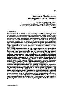
Figure 1. Pathophysiologic Relevance of Cytokines in Chronic Liver Disease. Most types of cells in the liver, including Kupffer cells, hepatocytes, and stellate cells, either synthesize or respond to cytokines. In the early phase of chronic liver disease, specific agents such as viruses, ethanol, and toxins may stimulate the production of cytokines. In the late phase, endotoxin may be the key agent stimulating cytokine production. Clinical features of chronic liver disease that are mediated by cytokines include cachexia, cholestasis, fibrosis, synthesis of acute-phase proteins, and hypergammaglobulinemia. Whereas proinflammatory cytokines such as tumor necrosis factor a (TNF-a) and interleukin-6 are mainly involved in cholestasis and the synthesis of acute-phase proteins, transforming growth factor b (TGF-b) released by activated Kupffer cells and hepatocytes may be one of the critical cytokines involved in fibrosis. In patients with progressive liver disease, the balance between proinflammatory and antiinflammatory cytokines may be shifted toward the proinflammatory axis, thus the counteracting antiinflammatory cytokines are unable to control inflammation and fibrosis.
old for TNF-induced cell death is reduced in hepatocytes that are deficient in these proteins.28 Thus, liver injury requires at least two “hits”: one that increases the exposure of hepatocytes to TNF-a and another that interferes with the hepatocyte’s normal ability to protect itself from TNF-a–induced cell death (Fig. 3).
The ultimate effect of TNF-a on hepatocytes in vivo is strongly influenced by other cytokines in liver tissue. For example, in mice that are deficient in interleukin-6 increased production of TNF-a induced by partial hepatectomy promotes the death of hepatocytes instead of stimulating their proliferation.9 Similarly, interruption of the gene for the antiinflammatoVol ume 343
Numb e r 2 0
Downloaded from www.nejm.org at LSU HEALTH SCIENCES CENTER LIBRARY on March 29, 2006 . Copyright © 2000 Massachusetts Medical Society. All rights reserved.
·
1469
The Ne w E n g l a nd Jo u r n a l o f Me d ic i ne
Proliferation A
Apoptosis
TNF-a
TNF-a
B
Epidermal growth factor
Cell membrane
SEK-1
Caspase 8 Cytoplasm
Jun N-terminal kinase
Apoptosis
Bid Caspase 3
Mitochondria
Nucleus c-Jun Cytochrome c oxidase Proliferation Necrosis C
Survival Factors D
TNF-a
Sphingomyelinase Ceramide Mitochondria
Reactive oxygen species
Necrosis
Lipid peroxidation
TNF-a
Mitochondria Reactive oxygen species
Nuclear factor-kB
Survival factors MnSOD, Bfl-1, Bcl-xL
Adaptor protein 1
Figure 2. Hepatocyte Responses to Tumor Necrosis Factor a (TNF-a). The interaction between TNF-a and its receptors on hepatocyte membranes causes the receptors to aggregate, which leads to the recruitment of various adaptor proteins that activate downstream kinases (Panel A), proteases (e.g., caspase 8) (Panel B), sphingomyelinase (Panel C), and transcription factors that regulate cell-survival factors (Panel D). TNF-a initiates a stress-related protein kinase cascade involving Jun N-terminal kinase kinase 1 (or SEK-1) and Jun N-terminal kinase that culminates in the phosphorylation of the proto-oncogene c-Jun, helping mitogens such as epidermal growth factor to promote proliferation (Panel A). TNF-a activates caspase 8, which cleaves the cytosolic protein Bid, a member of the BH3 subfamily of Bcl-2–related proteins. This truncated form of Bid is redistributed to mitochondrial membranes and permits the release of mitochondrial factors, including cytochrome c oxidase, that activate caspase 3 and cause apoptosis (Panel B). The induction of sphingomyelinase by TNF-a increases ceramide, an inhibitor of the activity of the mitochondrial electron-transport chain, leading to increased mitochondrial production of reactive oxygen species, which in turn promote lipid peroxidation and cellular necrosis (Panel C). TNF-related increases in reactive oxygen species also contribute to the activation of oxidant-sensitive transcription factors (e.g., nuclear factor-kB and adaptor protein 1) that are necessary for the synthesis of cell-survival factors that protect mitochondria (e.g., antiapoptotic proteins such as Bfl-1 and Bcl-xL and oxidant-detoxifying enzymes such as manganese superoxide dismutase [MnSOD]) (Panel D). Thus, hepatocytes respond to TNF-a by modulating signals that can lead to apoptosis and necrosis, as well as signals that promote cellular survival and proliferation.
1470 ·
Novem b er 16 , 2 0 0 0 Downloaded from www.nejm.org at LSU HEALTH SCIENCES CENTER LIBRARY on March 29, 2006 . Copyright © 2000 Massachusetts Medical Society. All rights reserved.
MECH A NIS MS OF D IS EAS E
ry cytokine interleukin-10 exacerbates TNF-mediated liver injury in mice.29 Conversely, mice that are deficient in interleukin-12,30 interferon-g,31 or interleukin-1832 are protected against TNF-a–induced liver damage.
Hepatocyte
Cytokine Studies in Patients with Alcoholic Liver Disease
The translocation of bacterial products from the intestinal lumen to the mesenteric circulation and its lymphatics induces regional and systemic production of TNF-a and other proinflammatory cytokines. Serum concentrations of TNF-a33,34 and several TNFinducible cytokines, such as interleukin-1,35 interleukin-6,36 and interleukin-8,37 are also increased initially in hospitalized patients with alcoholic steatohepatitis and decline during recovery. This finding, coupled with evidence that long-term ingestion of alcohol increases intestinal permeability38 and that patients with the highest serum cytokine concentrations have the highest rate of in-hospital mortality,34,36 indicates that intestinally derived endotoxin and endotoxin-induced cytokines, such as TNF-a, have a role in the pathogenesis of steatohepatitis. Furthermore, serum concentrations of both TNF-a and soluble TNF receptors are correlated with the degree of endotoxemia and the stage of liver disease in patients with alcoholic liver disease.39
Mitochondria
TNF-a
First hit
Mitochondria Increased permeability Increased reactive oxygen species
Adaptation Apoptosis
X
TNF-a and Alcoholic Liver Disease in Animals
The possibility that increased production of TNF-a is the consequence, rather than the cause, of alcoholrelated liver injury cannot be ruled out. Indeed, the former possibility is suggested by evidence that the serum concentrations of various cytokines are increased in patients with acute or chronic liver diseases.39-41 On the other hand, close temporal associations among Kupffer-cell activation, increased transcription of the genes for TNF-a and related cytokines, hepatic inflammation, and liver-cell death have been reported in rodents with alcohol-induced steatohepatitis.42,43 Furthermore, alcohol-associated liver injury is inhibited when the animals are treated with poorly ab-
Second hit
Mitochondria Decreased permeability Decreased reactive oxygen species Partial depolarization
Decreased ATP Figure 3. Two-Hit Model of the Progression of Fatty Liver Disease. The earliest stages of fatty liver disease involve increased exposure of hepatocytes to tumor necrosis factor a (TNF-a). TNF-a initiates various intracellular signals that increase mitochondrial permeability and the release of reactive oxygen species. Left unchecked, these responses promote hepatocyte apoptosis. Fortunately, most healthy hepatocytes use some of the potentially lethal signals (e.g., reactive oxygen species) to activate a multifaceted response that permits the hepatocytes to survive. Secondary insults (second hits) that inhibit this adaptation also result in apoptosis. Even when adaptation is successful and hepatocytes remain viable, they become extremely vulnerable to other insults that partially depolarize the mitochondrial inner membrane, dying by necrosis when the mitochondrial membrane potential collapses.
Viable but vulnerable
Necrosis
Vol ume 343
Numb e r 2 0
Downloaded from www.nejm.org at LSU HEALTH SCIENCES CENTER LIBRARY on March 29, 2006 . Copyright © 2000 Massachusetts Medical Society. All rights reserved.
·
1471
The Ne w E n g l a nd Jo u r n a l o f Me d ic i ne
sorbed oral antibiotics44 or lactobacillus45 in order to decrease endotoxemia or when they are treated with antibodies that neutralize the action of TNF-a.46 The recent finding that exposure to alcohol does not induce steatohepatitis in mice in which the gene for type I TNF receptor is disrupted47 constitutes the best evidence that TNF-a is a key pathogenic factor in alcohol-related liver injury. Even though TNF-a appears to be necessary for the development of alcohol-related steatohepatitis, however, increased production of this cytokine is not sufficient to cause liver injury. The importance of other factors in the pathogenesis of alcoholic liver disease is supported by evidence that the disease is also inhibited in rats treated with gadolinium chloride to deplete the liver of Kupffer cells. After the administration of gadolinium chloride, hepatic and serum concentrations of TNF-a increase dramatically,48 but liver injury does not occur.49 Long-Term Alcohol Ingestion and Hepatocyte Vulnerability to TNF-a
Unlike normal hepatocytes, which are resistant to TNF-a–induced apoptosis, hepatocytes from rats with alcohol-induced fatty livers die quickly after in vitro exposure to small amounts of TNF-a.50 Several mechanisms have been implicated, many of which involve hepatocyte mitochondria, organelles that play a central part in TNF-induced apoptosis and necrosis.51 In alcohol-induced fatty livers, the mitochondria have ultrastructural abnormalities, including swelling and disruption of the inner membrane.52 These anatomical abnormalities are correlated with alcohol-related inhibition of mitochondrial respiration,52 increased generation of mitochondrial oxidants,53 depletion of mitochondrial glutathione,54 and multiple mitochondrial DNA deletions similar to those that occur during aging.55 Although viable hepatocytes may have these mitochondrial abnormalities, they are likely to potentiate vulnerability to TNF-a–induced cell death. The molecular mechanisms involved in cell death remain uncertain. Long-term exposure to alcohol may promote hepatocyte apoptosis56 or necrosis.10,57 Perhaps long-term exposure to alcohol potentiates both processes by depleting antioxidants, such as mitochondrial glutathione, that protect hepatocytes from necrosis and apoptosis,50 while inhibiting the production of nuclear factor-kB, an antiapoptotic transcription factor, in response to increased hepatic production of TNF-a.58 CYTOKINES AND OBESITY-RELATED FATTY LIVER DISEASES Similarities between Alcoholic and Nonalcoholic Fatty Liver Diseases
Obesity and alcohol abuse are associated with the same spectrum of liver diseases.59 The time course for the progression of disease and the relative risk of cirrhosis are also similar in obesity-related liver dis1472 ·
ease and alcoholic liver disease.60 The similar pathologic features and natural histories of the two types of fatty liver disease suggest that common pathogenic mechanisms may be involved. Distinct, but mutually enhancing, mechanisms may also be involved, because a fatty liver is more common in patients with obesity and alcohol abuse than in those with either alone. Moreover, obesity is an independent risk factor for cirrhosis in patients who abuse alcohol.11 Thus, elucidation of the mechanisms that cause obesity-related fatty liver disease is likely to clarify the role of pluripotent cytokines, such as TNF-a, in promoting alcohol-induced liver damage. Liver Disease in Genetically Obese Rats and Mice
Studies of genetically obese ob/ob mice and fa/fa rats have provided information about the pathogenesis of obesity-related fatty liver disease. Both rodent strains have spontaneous mutations that either diminish production of the appetite-suppressing hormone leptin (in ob/ob mice) or inactivate the leptin receptor (in fa/fa rats).61,62 Like many obese humans, obese ob/ob mice and fa/fa rats have insulin resistance, hyperglycemia, hyperlipidemia, and fatty livers.63 These rodents also have several immunologic abnormalities, including phagocyte dysfunction, altered transcription of cytokine genes (including constitutive increases in TNF-a), and excessive production of prostanoids by hepatic and peritoneal macrophages.64-66 Just as abnormalities of hepatocyte mitochondria have been found in alcohol-induced fatty livers, ultrastructural and functional alterations of hepatocyte mitochondria have been found in the fatty livers of ob/ob mice.67,68 Also, ob/ob mice with fatty livers, like patients and rodents with alcohol-induced fatty livers, have little evidence of associated hepatitis until they are subjected to another insult.69 Nevertheless, even in ob/ob mice with early fatty liver disease, hepatocytes synthesize both proapoptotic and antiapoptotic proteins that are not detected in hepatocytes from normal mice,69 suggesting that apoptotic stress contributes to obesity-related fatty liver disease. Hepatic accumulation of fat may be the simplest explanation of obesity-related hepatomegaly, but the administration of drugs that stimulate the proliferation of peroxisomes also increases the ratio of liver weight to body weight in mice.70 Hepatomegaly occurs because the rate of hepatocyte apoptosis becomes insufficient to match the rate of proliferation of hepatocytes. Drugs that stimulate peroxisome-proliferator–activated receptor g, such as thiozolidinediones, stimulate the production of mitochondrial uncoupling protein in adipocytes.71 Although uncoupling protein is barely detectable in hepatocytes from normal adults,72 hepatic synthesis of uncoupling protein 2 increases after the induction of peroxisome-proliferator–activated receptor a in hepatocytes.73 The rate of production and activity of uncoupling protein 2
Novem b er 16 , 2 0 0 0 Downloaded from www.nejm.org at LSU HEALTH SCIENCES CENTER LIBRARY on March 29, 2006 . Copyright © 2000 Massachusetts Medical Society. All rights reserved.
MECH A NIS MS OF D IS EAS E
are also increased in hepatocytes from obese ob/ob mice.68 Increased uncoupling protein 2 in mitochondria depolarizes the inner mitochondrial membrane, increasing the activity of the electron-transport chain while limiting the production of superoxide anion and the accumulation of calcium.74 Thus, synthesis of uncoupling protein 2 in a fatty liver may help inhibit hepatocyte apoptosis,75 and this may explain why the activation of peroxisome-proliferator–activated receptor a increases hepatocyte survival. However, because cells with increased uncoupling-protein activity have partially depolarized mitochondria, they may also be more vulnerable to loss of the mitochondrial innermembrane potential, with consequent depletion of ATP and necrosis, if exposed to certain secondary insults such as endotoxin or TNF-a.64,68,76,77 Increased production of uncoupling protein 2 has been reported in hepatocytes from some patients with nonalcoholic steatohepatitis or alcoholic hepatitis.78 Although it remains uncertain whether uncoupling protein 2 contributes to the pathogenesis of these diseases, both animals and patients with alcohol-induced liver disease or nonalcoholic steatohepatitis have a diminished capacity to replenish liver ATP stores after transient, experimentally induced depletion of ATP.68,79 Thus, uncoupling protein 2 may be one component of a general adaptive response that preserves the viability of hepatocytes in fatty livers but also increases the vulnerability of these cells to subsequent insults. TNF-a AND PROGRESSION FROM STEATOHEPATITIS TO CIRRHOSIS
Transient reconfiguration of the extracellular matrix of the injured liver, which permits the infiltration of inflammatory cells, facilitates the local accumulation of growth factors, and accommodates regenerating hepatocytes, is a critical component of healing after liver injury.80 After acute liver injury from various insults — for example, toxins, viral infections, or surgical trauma — the resorption and deposition of the components of the matrix are balanced, and therefore there is no accumulation of fibrous tissue. However, chronic inflammatory conditions, such as alcoholic and nonalcoholic steatohepatitis, alter the composition of the matrix and upset the balance between the synthesis and degradation of the matrix. Consequently, fibrosis occurs, compromising portal venous blood flow, which in turn both compromises hepatic regeneration and promotes the portosystemic shunting of blood that leads to some of the clinical manifestations of advanced liver disease.81 TNF-a is involved in the pathogenesis of cirrhosis, but additional factors must also be present.6 Otherwise, cirrhosis would be a universal consequence of liver injury. Although there is little doubt that cirrhosis is one of the most clinically relevant outcomes of steatohepatitis, a detailed discussion of the mechanisms that regulate the activation of stellate cells,
matrix-gene expression, and matrix remodeling is beyond the scope of this review. TNF-a promotes each of these responses.82,83 The remaining challenge is to differentiate the cellular and environmental factors that interact with TNF-a to promote normal matrix remodeling from the factors that promote fibrosis. Although the mechanisms are not well understood, steatohepatitis modifies the response of hepatic stellate cells to injury-related cytokines so that both transcriptional and post-transcriptional mechanisms that promote the deposition of type 1 collagen are induced preferentially.82,83 In addition, the microenvironment becomes more conducive to the survival of stellate cells, because the number of stellate cells increases as cirrhosis evolves.6 Moreover, even after cirrhosis is well established, the ongoing production of TNF-a and related inflammatory cytokines modulates the expression of enzymes, such as inducible nitric oxide synthase, that regulate the production of vasoactive molecules, which mediate the hemodynamic abnormalities of cirrhosis, such as portosystemic shunting and the hepatorenal syndrome.84,85 INHIBITION OF TNF-a AND TREATMENT OF STEATOHEPATITIS
Current treatments for liver disease related to use of alcohol or obesity focus on reducing alcohol intake and obesity, respectively. Abstinence from alcohol prolongs the survival of patients with alcoholic liver disease, whether or not they have cirrhosis, but usually does not prevent the progression to cirrhosis.11,15 Although there is some evidence that abstinence improves the regenerative capacity of the liver in patients with cirrhosis, once cirrhosis has developed, it persists despite the cessation of alcohol use. Similarly, weight loss is an imperfect cure for obesityrelated liver disease. Weight reduction may decrease serum aminotransferase concentrations in obese patients,86 but rapid or extreme weight loss appears to promote the transition from steatosis to cirrhosis.87 Thus, although there is little doubt that obesity is a risk factor for liver disease, weight reduction is not always an effective treatment and may even exacerbate the disease. On the basis of data from studies of alcohol- and obesity-related liver disease in animals, alternative therapies have been proposed. Treatment with various antioxidants (Table 2) decreases alcohol-induced liver damage in rats11,88-91 and improves the fatty liver that develops in rats fed choline- and methioninedeficient diets,92 but so far, the benefits have been inconsistent in the small groups of patients with alcoholic liver disease who have been treated with similar agents.93-97 Few clinical trials of putative antioxidants have been performed in patients with nonalcoholic steatohepatitis.97,98 However, evidence that TNF-a mediates insulin resistance in ob/ob mice with fatty livers99 and the strong correlation between type 2 diVol ume 343
Numb e r 2 0
Downloaded from www.nejm.org at LSU HEALTH SCIENCES CENTER LIBRARY on March 29, 2006 . Copyright © 2000 Massachusetts Medical Society. All rights reserved.
·
1473
The Ne w E n g l a nd Jo u r n a l o f Me d ic i ne
TABLE 2. POTENTIALLY BENEFICIAL TREATMENTS IN ANIMALS OR CLINICAL TRIALS.*
IN
STUDIES
THERAPY
PURPOSE
Antibiotics, lactobacillus
Prevent gut-derived endotoxemia Inhibit TNF-a activity Inhibit inflammation Decrease production of reactive oxygen species Reduce liver lipids Improve insulin sensitivity
Anti-TNF antibodies, TNF-receptor antagonists Glucocorticoids Glutathione prodrugs (S-adenosylmethionine, betaine, choline), vitamin E, silymarin, propylthiouracil Ursodiol, gemfibrozil Leptin, thiozolidinediones
*TNF denotes tumor necrosis factor.
gression of liver diseases. The importance of cytokines as effector molecules in liver damage has been particularly well demonstrated in patients and animals with alcoholic or nonalcoholic liver diseases ranging from steatosis to cirrhosis. Thus, antagonism of TNF-a and other injury-related cytokines merits evaluation as a treatment for these diseases. However, since the same cytokines are also necessary for the regeneration of tissue after the liver has been injured, cytokine antagonism is not without risk in patients with liver disease, and complete inhibition of TNF-a activity might impair hepatic recovery. The cellular signals that mediate the various actions of TNF-a must be delineated so that new therapeutic agents can be developed to inhibit TNF-initiated cell-death signals preferentially. Supported by grants from the Austrian Science Fund (P 12790) and the National Institutes of Health (RO1 AA10154 and DK53792).
abetes and nonalcoholic steatohepatitis have generated support for trials of insulin-sensitizing drugs. To date, this approach has proved effective only in transgenic mice with steatohepatitis. Mice with adipocyte-targeted overexpression of active sterol regulatory element–binding protein (SREP) 1c are born with lipodystrophy, and severe insulin resistance and steatohepatitis develop in these animals at a young age.101 They have a paucity of white adipose tissue and are therefore deficient in adipose gene products, such as peroxisome-proliferator–activated receptor g and leptin.101 Treatment of SREP transgenic mice with leptin improves both their insulin resistance and steatohepatitis.102 Evidence of the important role that TNF-a has in animals with steatohepatitis suggests that TNF-a is another potential therapeutic target in patients with the disease. Various treatments that decrease the absorption of intestinal endotoxin44,45 or inhibit the activity of TNF-a,46,47 an endotoxin-inducible cytokine, prevent alcohol-induced liver disease in animals. Treatment with glucocorticoids has been shown to be effective in carefully selected patients with severe, acute alcohol-induced steatohepatitis (excluding those with active bacterial infections, insulin-requiring diabetes, active gastrointestinal bleeding, or acute pancreatitis).16 No trials of other antiinflammatory or anticytokine therapies in patients with alcoholic or obesity-related liver disease have been reported. 100
CONCLUSIONS
Endotoxin-inducible cytokines such as TNF-a, TNF-regulated cytokines, and cytokines that modulate the synthesis and biologic actions of TNF are produced by many cells within the liver and other organs. This extensive cytokine network mediates various aspects of liver injury and repair. Consequently, cytokines ultimately control the pathophysiology and pro1474 ·
We are indebted to Dr. C.A. Dinarello, University of Colorado Health Sciences Center, Denver, and Dr. D. Schuppan, University of Erlangen, Erlangen, Germany, for their critical reading of the manuscript; and to Dr. A. Kaser, University Hospital Innsbruck, Innsbruck, Austria, for helpful comments.
REFERENCES 1. Dinarello CA. Biologic basis for interleukin-1 in disease. Blood 1996; 87:2095-147. 2. Tracey KJ, Cerami A. Tumor necrosis factor, other cytokines and disease. Annu Rev Cell Biol 1993;9:317-43. 3. Mizuhara H, O’Neill E, Seki N, et al. T cell activation-associated hepatic injury: mediation by tumor necrosis factors and protection by interleukin-6. J Exp Med 1994;179:1529-37. 4. Orange JS, Salazar-Mather TP, Opal SM, Biron CA. Mechanisms for virus-induced liver disease: tumor necrosis factor-mediated pathology independent of natural killer and T cells during murine cytomegalovirus infection. J Virol 1997;71:9248-58. 5. Trauner M, Meier PJ, Boyer JL. Molecular pathogenesis of cholestasis. N Engl J Med 1998;339:1217-27. 6. Friedman SL. Molecular regulation of hepatic fibrosis, an integrated cellular response to tissue injury. J Biol Chem 2000;275:2247-50. 7. Akerman P, Cote P, Yang SQ, et al. Antibodies to tumor necrosis factoralpha inhibit liver regeneration after partial hepatectomy. Am J Physiol 1992;263:G579-G585. 8. Yamada Y, Kirillova I, Peschon JJ, Fausto N. Initiation of liver growth by tumor necrosis factor: deficient liver regeneration in mice lacking type I tumor necrosis factor receptor. Proc Natl Acad Sci U S A 1997;94:1441-6. 9. Cressman DE, Greenbaum LE, DeAngelis RA, et al. Liver failure and defective hepatocyte regeneration in interleukin-6-deficient mice. Science 1996;274:1379-83. 10. MacSween RNM, Burt AD. Histologic spectrum of alcoholic liver disease. Semin Liver Dis 1986;6:221-32. 11. Lieber CS. Alcoholic liver disease: new insights in pathogenesis lead to new treatments. J Hepatol 2000;32:Suppl:113-28. 12. Crabb DW. Ethanol oxidizing enzymes: roles in alcohol metabolism and alcoholic liver disease. Prog Liver Dis 1995;13:151-72. 13. Marsano LS, Pena LR. The interaction of alcoholic liver disease and hepatitis C. Hepatogastroenterology 1998;45:331-9. 14. McClain CJ, Hill DB, Schmidt J, Diehl AM. Cytokines and alcoholic liver disease. Semin Liver Dis 1993;13:170-82. 15. Pares A, Caballeria J, Bruguera M, Torres M, Rodes J. Histologic course of alcoholic hepatitis: influence of abstinence, sex and extent of hepatic damage. J Hepatol 1986;2:33-42. 16. Imperiale TF, McCullough AJ. Do corticosteroids reduce mortality from alcoholic hepatitis? A meta-analysis of the randomized trials. Ann Intern Med 1990;113:299-307. 17. Decker K. Biologically active products of stimulated liver macrophages (Kupffer cells). Eur J Biochem 1990;192:245-61. 18. Trautwein C, Rakemann T, Niehof M, Rose-John S, Manns MP. Acute-phase response factor, increased binding, and target gene transcription during liver regeneration. Gastroenterology 1996;110:1854-62.
Novem b er 16 , 2 0 0 0 Downloaded from www.nejm.org at LSU HEALTH SCIENCES CENTER LIBRARY on March 29, 2006 . Copyright © 2000 Massachusetts Medical Society. All rights reserved.
MECH A NIS MS OF D IS EAS E
19. Feingold KR , Soued M, Grunfeld C. Tumor necrosis factor stimulates DNA synthesis in the liver of intact rats. Biochem Biophys Res Commun 1988;153:576-82. 20. Diehl AM, Yin M, Fleckenstein J, et al. Tumor necrosis factor-alpha induces c-jun during the regenerative response to liver injury. Am J Physiol 1994;267:G552-G561. 21. Magnusson C, Vaux DL. Signalling by CD95 and TNF receptors: not only life and death. Immunol Cell Biol 1999;77:41-6. 22. Schulze-Osthoff K, Bakker AC, Vanhaesebroeck B, Beyaert R, Jacob WA, Fiers W. Cytotoxic activity of tumor necrosis factor is mediated by early damage of mitochondrial functions: evidence for the involvement of mitochondrial radical generation. J Biol Chem 1992;267:5317-23. 23. Higuchi M, Aggarwal BB, Yeh ET. Activation of CPP32-like protease in tumor necrosis factor-induced apoptosis is dependent on mitochondrial function. J Clin Invest 1997;99:1751-8. 24. Leist M, Gantner F, Bohlinger I, Germann PC, Tiegs G, Wendel A. Murine hepatocyte apoptosis induced in vitro and in vivo by TNF-alpha requires transcriptional arrest. J Immunol 1994;153:1778-88. 25. Bellas RE, FitzGerald MJ, Fausto N, Sonenshein GE. Inhibition of NF-kappa B activity induces apoptosis in murine hepatocytes. Am J Pathol 1997;151:891-6. 26. Manna SK, Zhang HJ, Yan T, Oberley LW, Aggarwal BB. Overexpression of manganese superoxide dismutase suppresses tumor necrosis factorinduced apoptosis and activation of nuclear transcription factor-kappaB and activated protein-1. J Biol Chem 1998;273:13245-54. 27. Kim YM, Talanian RV, Billiar TR. Nitric oxide inhibits apoptosis by preventing increases in caspase-3-like activity via two distinct mechanisms. J Biol Chem 1997;272:31138-48. 28. Rai RM, Lee FYJ, Rosen A, et al. Impaired liver regeneration in inducible nitric oxide synthase-deficient mice. Proc Natl Acad Sci U S A 1998;95:13829-34. 29. Berg DJ, Kuhn R, Rajewsky K, et al. Interleukin-10 is a central regulator of the response to LPS in murine models of endotoxic shock and the Shwartzman reaction but not endotoxin tolerance. J Clin Invest 1995;96: 2339-47. 30. Tanaka Y, Takahashi A, Watanabe K, et al. A pivotal role of IL-12 in Th1-dependent mouse liver injury. Int Immunol 1996;8:569-76. 31. Car BD, Eng VM, Schnyder B, et al. Interferon gamma receptor deficient mice are resistant to endotoxic shock. J Exp Med 1994;179:143744. 32. Sakao Y, Takeda K, Tsutsui H, et al. IL-18-deficient mice are resistant to endotoxin-induced liver injury but highly susceptible to endotoxin shock. Int Immunol 1999;11:471-80. 33. Bird GLA, Sheron N, Goka AKJ, Alexander GL, Williams RS. Increased plasma tumor necrosis factor in severe alcoholic hepatitis. Ann Intern Med 1990;112:917-20. 34. Felver ME, Mezey E, McGuire M, et al. Plasma tumor necrosis factor alpha predicts decreased long-term survival in severe alcoholic hepatitis. Alcohol Clin Exp Res 1990;14:255-9. 35. McClain CJ, Cohen DA, Dinarello CA, Cannon JG, Shedlofsky SI, Kaplan AM. Serum interleukin-1 (IL-1) activity in alcoholic hepatitis. Life Sci 1986;39:1479-85. 36. Hill DB, Marsano L, Cohen D, Allen J, Shedlofsky S, McClain CJ. Increased plasma interleukin-6 concentrations in alcoholic hepatitis. J Lab Clin Med 1992;119:547-52. 37. Sheron N, Bird G, Koskinas J, et al. Circulating and tissue levels of the neutrophil chemotaxin interleukin-8 are elevated in severe acute alcoholic hepatitis, and tissue levels correlate with neutrophil infiltration. Hepatology 1993;18:41-6. 38. Fukui H, Brauner B, Bode JC, Bode C. Plasma endotoxin concentrations in patients with alcoholic and non-alcoholic liver disease: reevaluation with an improved chromogenic assay. Hepatology 1991;12:162-9. 39. Hanck C, Rossol S, Bocker U, Tokus M, Singer MV. Presence of plasma endotoxin is correlated with tumour necrosis factor receptor levels and disease activity in alcoholic cirrhosis. Alcohol Alcohol 1998;33:606-8. 40. Tilg H, Wilmer A, Vogel W, et al. Serum levels of cytokines in chronic liver diseases. Gastroenterology 1992;103:264-74. 41. Tilg H, Vogel W, Wiedermann CJ, et al. Circulating interleukin-1 and tumor necrosis factor antagonists in liver disease. Hepatology 1993;18: 1132-8. 42. Tsukamoto H, Towner SJ, Ciofalo LM, French SW. Ethanol-induced liver fibrosis in rats fed high fat diets. Hepatology 1986;6:814-22. 43. Nanji AA, Khettry U, Sadrzadeh SMH, Tamanaka T. Severity of liver injury in experimental alcoholic liver disease: correlation with plasma endotoxin, prostaglandin E2, leukotriene B4, and thromboxane B2. Am J Pathol 1993;142:367-73. 44. Adachi Y, Moore LE, Bradford BU, Gao W, Thurman RG. Antibiotics prevent liver injury in rats following long-term exposure to ethanol. Gastroenterology 1995;108:218-24. 45. Nanji AA, Khettry U, Sadrzadeh SM. Lactobacillus feeding reduces
endotoxemia and severity of experimental alcoholic liver (disease). Proc Soc Exp Biol Med 1994;205:243-7. 46. Iimuro Y, Gallucci RM, Luster MI, Kono H, Thurman RG. Antibodies to tumor necrosis factor alfa attenuate hepatic necrosis and inflammation caused by chronic exposure to ethanol in the rat. Hepatology 1997; 26:1530-7. 47. Yin M, Wheeler MD, Kono H, et al. Essential role of tumor necrosis factor alpha in alcohol-induced liver injury in mice. Gastroenterology 1999;117:942-52. 48. Rai RM, Loffreda S, Karp CL, Yang SQ, Lin HZ, Diehl AM. Kupffer cell depletion abolishes induction of interleukin-10 and permits sustained overexpression of tumor necrosis factor alpha messenger RNA in the regenerating rat liver. Hepatology 1997;25:889-95. 49. Rai RM, Yang SQ, McClain C, Karp CL, Klein AS, Diehl AM. Kupffer cell depletion by gadolinium chloride enhances liver regeneration after partial hepatectomy in rats. Am J Physiol 1996;270:G909-G918. 50. Fernandez-Checa JC, Kaplowitz N, Garcia-Ruiz C, et al. GSH transport in mitochondria: defense against TNF-induced oxidative stress and alcohol-induced defect. Am J Physiol 1997;273:G7-G17. 51. Kroemer G, Dallaporta B, Resche-Rignon M. The mitochondrial death/life regulator in apoptosis and necrosis. Annu Rev Physiol 1998;60: 619-42. 52. Bruguera M, Bertran A, Bombi JA, Rodes J. Giant mitochondria in hepatocytes: a diagnostic hint for alcoholic liver disease. Gastroenterology 1977;73:1383-7. 53. Pitkanen S, Robinson BH. Mitochondrial complex I deficiency leads to increased production of superoxide radicals and induction of superoxide dismutase. J Clin Invest 1996;98:345-51. 54. Garcia-Ruiz C, Morales A, Ballesta A, Rodes J, Kaplowitz N, Fernandez-Checa JC. Effect of chronic ethanol feeding on glutathione and functional integrity of mitochondria in periportal perivenous rat hepatocytes. J Clin Invest 1994;94:193-201. 55. Fromenty B, Pessayre D. Impaired mitochondrial function in microvesicular steatosis: effects of drugs, ethanol, hormones and cytokines. J Hepatol 1997;26:Suppl 2:43-53. 56. Benedetti A, Brunelli E, Risicato R, Cilluffo T, Jezequel AM, Orlandi F. Subcellular changes and apoptosis induced by ethanol in rat liver. J Hepatol 1988;6:137-43. 57. Nanji AA, Zhao S, Sadrzadeh SMH, Dannenberg AJ, Tahan SR , Waxman DJ. Markedly enhanced cytochrome P450 2E1 induction and lipid peroxidation is associated with severe liver injury in fish oil-ethanol-fed rats. Alcohol Clin Exp Res 1994;18:1280-5. 58. Zeldin G, Yang SQ, Yin M, Lin HZ, Rai R, Diehl AM. Alcohol and cytokine-inducible transcription factors. Alcohol Clin Exp Res 1996;20: 1639-45. 59. Sheth SG, Gordon FD, Chopra S. Nonalcoholic steatohepatitis. Ann Intern Med 1997;126:137-45. [Erratum, Ann Intern Med 1997;127:658.] 60. Matteoni C, Younossi ZM, Gramlich T, Boparai N, Liu YC, McCullough AJ. Nonalcoholic fatty liver disease: a spectrum of clinical and pathological severity. Gastroenterology 1999;116:1413-9. 61. Pelleymounter MA, Cullen MJ, Baker MB, et al. Effects of the obese gene product on body weight regulation in ob/ob mice. Science 1995;269: 540-3. 62. Phillips MS, Liu Q, Hammond HA, et al. Leptin receptor missense mutation in the fatty Zucker rat. Nat Genet 1996;13:18-9. 63. Shulman GI. Cellular mechanisms of insulin resistance in humans. Am J Cardiol 1999;84:3J-10J. 64. Yang SQ, Lin HZ, Lane MD, Clemens M, Diehl AM. Obesity increases sensitivity to endotoxin liver injury: implications for the pathogenesis of steatohepatitis. Proc Natl Acad Sci U S A 1997;94:2557-62. 65. Loffreda S, Yang SQ, Lin HZ, et al. Leptin regulates proinflammatory immune responses. FASEB J 1998;12:57-65. 66. Lee F-Y, Li Y, Yang EK, et al. Phenotypic abnormalities in macrophages from leptin-deficient, obese mice. Am J Physiol 1999;276:C386-C394. 67. Katyare SS, Howland JL. Enhanced oxidative metabolism in liver mitochondria from genetically obese mice. Arch Biochem Biophys 1978;188: 15-20. 68. Chavin KD, Yang SQ, Lin HZ, et al. Obesity induces expression of uncoupling protein-2 in hepatocytes and promotes liver ATP depletion. J Biol Chem 1999;274:5692-700. 69. Rashid A, Wu T-C, Huang CC, et al. Mitochondrial proteins that regulate apoptosis and necrosis are induced in mouse fatty liver. Hepatology 1999;29:1131-8. 70. Roberts RA, James NH, Woodyatt NJ, Macdonald N, Tugwood JD. Evidence for the suppression of apoptosis by the peroxisome proliferator activated receptor alpha (PPAR alpha). Carcinogenesis 1998;19:43-8. 71. Aubert J, Champigny O, Saint-Marc P, et al. Up-regulation of UCP-2 gene expression by PPAR agonists in preadipose and adipose cells. Biochem Biophys Res Commun 1997;238:606-11. 72. Fleury C, Neverova M, Collins S, et al. Uncoupling protein-2: a novel
Vol ume 343
Numb e r 2 0
Downloaded from www.nejm.org at LSU HEALTH SCIENCES CENTER LIBRARY on March 29, 2006 . Copyright © 2000 Massachusetts Medical Society. All rights reserved.
·
1475
The Ne w E n g l a nd Jo u r n a l o f Me d ic i ne
gene linked to obesity and hyperinsulinemia. Nat Genet 1997;15:26972. 73. Kelly LJ, Vicario PP, Thompson GM, et al. Peroxisome proliferatoractivated receptors gamma and alpha mediate in vivo regulation of uncoupling protein (UCP-1, UCP-2, UCP-3) gene expression. Endocrinology 1998;139:4920-7. 74. Boss O, Muzzin P, Giacobino JP. The uncoupling proteins, a review. Eur J Endocrinol 1998;139:1-9. 75. Diehl AM, Hoek JB. Mitochondrial uncoupling: role of uncoupling protein anion carriers and relationship to thermogenesis and weight control “the benefits of losing control.” J Bioenerg Biomemb 1999;31:493506. 76. Faggioni R , Fantuzzi G, Gabay C, et al. Leptin deficiency enhances sensitivity to endotoxin-induced lethality. Am J Physiol 1999;276:R136R142. 77. Takahashi N, Waelput W, Guisez Y. Leptin is an endogenous protective protein against the toxicity exerted by tumor necrosis factor. J Exp Med 1999;189:207-12. 78. Rashid A, Koteish AK, Cortez-Pinto H, et al. Subcellular localization of uncoupling protein (UCP)2 and evidence for increased UCP2 expression in nonalcoholic steatohepatitis. Hepatology 1999;30:Suppl:405A. abstract. 79. Cortez-Pinto H, Chatham J, Chacko VP, Arnold C, Rashid A, Diehl AM. Alterations in liver ATP homeostasis in human nonalcoholic steatohepatitis: a pilot study. JAMA 1999;282:1659-64. 80. Michalopoulos GK, DeFrances MC. Liver regeneration. Science 1997; 276:60-6. 81. Bosch J, Garcia-Pagan JC. Complications of cirrhosis. I. Portal hypertension. J Hepatol 2000;32:Suppl:141-56. 82. Brenner DA, O’Hara M, Angel P, Chojkier M, Karin M. Prolonged activation of jun and collagenase genes by tumor necrosis factor-alpha. Nature 1989;337:661-3. 83. Houglum K, Buck M, Adir V, Chojkier M. LAP (NF-IL6) transactivates the collagen alpha 1(I) gene from a 5' regulatory region. J Clin Invest 1994;94:808-14. 84. Wiest R , Groszmann RJ. Nitric oxide and portal hypertension: its role in the regulation of intrahepatic and splanchnic vascular resistance. Semin Liver Dis 1999;19:411-26. 85. Martin P-Y, Ginès P, Schrier RW. Nitric oxide as a mediator of hemodynamic abnormalities and sodium and water retention in cirrhosis. N Engl J Med 1998;339:533-41. 86. Palmer M, Schaffner F. Effect of weight reduction on hepatic abnormalities in overweight patients. Gastroenterology 1990;99:1408-13. 87. Capron JP, Delamarre J, Dupas JL, Braillon A, Degott C, Quenum C. Fasting in obesity: another cause of liver injury with alcoholic hyaline? Dig Dis Sci 1982;27:265-8.
1476 ·
88. Feo F, Pascale R, Garcea R, et al. Effect of the variations of S-adenosyl-L-methionine liver content on fat accumulation and ethanol metabolism in ethanol-intoxicated rats. Toxicol Appl Pharmacol 1986;83:331-41. 89. Garcia-Ruiz C, Morales A, Colell A, et al. Feeding S-adenosylL-methionine attenuates both ethanol-induced depletion of mitochondrial glutathione and mitochondrial dysfunction in periportal and perivenous rat hepatocytes. Hepatology 1995;21:207-14. 90. Nanji AA, Yang EK, Fogt F, Sadrzadeh SM, Dannenberg AJ. Medium chain triglycerides and vitamin E reduce the severity of established experimental alcoholic disease. J Pharmacol Exp Ther 1996;277:1694-700. 91. Barak AJ, Beckenhauer HC, Badakhsh S, Tuma DJ. The effect of betaine in reversing alcoholic steatosis. Alcohol Clin Exp Res 1997;21:1100-2. 92. Weltman MD, Farrell GC, Liddle C. Increased hepatocyte CYP2E1 expression in a rat nutritional model of hepatic steatosis with inflammation. Gastroenterology 1996;111:1645-53. 93. Orrego H, Blake JE, Blendis LM, Compton KV, Israel Y. Long-term treatment of alcoholic liver disease with propylthiouracil. N Engl J Med 1987;317:1421-7. 94. Ferenci P, Dragosics B, Dittrich H, et al. Randomized controlled trial of silymarin treatment in patients with cirrhosis of the liver. J Hepatol 1989;9:105-13. 95. Vendemiale G, Altomare E, Trizio T, et al. Effects of oral S-adenosylL-methionine on hepatic gluthathione in patients with liver disease. Scand J Gastroenterol 1989;24:407-15. 96. de la Maza MP, Petermann M, Bunout D, Hirsch S. Effects of longterm vitamin E supplementation in alcoholic cirrhotics. J Am Coll Nutr 1995;14:192-6. 97. Velussi M, Cernigoi AM, De Monte A, Dapas F, Caffau C, Zilli M. Long-term (12 months) treatment with an anti-oxidant drug (silymarin) is effective on hyperinsulinemia, exogenous insulin need and malondialdehyde levels in cirrhotic diabetic patients. J Hepatol 1997;26:871-9. 98. Abdelmalek M, Ludwig J, Lindor KD. Two cases from the spectrum of nonalcoholic steatohepatitis. J Clin Gastroenterol 1995;20:127-30. 99. Uysal KT, Wiesbrock SM, Marino MW, Hotamisligil GS. Protection from obesity-induced insulin resistance in mice lacking TNF-alpha function. Nature 1997;389:610-4. 100. Marchesini G, Brizi M, Moreselli-Labate AM, et al. Association of nonalcoholic fatty liver disease with insulin resistance. Am J Med 1999; 107:450-5. 101. Shimomura I, Hammer RE, Richardson JA, et al. Insulin resistance and diabetes mellitus in transgenic mice expressing nuclear SREBP-1c in adipose tissue: model for congenital generalized lipodystrophy. Genes Dev 1998;12:3182-94. 102. Shimomura I, Hammer RE, Ikemoto S, Brown MS, Goldstein JL. Leptin reverses insulin resistance and diabetes mellitus in mice with congenital lipodystrophy. Nature 1999;401:73-6.
Novem b er 16 , 2 0 0 0 Downloaded from www.nejm.org at LSU HEALTH SCIENCES CENTER LIBRARY on March 29, 2006 . Copyright © 2000 Massachusetts Medical Society. All rights reserved.