THE JOURNAL OF BIOLOGICAL CHEMISTRY © 2003 by The American Society for Biochemistry and Molecular Biology, Inc.
Vol. 278, No. 14, Issue of April 4, pp. 11888 –11896, 2003 Printed in U.S.A.
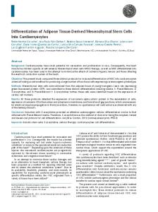
Matrix Metalloproteinases Are Differentially Expressed in Adipose Tissue during Obesity and Modulate Adipocyte Differentiation* Received for publication, September 9, 2002, and in revised form, December 27, 2002 Published, JBC Papers in Press, January 15, 2003, DOI 10.1074/jbc.M209196200
Carine Chavey‡§, Bernard Mari¶, Marie-Noe¨lle Monthouel‡, Ste´phanie Bonnafous‡, Patrick Anglard储, Emmanuel Van Obberghen‡, and Sophie Tartare-Deckert‡** From ‡INSERM, Unite´ 145, Institut Fe´de´ratif de Recherche (IFR) 50, 06107 Nice, France, ¶INSERM, Unite´ 526, IFR 50, 06107 Nice, France, and 储Institut de Ge´ne´tique et de Biologie Mole´culaire, Unite´ Mixte de Recherche 7104, 67404 Illkirch, France
Matrix metalloproteinases (MMPs) are essential for proper extracellular matrix remodeling, a process that takes place during obesity-mediated adipose tissue formation. Here, we examine expression profiles and the potential role of MMPs and their tissue inhibitors (TIMPs) in adipose tissue remodeling during obesity. Expression patterns are studied by Northern blot and real-time PCR in two genetic models of obesity (ob/ob and db/db mice) and in a diet-induced model of obesity (AKR mice). Of the MMPs and TIMPs studied, mRNA levels for MMP-2, MMP-3, MMP-12, MMP-14, MMP-19, and TIMP-1 are strongly induced in obese adipose tissues compared with lean tissues. In contrast, MMP-7 and TIMP-3 mRNAs are markedly decreased in obesity. Interestingly, enzymatic activities of MMP-12 and of a new identified adipocyte-derived 30-kDa metalloproteinase are enhanced in obese adipose tissue fractions, demonstrating that MMP/TIMP balance is shifted toward increased matrix degradation in obesity. Finally, we analyze the modulation of MMP-2, MMP-19, and TIMP-1 during 3T3-L1 preadipocyte differentiation, and we explore the effect of inhibition of MMP activity on in vitro adipogenesis. We find that the synthetic MMP inhibitor BB-94 (Batimastat) decreases adipose conversion of 3T3-L1 and primary rat preadipocytes. BB-94 represses differentiation without affecting mitotic clonal expansion but prevents the early expression of CCAAT/enhancer-binding protein , a transcription factor that is thought to play a major role in the adipogenic program. Such findings support a role for the MMP/TIMP system in the control of proteolytic events and adipogenesis during obesity-mediated fat mass development.
Obesity is a nutritional disorder characterized by excess of adipose tissue (1). Adipose tissue mass is a reflection of the number of adipocytes and their amount of fat stored. The development of obesity is associated with coordinated cellular processes, including adipocyte hypertrophy followed by recruitment of adipocyte precursors, and new fat cell differentiation * This work was supported in part by INSERM, the Association pour la Recherche sur le Cancer, and a grant from Groupe Merck-Lipha (Lyon, France). The costs of publication of this article were defrayed in part by the payment of page charges. This article must therefore be hereby marked “advertisement” in accordance with 18 U.S.C. Section 1734 solely to indicate this fact. § Recipient of a doctoral fellowship from the Ministe`re de l’Enseignement Supe´rieur et de la Recherche (France). ** To whom correspondence should be addressed: INSERM Unite´ 385, Faculte´ de Me´decine, ave. de Valombrose, 06107 Nice Ce´dex 2, France. Tel.: 33-4-93-37-77-90; Fax: 33-4-93-81-14-04; E-mail: tartare@ unice.fr.
(2, 3). These processes are also accompanied by neovascularization that is essential for generation and proper function of the tissue (4). It is generally accepted that such multiple events include dynamic changes of cell-matrix interactions and extensive extracellular matrix (ECM)1 remodeling, and that modifications in proteolytic activities within the adipose microenvironment might occur during the development of the fat depot. Although numerous studies have examined the regulation and intracellular events that occur during adipogenesis, only limited information is available about molecular mechanisms underlying ECM remodeling, proteolytic events, and cell-matrix interactions during obesity-related fat mass development. In a course of differential gene expression analysis between lean and obese adipose tissues, we recently identified osteonectin/ SPARC, a protein involved in dynamic interactions between cell and matrix, up-regulated in obesity (5). This suggested that alteration in the expression level of matrix proteins may contribute to the development of obesity-associated adipose tissue growth. A balance between the opposing activities of proteinases and their inhibitors controls pericellular proteolytic events. Among enzymes implicated in the degradation of matrix molecules and in the generation of bioactive factors, the matrix metalloproteinase (MMP) family is considered to be primarily responsible for these processes (6). MMPs comprise a large family of structurally related zinc-dependent proteinases that has been classified into subgroups on the basis of their structure, substrate specificity, and cellular localization. These subgroups are collagenases, gelatinases, stromelysins, membrane-type MMPs (MT-MMPs), and other MMPs (7, 8). MMPs participate in many physiological and pathological processes such as embryonic development, angiogenesis, wound repair, reproductive cycling, and metastasis (6, 7). In addition to their direct influence on the degradation of structural matrix molecules, MMPs have also been implicated in the generation of bioactive molecules. They can mediate the release and/or activation of sequestered growth factors and the cleavage of cell surface adhesion receptors (7). The activity of MMPs is regulated at the level of gene transcription, proenzyme activation, and via inhibition of their activity by endogenous inhibitors, the tissue inhibitors of MMPs (TIMPs) (9). TIMPs are a family of four secreted proteins (TIMP-1 to TIMP-4) that selectively inhibit MMPs in a 1:1 stoichiometric manner. Interestingly, TIMPs can exert biolog-
1 The abbreviations used are: ECM, extracellular matrix; MMP, matrix metalloproteinase; TIMP, tissue inhibitor of metalloproteinase; C/EBP, CCAAT/enhancer-binding protein ; PPAR␥, peroxisome proliferator-activated receptor ␥; S-V, stromal-vascular; ADAM, a disintegrin and metalloprotease; MT-MMP, membrane-type matrix metalloproteinase; DMEM, Dulbecco’s modified Eagle’s medium.
11888
This paper is available on line at http://www.jbc.org
MMPs and Obesity ical activities independent of their MMP-blocking action. The balance between MMPs and TIMPs is a critical determinant of ECM integrity and function, and alterations in MMP/TIMPmediated proteolysis may contribute to many pathological states. The role of matrix remodeling and proteolytic pathways that occur during adipogenesis and adipose tissue formation has just begun to unfold. Recent studies have indicated that matrix degradation might be essential for adipogenesis. It has been proposed that serine proteases of the plasminogen system positively regulated adipocyte differentiation in vitro, as well as in vivo during mammary gland involution (10). Other studies have demonstrated that mature fat cells and cultured adipocytes secreted gelatinases A and B (MMP-2 and MMP-9, respectively), and that their proteolytic activities were induced during adipocyte differentiation (11, 12). Further, elevated expression of MMP-2 was reported in adipose tissue of obese mice (13). Interestingly, treatment of 3T3-F442A preadipocytes with synthetic MMP inhibitors and by neutralizing antibodies decreased differentiation, suggesting that MMP activities were required for adipocyte conversion (12). In contrast, another study showed that addition of TIMP-1 or MMP inhibitor GM6001 accelerated 3T3-L1 adipocyte differentiation (14). In addition, this study also demonstrated that stromelysin-1 (MMP-3) determined the rate of adipose reconstitution during mammary gland involution in mice (14). Moreover, on a high fat diet, stromelysin-3 (MMP-11)-deficient mice developed adipocyte hypertrophy compared with wild-type mice (15). Together, these reports revealed a novel function for MMPs as modulators of adipogenesis. However, their expression profile and role in the cellular microenvironment during obesity-mediated adipose tissue development remain poorly defined. Here, we examine the cellular regulation of MMPs and their endogenous inhibitors in white adipose tissue from two rodent genetic models of obesity and from a diet-induced model of obesity, and we further explore the effect of pharmacological inhibition of metalloproteinases on adipogenesis. Our data show that obesity is associated with profound changes in the MMP/TIMP balance and support a potential role for these matrix proteins in the control of remodeling events and adipogenesis during obesity-mediated fat mass development. MATERIALS AND METHODS
Animals and Protocols—Care of animals was performed in accordance with institutional guidelines. Male C57BL/6, ob/ob, db/db, and AKR mice were obtained at 6 weeks of age from Harlan (Gannat, France). Male Wistar rats (150 –200 g) were from Iffa-Credo (L’Arbresle, France). Animals were maintained in a temperature-controlled facility (22 °C) on a 12-h light-dark cycle with regular unrestricted diet. Induction of obesity by high fat diet in AKR mice was carried out as described previously (5). Animals were killed by cervical dislocation, and epididymal fat pads were rapidly dissected and processed for RNA or protein analysis. Total RNA Extraction and Northern Blot—Total RNA from epididymal white adipose tissue or cells was isolated using the TRIzol reagent following the instructions from the manufacturer (Invitrogen). Northern blotting was conducted as previously described (5). Briefly, 10 g of denatured RNA was resolved by electrophoresis on 1.2% agarose gels and transferred to positively charged nylon membrane. Blots were hybridized overnight at 42 °C with specific 32P-labeled cDNA probes. Membranes were exposed to phosphor screen for 4 –24 h, and the signals were scanned with a STORM 840 and quantified using ImageQuant 5.0 software (Molecular Dynamics, Amersham Biosciences). Blots were stripped and rehybridized with an 18 S oligonucleotide probe as an indicator of RNA integrity and loading. In some experiments data were normalized to the signal generated from 18 S probe. Real-time Quantitative PCR—After treatment with DNase I, 1 g of total RNA was reverse transcribed using random priming and Superscript II reverse transcriptase (Invitrogen), according to the instructions from the manufacturer. Quantitative PCR was performed by monitoring in real time the increase in fluorescence of the SYBR Green
11889
TABLE I Primers used for amplification of MMP-2, TIMP-3, and Wnt-10b PCR products Gene product
MMP-2 TIMP-3 Wnt-10b 18 S
Primers
Sequence (5⬘–3⬘)
Forward Reverse Forward Reverse Forward Reverse Forward Reverse
GCCTCATACACAGCGTCAATCTT CGGTTTATTTGGCGGACAGT TGACAGGGCGCGTGTATGAAGG GTGGTAGCGGTAATTGAGGCC AATGCGGATCCACAACAACA TTCCATGGCATTTGCACTTC TCGGAACTGAGGCCATGATT CCTCCGACTTTCGTTCTTGATT
dye on an ABI PRISM 7000 Sequence Detector System (Applied Biosystems) according to the instructions from the manufacturer. Genespecific primers (Invitrogen) were designed using the Primer Express software from Applied Biosystems (Table I). Relative expression level of the target gene in obese adipose tissue was plotted as -fold change compared with lean control. 18 S rRNA was used for normalization. Each real-time quantitative PCR assay was performed twice using triplicate samples. cDNA—Mouse cDNA clones for matrilysin (MMP-7), stromelysin-2 (MMP-10), and MMP-19 were gifts from T. Ny (Umeå University, Umeå, Sweden). cDNA probes for collagenase-2 (MMP-8) and murine collagenase-like A and B (Mcol-A and -B, respectively) were gifts from C. Lo´ pez-Otı´n (University of Oviedo, Oviedo, Spain). Rat cDNA fragments for gelatinase A (MMP-2), gelatinase B (MMP-9), stromelysin-1 (MMP-3), stromelysin-3 (MMP-11), MT1-MMP (MMP-14), collagenase-3 (MMP-13), TIMP-1, TIMP-2, and TIMP-3 were used to probe for the corresponding mRNAs. cDNA probes for mouse metalloelastase (MMP-12) and TIMP-4 were obtained by reverse transcriptase-PCR from mouse epididymal white adipose tissue RNA as described for other probes (5). cDNA for aP2 was provided by C. Dani (CNRS, UMR 6543, Nice, France). cDNAs were labeled to high specific activity by random priming and used as probes for Northern blot analysis. Adipose Tissue Fractionation—Epididymal fat pads from male Wistar rats or from male C57BL/6 and ob/ob mice were minced and digested with the Liberase Blendzyme 3 (Roche Molecular Biochemicals) as previously described (5). Isolated adipocytes were separated from stromal-vascular cells (S-V) by centrifugation. The floating top layer of adipocytes and the S-V pellet were washed three times in Krebs-Ringer bicarbonate Hepes pH 7.4 buffer and processed for RNA or protein preparation. Adequate cellular fractionation was confirmed by analyzing the expression of S-V and adipogenic markers (Wnt-10b and aP2, respectively). For primary rat preadipocyte culture, the S-V fraction was filtered through a 30-m nylon mesh, centrifuged at 500 ⫻ g for 5 min, and plated at 50% confluence in the presence of Dulbecco’s modified Eagle’s medium (DMEM) supplemented with 10% fetal calf serum and antibiotics. Substrate Zymography—Freshly isolated adipocytes or S-V cells were lysed in a buffer containing 1% Triton X-100, 150 mM NaCl, and 20 mM Tris, pH 7.4, supplemented with a protease inhibitor mixture (Complete EDTA-free, Roche Molecular Biochemicals) at 4 °C under agitation for 30 min. Lysates were clarified by brief spinning, and protein concentration was evaluated by bicinchoninic acid technique (BCA protein assay kit, Pierce). 40 g of nonreduced protein sample was loaded on 10% SDS-polyacrylamide gels containing 1 mg/ml ␣-casein (Sigma) or type I collagen prepared from rat tail tendon. Following electrophoresis, proteins were renatured by incubating gels in 2.5% Triton X-100 for 2 h at 37 °C. Gels were then washed three times in distilled water and incubated in substrate buffer (50 mM Tris, pH 7.4, and 5 mM CaCl2) at 37 °C for 24 h (collagen zymography) or 48 h (␣-casein zymography) with gentle shaking. Gels were stained with 0.1% Coomassie Blue R-250 (Sigma) and destained in 7% acetic acid. Enzymatic activities appear as cleared bands in a dark background. In separate experiments, gels were incubated at 37 °C in substrate buffer containing the hydroxamate MMP inhibitor BB-94 (British Biotech, Oxford, United Kingdom; 10 M) or EDTA (1 mM) to verify that zymogen activities were attributable to MMPs. Adipocyte Differentiation—3T3-L1 preadipocytes (American Type Culture Collection) and primary rat preadipocytes prepared as described above were propagated in DMEM supplemented with 10% fetal calf serum, 50 units/ml penicillin, and 50 g/ml streptomycin and allowed to reach confluence. After 2 days (day 0), the differentiation was initiated by addition of a hormonal mixture composed of 2 M insulin, 1 M dexamethasone, and 0.25 mM isobutylmethylxanthine in DMEM
11890
MMPs and Obesity TABLE II Nomenclature of MMPs studied
MMP designation
Common name
MMP-2 MMP-3 MMP-7 MMP-8 MMP-9 MMP-10 MMP-11 MMP-12 MMP-13 MMP-14 MMP-19 No designation No designation
Gelatinase A Stromelysin-1 Matrilysin Collagenase-2 Gelatinase B Stromelysin-2 Stromelysin-3 Metalloelastase Collagenase-3 MT1-MMP RASI-1 Mcol-A Mcol-B
plus 10% fetal bovine serum with or without 10 M BB-94. Three days later (day 3), the induction medium was replaced by DMEM supplemented with 10% fetal bovine serum plus insulin only, and cells were then fed every 2 days. Adipogenesis was scored by analysis of the expression of adipocyte-specific genes (aP2 and peroxisome proliferatoractivated receptor ␥ (PPAR␥)) and by lipid accumulation using microscopic analysis or oil red O staining. In standard conditions, cytoplasmic lipid droplets were visible by day 4, and cells were fully differentiated by day 6. For BB-94 treatment, control cells were exposed to an identical concentration of vehicle (Me2SO). Where indicated, cell differentiation was initiated in the presence of rosiglitazone (0.01 M). Cell number at various stages of differentiation was determined by trypsinizing cell monolayers from six-well culture plates followed by counting with a Coulter Counter (Coulter Electronics). Data are expressed as the mean ⫾ S.D. Nuclear Extracts and Western Blot—Nuclear extracts from 3T3-L1 cells were prepared at the indicated times according to the method of Schreiber et al. (16) with the following modifications. Cell monolayers were washed twice in ice-cold phosphate-buffered saline, pH 7.4, scraped, and collected by centrifugation at 1500 ⫻ g for 5 min. Cell pellet was lysed in cold hypotonic buffer (10 mM HEPES, pH 7.9, 10 mM KCl, 0.1 mM EDTA, 0.1 mM EGTA, 1 mM dithiothreitol, and protease inhibitor mixture) supplemented with 10% IGEPAL CA-630 (Sigma). After 15 min on ice, the homogenate was centrifuged at 3000 ⫻ g for 30 s. Nuclear pellet was resuspended in ice-cold hypertonic buffer (20 mM HEPES, pH 7.9, 0.4 M NaCl, 1 mM EDTA, 1 mM EGTA, 1 mM dithiothreitol, and protease inhibitor mixture) and vortexed every 5 min during 30 min on ice. The supernatant was collected after centrifugation at 12,000 ⫻ g for 5 min. The resulting nuclear extracts (30 g) were separated by SDS-polyacrylamide gel electrophoresis and analyzed by immunoblot using a polyclonal antibody to C/EBP, or a monoclonal antibody to PPAR␥ from Santa Cruz Biotechnology, Inc. Immunoreactive proteins were revealed by enhanced chemiluminescence using ECLTM (Amersham Biosciences). Western blotting was also performed on adipocytes and S-V cell lysates to evaluate MMP-12 expression. Each cellular fraction was mixed with Laemmli buffer (3% SDS, 70 mM Tris, pH 7, 11% glycerol) and protein concentration was assayed by bicinchoninic acid technique (BCA protein assay kit, Pierce). Samples were separated by SDS-polyacrylamide gel electrophoresis and analyzed by immunoblot using a monoclonal antibody against MMP-12 (R&D Systems, Inc.). RESULTS
Expression of MMP and TIMP mRNAs in Genetic Models of Obesity—To test the hypothesis that obesity-linked adipose tissue growth might be associated with alterations in MMP/ TIMP balance, we first evaluated expression of 13 MMPs in epididymal white adipose tissue of genetically obese ob/ob and db/db mice and their lean controls (see Table II for MMP nomenclature). The extreme obesity of these two strains results from leptin signaling disruption, and they represent well studied models of obesity. ob/ob mice lack functional leptin, whereas db/db mice have no functional leptin receptor (17, 18). Northern blot analysis showed that MMP-2, MMP-3, MMP-12, MMP14, and MMP-19 mRNA levels were markedly increased in adipose tissue of both ob/ob and db/db mice compared with their lean littermates (C57BL/6) (Fig. 1A). In contrast, MMP-7
FIG. 1. Expression of MMP and TIMP mRNAs in genetic models of obesity. 10 g of total RNA isolated from epididymal fat pads of 17-week-old ob/ob mice (n ⫽ 3), 24-week-old db/db mice (n ⫽ 3), and their lean controls (C57BL/6) (n ⫽ 8) was analyzed by Northern blot. mRNA level of the indicated MMPs (A) and TIMPs (B) was measured using 32P-labeled appropriate cDNA probe. 18 S rRNA is shown as a control for loading and integrity of RNA. Data shown are representative of at least three independent experiments.
transcript was strongly reduced in obese adipose tissues. MMP-9, MMP-11, and MMP-13 transcripts were detected in fat tissues, but levels were similar between lean and obese mice (data not shown). MMP-8, Mcol-A, Mcol-B, and MMP-10 transcripts were not detected in adipose tissue by Northern blot analysis (data not shown). Because activity of mature MMPs and activation of proenzymes are regulated by their physiological inhibitors, the TIMPs, we compared expression pattern of the four members of the TIMP family (TIMP-1 to TIMP-4) described to date in adipose tissue of lean and obese mice. As previously documented, all TIMPs were found to be expressed in white fat (19). TIMP-1 mRNA expression level was strongly elevated in adipose tissue of ob/ob and db/db mice, whereas TIMP-3 mRNA level was decreased (Fig. 1B). No significant changes in TIMP-2 and TIMP-4 transcripts were observed between lean and obese adipose tissues. Expression of MMP and TIMP mRNAs in Diet-induced Obesity—To further characterize the regulation of MMPs and TIMPs in obesity, we analyzed their expression in wild-type AKR mice that developed obesity on high fat diet. In the following experiments, we focused mainly on MMP-2, MMP-7, MMP-12, MMP-19, TIMP-1, and TIMP-3 that were the most differentially expressed in fatty tissues studied. AKR mice were fed a high fat diet or control chow for 12 weeks. Fat pads were dissected and weighed (Fig. 2A). As observed previously,
MMPs and Obesity
FIG. 2. Expression of MMP and TIMP mRNAs in diet-induced obesity. The obesity-prone AKR strain was fed with regular diet (chow) or high fat diet (high fat). A, after 12 weeks, animals (n ⫽ 5/group) were euthanized and the epididymal fat pads were removed and weighed. B, total RNA was extracted and expression of MMP-7, -12, -19, and TIMP-1 was analyzed by Northern blot. 18 S rRNA is shown as a control for loading and integrity of RNA. C, mRNA expression level of MMP-2 and TIMP-3 was analyzed by real-time quantitative PCR. mRNA expression data was normalized to 18 S rRNA level in the corresponding sample. Values are the mean ⫾ S.D. of three independent experiments.
fat pads from fat-fed mice weighed ⬃4.5-fold more than those of control mice (Fig. 2A) (5). Northern blot analysis showed that MMP-12, MMP-19, and TIMP-1 transcripts were elevated in adipose tissue of fat-fed mice as compared with chow-fed, whereas MMP-7 mRNA level strongly decreased in adipose tissue from obese mice (Fig. 2B). In parallel experiments, TIMP-2 and TIMP-4 transcripts remained constant, whereas MMP-2 and TIMP-3 transcripts were below the detection level in these tissues (data not shown). Using a more sensitive assay, we examined the amount of MMP-2 and TIMP-3 mRNAs in diet-induced obesity. Adipose tissue from fat-fed mice showed a 2.5-fold increase in MMP-2 mRNA level and a 2-fold decrease in TIMP-3 mRNA level, as assessed by real-time quantitative PCR (Fig. 2C). These data demonstrate that expression of MMP-2, MMP-7, MMP-12, MMP-19, TIMP-1, and TIMP-3 was modulated in mice with diet-induced obesity. Our observations are consistent with those obtained in ob/ob and db/db mice in whom obesity is inherited trait (Fig. 1). These findings collectively show that obesity is associated with profound changes in expression of a subset of MMPs and TIMPs in adipose tissue. Expression of MMP and TIMP mRNAs in Adipocytes and Stromal-Vascular Cells Isolated from Lean and Obese Adipose Tissue—In addition to mature fat cells, white adipose tissue
11891
contains adipocyte precursors and a variety of other cell types such as fibroblasts, smooth muscle cells, endothelial cells, and macrophages. We next determined the source of MMPs and TIMPs expression in adipose tissue. Fat pads from lean (C57BL/6) and ob/ob mice were divided into adipocytes and non-adipose cells. Total RNA of these two fractions was analyzed by Northern blot and real-time quantitative PCR (Fig. 3). Adequate separation of the two cell fractions was confirmed by quantitative PCR assay for the S-V marker Wnt-10b, and by Northern blotting for the adipocyte marker aP2 (Fig. 3C) (20, 21). All transcripts tested were detected in both adipocytes and their associated S-V cells. However, except for MMP-19, expression of all transcripts was mainly associated with the S-V compartment. Fig. 3A shows that in ob/ob-derived cells MMP-7 mRNA was decreased by a 1.9-fold (adipocytes) and by a 4.6fold (non-adipose cells) compared with lean counterparts, whereas MMP-12 transcript was strongly induced in the two obese fractions (4.4- and 17.8-fold in adipocytes and S-V cells, respectively). TIMP-1 transcript was, respectively, 2.6- and 4.3-fold higher in obese adipocytes and S-V cells than in lean ones. MMP-19 transcript was increased approximately by 1.4fold in the two fractions isolated from ob/ob mice. Fig. 3B shows real-time PCR analyses of MMP-2 and TIMP-3 transcript distribution in different cell fractions. MMP-2 mRNA level was increased by 2.2-fold in obese adipocytes, whereas the amount was decreased by 3-fold in S-V cells isolated from obese fat tissue. In ob/ob-derived cells, TIMP-3 transcript was markedly decreased in adipocytes (3.6-fold) and in S-V cells (8.2-fold) compared with lean controls. Note that modulation of MMP-7, MMP-12, TIMP-1, and TIMP-3 expression was to a greater degree in S-V cells than in adipocytes. These results indicate that the S-V fraction of adipose tissue is the main source of MMP and TIMP expression, and that modulation of their expression profile in obesity occurs mainly in these cells. MMP Enzymatic Activities Associated with Adipocytes and Stromal-Vascular Cells Isolated from Lean and Obese Adipose Tissue—We next examined MMP activities in adipocytes and non-adipose cells from lean (C57BL/6) and ob/ob adipose tissue to determine whether modulation of mRNA steady state levels is reflected by changes of corresponding proteins. Cell-associated metalloproteinase activities were assessed by type I collagen and ␣-casein substrate gel zymography (Fig. 4). Collagen zymography allowed the detection of enzymatic activities at ⬃82–92 and 62–72 kDa that are consistent with latent proforms and active forms of MMP-9 and MMP-2, respectively (Fig. 4A). Activities were high in both cell fractions, and there was no significant difference between lean and obese cells. Note that a similar pattern of activities was observed in gelatin substrate zymography (data not shown). Using ␣-casein, a preferential substrate for MMP-12 (22), we showed that S-V cells contained a caseinolytic activity migrating at 22 kDa. The molecular mass of this activity corresponds to that expected for the active form of MMP-12. This activity was detectable only in the non-adipose fraction and was strongly increased in obese cells compared with lean ones (Fig. 4B). Under conditions of sample preparation used here, we did not observe the activity of the proenzyme form of MMP-12. Immunoblot analysis further confirmed the identity of this caseinolytic band as MMP-12 (Fig. 4C). In addition, a prominent ⬃30-kDa caseinase was detected in both adipocytes and S-V cells (Fig. 4D). Interestingly, this activity was higher in adipocytes from ob/ob mice compared with that of lean mice, suggesting that increase of this activity is adipocyte-specific. Further, the 30-kDa caseinase was inhibited by BB-94 (Fig. 4D, lower panel), which is consistent with a metalloproteinase activity.
11892
MMPs and Obesity
FIG. 4. MMP enzymatic activities in adipocytes and S-V cells of adipose tissue from lean and obese mice. Epididymal fat pads from 13-week-old obese ob/ob mice (n ⫽ 3) and their lean control (C57BL/6) (n ⫽ 8) were divided into adipocytes and stromal cells. 40 g of nonreduced protein extract from each sample was fractionated on 10% SDSpolyacrylamide gel copolymerized with 1 mg/ml type I collagen (A) or ␣-casein (B and D), and subjected to substrate gel zymography as described under “Materials and Methods” or were separated on 13.5% SDS-polyacrylamide gel for immunoblot analysis using a monoclonal antibody to MMP-12 (C). In D, gel was divided into sections for digestion in substrate buffer in absence (top) or presence of the MMP inhibitor BB-94 (10 M) (bottom). Representative Coomassie Blue staining of gels is shown. Bands corresponding to specific MMPs are shown. Asterisks indicate active forms of MMP-2 and MMP-9. These data are representative of two independent experiments.
FIG. 3. Expression of MMP and TIMP mRNAs in adipocytes and S-V cells of adipose tissue from lean and obese mice. Adipocytes (adip) from 13-week-old ob/ob mice (n ⫽ 4) and lean controls (C57BL/6) (n ⫽ 10) were separated from stromal-vascular fraction and total RNA was prepared from each cellular fraction. A, quantification of mRNA levels for MMP-7, -12, and -19 and TIMP-1 analyzed by Northern blot. The intensity of hybridization signal was calculated from the ImageQuant program and normalized against the relative level of 18 S rRNA in the corresponding sample. B, mRNA level for MMP-2 and TIMP-3 was determined by real-time quantitative PCR. mRNA expression data were normalized to 18 S rRNA level in the corresponding sample. C, expression of adipocyte and S-V cell-specific markers. Wnt10b mRNA was analyzed by quantitative PCR, and aP2 mRNA was measured by Northern blot. Values are the mean ⫾ S.D. of three independent experiments.
Together, these data confirm at the protein level the overexpression of MMP-12 in obese tissue and allow the observation of a ⬃30-kDa metalloproteinase activity in obese adipose tissue.
Synthetic MMP Inhibitor BB-94 Prevents Adipocyte Differentiation—In an effort to understand the role of MMPs in adipose tissue mass formation in vivo, we investigated the expression pattern of MMP-2, MMP-7, MMP-12, MMP-19, TIMP-1, and TIMP-3 during adipocyte differentiation in vitro. Growth-arrested 3T3-L1 preadipocytes (day 0) were induced to differentiate by addition of adipogenic mixture. Adipose conversion was monitored by the analysis of the adipocyte lipid-binding protein aP2 expression. Northern blot analysis showed that MMP-2 is strongly expressed in committed preadipocytes (Fig. 5A). After a strong decline within 1 day after induction of differentiation, expression of MMP-2 was induced at day 2, reached maximal level at day 3, and then returned to a low level at day 6 after induction. In contrast, MMP-19 mRNA level increased throughout the differentiation process, whereas TIMP-1 transcript is highly expressed in preadipocytes and decreased during adipocyte conversion. Expression of MMP-7, MMP-12, and TIMP-3 mRNAs was below the detection level (data not shown). These observations prompted us to examine the effect of pharmacological inhibition of MMPs on adipogenesis in vitro. Modulation of preadipocyte differentiation by MMP inhibitors has been reported recently (12, 14). However, the precise molecular mechanisms involved remain unknown. We wanted to
MMPs and Obesity
FIG. 5. Inhibition of MMP activities prevents adipocyte differentiation. A, 2-day postconfluent 3T3-L1 preadipocytes were induced to differentiate with medium containing dexamethasone, isobutylmethylxanthine, and insulin (MIX). Total cellular RNA was isolated at different days after the induction of differentiation, and 10 g of RNA was subjected to Northern blot analysis using cDNA probe for MMP-2, MMP-19, TIMP-1, and aP2. B, lipid accumulation in differentiating preadipocyte cells. Primary rat preadipocytes or 3T3-L1 preadipocytes were differentiated or not with the standard mix in presence or absence of BB-94 (10 M). Cells were stained with oil red O and analyzed microscopically (magnification, ⫻200) on day 6 of the differentiation program. Representative fields of primary cells (top) and 3T3-L1 cells (bottom) are shown. C and D, expression of adipocyte markers. C, 2-day postconfluent (day 0) 3T3-L1 preadipocytes were induced to differentiate with the standard mix in absence or presence of BB-94 (10 M). aP2 mRNA expression was analyzed by Northern blot. 18 S rRNA is shown as a control for loading and RNA integrity. D, 3T3-L1 preadipocyte differentiation was induced in absence (control) or presence of BB-94 (10 M) or rosiglitazone (0.1 M). At different days after the induction of differentiation, nuclear extracts were prepared and examined for PPAR␥ expression by Western blot and enhanced chemiluminescence detection. Data are representative of at least three independent experiments.
11893
extend the above findings by first studying the effect of BB-94, a broad spectrum MMP inhibitor on 3T3-L1 and primary rat preadipocyte differentiation. Cell differentiation was induced by the addition of the adipogenic medium in the presence or absence of 10 M BB-94 (Fig. 5B). Differentiation was assessed by staining with oil red O to detect intracellular triglyceride deposit accumulation and by adipocyte-specific gene induction analysis. Undifferentiated preadipocytes cultured for the same period and stained with oil red O is shown for comparison. Cells cultured in the presence of the induction medium accumulated lipid droplets and adopted morphological characteristics of adipocytes. In contrast, BB-94 treatment throughout the differentiation protocol severely decreased the number of 3T3-L1 and primary differentiated cells (Fig. 5B). The effect of another MMP inhibitor, GM6001, was also investigated. Similar to our observations with BB-94, 3T3-L1 preadipocyte differentiation was inhibited in presence of GM6001 (data not shown). We also examined the expression of adipocyte markers in differentiating 3T3-L1 exposed to BB-94. Northern blot analysis was performed using the late adipogenic marker aP2 as probe (Fig. 5C). In vehicle-treated cells, aP2 transcript was induced at day 3 and reached maximal level at day 6 after induction of differentiation (Fig. 5C). Expression of aP2 was delayed in BB-94-treated cells, and the level of expression of this mRNA never achieved that of vehicle-treated cells. The expression of PPAR␥, a master regulator of adipocyte gene expression, was also analyzed (Fig. 5D). Addition of the thiazolidinedione rosiglitazone, a synthetic agonist for PPAR␥, during the differentiation protocol is shown as a positive control for PPAR␥ expression. Nuclear extracts were prepared from undifferentiated and differentiated cells and analyzed by Western blot. As expected, PPAR␥ expression markedly increased in control and rosiglitazone-treated cells at day 3 and was maximal at day 6 after induction. In BB-94-treated cells, expression of PPAR␥ was undetectable at day 3 and was ⬃80% lower compared with control cells at the end of the protocol. These results indicate that BB-94 alters expression of adipocyte-specific proteins, and are consistent with the visualized inhibition of fat cell differentiation. Further, the anti-adipogenic effect of BB-94 on 3T3-L1 and primary culture of preadipocytes is in total agreement with those observed in 3T3-F442A preadipocyte cell line (12). Synthetic MMP Inhibitor BB-94 Blocks Early Induction of C/EBP but Not Mitotic Clonal Expansion—To gain further insight into the mechanism by which BB-94 affects adipogenesis, we next examined the effect of BB-94 on two early differentiation-associated events, mitotic clonal expansion and C/EBP expression (23). During the first 2 days after induction, 3T3-L1 preadipocytes synchronously undergo mitotic clonal expansion that is an essential step of the differentiation program (24, 25). As illustrated in Fig. 6A, this process did not appear to be altered by BB-94 treatment. The increase in cell number following induction of differentiation was the same whether BB-94 was present or not. This demonstrates that BB-94 induces inhibition of adipogenesis without affecting mitotic clonal expansion. By contrast, 24 h after the induction of differentiation, expression of the two nuclear C/EBP forms (18 and 38 kDa) was markedly reduced by BB-94 treatment (Fig. 6B). This observation indicates that MMP inhibitor represses adipocyte differentiation by inhibiting early induction of the C/EBP transcription factor. Rosiglitazone Rescues the Effect of BB-94 on Adipocyte Differentiation—Finally, we tested whether rosiglitazone could rescue differentiation of 3T3-L1 in presence of BB-94. Rosiglitazone is a potent activator of PPAR␥ and thereby forces differentiation by directly trans-activating the activity of endog-
11894
MMPs and Obesity TABLE III Rosiglitazone rescues the effect of BB-94 on adipocyte differentiation Two-day postconfluent 3T3-L1 preadipocytes were induced or not to differentiate with medium containing dexamethasone, Isobutylmethylxanthine, and insulin (Mix) in the presence or absence of BB-94 (10 M). At day 0 of differentiation, rosiglitazone (0.01 M) was added or not added to the differentiation medium. At day 7, adipogenesis was monitored by analyzing microscopically the accumulation of cytoplasmic lipid droplets. ⫺ symbol indicates ⬍ 5% differentiation by day 7; ⫹ symbols indicate the relative number of preadipocytes differentiated into adipocytes. ND, not determined. Results are representative of three individual experiments. Relative extent of differentiation Treatment
None Mix Mix ⫹ BB-94
FIG. 6. The metalloproteinase inhibitor BB-94 blocks early induction of C/EBP but not mitotic clonal expansion. A, analysis of mitotic clonal expansion in differentiating 3T3-L1 preadipocytes in the absence or presence of BB-94 (10 M). At different days after the induction of differentiation (days 0 –5), cells were trypsinized and counted using a Coulter counter. Values are the mean ⫾ S.D. of two independent experiments performed in triplicate. B, 3T3-L1 preadipocyte differentiation was induced in absence (control) or presence of BB-94 (10 M) for 24 h. Nuclear extracts (30 g) from 2-day postconfluent 3T3-L1 preadipocytes (day 0) or from 24-h differentiated cells were analyzed by Western blot using a C/EBP antibody and enhanced chemiluminescence detection. Data shown are representative of three independent experiments. The position of the two C/EBP isoforms (38 and 18 kDa) is shown by arrow.
enous PPAR␥. 3T3-L1 preadipocytes were subjected to the differentiation program in the presence or absence of BB-94 and rosiglitazone, and the extent of differentiation was compared between different treatments (Table III). Addition of rosiglitazone to the induction medium increased the number of differentiated cells. As expected, in the absence of rosiglitazone, BB-94 blocked differentiation triggered by the adipogenic medium. However, induction of differentiation in presence of rosiglitazone restored adipocyte differentiation to levels seen in the absence of BB-94. This result indicates that rosiglitazone can overcome the inhibitory effect of BB-94 on adipocyte differentiation and confirms that BB-94 inhibits the differentiation program at a step that precedes PPAR␥ activation. DISCUSSION
In this study we have described the expression pattern and activation of a set of MMPs in the adipose tissue during obesity, and we have provided further evidence for involvement of metalloproteinase activities in adipocyte differentiation. From our observations, we propose that the MMP/TIMP system might play an integral role in obesity-mediated adipose tissue growth. By studying differences between obese and lean fat tissues, we found that the expression of five MMPs (MMP-2, MMP-3,
None
Rosiglitazone
⫺ ⫹⫹ ⫺
ND ⫹⫹⫹ ⫹⫹⫹
MMP-12, MMP-19, MMP-14) and one TIMP (TIMP-1) was upregulated in obese adipose tissues, whereas that of MMP-7 and TIMP-3 was down-regulated. We describe the altered expression profile of MMPs and TIMPs in several models of experimental obesity, including genetic models of obesity caused by mutations in the leptin receptor or leptin itself. Leptin is an adipocyte-derived hormone that plays an important role in the regulation of energy balance (26). More recently, this hormone has also been implicated in angiogenesis and in matrix remodeling by modulating MMP and TIMP expression (27, 28). However, our data clearly demonstrate that regulation of MMP and TIMP expression in adipose tissue during obesity is independent on a functional leptin signaling system. However, this does not preclude the possibility that, in diet-induced obesity, leptin could be involved in MMP/TIMP regulation. Moreover, although all MMPs and TIMPs transcripts analyzed were detected in the adipocyte fraction of the adipose tissue, we found that, except for MMP-19, they were predominantly expressed by surrounding S-V cells. This observation suggests that obesity-linked matrix remodeling events are associated with the expression of a complex mix of MMPs that is mainly orchestrated by the S-V compartment of the adipose tissue. Further studies are required to precisely define sites of MMPs release in remodeling adipose tissue during obesity. Interestingly, a similar situation is observed during tumor establishment, where it is not only tumor cells that overexpress MMPs, but also surrounding, normal stromal cells. During the preparation of this manuscript, one study reported the analysis of MMPs and TIMPs expression in a model of nutritionally induced obesity (29). Similar results were reported concerning the increased amount of MMP-3, MMP-12, MMP-14, and TIMP-1 and decreased amount of MMP-7 in obesity. However, this work also reported increased levels of MMP-11 and MMP-13 mRNAs and diminished levels of MMP-9 and TIMP-4 mRNAs. In contrast, we found no significant modulation of these latter transcripts associated with obesity. Whether these differences in expression pattern are related to different experimental approaches (semiquantitative reverse transcriptase-PCR versus Northern blot and real-time quantitative PCR in our present study) or reflect differences in models of obesity studied remains to be established. Using a combination of collagen and ␣-casein zymography, at least four activities were detected in adipocytes and S-V cells. On the basis of apparent molecular sizes, substrate specificities, Western blotting, and sensitivity to inhibition by BB-94, we concluded that these activities represent MMPs. We identified MMP-2, MMP-9, MMP-12, and a novel 30-kDa activity. To our knowledge this 30-kDa caseinase is not attributable to a previously identified enzymatic activity, and thus may reflect a novel adipocyte-derived metalloproteinase. Interestingly, enzy-
MMPs and Obesity matic level of MMP-12 and of the ⬃30-kDa metalloproteinase is elevated in obesity. We are currently investigating the nature of this adipocyte-associated metalloproteinase activity. MMP-12 (also known as macrophage metalloelastase) is a macrophagespecific MMP and is required for macrophage-mediated ECM proteolysis and tissue invasion (30). In the context of adipose tissue, MMP-12 might be expressed by macrophages, which are cellular components of the tissue, or by preadipocytes, which are known to function as macrophage-like cells (31). At present, we do not know the biological significance of elevated MMP-12 activity in obese adipose tissue. In this report, MMP and TIMP expression was primarily studied at the transcript level, which does not necessarily reflect the level of protein and/or activity. However, the study of MMP activity in vivo is complex, because there is a lack of sensitive and specific assays and the activated form of some MMPs can be lost during adipose tissue extraction and sample preparations. However, in the case of MMP-12, the induction of its transcript correlated with the induction of caseinase activity, as shown by zymography. In agreement with previous studies, we found that adipocytes produced high levels of MMP-2 gelatinase activity (12, 13). However, the level of its activity was not modulated in obesity, whereas MMP-2 transcript was up-regulated at least in the adipocyte fraction. Note that the presence of active MMP-2 in both adipocyte and S-V cells is associated with a corresponding expression of MMP-14 (MT1-MMP) and TIMP-2, which are required for MMP-2 activation (7). Together, our data suggest that changes in expression of MMPs associated with obesity lead to an increase of proteolytic activities, and that release and activation of MMPs are likely to create a proteolytic environment where different molecules can be degraded. Of the MMPs studied, MMP-7 (also known as matrilysin) exhibits a unique expression pattern in obesity. MMP-7 is an epithelial-specific MMP that has been involved in promoting tumorigenesis and epithelial cell apoptosis (32). Expression of two natural inhibitors of metalloproteinases was also modulated in adipose tissue during obesity. The differential regulation of TIMP-1 and TIMP-3 might have biological significance in adipose tissue homeostasis. Indeed, TIMP-1 and TIMP-3 have divergent effects on cell proliferation and death. TIMP-1 has growth factor-like activity in a variety of cell types and can inhibit apoptosis through a non-MMP inhibitory pathway (33, 34). In contrast to TIMP-1 and depending on the cell types examined, TIMP-3 promotes apoptosis (35). Thus, it is tempting to speculate that the concomitant modulation of TIMP-1, TIMP-3, and MMP-7 in obesity would result in suppression of apoptosis and might be involved in maintenance of adipose tissue homeostasis. In addition, low level of TIMP-3 might increase MMP activities and thus be involved in maintaining a high proteolytic index within the adipose microenvironment. Alternatively, TIMP-3 suppression might influence the activity of the ADAM (a disintegrin and metalloprotease) family of metalloproteinases because reports indicated that TIMP-3 exhibits inhibitory activity against several ADAMs that are not inhibited by other TIMPs, such as tumor necrosis factor ␣ convertase (ADAM-17) (7, 36). In support of this, Kawaguchi et al. (40) recently found that female transgenic mice expressing ADAM-12 become obese, demonstrating that an ADAM metalloproteinase might also regulate adipogenesis. Because of the multitude of functional properties attributed to the MMP system, it is possible that MMP activities affect multiple processes, acting in concert to lead to the development of adipose mass. MMPs could increase matrix plasticity, thereby facilitating adipose tissue remodeling and/or adipocyte hypertrophy. Indeed, adipocytes produce a complex mixture of
11895
ECM and are enveloped by a basement membrane (37). It is likely that MMP-2, MMP-3, MMP-14, and MMP-19 participate in the degradation of the adipocyte ECM, as they have been shown to degrade collectively basement membrane components. Further, it is conceivable to suggest that other factors may constitute physiological targets of MMP-mediated cleavage within the adipose microenvironment during obesity. MMPs might control the release of transforming growth factor-, an ECM sequestered growth factor, the processing of the membrane-bound tumor necrosis factor ␣, the cleavage of the matricellular protein SPARC, and the bioavailability of adipogenic factors such as insulin-like growth factor-1 (7). In this regard, it is interesting to note that some of these molecules have been implicated in the development and pathophysiology of obesity (5, 38, 39). A pivotal role of MMPs in angiogenesis has also been documented, suggesting that MMPs may play an additional role in obesity-related adipose tissue growth. Finally, MMPs may have additional functions in obesityassociated adipose tissue growth such as supporting the differentiation of adipose precursor cells. Recent studies have documented the change of MMP gene expression and activities during adipocyte differentiation (11, 12, 14). Consistent with these data, we showed that MMP-2 and TIMP-1 are differentially regulated during adipocyte differentiation. Moreover, we demonstrated that MMP-19 mRNA expression is detectable only in mature 3T3-L1 adipocytes. This prompted us to investigate the role of metalloproteinase activity in adipocyte conversion. In agreement with previous studies performed in 3T3F442A preadipocytes (12, 29), we found that BB-94 prevents the differentiation of 3T3-L1 and primary preadipocyte cultures, suggesting that MMP activities are likely important for adipocyte differentiation. We cannot rule out the possibility that the peptide hydroxamate BB-94 may also influence adipocyte differentiation by inhibiting other related metalloproteinases such as members of the ADAM family (7). However, it has been shown that preadipocyte treatment with MMP-2 and MMP-9 neutralizing antibodies also decreased adipocyte differentiation (12). In addition, a recent study has revealed that daily injection of galardin, a synthetic MMP inhibitor, impaired adipose tissue development in mice (41). These observations further support the hypothesis that the proteolytic activity of MMPs is critical for adipose tissue development. More interestingly, we observed that BB-94 acted early in the differentiation program, at the level of C/EBP expression, and without affecting the mitotic clonal phase of the program. C/EBP gene is expressed at a relatively early stage of adipogenesis and is involved in the activation of the terminal adipogenic transcription factors, C/EBP␣ and PPAR␥ (23, 42). Consistent with this, we also found that the thiazolidinedione rosiglitazone can overcome the inhibitory effect of BB-94, demonstrating that BB-94 inhibits the differentiation program at a step that precedes PPAR␥ activation. Our observations are consistent with a role for MMP activities at an early step of adipocyte differentiation and suggest that adipogenesis requires the activation or release of factors that might play a role in this process. In conclusion, we have revealed that obesity is associated with specific changes in the MMP/TIMP balance and that MMP activities modulate adipocyte differentiation at an early stage of the program. Our data illustrate a potential role for these matrix proteins in the control of proteolytic events within the adipose tissue. Acknowledgments—We thank Georges Manfroni for expertise in animal care, Franc¸ oise Cottrez for assistance in real-time PCR analyses, and Marcel Deckert and Anne Johnston for discussion and critical reading. We also thank Christian Dani, Tor Ny, and Carlos Lo´ pez-Otı´n for providing cDNAs.
11896
MMPs and Obesity REFERENCES
1. 2. 3. 4. 5. 6. 7. 8. 9. 10. 11. 12. 13.
14. 15. 16. 17. 18.
19. 20. 21.
22.
Kopelman, P. G. (2000) Nature 404, 635– 643 Spiegelman, B. M., and Flier, J. S. (2001) Cell 104, 531–543 Ailhaud, G., Grimaldi, P., and Ne´ grel, R. (1992) Annu. Rev. Nutr. 12, 207–233 Crandall, D. L., Hausman, G. J., and Kral, J. G. (1997) Microcirculation 4, 211–232 Tartare-Deckert, S., Chavey, C., Monthouel, M. N., Gautier, N., and Van Obberghen, E. (2001) J. Biol. Chem. 276, 22231–22237 Vu, T. H., and Werb, Z. (2000) Genes Dev. 14, 2123–2133 Sternlicht, M. D., and Werb, Z. (2001) Annu. Rev. Cell Dev. Biol. 17, 463–516 McCawley, L. J., and Matrisian, L. M. (2001) Curr. Opin. Cell Biol. 13, 534 –540 Brew, K., Dinakarpandian, D., and Nagase, H. (2000) Biochim. Biophys. Acta 1477, 267–283 Selvarajan, S., Lund, L. R., Takeuchi, T., Craik, C. S., and Werb, Z. (2001) Nat. Cell Biol. 3, 267–275 Brown, L. M., Fox, H. L., Hazen, S. A., LaNoue, K. F., Rannels, S. R., and Lynch, C. J. (1997) Am. J. Physiol. 272, C937–C949 Bouloumie, A., Sengenes, C., Portolan, G., Galitzky, J., and Lafontan, M. (2001) Diabetes 50, 2080 –2086 Lijnen, H. R., Maquoi, E., Holvoet, P., Mertens, A., Lupu, F., Morange, P., Alessi, M. C., and Juhan-Vague, I. (2001) Thromb. Haemostasis 85, 1111–1116 Alexander, C. M., Selvarajan, S., Mudgett, J., and Werb, Z. (2001) J. Cell Biol. 152, 693–703 Lijnen, H. R., Van Hoef, B., Frederix, L., Rio, M. C., and Collen, D. (2002) Thromb. Haemostasis 87, 530 –535 Schreiber, E., Matthias, P., Muller, M. M., and Schaffner, W. (1989) Nucleic Acids Res. 17, 6419 Zhang, Y., Proenca, R., Maffei, M., Barone, M., Leopold, M., and Friedman, J. (1994) Nature 372, 425– 432 Chen, H., Charlat, O., Tartaglia, L. A., Woolf, E. A., Weng, X., Ellis, S. J., Lakey, N. D., Culpepper, J., Moore, K. J., Breitbart, R. E., Duyk, G. M., Tepper, R. I., and Morgenstern, J. P. (1996) Cell 84, 491– 495 Fata, J. E., Leco, K. J., Moorehead, R. A., Martin, D. C., and Khokha, R. (1999) Dev. Biol. 211, 238 –254 Bernlohr, D. A., Bolanowski, M. A., Kelly, T. J., Jr., and Lane, M. D. (1985) J. Biol. Chem. 260, 5563–5567 Bennett, C. N., Ross, S. E., Longo, K. A., Bajnok, L., Hemati, N., Johnson, K. W., Harrison, S. D., and MacDougald, O. A. (2002) J. Biol. Chem. 277, 30998 –31004 Curci, J. A., Liao, S., Huffman, M. D., Shapiro, S. D., and Thompson, R. W.
(1998) J. Clin. Invest. 102, 1900 –1910 23. Cao, Z., Umek, R. M., and McKnight, S. L. (1991) Genes Dev. 5, 1538 –1552 24. MacDougald, O. A., Lane, M. D., and McKnight, S. L. (1995) Annu. Rev. Biochem. 64, 345–373 25. Gregoire, F. M., Smas, C. M., and Sul, H. S. (1998) Physiol. Rev. 78, 783– 809 26. Friedman, J. M., and Halaas, J. L. (1998) Nature 395, 763–770 27. Sierra-Honigmann, M. R., Nath, A. K., Murakami, C., Garcia-Cardena, G., Papapetropoulos, A., Sessa, W. C., Madge, L. A., Schechner, J. S., Schwabb, M. B., Polverini, P. J., and Flores-Riveros, J. R. (1998) Science 281, 1683–1686 28. Park, H. Y., Kwon, H. M., Lim, H. J., Hong, B. K., Lee, J. Y., Park, B. E., Jang, Y., Cho, S. Y., and Kim, H. S. (2001) Exp. Mol. Med. 33, 95–102 29. Maquoi, E., Munaut, C., Colige, A., Collen, D., and Lijnen, H. R. (2002) Diabetes 51, 1093–1101 30. Shipley, J. M., Wesselschmidt, R. L., Kobayashi, D. K., Ley, T. J., and Shapiro, S. D. (1996) Proc. Natl. Acad. Sci. U. S. A. 93, 3942–3946 31. Cousin, B., Munoz, O., Andre, M., Fontanilles, A. M., Dani, C., Cousin, J. L., Laharrague, P., Casteilla, L., and Penicaud, L. (1999) FASEB J. 13, 305–312 32. Powell, W. C., Fingleton, B., Wilson, C. L., Boothby, M., and Matrisian, L. M. (1999) Curr. Biol. 9, 1441–1447 33. Guedez, L., Stetler-Stevenson, W. G., Wolff, L., Wang, J., Fukushima, P., Mansoor, A., and Stetler-Stevenson, M. (1998) J. Clin. Invest. 102, 2002–2010 34. Hayakawa, T., Yamashita, K., Tanzawa, K., Uchijima, E., and Iwata, K. (1992) FEBS Lett. 298, 29 –32 35. Baker, A. H., Zaltsman, A. B., George, S. J., and Newby, A. C. (1998) J. Clin. Invest. 101, 1478 –1487 36. Amour, A., Slocombe, P. M., Webster, A., Butler, M., Knight, C. G., Smith, B. J., Stephens, P. E., Shelley, C., Hutton, M., Knauper, V., Docherty, A. J., and Murphy, G. (1998) FEBS Lett. 435, 39 – 44 37. Pierleoni, C., Verdenelli, F., Castellucci, M., and Cinti, S. (1998) Eur. J. Histochem. 42, 183–188 38. Alessi, M. C., Morange, P., and Juhan-Vague, I. (2000) Horm. Metab. Res. 32, 504 –508 39. Hotamisligil, G. S. (1999) J. Intern. Med. 245, 621– 625 40. Kawaguchi, N., Xu, X., Tajima, R., Kronqvist, P., Sundberg, C., Loechel, F., Albrechtsen, R., and Wewer, U. M. (2002) Am. J. Pathol. 160, 1895–1903 41. Lijnen, H. R., Maquoi, E., Hansen, L. B., Van Hoef, B., Frederix, L., and Collen, D. (2002) Arterioscler. Thromb. Vasc. Biol. 22, 374 –379 42. Rosen, E. D., and Spiegelman, B. M. (2000) Annu. Rev. Cell Dev. Biol. 16, 145–171