Curr Urol Rep DOI 10.1007/s11934-013-0336-7
LOWER URINARY TRACT SYMPTOMS AND VOIDING DYSFUNCTION (G BADLANI AND H GOLDMAN, SECTION EDITORS)
Management Options for Women with Uterine Prolapse Interested in Uterine Preservation Nathan Kow & Howard B. Goldman & Beri Ridgeway
# Springer Science+Business Media New York 2013
Abstract A variety of nonsurgical and surgical treatment options exist for the treatment of pelvic organ prolapse. While nonsurgical management is often selected as first-line treatment, many women eventually elect to undergo surgical management. Traditionally, prolapse repair often includes concomitant hysterectomy; however, women increasingly desire uterine preservation for a myriad of reasons. Multiple surgical procedures have been described to correct apical prolapse while preserving the uterus. Many studies suggest similar anatomic and functional outcomes compared to prolapse procedures with concomitant hysterectomy. Potential benefits include decreased operative time and avoidance of hysterectomy-specific complications, although there are several unique issues to consider if the uterus is retained. Surgeons must provide adequate counseling and preoperative evaluation before proceeding with uterine preservation. Keywords Uterine preservation . Hysteropexy . Pelvic organ prolapse . Management options . Apical prolapse
Introduction Pelvic organ prolapse (POP) is a major health issue with approximately 7 % of American women electing to have N. Kow : H. B. Goldman : B. Ridgeway From the Center of Urogynecology and Pelvic Floor Disorders, Obstetrics, Gynecology & Women’s Health Institute, Cleveland Clinic and the Glickman Urological Institute, Cleveland Clinic, Cleveland, OH, USA N. Kow (*) Fellow, Female Pelvic Medicine and Reconstructive Surgery, Center of Urogynecology and Pelvic Floor Disorders, Desk A-81, Obstetrics, Gynecology & Women’s Health Institute, Cleveland Clinic, 9500 Euclid Avenue, Cleveland, OH 44195, USA e-mail:
[email protected]
reconstructive surgery. [1] Additionally, POP represents the third leading cause of hysterectomy for benign disease. [2] Traditionally, surgical treatment for pelvic organ prolapse has included concomitant hysterectomy, even in the absence of uterine disease. While there are no randomized trials to determine the utility of hysterectomy at the time of prolapse repair, it has become a routine component of reconstructive procedures regardless of surgical route. Reasons for performing concomitant hysterectomy often include the potential for better access and exposure to the pelvis. Long-term benefits of hysterectomy include lower life-time risk of cervical and uterine disease. From a biologic perspective, the uterus has a singular function of carrying a fetus during a pregnancy. While this is understood, a growing number of women worldwide desire uterine conservation for a variety of reasons other than to preserve the ability for future pregnancy. Reasons for retaining the uterus include the ability for future pregnancy, gender identity, cultural beliefs, and personal preference. In the 2005–2006 National Health and Nutrition Examination Survey (NHANES) cohort, the Pelvic Floor Disorders Network (PFDN) determined the prevalence of pelvic organ prolapse among women age 20–39 as 1.6 % [3] Furthermore, Smith et al. documented the incidence of firsttime surgery for pelvic organ prolapse for women under the age of 30 years to be 0.03–1.44 % [4] While traditional surgical treatment options have included hysterectomy, it is important to recognize that uterine-sparing options exist and are desired by both premenopausal and postmenopausal women. The objectives of this article are to review the current evidence for the nonsurgical and surgical management options performed for patients considering uterine preservation and to discuss the unique considerations in women who undergo surgical management (Table 1).
Curr Urol Rep Table 1 Preoperative considerations in women considering uterine preservation Future pregnancy Obstetrical history including mode of delivery Risk assessment for cervical disease Recent normal pap smears Negative testing for oncogenic HPV or History of prior HPV vaccine Risk assessment for uterine disease Normal menstrual cycles in premenopausal women No abnormal uterine bleeding No history of postmenopausal bleeding Ultrasound (recommended if history of abnormal uterine bleeding) Endometrial biopsy (recommended if history of abnormal uterine bleeding) Counseling regarding need for continued screening for gynecologic disease
Nonsurgical Options For patients seeking treatment for pelvic organ prolapse who do not desire surgical management, two options are available. Pelvic floor physical therapy and pessary provide nonsurgical options that have distinct advantages. Both treatments are noninvasive and can be discontinued at any time. Additionally, their complication profile is very low.
Pessaries are devices placed transvaginally in order to restore pelvic support. While many types of pessaries are available, they are generally categorized into two functional groups: support and space-filling. Pessaries should be fitted based on the patient’s needs and activity levels. Utilization of water-soluble lubricants can aid in placement of a pessary. Initial estimation of size should be performed prior to initial placement. The correct pessary size should fulfill two criteria: patient comfort and restoration of pelvic support. Choice of pessaries is usually based on experience, although there are some studies that suggest that the ring pessary can be fitted in a high percentage of women. In one study, the authors described their protocol using ring pessaries as their first choice, followed by Gellhorn pessaries if satisfactory fit was not achieved. Ring pessaries were successful to a varying degree depending on the stage of prolapse. In general, they showed that women with stage stage II (100 %) and stage III (71 %) prolapse were successfully fitted with a ring pessary. Women with stage IV prolapse frequently required Gellhorn pessaries (64 %). [6] Pessary maintenance varies among practitioners, but in general, it is important to reevaluate the patient at set intervals in order to ensure proper placement, patient satisfaction, and to examine the vaginal epithelium for evidence of irritation or ulcerations.
Surgical Options Pelvic Floor Physical Therapy Vaginal Procedures Pelvic floor physical therapy (PFPT) is a first-line treatment modality for pelvic organ prolapse that generally involves learning to strengthen the levator ani muscles. This treatment modality has been suggested both for prevention and for treatment of prolapse. While there are very few studies comparing PFPT to other treatment modalities, a recent Cochrane Review has shown some benefit regarding the use of PFPT for the treatment and prevention of prolapse. [5] The benefits include improvements in both anatomic and functional outcomes, although only short-term data exist. One major criticism is the lack of standardization regarding PFPT, including the level of intensity and duration of a program before results can be achieved. Furthermore, larger trials with longer followup are required to determine the extent of benefit that can be achieved with PFPT. Still, it remains a safe treatment modality that may have potential benefits. Pessary In the case of women who are considering uterine preservation, a properly fitted pessary can represent a long-term treatment option or an interim treatment option for those women who are considering prolapse repair with uterine preservation.
Historically, uterine-sparing prolapse procedures can be traced back to the late nineteenth century. Native tissue repairs that have been described include the Watkins interposition, the Manchester procedure, the transvaginal uterosacral ligament suspension, and the sacrospinous hysteropexy. [7] Variations of the sacrospinous hysteropexy also exist that utilize mesh augmentation. While not commonly performed today, the Watkins interposition, Manchester procedure and the uterosacral ligament suspension all represent the evolution of uterine sparing techniques. Data supporting these procedures are scant, but suggest acceptable outcomes and morbidity as a uterine sparing technique. The Watkins interposition was first described in 1899 and involves the separation of the uterus from the vagina using a circumferential incision. An anterior colpotomy is created and the uterine fundus is grasped and anteverted through this defect and subsequently sutured to the anterior vaginal wall. The posterior wall is opened from the cervix downward for approximately one inch in order to displace the cervix upwards. Finally, the posterior incision is closed. [8] The Manchester procedure was first described in 1888 and was initially described for treatment of cervical elongation.
Curr Urol Rep
The procedure involves amputation of the cervix with subsequent suturing of the remaining cervix and the cardinal ligaments. This effectively retracts the cervix posteriorly while elevating the uterus anteriorly. Cure rates between 94 and 95.7 % have been reported, and there has been noted to be lower operative time and blood loss as compared to patients undergoing vaginal hysterectomy with anterior and posterior colporrhaphy. [9–11] Several concerns regarding the Manchester procedure include a reoperation rate of 21 %, including reoperation for prolapse, as well as uterine and cervical disease. [10] Recently, a study of 81 patients who underwent modified Manchester procedure with a follow-up of 1 year, reported a 50 % recurrence rate of the anterior compartment. [12] Finally, because the Manchester procedure requires cervical amputation, is has been associated with infertility, miscarriage and preterm delivery. Multiple variations of the uterosacral ligament suspension have been described, but the primary anchoring point remains the same. The uterosacral ligament suspension consists of creating a posterior colpotomy, dividing the uterosacral ligaments from the cervix, plicating them across the midline, and reinserting into the cervix. Subsequently, the vesicocervical junction is incised and the cardinal ligaments are plicated in the midline to deviate the cervix upwards. One case series on vaginal uterosacral ligament suspension reported an anatomic cure rate of 85 %. Overall complication rate was 30 %, including 3 (15 %) patients requiring hysterectomy for abnormal bleeding. [13] Sacrospinous hysteropexy is the vaginal procedure that has been best studied. The procedure consists of performing an extraperitoneal dissection of the pararectal space to gain access to the sacropinous ligament. The posterior cervix is then attached to the sacrospinous ligament utilizing permanent or delayed absorbable sutures. Various techniques for suture placement have been described including free suturing, reusable ligature carriers (Miyazaki hook or Deschamps ligature carrier), or disposable suturing devices such as the Capio (Boston Scientific, Natick, MA). Outcome and functional data on sacrospinous hysteropexy appear to be similar to vault suspension with vaginal hysterectomy. Objective cure rate have been reported to be 62–100 % while subjective cure rates range between 78–91.8 %. [14–21] Several studies have begun to look at functional outcomes for sacrospinous hysteropexy, including sexual function and urinary symptoms. One randomized trial that included 10 women undergoing sacrospinous hysteropexy utilized face-to-face questionnaires regarding sexual function. Sexual interest, frequency of sexual intercourse and sexual satisfaction were not different pre- and postoperatively, suggesting that sexual function is not negatively affected by sacrospinous hysteropexy. Recently, a multicenter randomized control trial of sacrospinous hysteropexy compared with vault suspension after vaginal hysterectomy showed significantly shorter length of
hospital stay (3 days vs. 4 days; p=0.03) and length of time to return to work in the uterine sparing group (57 days vs. 80 days; p=0.08). Functional data at one year, including the urinary distress inventory (UDI) and incontinence impact questionnaire (IIQ) both showed similar improvements after surgery. Surgery for recurrent prolapse after a sacrospinous hysteropexy was performed in 11 % (4/35) of patients compared to 7 % (2/31) of patients who underwent vault suspension with concomitant hysterectomy (risk difference, 5 %; 95 % CI=−9 to 19). [22•] Mesh-augmented sacrospinous hysteropexy is a modification of the native tissue repair that utilizes polypropylene mesh to augment the repair. While several different mesh kits were available, many were voluntarily removed from the market after the FDA issued a warning regarding the use of vaginal mesh for prolapse repair. [23] Very few studies exist that report outcomes after mesh-augmented sacrospinous hysteropexy,. The Prolift mesh system (Women’s Health and Urology ⁄ Ethicon, Somerville, NJ, USA) has been most studied. In a prospective study of 100 women undergoing Prolift sacrospinous hysteropexy, objective success rates were 75 % and subjective success rates 85 %, with erosion rates of 11 %. [24] More recently, a retrospective cohort study comparing Total Prolift colpopexy and Total Prolift hysteropexy was published. Postoperative POP-Q measurements were similar, except for a higher C point in the colpopexy group (-7.9 cm vs. -6.8 cm, p=0.05). Additionally, mesh erosion rates were noted to be similar (8 % vs. 13 %, p=0.7). [25] While Prolift products are no longer available, the Uphold mesh system (Boston Scientific, Natick, MA, USA) remains available as an option for mesh-augmented sacrospinous hysteropexy. In one study of 53 patients who underwent hysteropexy utilizing the Uphold mesh system with a median follow-up of 11.8 (0.4–30.9) months, improvement was observed in all pertinent POP-Q points with a combined anterior–apical recurrence rate of 1.89 %. Subjective outcomes measured using validated questionnaires showed improvements. Mesh exposure was noted in one patient (1.9 %). [26•] Currently, a prospective cohort study comparing Uphold hysteropexy and laparoscopic sacrohysteropexy is underway (ClinicalTrails.gov NCT01377142). Of all the previously described uterine sparing vaginal procedures, native tissue sacrospinous hysteropexy and mesh-augmented sacrospinous hysteropexy represent the more common vaginal procedures performed today. While the literature supporting these procedures are not robust, with the currently available evidence, sacrospinous hysteropexy with or without mesh augmentation appears to be an acceptable procedure for uterine preservation. Abdominal Procedures Abdominally performed uterine sparing prolapse procedures have similarly evolved. The open sacrohysteropexy was first
Curr Urol Rep
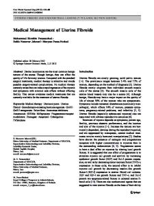
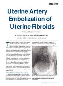
described in 1955 and described the attachment of a band of external oblique fascia from the cervix to the sacral periosteum. Subsequently the utilization of mesh for suspension of the vaginal apex was introduced and continues to be used today. While descriptions of open sacrohysteropexy differ in material type, size, and site of attachment, the overall technique is similar. Mesh is placed between the vagina and/or cevico-uterine junction and subsequently affixed to the sacral promonotory. Some authors utilize a single piece while others advocate for a dual-mesh system. Single-mesh sacrohysteropexy utilizes a single piece of mesh that is often affixed to the posterior vagina, therefore avoiding placement of mesh on the anterior vagina. Dualmesh sacrohysteropexy utilizes the posterior mesh and an additional piece of mesh that is placed on the anterior vaginal wall. Bilateral windows in the broad ligament are created to allow for the anterior mesh arms to be placed underneath the uterus (Fig. 1). Subsequently, the two mesh arms are affixed to the anterior longitudinal ligament of the sacrum (Fig. 2). Despite the varied techniques, the overall evidence supporting sacrohysteropexy suggests good success rates. While no randomized controlled trials have been published, multiple retrospective and prospective cohorts have reported anatomic and subjective cure rates of 91–100 % and 77– 100 %, respectively. [27–33] Most recently, an ambispective cohort study compared 20 women undergoing abdominal sacrohysteropexy to nine women undergoing sacrocolpopexy with hysterectomy. [34] Subjective outcomes (89 % vs. 87 %) were similar, although a lower mean operating time was seen in the hysteropexy cohort. Quality of life was assessed using the PFDI-20 and PFIQ, both of which showed improvement. While subjective outcomes did not differ, anterior failure (55 %) was higher in the hysteropexy cohort. Reoperation
Fig. 1 Bilateral windows in the broad ligament are created to allow for the anterior mesh arms to be placed underneath the uterus
Fig. 2 Two mesh arms are affixed to the anterior longitudinal ligament of the sacrum
rate was higher in the hysterectomy group (2/8, 25 %, 1 patient with recurrent prolapse, 1 patient with uterine pathology) compared with the hysteropexy group (2/18, 11 %, 2 patients with mesh erosion). Four patients (22 %) required postoperative work up for abnormal uterine bleeding or post menopausal bleeding. Complications were rare, with only three mesh erosions, all in the hysterectomy group. Laparoscopic/Robotic Procedures Advances in minimally invasive techniques have yielded several laparoscopic approaches including laparoscopic ventrosuspension, laparoscopic uterosacral hysteropexy, and laparoscopic sacrohysteropexy. Of the three procedures, the majority of the literature has focused on laparoscopic uterosacral hysteropexy and sacrohysteropexy. Laparoscopic uterosacral hysteropexy involves placing sutures through the uterosacral ligaments to suspend the cervix with permanent sutures. The location on the uterosacral ligaments varies, but overall focuses on shortening the uterosacral ligaments in the process of suspending the cervix. Several authors advocate making ureteral releasing incisions in the peritoneum to decrease the risk of ureteral kinking. Evidence suggests the laparoscopic uterosacral ligament suspension is safe and effective. Objective cure rates have been reported to be 79 to 81 %. [35–38] Most recently, a retrospective cohort study comparing 104 laparoscopic uterosacral hysteropexies with 160 laparoscopic hysterectomy and uterosacral colpopexies demonstrated the failure rate (descent to stage 2 in any compartment) was significantly higher in the hysteropexy group (59 % vs. 41 %, p