Review
CME Management of interstitial lung disease in systemic sclerosis
Interstitial lung disease (ILD) is very common in systemic sclerosis (SSc) and the leading cause of death. High-resolution computed tomography is a sensitive tool for the diagnosis of ILD in SSc and is abnormal in up to 90% of patients. The most common radiographic and histopathologic pattern seen in these patients is one of nonspecific interstitial pneumonia. Despite the high incidence of disease, the prognosis for most patients is good and those who progress tend to do so early in the course of disease. Treatment options are limited by a paucity of placebo-controlled trials. Data for use of cyclophosphamide and mycophenolate mofetil exist and there is an ongoing trial comparing these two treatments. Although there are limited data to guide us on how to care for these patients, this article proposes a management algorithm. There is ongoing research on new biological treatments as well as the use of biomarkers to predict the course of disease and response to treatment and these may shape the future management of SSc-ILD. KEYWORDS: biomarkers n cyclophosphamide n interstitial lung disease n mycophenolate mofetil n nonspecific interstitial pneumonia n scleroderma n systemic sclerosis
Medscape: Continuing Medical Education Online
Joshua J Solomon1 & Kevin K Brown†1 1 Autoimmune Lung Center & Interstitial Lung Disease Program, National Jewish Health, 1400 Jackson Street, Denver, CO 80206, USA † Author for correspondence: Tel.: +1 303 398 1621 Fax: +1 303 398 1040
[email protected]
This activity has been planned and implemented in accordance with the Essential Areas and policies of the Accreditation Council for Continuing Medical Education through the joint sponsorship of Medscape, LLC and Future Medicine Ltd. Medscape, LLC is accredited by the ACCME to provide continuing medical education for physicians. Medscape, LLC designates this Journal-based CME activity for a maximum of 1 AMA PRA Category 1 Credit(s)™. Physicians should claim only the credit commensurate with the extent of their participation in the activity. All other clinicians completing this activity will be issued a certificate of participation. To participate in this journal CME activity: (1) review the learning objectives and author disclosures; (2) study the education content; (3) take the post-test with a 70% minimum passing score and complete the evaluation at w ww.medscape.org/journal/ijcr; (4) view/print certificate. Release date: 27 September 2011; Expiration date: 27 September 2012
Learning objectives Upon completion of this activity, participants should be able to: • Distinguish appropriate initial tests for ILD among patients with SSc • Assess how to follow the course of pulmonary disease in patients with SSc • Analyze treatments for ILD among patients with SSc • Evaluate the prognosis of ILD in the setting of SSc 10.2217/IJR.11.43 © 2011 Future Medicine Ltd
Int. J. Clin. Rheumatol. (2011) 6(5), 503–515
ISSN 1758-4272
503
Review
CME
Solomon & Brown
Financial & competing interests disclosure CME Author Charles P Vega, MD, Associate Professor; Residency Director, Department of Family Medicine, University of California, Irvine, USA. Disclosure: Charles P Vega, MD, has disclosed no relevant financial relationships. Authors and Disclosures Joshua J Solomon, Autoimmune Lung Center & Interstitial Lung Disease Program, National Jewish Health, 1400 Jackson Street, Denver, CO 80206, USA. Disclosure: Joshua J Solomon has disclosed no relevant financial relationships. Kevin K Brown, Autoimmune Lung Center & Interstitial Lung Disease Program, National Jewish Health, 1400 Jackson Street, Denver, CO 80206, USA. Disclosure: Kevin K Brown has disclosed no relevant financial relationships. Editor Elisa Manzotti, Editorial Director, Future Science Group, London, UK. Disclosure: Elisa Manzotti has disclosed no relevant financial relationships.
Systemic sclerosis (SSc) is a heterogeneous com plex of diseases characterized by multiorgan involvement, endothelial dysfunction, excessive collagen production and immune system abnor malities [1] . Clinically, patients can have diverse systemic manifestations with any combination of skin, pulmonary, cardiac, renal, musculo skeletal and gastrointestinal involvement. SSc is an uncommon disorder that has an incidence of 18.7 new cases per million people per year and a prevalence of 242 cases per million people. It is a female predominant disease with a 3 to 4:1 female-to-male ratio and blacks have a higher incidence rate and more severe disease when compared with whites [2] . Our current understanding of the natural evolution of SSc has been facilitated by the 1980 SSc classification criteria, which provided uniform diagnostic criteria (Box 1) [3] . The dis ease is subdivided based on the pattern of skin involvement, with diffuse, limited and even absent skin manifestations.
SSc & lung involvement Lung involvement in SSc is extremely common and clinically important. With improvement in the management of scleroderma renal dis ease, pulmonary disease has become the leading cause of death (it is the primary contributor in 33% of cases) [4] . In early autopsy series, up to 100% of patients had some type of pulmonary involvement [5,6] . When unselected patients are screened with high-resolution computed tomog raphy (HRCT), up to 90% of patients will have SSc-associated interstitial lung disease (SScILD) [7] , while 40–75% will have physiologic abnormalities on pulmonary function testing (PFT) [8,9] . Clinically significant pulmonary disease appears early in the course of SSc, with 25% of patients developing lung disease within 504
Int. J. Clin. Rheumatol. (2011) 6(5)
3 years of diagnosis as defined by physiologic, radiographic or bronchoalveolar lavage (BAL) abnormalities [10] . Pulmonary involvement is associated with African–American ethnicity, skin score, serum creatinine and creatinine phosphokinase levels, hypothyroidism and cardiac involvement [10,11] . Other predictors include genetic factors [12] , patterns of antibody positivity (antitopoisom erase antibodies predict lung involvement [11] and anticentromere and anti-RNA polymerase III antibodies appear protective [10,13]) and the pattern of skin disease (patients with diffuse SSc have a higher incidence of lung disease and those with limited SSc have a higher incidence of pulmonary hypertension [14–16] ). African– American patients with SSc-ILD are particu larly sensitive to the development of severe disease with a younger age of onset, higher antitopoisomerase levels, lower levels of forced vital capacity (FVC%) and diffusion capacity for carbon monoxide (DLCO%), and associ ated cardiac involvement at presentation when compared with white patients [8,11] . Pulmonary function tests Screening PFT will show a reduced FVC% in 40–75% of patients, with 15% having a severe reduction [8,9,17] . DLCO% is reduced in 96% of patients with any other physiologic abnor malities [18] and correlates with the extent of lung disease on HRCT [19] . Patients with the CREST variant of SSc (those with subcutane ous calcinosis [C], Raynaud’s phenomenon [R], esophageal dysmotility [E], sclerodactyly [S] and telangiectasias [T]) have a higher FVC% and lower DLCO%, ref lecting the higher incidence of pulmonary vascular disease [20] . Both FVC% and DLCO% are markers of dis ease severity with lower values associated with future science group
CME
Management of interstitial lung disease in systemic sclerosis
Review
Box 1. Systemic sclerosis diagnostic criteria. Patient must have: –– Proximal scleroderma –– Or two or more of the following: –– Sclerodactyly –– Digital pitting scars of the fingertips or loss of substance of the distal finger pad –– Bilateral basilar pulmonary fibrosis
shorter survival [8,21] . A declining DLCO% is the single most significant marker of poor out come [18] . Bronchoalveolar lavage An abnormal BAL count is defined as a neu trophil count of ≥3% or an eosinophil count of ≥2%, and 38–72% of selected patients with SSc will have abnormalities [22,23] . Even in patients with a normal HRCT of the chest, close to 50% of patients will have abnormal BAL cell counts [24] . Early studies suggested that patients with a neutrophilic BAL had a more rapid decline in their FVC% and DLCO% when compared with those with a normal BAL [25,26] . Owing to this early association with disease progression, as well as uncontrolled data suggesting improved survival in patients with a neutrophilic BAL who received cyclophosphamide (CYC) [26] , BAL was frequently recommended during the evaluation of SSc-ILD patients and was an inclusion crite ria for the Scleroderma Lung Study I (SLS I), a multicenter, randomized double-blinded study of CYC in SSc-ILD [27] . However, additional well-controlled studies determined that this prior association between BAL cellularity and progression was likely an epiphenomena, with neutrophilia representing more extensive disease, as seen on chest imaging. The findings from BAL counts do not appear to add to the prognos tic information when noninvasive testing such as pulmonary physiology and HRCT are avail able [28,29] . However, BAL is useful in exclud ing infection when it is clinically suspected, and although limited to the research setting at pres ent, BAL may have future utility in the measure of biomarkers present in the alveolar lining fluid. High-resolution computed tomography High-resolution computed tomography is the standard method for noninvasive diagnosis of SSc-ILD. The true incidence of HRCT abnor malities is difficult to determine as most stud ies use patients who are referred for respiratory symptoms, introducing a significant bias. Up to half of the HRCTs conducted in patients with normal lung volumes and mild reductions future science group
in DLCO% show abnormalities [14] . Even in patients with limited SSc and anticentromere antibodies (and thus have a low risk of devel oping ILD), a third will have abnormalities on HRCT [14] . Although 55–91% of patients will have imaging abnormalities [7,14,30–32] , the extent is generally limited, with an average of 13% of the lung parenchyma involved [31,33] . Despite its sensitivity for locating lung dis ease, HRCT has limitations. HRCT can be nor mal in patients with PFT abnormalities and an abnormal chest exam (i.e., crackles), and a num ber of these patients go on to develop abnormal HRCT scans at follow-up [14] . Patients can have histopathologic evidence of disease (inflamma tion and fibrosis) in areas of ‘normal’ lung when using HRCT [34] . Despite these limitations, the presence of a normal HRCT at baseline predicts a good prognosis, with 85% of these patients still having a normal HRCT at a mean follow-up of 5 years [14] . Specific patterns of HRCT abnormalities are recognized and, similar to the patterns seen in the idiopathic interstitial pneumonias, these pat terns can generally predict the underlying histo pathology. SSc patients most frequently will have a nonspecific interstitial pneumonia (NSIP) pat tern [33] , characterized by a greater proportion of ground-glass opacity (GGO) and a lesser degree of coarse reticulation with rare honeycomb cys tic change [35] . This pattern was seen in 59% of patients in a large series [18] . Radiographic progression occurs; there is replacement of ground-glass abnormalities by traction bronchiectasis/bronchiolectasis and/or honeycomb change over time [14] . Up to twothirds of patients with GGO will progress to fibrosis [24] . The reason for progression in areas of GGO may be that they actually represent a fine intralobular fibrosis below the resolution of HRCT [21] , as surgical biopsies of these areas show fibrosis in up to 50% of cases, usually asso ciated with traction bronchiectasis or bronchio lectasis on HRCT [36] . Prognosis depends not only on the extent of ground-glass abnormalities, but also the amount of associated reticulation, and stability or progression in HRCT pattern, in spite of treatment, is common. www.futuremedicine.com
505
Review
CME
Solomon & Brown
Pathology Early reports of the pathologic changes in SSc-ILD show a mixed pattern of fibrosis and inflamma tion in the majority of cases. The pattern consisted of interstitial and alveolar cellular inflammation with alveolar wall fibrosis. When compared with cryptogenic fibrosing alveolitis in older studies, the only noted differences were more lymphoid aggre gates in patients with SSc [34] . After Katzenstein and Fiorelli described the features of NSIP in 1994 [37] , a re-evaluation of patients with SSc-ILD revealed a significant number with features con sistent with a diagnosis of NSIP [38] . In the largest study to date, 77% of patients with SSc-ILD had a histological pattern of NSIP, the majority of which were fibrotic NSIP [18] . More recently, in a minor ity of patients with a usual interstitial pneumonia (UIP) pattern on biopsy, more germinal centers and inflammation and fewer fibroblast foci were seen when compared with subjects with idiopathic pulmonary fibrosis [39] . Biomarkers There has been interest in finding biomarkers that could predict the development of fibrosis, the clinical course and the response to therapy. A number of small studies have evaluated a few potential biomarkers that can be measured in biologic fluids. Krebs von den Lungen 6 antigen and surfactant proteins A and D are produced by type II alveolar cells in the lung, are measurable in the blood and BAL, and have been correlated with the presence of ILD in patients with SSc [40,41] . Elevated serum levels of pulmonary and activation-regulated chemokine were correlated with ILD severity in SSc and levels of this chemo kine in BAL fluid were negatively correlated with total lung capacity and DLCO% [42,43] . Treatment Because of the presence of significant amounts of inflammatory cell infiltrate in patients with SScILD, a number of anti-inflammatory agents have been investigated. Corsticosteroids have histori cally been used but their efficacy has never been proven, and their use has been associated with the development of scleroderma renal crisis in higher doses [44,45] . D-penicillamine was traditionally used in the 1970s and 1980s and a retrospective analysis suggested that D-penicillamine led to an improvement in DLCO% [46] , although its efficacy has not been confirmed with prospective placebo-controlled trials. IFN‑g, which has been evaluated in many open-label studies, was studied in a prospective trial and no effect on patients with SSc-ILD was noted [47] . The endothelin 506
Int. J. Clin. Rheumatol. (2011) 6(5)
antagonist bosentan was recently evaluated in a prospective, double-blind, multicentered treat ment trial and was found not to be useful in the treatment of SSc-ILD [48] . There is significantly more robust data examin ing the use of CYC. Studies dating back to 1993 showed that SSc-ILD patients treated with CYC and prednisone had a significant improvement in FVC% at 6 and 12 months, and the improve ment seen with CYC was suggested to be superior to other immunosuppressive medications [49–51] . In 2000, a retrospective cohort study found that patients with a neutrophilic BAL who were treated with CYC were more likely to have stabilization or improvement in FVC% and DLCO% than those who were not treated [26] . This preliminary data led to two prospective, randomized, placebocontrolled trials that have shed light on the role of CYC in the treatment of SSc-ILD. The first was SLS I, a 13-center, double-blind, placebocontrolled trial looking at oral CYC in patients with active symptomatic SSc lung disease [27] . After 1 year of treatment, a small but significant treatment effect on FVC% as well as significant improvements in dyspnea scores and skin thick ness were seen. More adverse events were seen in the CYC group but no significant increase in serious adverse events was seen. Patients with more fibrotic lung disease (as defined by chest imaging) at baseline noted a greater benefit, and may have been under-represented in the trial [52] . The beneficial effects returned to placebo levels 1 year after cessation of therapy with the excep tion of the improvements in dyspnea [53] . A second trial (Fibrosing Alveolitis in Scleroderma Trial [FAST]) in which subjects received 6 months of intravenous CYC followed by oral azathioprine suggested a trend towards an FVC% benefit, but no statistically significant difference in FVC%, DLCO%, HRCT appearance or dyspnea scores was demonstrated [54] . Mycophenolate mofetil (MM) has been used with increasing frequency in patients with SScILD. An uncontrolled small study looking at 13 patients with recent onset SSc-ILD treated with antithymocyte globulin followed by MM showed improvement in skin scores and stable PFTs (FVC% and DLCO%) over the year of treatment [55] . MM with low-dose prednisone improved pulmonary function and HRCT find ings in a small prospective, open-label trial with five [56] and nine patients showing improve ment in PFTs with minimal side effects in a recent retrospective review [57] . Another review of 17 patients with SSc-ILD treated for up to 2 years with MM showed stable pulmonary future science group
CME
Management of interstitial lung disease in systemic sclerosis
function in the majority of patients [58] . A ret rospective analysis of patients with SSc treated with MM found a lower incidence of pulmonary fibrosis and better survival when compared with other immunosupressive regimens [59] . Finally, a retrospective analysis of 13 patients treated with MM found a reversal in the pretreatment FVC% decline and a stabilization of the pre treatment DLCO% decline after 12 months of treatment [60] . Owing to this preliminary data with MM and the lack of sustained improve ment seen with CYC, the Scleroderma Lung Study II (SLS II) is an ongoing prospective, multicentered, placebo-controlled trial inves tigating the effects of 1 year of treatment with CYC versus 2 years of treatment with MM. Other agents have been evaluated in smaller trials. Owing to their ability to affect profibrotic pathways through inhibition of both TGF-b and PDGF signaling, the tyrosine kinase inhibitors are an attractive treatment option for SSc. In two open-label trials of the tyrosine kinase inhibitor imatinib, a total of 36 patients with SSc-ILD had stable FVC% over 12 months of therapy with imatinib (with one trial showing a significant improvement in DLCO%) [61,62] . Owing to the possible pathogenic role of B cells in SSc, anti-CD20 therapy has been tried. Three small, open-label trials in a total of 32 patients with SSc treated with rituximab (a monoclonal antibody against CD20) showed no signficant decline in physiologic variables over 6 months of follow-up [63–65] . In an open-label, randomized trial of 14 patients with SSc-ILD, treatment with rituximab resulted in significant improvements in FVC% and DLCO% while patients treated with standard therapy (consisting of a combina tion of prednisone, bosentan, CYC and MM) showed a decline in these indices [66] . Finally, basiliximab (a monoclonal antibody against CD25) stabilized the PFTs of ten patients with SSc, eight of whom had ILD [67] . These prom ising results will hopefully translate to future larger, prospective, randomized controlled trials. When medical therapy fails to stem progres sive loss of lung function, lung transplantation can be lifesaving. Although there is a perception among some physicians that patients with SScILD will have poor post-transplant outcomes due to concomitant gastroesophageal disease, renal disease or skin fibrosis, comparative stud ies show the 2‑ and 5‑year outcomes in these patients to be similar to those who have under gone a lung transplant for other conditions (sur vival rates of 72 and 55%, respectively) [68,69] . Relative contraindications include significant future science group
Review
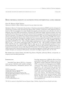
skin breakdown from severe cutaneous disease, a creatinine clearance of less than 50 ml/min, severe reflux disease with aspiration and cardiac involvement with arrhythmias [69] . Prognosis Patients with SSc-ILD have an estimated survival of 85% at 5 years [70] . Progression to respiratory failure is an uncommon but dreaded complica tion. End-stage lung disease, defined as death or leading to a need for oxygen or continuous medication for pulmonary arterial hypertension, is seen in only 4% of patients at 5 years [21] , while severe restrictive lung disease, defined by a FVC% of ≤50%, is seen in 13% [8] . For patients who develop restrictive lung dis ease, a decline in lung function occurs earlier; the greatest decline in lung function in these patients occurs within the first 2 years [8] . Predictors of severe restrictive lung disease include male gen der, PFTs at diagnosis (FVC% and DLCO%) and age (a higher incidence in younger patients) [8,21] . Furthermore, survival does not differ between those with a pathologic pattern of NSIP and those with UIP, a notable difference from patients with an idiopathic interstitial pneumo nia. Both groups have an 82–90% 5‑year sur vival rate and 29–69% 10‑year survival rate [18] . It also appears that neither the subtype of SSc (limited versus diffuse) nor the degree of fibrosis on HRCT affects the likelihood of progression [35] . Mortality is higher in African–Americans [8] and those with a lower FVC% and DLCO% at presentation [18] . Patients presenting with a FVC% of ≤50% have a 10‑year survival rate of 40–50% [8] . When followed over time, changes in DLCO% at 3 years was associated with a decreased survival, an association not seen with changes in FVC% or DLCO% at 1 year [18] . Recently, Goh et al. developed a prognostic algorithm for patients with SSc-ILD (Figure 1) [31] . The algorithm relies solely on HRCT scor ing cases with minimal or extensive disease, with recourse to a FVC% cutoff in cases of an indeter minate extent of disease. This staging system was shown to be easy to use and predictive of mortality.
Evaluating patients for lung disease Clinicians should consider PFTs and a HRCT scan of the chest in all patients with SSc to help with the early identification of those at risk for the development of clinically significant respira tory disease. Normal initial testing portends a good prognosis; only 15% of those with a normal HRCT will develop clinically significant lung involvement at 5 years. www.futuremedicine.com
507
Review
CME
Solomon & Brown
HRCT extent of disease
20%
PFTs
FVC >70%
Limited disease
FVC