MAJOR ARTICLE
Pharmacodynamics of Liposomal Amphotericin B and Flucytosine for Cryptococcal Meningoencephalitis: Safe and Effective Regimens for Immunocompromised Patients Lucy O’Connor,1 Joanne Livermore,1,2 Andrew D. Sharp,1,2 Joanne Goodwin,1,2 Lea Gregson,1,2 Susan J. Howard,1,2 Timothy W. Felton,1 Julie A. Schwartz,3 Michael N. Neely,4 Thomas S. Harrison,5 John R. Perfect,6 and William W. Hope1,2 1 School of Translational Medicine, The University of Manchester, Manchester Academic Health Science Centre, National Institute for Health Research Translational Research Facility in Respiratory Medicine, University Hospital of South Manchester National Health Service, Foundation Trust, and 2 Antimicrobial Pharmacology and Therapeutics, Department of Molecular and Clinical Pharmacology, Institute of Translational Medicine, University of Liverpool, United Kingdom; 3Charles River Laboratories, Davis, California; 4Laboratory of Applied Pharmacokinetics, University of Southern California, Los Angeles; 5Department of Infectious Diseases, St George’s Hospital Medical School, London, United Kingdom; and 6Department of Medicine, Duke University, Durham, North Carolina
Background. Cryptococcal meningoencephalitis is a lethal infection with relatively few therapeutic options. The optimal dosage of liposomal amphotericin B (LAmB) alone or in combination with flucytosine is not known. Methods. A murine model of cryptococcal meningoencephalitis was used. The fungal density in the brain was determined using quantitative cultures. Pharmacokinetic–pharmacodynamic relationships were determined for LAmB and flucytosine administered alone. The effect of the combination was described using the Greco model and a mathematical model. The results were bridged to humans. Results. Inoculation resulted in hematogenous dissemination and logarithmic growth within the central nervous system. There was histological evidence of multifocal infection throughout the brain. Both LAmB and flucytosine produced a dose-dependent reduction in fungal burden. The effect of the combination of agents in the brain was additive. Bridging studies suggested that a human dosage of LAmB 3 mg/kg/d resulted in a submaximal antifungal effect. Regimens of LAmB 6 mg/kg/d alone, LAmB 3 mg/kg/d plus flucytosine 50 mg/kg/d, and LAmB 3 mg/kg/d plus flucytosine 100 mg/kg/d all resulted in near-maximal antifungal activity. Conclusions. Potential regimens for further study in clinical trials include LAmB 6 mg/kg/d alone, LAmB 3 mg/kg/d plus flucytosine 50 mg/kg/d, and LAmB 3 mg/kg/d plus flucytosine 100 mg/kg/d. Keywords.
Cryptococcus; meningitis; meningoencephalitis; liposomal amphotericin B; flucytosine.
Cryptococcal meningoencephalitis is a frequently lethal opportunistic infection. The global burden of disease is intricately linked with the human immunodeficiency virus (HIV)/AIDS epidemic, especially in sub-Saharan Africa [1]. Cryptococcal meningoencephalitis also
Received 20 November 2012; accepted 24 January 2013; electronically published 18 April 2013. Correspondence: William Hope, BMBS, FRACP, FRCPA, PhD, Department of Molecular and Clinical Pharmacology, Antimicrobial Pharmacodynamics & Therapeutics, University of Liverpool, 1.09 Sherrington Building, Ashton Street, Liverpool L69 3GE, United Kingdom. (
[email protected]). The Journal of Infectious Diseases 2013;208:351–61 © The Author 2013. Published by Oxford University Press on behalf of the Infectious Diseases Society of America. All rights reserved. For Permissions, please e-mail:
[email protected]. DOI: 10.1093/infdis/jit164
occurs in other immunocompromised patients, such as solid organ transplant recipients. Treatment includes an intensive period of induction therapy that usually includes an amphotericin B formulation [2]. All amphotericin B formulations cause dose-dependent toxicity [3] that may lead to interrupted antifungal therapy and potentially poorer clinical outcomes. A further understanding of antifungal regimens for cryptococcal meningoencephalitis that are safe and effective is urgently needed. Lipid formulations of amphotericin B are recommended as first-line agents for treating cryptococcal meningoencephalitis; however, there is uncertainty regarding optimal regimens. For example, a comparative study of liposomal amphotericin B (LAmB) 3 mg/kg/d
Combination Therapy for Cryptococcal Meningoencephalitis
•
JID 2013:208 (15 July)
•
351
vs 6 mg/kg/d vs amphotericin B deoxycholate showed no differences between any of these regimens [4]. Conversely, a small study using 4 mg/kg/d of LAmB resulted in a significantly earlier cerebrospinal fluid (CSF) culture conversion than treatment with 0.7 mg/kg/d of amphotericin B deoxycholate [5]. Therefore, there is no known optimal initial dosage of a lipid formulation for cryptococcal meningoencephalitis. Furthermore, the potential benefit of flucytosine for patients with cryptococcal meningoencephalitis is not known nor is the smallest effective dose that can be used in combination with LAmB [6]. Here, we use a previously described murine model of cryptococcal meningoencephalitis [7] to define the pharmacokinetics and pharmacodynamics of LAmB and flucytosine alone and in combination. We bridged the results to humans in order to provide further insight into combination regimens that are likely to be safe and effective. These results can now be used to aid in the design of clinical trials to further understand the optimal use of these compounds in humans with cryptococcal meningoencephalitis. METHODS Organism
A well-characterized clinical isolate of Cryptococcus neoformans var. grubii, H99 (American Type Culture Collection [ATCC 208821]), was used (LGC Standards). The isolate was stored on beads at −80°C. Seventy-two hours prior to inoculation, the organism was plated to Sabouraud dextrose agar (Oxoid, Basingstoke, UK) containing chloramphenicol (Sigma-Aldrich Company Ltd., UK) and incubated at 30°C for 48 hours. Twenty-four hours prior to inoculation, several colonies were placed into 10 mL of Sabouraud dextrose broth (Oxoid) and incubated at 37°C on a shaker. Immediately prior to inoculation, the fungal suspension was centrifuged and washed 3 times with phosphate buffered saline (PBS; GIBCO, Invitrogen, Paisley, UK). The final fungal inoculum was prepared by resuspension and dilution in PBS. A hemocytometer was used to achieve the desired concentration of 3.8 × 10⁵ CFU/mL.
flucytosine (Valeant Pharmaceuticals Ltd., Basingstoke, UK) was used for all murine experiments but administered orally. Murine Model of Cryptococcal Meningoencephalitis
A previously described model of cryptococcal meningoencephalitis was used [7]. Male CD1 mice (Charles River Ltd, Kent, UK) were housed in groups of 5. Mice were inoculated via a lateral tail vein with 9.5 × 10⁴ cryptococcal organisms in a 0.25-mL suspension. Histology and Immunohistochemistry
Brains were fixed in 10% neutral buffered formalin, cryoprotected, and embedded in OCT freezing medium (Tissue–Tek® O.C.T. Compound); 5-µm-thick sections were then cut. Tissue sections were treated with peroxidase solution (Envison+ kit, Dakocytomation, Carpinteria, California) to block endogenous peroxidase activity. A 1.5% goat serum protein block was applied to reduce nonspecific binding. Sections were incubated with an anti-Cryptococcus monoclonal antibody (Mybiosource, San Diego, California; 1:1000 dilution). Slides were reacted with a peroxidase-labeled anti-mouse polymer (Envison+ kit, Dakocytomation). Antibody–antigen complexes were visualized using 3, 3′-diaminobenzidine chromogen substrate (Dakocytomation) for the peroxidase reaction. Slides were counterstained with hematoxylin (Richard-Allan Scientific, Kalamazoo, Michigan).
Minimum Inhibitory Concentrations
The in vitro susceptibility of H99 to amphotericin B and flucytosine was determined using Clinical and Laboratory Standards Institute methodology (M27-A3) [8]. The minimum inhibitory concentration (MIC) of each agent was determined over the course of 5 independently conducted experiments. Antimicrobial Agents
LAmB for injection was reconstituted according to the manufacturer’s instructions. The desired concentrations were obtained by dilution of the stock solution with 5% glucose (Baxter, Thetford, UK). The intravenous clinical preparation of 352
•
JID 2013:208 (15 July)
•
O’Connor et al
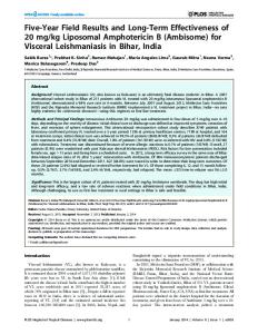
Figure 1. The rate of change of fungal density in the cerebrum of mice infected with Cryptococcus neoformans at time zero. Data are pooled from 5 experiments and are mean ± SD of 3–4 mice. The solid line is the fit of the model dN/dt = Kgmax*(N/POPMAX)*N to the data, where N is the number of organisms, Kgmax is the growth constant, and POPMAX is the maximal fungal density in the brain. The estimates for the parameter values are Kgmax = 0.08 log10CFU/g brain/h and POPMAX = 6.41 log10CFU/g brain and the number of organisms in the brain immediately after inoculation of 2.00 log10CFU/g brain; r2 = 0.88. The corresponding histological sections stained with hematoxylin and eosin and anti–C. neoformans antibody and taken on days 1, 3, 5, and 7 post infection are shown. A high power section shows the formation of cyst-like structures with leaching of cryptococcal capsular material into the contiguous parenchyma.
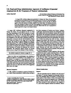
Figure 2. The pharmacokinetics of liposomal amphotericin B (LAmB) 3, 10, and 20 mg/kg/d intravenously in plasma and cerebrum. Data are mean ± SD, and the solid line is the fit of the model to the data.
Pharmacokinetics and Pharmacodynamics of LAmB and Flucytosine as Monotherapy
Therapy with LAmB and flucytosine commenced 24 hours post inoculation. Informative parts of the exposure–response relationships were defined in preliminary dose-finding experiments.
Dosages of LAmB 3, 10, and 20 mg/kg were administered every 24 hours via a lateral tail vein in a volume of 0.25 mL. The respective controls received 0.25 mL of 0.9% sodium chloride once daily. The intravenous formulation of flucytosine 25, 50, and 100 mg/kg (ie, 75, 150, and 300 mg/kg/d) was administered
Combination Therapy for Cryptococcal Meningoencephalitis
•
JID 2013:208 (15 July)
•
353
every 8 hours by oral gavage in a volume of 0.25 mL. Control mice received 0.25 mL of the vehicle, 0.9% sodium chloride, every 8 hours by oral gavage. The concentration–time profiles of LAmB in plasma and cerebrum were defined using cohorts of mice (n = 3 for each dose– timepoint combination). Mice were sacrificed at 1, 2, 4, 6, and 24 hours post dose. Plasma was obtained by cardiac puncture and stored at −80°C. The brain was removed, placed in a sterile plastic bag, and stored at −80°C. A similar approach was used for flucytosine except a different sampling strategy was used (plasma and cerebrum samples were obtained 0.5, 1, 3, and 8 hours post dose). The temporal change in cerebral fungal burden following treatment with both agents was defined using a serial sacrifice design. Cohorts of mice (n = 3 for each regimen at each timepoint) were sacrificed at 1, 24, 72, 120, and 168 hours post infection. The pharmacodynamic relationships were defined over 7 experiments. The entire brain was removed and homogenized in 1 mL PBS; serial 10-fold dilutions were plated to enumerate the fungal density in cerebrum. Pharmacodynamics of LAmB and Flucytosine in Combination
The pharmacodynamics of the combination of LAmB and flucytosine were defined in a single experiment with 116 mice. The combination experiment was designed following monotherapy experiments. Cohorts of mice (n = 12) received the following dosage regimens: LAmB 3 mg/kg/d alone; LAmB 20 mg/kg/d alone; flucytosine 25 mg/kg every 8 hours (ie, 75 mg/kg/d) alone; flucytosine 100 mg/kg every 8 hours (ie, 300 mg/kg/d) alone; LAmB 3 mg/kg/d with flucytosine 25 mg/kg every 8 hours; LAmB 3 mg/kg/d in combination with flucytosine 100 mg/kg every 8 hours; LAmB 20 mg/kg/d in combination with flucytosine 25 mg/kg every 8 hours; and LAmB 20 mg/kg in combination with flucytosine 100 mg/kg every 8 hours. Mice in the control group received 0.25 mL of the vehicle, 0.9% sodium chloride every 8 hours by oral gavage. Cohorts of mice (n = 4 for each regimen at each timepoint) were sacrificed at 72, 120, and 168 hours post infection. In addition, 2 groups of control mice (n = 4) were sacrificed at 1 and 24 hours post inoculation. Plasma samples were collected by cardiac puncture (as above) after terminal anesthesia with 5% isoflurane. Mice were then sacrificed by cervical dislocation, and whole brains were excised aseptically for quantitative culture (as above). Sampling times were chosen to obtain trough concentrations of each antimicrobial agent in plasma and cerebrum samples immediately prior to dosing on the second, fourth, and sixth days post initiation of therapy in the murine model. Trough concentrations from combination therapy were compared with data from the monotherapy experiments to examine the possibility of a pharmacokinetic interaction. Measurement of Antifungal Drug Concentrations in Plasma and Cerebrum
The total concentrations of amphotericin B and flucytosine in plasma and cerebrum samples were measured by high354
•
JID 2013:208 (15 July)
•
O’Connor et al
performance liquid chromatography (Shimadzu Prominence; Shimadzu, Milton Keynes, UK). For amphotericin B, the internal standard was piroxicam (Sigma-Aldrich Company Ltd.). A standard curve was constructed over the concentration range 0.05–100 mg/L. Plasma and cerebrum homogenate were mixed with 300 µL methanol containing the internal standard, and 50 µm was injected onto a 5-µm 50 × 20 mm column (Varian Pursuit C18, Varian Inc., UK). The mobile phase consisted of a starting concentration of 80% of 0.1% aqueous formic acid (v/v; Fisher Scientific, Loughborough, UK) and 20% of 0.1% formic acid in acetonitrile (v/v; Fisher Scientific), progressing with a gradient to a 30:70 mix over 7 minutes. The run time was 9.5 minutes, with a flow rate of 0.8 mL/min. Amphotericin B and the internal standard eluted after 4.0 minutes and 2.3 minutes, respectively, and were detected using ultraviolet absorbance at 385 nm. The coefficient of variation was