Magnesium in Single Skeletal Muscle Cells of Balanus ERNEST PAGE, BERT A. MOBLEY, MARGARET JOHNSON, and JUDY E. UPSHAW From the Departments of Medicine and Physiology, The Pritzker School of Medicine, The University of Chicago, Chicago, Illinois 60637
1 mmoles Single skeletal muscle cells of Balanus contain 48 magnesium/kg dry weight. Although 28 Mg can be shown either to enter the 0.3 % cells or to be bound to the cell surface within less than 10 min, only 2.1 of cellular or cell surface Mg exchanges with this isotope even after several hours. Glycerinated cells washed out in Tris buffer at low ionic strength retain -70 % of the Mg present in intact cells. About 85 % of this Mg is removed by extraction with KC1 or NaCl at concentrations of K and Na which prevail in intact cells, as well as by pyrophosphate, Tris-ATP, or reduction of the ionized Mg concentration to 1 iM. Lowering the ionized Mg concentration to 0.1 uM does not further reduce the Mg content of glycerinated cells. The pH dependence of KCl-inextractable Mg suggests that more than one class of binding sites is involved. A significant fraction of the KCl-inextractable Mg bound to glycerinated cells fails to exchange with 28Mg even after long equilibration. It is suggested that this fraction may be actin-bound Mg incorporated into the thin filaments during the polymerization of actin. ABSTRACT
Magnesium ion plays an important role in the myofibrillar processes of contraction and relaxation (Weber and Winicur, 1961; Weber and Herz, 1963; Weber, Herz, and Reiss, 1969; Dancker, 1970; Hotta and Bowen, 1970). Nevertheless, remarkably little is known about the state of Mg in striated muscle cells and about the behavior of this ion under various conditions. Early studies by Bozler (1955) and Hasselbach (1957) indicated that a fraction of cellular Mg is bound to the myofibrils. Measurements of the exchange of Mg with 2 8Mg in frog skeletal muscles (Gilbert, 1960) showed that the sarcolemma is permeable to Mg, but that as much as 75% of the Mg in these muscles fails to exchange with 2 8Mg even after a very long equilibration. The giant skeletal muscle cells of Balanus, introduced by Hoyle and Smyth (1963), have recently been used for extensive studies of ion transport and electrophysiological phenomena (Hagiwara and Naka, 1964; Hagiwara and Takahashi, 1967; Hagiwara, Takahashi, and Junge, 1968; Brinley, 1968; I88
The Journal of General Physiology
E. PAGE ET AL.
Magnesium in Single Balanus Skeletal Muscle Cells
189
McLaughlin and Hinke, 1966, 1968). These cells are up to 0.2 cm in diameter and several centimeters long. They are composed of a sarcolemma enclosing a mass of striated myofibrils, with relatively insignificant amounts of mitochondria, sarcoplasmic reticulum, and nuclei (McNeill and Hoyle, 1967; Selverston, 1967). Because of its large size, this preparation presents an opportunity to study the properties of Mg in single striated muscle cells. In the experiments described here we have taken advantage of this opportunity to measure the content, exchange, and binding of Mg in intact and glycerinated cells. Portions of this work have been previously reported (Page, Mobley, and Lewis, 1970). METHODS
Barnacles of the genus Balanus obtained from Puget Sound (C. Vandersluys, Friday Harbor, Wash.) were shipped by air to Chicago and maintained in an aquarium (Aquarium Systems, Inc., Wickliffe, Ohio) containing seawater at 120C. Multiple single muscle cells were isolated from the depressor scutorum rostralis at their tendinous insertion; their other (muscular) insertion into the shell was left intact until the end of the experiment. Single muscle cells thus dissected were the starting material for two types of experiments: (a) incubation of intact cells with radioactive tracers in solutions of different chemical composition, followed by chemical and radioactive analyses; and (b) glycerination of the muscle, followed by equilibration with radioactive or nonradioactive solutions of variable chemical composition. Experiments with Intact Cells Single cells of a cell bundle were freed from their connective tissue attachments to one another by blunt dissection leaving intact their muscular insertion into the shell. This procedure yielded a muscle bundle containing many cells freely floating at their tendinous ends. Each cell could be excised by holding its free tendinous end with a forceps and cutting its muscular insertion near the shell with a pair of fine scissors. The muscle bundle was incubated at 22-24°C in a small glass vessel containing a solution of the desired composition for as long as required by the experimental design. The vessel was stirred with a magnetic stirrer. At the designated time, cells were removed, drained against the side of the vessel without blotting, and weighed immediately on a Cahn recording microbalance. Alternate muscles were (a) extracted without drying for the chemical determination of Mg, and, when applicable, for measurement of radioactivity; or (b) dried in a vacuum oven at 90°C, reweighed, and, in selected instances, ashed for the subsequent determination of their K and Na contents. Correction for extracellular volume and for surface contamination with the incubating solution was made by including inulin-14C or sucrose- 14C in the bathing medium and comparing the 14C content of the muscle extract with that of a unit volume of bathing solution. The intracellular content of Mg was measured in cells incubated in a control (artificial) seawater solution at pH 7.2, in otherwise identical solutions at pH 4 and
I9
THE JOURNAL
OF GENERAL
PHYSIOLOGY
VOLUME
57
1971
pH 10, in Ca- and Mg-free artificial seawater containing EDTA, and in artificial seawater whose Mg concentration had been increased by raising the external Mg concentration from 12 to 24 mM. The exchange of cellular Mg for 28 Mg was measured as a 2 8Mg influx by incubating the muscle bundle in the control seawater labeled with 28 Mg and sucrose-14 C and assaying the radioactivity of cells removed sequentially at short intervals over a period of 2-3 hr. Experiments with Glycerinated Cells For experiments with glycerinated cells, the individual cells of the depressor scutorum were dissected free in the control seawater solution and left attached to the shell as described above. The bundle was then placed in a special Lucite glycerinating chamber which was initially filled with seawater. In the chamber the shell was immobilized by clamping it with a sliding Lucite clamp; each cell was then stretched to approximately in situ length by tying a silk thread to its tendon, threading the tie through a small hole in a plate perforated with many small holes, and attaching the thread to an anchor in the compartment of the chamber on the other side of the perforated plate. After the muscles were thus fixed in place, the seawater was poured out of the chamber and replaced by glycerinating solution at 3-4°C. The chamber was rinsed with several volumes of glycerinating solution and the cells were left in this solution for 24 hr at 3-4°C. The chamber was then transferred to a freezer at - 140C for from 2 wk to as long as 6 wk. For studies of Mg content and exchange, glycerinated cells were cut free from the shell and treated as described below. Glycerinated cells prepared in this way contracted without exception on exposure to MgATP in the presence of traces of Ca ion as low as 10- 9 M(i.e., in the presence of 4 mM EGTA). All subsequent experiments on the glycerinated cells were done in a cold room at 3-4°C. After removal from the glycerinating solution cells were first washed free of glycerol for 15 min in a large volume of a solution which will be referred to as the primary rinse. They were then equilibrated for a period of from 16 to 36 hr in a solution which will be referred to as the secondary rinse. The primary rinse had the same ionic composition as that of the equilibrating solution. After equilibration with a solution of a given ionic composition, the Mg and other ions not firmly bound to the glycerinated cell were removed by a secondary rinse for 1 hr in a solution containing no Mg, and usually no K or Na, the solution being buffered with Tris or TES at the same pH as that used for equilibration. (In selected experiments, the Mg-free secondary rinse contained 150 mM KC1.) This experimental plan, which resembles that of Bozler (1955), was adopted to measure bound Mg without contamination from Mg in the aqueous solution filling the interstices between binding sites, or Mg in the solution adhering to the surface of the tissue. Preliminary experiments, in which the secondary rinse (with two or more changes of solution) was carried out for 30 and 60 min, respectively, showed that the washout of unbound Mg was complete within 30 min. Equilibration was done in polypropylene test tubes set circumferentially in a rack around a magnetic stirrer (Tri-R Instruments, Inc. [Rockville Center, N.Y.], Stirrer NS-7 and Table MT-72). This arrangement permitted the simultaneous stirring of
E. PAGE ET AL.
Magnesium in Single Balanus Skeletal Muscle Cells
191
eight test tubes each containing two cells in 10 ml of solution. The cells were suspended in the middle of the solution by a silk tie around their tendinous end and protected from contact with the magnetic spinbar in the bottom of the tube by taping the thread around their tendinous end to the outside of the tube. At the end of the secondary rinse the glycerinated cells were drained quickly against the glass side of the vessel, then weighed and analyzed as described for intact cells. In these experiments the volume of the incubation vessel was effectively infinite relative to that of the glycerinated cells. Solutions The composition of the artificial seawater for experiments with intact cells was (in mmoles/liter): NaC1l 450, KC1 8.0, CaCl 2 2H20 20, MgCl2 6H20 12, Tris maleate 2, N-acetylglycine 2. The Mg and Ca concentrations are those recommended by Hoyle and Smyth (1963) and used by Hagiwara and Naka (1964); these Mg and Ca concentrations are, respectively, about one-quarter times and twice those of normal seawater. The pH was brought to 7.2 by the addition of NaOH, the final Na concentration being 454 m. In selected experiments the seawater was modified in one of several ways including (a) isosmolal substitution of NaCl for all of the Mg in the solution, (b) raising the Mg concentration to 24 mM by isosmolal substitution of MgC12 for NaCI, (c) omitting both Mg and Ca and adding 2 mM sodium ethylenediaminetetraacetate (EDTA), again at constant total osmolality and pH, (d) lowering the pH to 4.0 by the addition of HCI, and (e) raising the pH to 10.0 by the addition of NaOH. The glycerinating solution was prepared by adding 0.242 g of Tris (hydroxymethyl) aminomethane (Trizmabase, Sigma) and an appropriate volume of 1 N HC1 to 500 ml of glycerol and by making up to a volume of 1000 ml with distilled water. The pH of the glycerinating solution and of all the solutions used for experiments on glycerinated muscles was adjusted to 7.2 at the experimental temperature of 3-4°C. Because the alkali metal cations have important effects on the Mg content of the glycerinated cells, the use of KOH or of NaOH in the adjustment of pH was avoided; upward and downward adjustments were instead made by using organic buffers without significantly altering the ionic strength. In later experiments, the glycerinating solution was buffered with TES buffer (N-tris (hydroxymethyl) methyl-2-aminoethanesulfonic acid) (Good et al. [1966]). For experiments in which the pH was systematically varied from 3.5 to 8.5, a series of buffers were prepared by adding the required amount of solid Tris base to a 5 mM solution of glycylglycine. A buffer containing 150 mM KC1 at pH 10.1 was prepared using histidine adjusted with KOH. All solutions were made with distilled water which was passed through a deionizer and then redistilled with an all-glass still. Analyses
After weighing, cells were either extracted without drying, or dried, reweighed, and transferred to 10 ml covered quartz crucibles (Thermal American Fused Quartz Co., Montville, N. J.) for ashing at 550 0 C in a muffle furnace. Cells were extracted for 48
192
THE JOURNAL
OF GENERAL PHYSIOLOGY
· VOLUME
57
1971
hr on a shaker in polypropylene test tubes with caps. The extractant was 1.0 ml of the same solution that served as a blank and diluent in the spectrophotometric determination of Mg and Ca. (This solution, which is a modification of that previously employed by Krames and Page (1968), is made as follows: 11.73 g La20 8 are dissolved in 50 ml of concentrated HC1; to this are added sequentially 58.4 g NaCl, 500 ml distilled H 2 0, and 50 g trichloroacetic acid, and sufficient distilled water to make I liter. The concentrated stock solution thus obtained is diluted 1:10 with distilled water.) In experiments with 28 Mg or 42K, a polypropylene test tube containing one cell immersed in 1 ml of extractant solution was counted in the well of a gamma scintillation counter and its radioactivity compared with that of 1 ml of the bathing solution. After extraction aliquots of the extractant were taken for measurement of total Mg and 4 C. Mg was determined on a Jarrell-Ash atomic absorption spectrophotometer (Jarrell-Ash Co., Waltham, Mass.). Previous experience has shown that if fresh muscles are dried and ashed or extracted, recovery of Mg is incomplete because of the formation of effectively insoluble phosphate compounds. In the present work it was observed that, unlike fresh muscle, glycerinated muscle can be dried and ashed with maximal recovery, presumably because the glycerination has extracted the potentially insoluble organic and inorganic phosphates. The techniques for compensating interferences with the Mg determination have been published by Krames and Page (1968). In all analyses the experimental sample presented to the atomic absorption spectrophotometer was immediately preceded and followed by one of a large selection of standards. The standards between which the experimental sample was bracketed gave a signal within 5 % of that of the sample. Between each sample and standard, the flame was rinsed with distilled water to preserve as nearly constant a flame temperature as possible. In this way, the stability of the aspiration and flame could be checked for each sample. Analyses were performed with an air-acetylene flame using standards between 0.25 ppm and 1.0 ppm of Mg, a range in which the instrument was very sensitive to Mg. SD) of replicate paired determinations was 0.995 + 0.005 at a The ratio (mean concentration of 1 ppm and 1.00 4 0.01 at 0.25 ppm. The corresponding figure for Ca (to which the instrument is less sensitive) was 1.00 - 0.02 at a concentration of 1 ppm. When intact cells were analyzed in this way, their Mg content per unit dry weight was observed to be the same for cells varying in dry weight over a fivefold range. Protein was measured by the method of Lowry et al. (1951). Techniques for measurement of 14C, K, and Na have been previously described (Page, 1962; Page and Page, 1968). Reagents The sources of reagents were as follows: Tris, TES, HEPES, N-acetyl-glycine, Tris maleate, Tris ATP, and sodium ATP from Sigma Chemical Company (St. Louis, Mo.), inulin-'4 C-carboxylic acid and sucrose-14 C from New England Nuclear Corp. (Boston, Mass.), 28 Mg from Brookhaven National Laboratory, and 42K from the Isoserve Division of Cambridge Nuclear Corporation (Cambridge, Mass.). The decay scheme of 28Mg has recently been revised by Alburger and Harris (1969).
E. PAGE
Magnesium in Single Balanus Skeletal Muscle Cells
ET AL.
'93
RESULTS
Experiments with Intact Cells CELLULAR Mg CONTENT
Fig. 1 is a plot of the cellular Mg contents of four muscles against the duration of incubation in the control seawater solution at 22-24°C. The figure shows that the Mg content of cells from a given barnacle remains constant for several hours. Gilbert (1960) has suggested that Mg is actively transported out of muscle cells; if so, it is conceivable that the effectiveness of the pump might diminish during prolonged incubation, with a resultant cellular accumulation of Mg. No evidence for such an accumulation was found. Iijij
r 80Y
60-
AId 40-
o:-'
4
.:· * -oo °
D~
0 .
o
= Oo ° .
-j J
o
20-
FIGURE 1. Cellular magnesium content of cells from four barnacles as a function of the duration of incubation at 2224°C, [Mg]o = 12 ms.
l) _j
o
E
0
20
40
60
80
100
120
40
160
DURATION OF INCUBATION (Min)
Fig. I shows significant differences in the Mg contents of cells from the same barnacle, and somewhat greater differences in the Mg contents of cells from different barnacles. The Mg contents of 77 cells from 8 barnacles have been averaged for all times to give a mean Mg content of 48 - 1 mmoles/kg dry weight (Table I). Since Gilbert (1961) reported that the Mg content of frog skeletal muscle decreased below and increased above physiological pH, we have examined the cellular Mg contents at pH 4.0 and 10.0; at both pH values cellular contents did not change with time of incubation; their mean values (Table I) fall within the distribution of the controls. Table I also shows that incubation in isosmolal solutions modified by omitting Mg or by raising the Mg concentration to twice that of the control seawater produced no significant change in the average Mg contents, nor did the Mg contents vary with time under these conditions (five control cells incubated in normal seawater ([Mg] = 12 mM) and taken from the same muscle bundle as the cells incubated in 24 mM Mg had a cellular Mg content of 42 ±- 3 mmoles/kg dry weight; the apparently
194
THE JOURNAL
OF GENERAL PHYSIOLOGY
- VOLUME 57
I971
TABLE I
Mg CONTENT OF CELLS UNDER VARIOUS CONDITIONS* Experimental conditions
No. of barnacles
No. of cells
- 1 4 2 4 4 4- 2 4- 2
8 1 1 1 1
77 12 10 10 9
57 4- 4 57 4 7 59 - 5
2 2 2
18 8 10
Cellular Mg content mmoles cell Mg/kg dry weight
Controls (seawater with 12 m Mg-free isosmolal seawater 24 mM Mg isosmolal seawater pH 4.0 pH 10.0 Mg-free, Ca-free, EDTA All cells Spontaneous contracture Not contracted
Mg)
48 45 41 44 48
* Except for experiments in Mg-free solutions, the cellular Mg content has been obtained by sub-
tracting the Mg content of the inulin space from the total Mg content of the fiber.
low value for the 24 mM Mg solutions is therefore not significant.) Table I also presents the mean cellular contents for cells incubated in isosmolal Ca-free, Mg-free seawater containing 2 mM EDTA. Nearly half the cells contracted spontaneously under these conditions. There was no decrease in the cellular content of Mg and no difference in the Mg contents of contracted and uncontracted cells from the same muscle bundle. (In fact, the Mg content per unit dry weight of this group of muscles was, for undetermined reasons, higher than in the controls.) At the same time, there were significant changes in the total contents of Na and K regardless of the contractile state, with the Na content increasing from control values of 259 i 15 (n = 10) to 503 4- 34 (n = 12), and the K content falling from 514 4- 10 to 444 + 28. EXCHANGE OF CELLULAR OR CELL SURFACE Mg WITH 28Mg
Fig. 2 presents a representative experiment showing the uptake of 21Mg by cells from one barnacle fiber bundle immersed in 28Mg-labeled control seawater. The figure shows that the equilibration of both the total and the cellular Mg attains a steady value by the time the first measurement can be made, and certainly within less than 20 min; thereafter it does not usually increase significantly. In such experiments with 29 cells from 4 barnacles 0.003 of cellular or cell surwe have found that an average of only 0.021 face Mg exchanges with 2 8Mg even after long incubations. If this Mg fraction is in fact intracellular, it is possible to calculate that the intracellular concentration of exchangeable Mg in barnacle muscle would not exceed 309 M/kg cell water. However, an alternative interpretation is that all or part of the isotopic Mg does not enter the cells but is instead bound to the cell surface. Although the fraction of cellular or cell surface Mg exchanging is very close
E. PAGE ET AL.
Magnesium in Single Balanus Skeletal Muscle Cells
195
0
w I-
m 5
0.08
0
..
-
.
.
Total Mg . Cell Mg o
LL 0.04 0 Z 0 I-
_ L
o
o
o 2 4 60 0 J100 120 40 DURATION OF INCUBATION IN ZaMg (Min )
FIGURE 2.
Fraction of total Mg (upper curve) and cellular or cell surface Mg (lower curve) equilibrated with 28Mg in the incubation medium as a function of time in 28Mg-
labeled seawater, [Mg]o = 12 mM. to zero, there is no doubt that a small but significant amount of Mg associated with the cell does exchange. The evidence for this was that in the four experiments (each of which involved analysis of multiple cells) the difference between the total content of 2Mg and the 28Mg content of the sucrose space was without exception a positive quantity; that is, there was invariably a small, but significant amount of 28Mg either in the cells or bound to the cell surface. The relatively small amount of radioactivity thus taken up by the cells precluded a more definitive kinetic study of its transport or binding. Experiments with Glycerinated Cells EFFECTS OF GLYCERINATION
ON CELL COMPOSITION AND PERMEABILITY
Table II summarizes some of the effects of glycerination on barnacle muscle cells. The data in the table allow one to refer the Mg content of glycerinated cells to that of intact cells. Such a calculation must take into account that the units of reference commonly used for expressing the ion content of muscle cells (the dry weight or the protein content) are unstable under the conditions used by us; that is, that the dry weight and protein content of intact cells are not identical to the corresponding quantities after glycerination, since glycerination extracts protein and other components of the dry weight. Table II also shows that glycerination destroys the selective permeability of the plasma membrane and of other membrane-limited systems. The evidence for the loss of selective permeability includes the increase in the sucrose space, the loss of K associated with the intact cell, and the fact that cells will contract on exposure to ATP under appropriate conditions. The conclusion that cellular membrane systems have lost their effectiveness as osmotic barriers is further supported by the finding that neither the sucrose space nor the total
I96
VOLUME 57
THE JOURNAL OF GENERAL PHYSIOLOGY
I1971
TABLE II
COMPARISON OF GENERAL FEATURES OF INTACT AND GLYCERINATED CELLS* 0
Intact cells (22-24 C)
Weight total water/wet weight Weight water in sucrose space/total water Weight protein/dry weight Mmoles K/kg dry weight Dry weight after glycerination
Glycerinated cells (40C)
0.796 0.172
0.003(48, 5) 0.009(48, 5)
0.919 4 0.003(43, 5) 0.93 - 0.02 (10, 1):
0.46 514
0.02 (12, 2) 10 (10, 1)
0.65 - 0.03 (10, 2) (5, 1) Trace 0.642 4 0.005(20, 1)
Dry weight before glycerination * First figure in parentheses gives total cells measured; the second figure gives number of barnacles. The data on glycerinated cells were obtained after washing out the glycerol in Tris buffer at 40C. : Sucrose spaces were measured after 72 hr of equilibration.
water content of glycerinated cells changed when the external osmolality was raised by the addition of 100 mM MgC12. CONTENT AND EXCHANGE OF BOUND Mg AT PHYSIOLOGICAL pH
As an initial approach to the study of Mg in glycerinated single muscle cells we utilized a technique similar to that applied by Bozler (1955) to whole frog skeletal muscles. In this approach glycerination, primary rinse, equilibration, and secondary rinse are all, unless otherwise indicated, done in Mg-free solution. (The term Mg-free solution as used here means a solution to which no Mg has been added; contamination with traces of Mg in other reagents was doubtless present. Measurement by atomic absorption spectrophotometry suggested that the contamination was usually of the order of 10-6 M and did not exceed 10- 5 M. Mg++ was controlled in selected experiments by the use of a Mg-EDTA buffer.) Bozler's technique has the advantage that it introduces no exogenous Mg into the tissue. Its disadvantage is that some of the Mg normally bound by cellular structures may be lost by exposure of the cell to Mg-free solutions. Table III gives the Mg contents of glycerinated cells equilibrated under various conditions. The table also shows the fraction of Mg which has exchanged with 2Mg after 24 hr at 3-40 C. As in the glycerinated frog muscle described by Bozler, equilibration with buffered or unbuffered distilled water to which no inorganic cations have been added leaves a relatively large amount of Mg bound to the glycerinated cells. It can be calculated from the data of Tables II and III that the 52 mM Mg/kg dry weight remaining in glycerinated cells equilibrated with Tris buffer (Table III) represents -70% of the Mg content of the intact (unglycerinated) cell. From 83-85% of the Mg in the glycerinated cell can be extracted by exposure to solutions contain-
E. PAGE ET AL.
Magnesimwn in Single Balanus Skeletal Muscle Cells
197
TABLE III
CONTENT AND EXCHANGE OF ENDOGENOUS Mg IN GLYCERINATED CELLS (4°C, pH 7.2)
Equilibrating solution
Duration of equilibration
Mg concentration
Mg content
Fraction of Mg exchanged with "Mg in 24 hr
mmolts/kg dry weight
Unbuffered distilled water (5) Tris buffer (4) 150 m KCI (4) 20 mm NaCl (4) Tris buffer + 28Mg (10) 150 ma KC1 + 2SMg (8) 1 mm Na 4P207 (6) 5 mm Tris ATP (4)
1 18 18 18 24 24 24 24
0* 0* 0* 0* 0.09-0.12 0.13-0.14 0* 0*
45 52 5.5 8.9 41 8.3 6.7 5.9
44444 4 4 4
6 9 0.2 0.4 3 0.4 0.7 0.8
0.9 - 0.1 0.411 4 0.003 -
Figures in parentheses give number of cells. * No Mg added.
ing K and Na at the approximate concentrations in which these two alkali metal cations occur in the intact cell: 150 mM KC1 and 20 mM NaC1, respectively; other concentrations were not examined. The residual Mg represents 10-12% of the Mg content of the intact cell. Exploratory experiments in which the muscles were extracted with KCI for 6, 18, and 24 hr showed that the amount of residual Mg was the same at each of the three equilibration times. A similar amount of Mg is extracted from glycerinated cells by lowering the ionized Mg concentration ([Mg++]) to 1 LM (Fig. 3), by Na pyrophosphate, or by Tris ATP (Table III). It was of interest to determine whether the extraction of bound Mg by a KCl solution of approximately physiological K concentration resulted in significant binding of K to the glycerinated cell. In order to examine this question, the experiment was repeated in the presence of 150 mM KCI labeled with 42K. In nine such cells from two barnacles the mean content of 4"K-labeled K was 100 3 mmoles/kg dry weight. Although this result is not statistically different from the number of equivalents of Mg extracted, the scatter of the data precludes a decision as to whether the stoichiometry of the cation exchange is exact. A stoichiometry in which the equivalents of K bound exactly match the equivalents of Mg extracted is not, in any case to be expected, because 150 mM KCi also extracts bound Ca: The Ca content of glycerinated cells washed out in Tris buffer was 24 - 3 mmoles/kg dry weight (n = 5) before extraction with KCI, and 4 1 mmoles/kg dry weight (n = 5) after extraction with KC1. The amount of Ca bound to glycerinated cells both before and after extraction with KCI can be reduced by incorporating 5 nnmM MgC12 in
198
THE JOURNAL
OF
GENERAL
PHYSIOLOGY
VOLUME 57
1I971
140120
--4
1oo00 'X 80
.,
60 /
' 40 E E
/ /!
l
20
,/////i'~
UF/
....... s_______,/
)mM KCI
--------I
.I
7
6
5
4 p Mg++
3
2
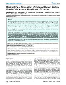
1
Mg content of glycerinated cells as a function of the Mg concentration of the incubation medium (3-4°C, pH 7.2). Upper curve, Mg content at low ionic strength in inorganic cation-free buffer. Lower curve, corresponding values in 150 mM KCI. Incubation time 24 hr. Values for pMg 6-7 were obtained in the presence of a magnesium-EDTA buffer. The number of experiments in the absence of KC1 at pMg 1, 2, 3, 4, 5, 6, and 7 were, respectively, 10, 10, 12, 6, 6, 8, and 6; in the presence of KCI there were six experiments at each point. FIGURE 3.
the glycerinating solution. Under these conditions the Ca content (millimoles per kilogram dry weight) after washing the cells out in inorganic cation-free 0.5 (n = 6); the corresponding figure after extraction Tris buffer was 7.5 with 150 mM KCl was 2.57 - 0.03 (n = 6). Exploratory experiments in which the muscles were equilibrated with 2 8Mg for 6 and 24 hr, respectively, showed that the fraction of bound Mg exchanged with 28 Mg did not increase between 6 and 24 hr. Table III shows that, to within the precision of the measurement, the Mg bound to the glycerinated cells washed out in low ionic strength Tris buffer (pH 7.2) exchanged with 2 8Mg. After removal of KCI-extractable Mg by exposure to 150 mM KC1 about 60% of the residual Mg was inexchangeable in 24 hr. While the absolute Mg content is subject to considerable variation between cells, the existence of a substantial fraction of inexchangeable Mg in KCl-extracted cells appears firmly established. The results shown in Table III indicate that the Mg content of glycerinated cells consists of at least two fractions: A small, tightly bound fraction which is only partially exchangeable at an external Mg concentration of 130 AM, that is, at a Mg concentration of the same order of magnitude as that of the exchangeable Mg in intact cells; and a much larger fraction, characterized by its extractability by KC1, NaCl, pyrophosphate, and Tris-ATP, as well as by the fact that it is exchangeable with 2 SMg.
E. PAGE ET AL.
Magnesium in Single Balanus Skeletal Muscle Cells
199
DEPENDENCE OF Mg CONTENT ON Mg CONCENTRATION
Fig. 3 is a plot of the Mg content against the external Mg concentration. The values for Mg concentrations of 0.1-10 M (pMg 7.0-5.0) refer to the ionized Mg concentration calculated for mixtures of MgCl 2 with EDTA, the total concentration of Mg being 1 mM. In the absence of KCI the Mg bound to the glycerinated cells approaches a value of about 80 mmoles/kg dry weight at the exchangeable Mg concentration (309 uM) which prevails in intact cells. At Mg concentrations higher than this value the Mg content increases in an approximately linear way. Below an external Mg concentration of 309 ,uM more than 90% of the Mg bound at 309 AM is progressively lost, until between pMg 6 and 7 the Mg content becomes independent of pMg. In the presence of 150 mM KC1 the Mg-binding curve is shifted to the right: Under these conditions the Mg content at an external Mg concentration of 100 M is as low as that found in 1 M Mg solution in the absence of KC1. pH DEPENDENCE OF Mg CONTENT
Fig. 4 is a plot of the Mg content of glycerinated cells equilibrated 24 hr in solutions whose pH was varied from 3.5 to 10.1. The experiments were done both in the presence and absence of 150 mM KCl; no exogenous Mg was added to the equilibrating solution. Under these conditions the Mg content in the absence of KC1 at a physiological pH was somewhat lower than usual, perhaps because a glycylglycine-Tris buffer was substituted for the usual
0
CD
cr 0', Y0
2
cn w -j
0
E
pH
FIGURE 4. pH dependence of Mg content in glycerinated cells(3-4 0 C). Upper curve, Mg content at low ionic strength in inorganic cation-free buffer. Lower curve, corresponding values in 150 mm KCI. Incubation time 24 hr.
200
THE JOURNAL
OF GENERAL PHYSIOLOGY
· VOLUME
57
I971
Tris or TES buffers. Nevertheless, the pH dependence of the Mg content is apparent. At pH 5.5 the Mg contents in the presence and absence of KC1 are approximately equal; at all other values of the pH the Mg content is strikingly less in the presence of KC1. In the absence of KC1 the curve has plateaus from pH 4.5 to 5.5 and from pH 7.5 to 8.5. In the presence of 150 mM KC1 there is again a plateau between pH 7.5 and 8.5, but the plateau which occurs from pH 4.5 to 5.5 in the absence of KC1 is shifted by 1 pH unit to pH 5.5-6.5. As the result of this shift the half-maximal value for the binding sites which saturate at pH 7.5-8.5 lies at about pH 7. At pH 10.1 the Mg content in the presence of 150 mM KC1 has decreased to the same magnitude as at pH 6.5. DISCUSSION
Residual Mg Present after Extraction with KCI Although about 70% of the Mg present in intact cells remains bound to glycerinated cells after rinsing the cells in distilled water or low ionic strength buffer, most of this Mg is lost on exposure to KC1 or NaCl at K or Na concentrations comparable to those which prevail in intact cells. Similar amounts of Mg are removed by anions which bind Mg, for example, by pyrophosphate or ATP. The nature of the cellular structures to which the KCl-extractable Mg is bound in the glycerinated cell remains undetermined. It seems probable that the residual Mg present after extraction with KCI is bound to myofibrillar protein. Fig. 3 shows that the Mg so bound is not further reduced when the Mg++ concentration is lowered from 10-6 to 10- ' M. The pH dependence (Fig. 4) suggests that at least two classes of binding sites may be involved; and the measurements of 28Mg exchange indicate with good statistical reliability that a significant fraction of KCl-inextractable Mg fails to exchange with 28 Mg. Weber, Herz, and Reiss (1969) have proposed that myofibrillar actin may contain Mg which is bound during the polymerization of actin. In this connection, the present finding that a fraction of Mg bound to KCl-extracted glycerinated cells fails to exchange with 2 8 Mg may be compared with the observation by Weber et al. (1969) that a part of myofibrillar Mg does not exchange with Ca. This comparison suggests that the 28Mg-inexchangeable fraction of Mg which remains in glycerinated cells after extraction with 150 m KC1 may be bound to actin. This work was supported by U.S. Public Health Service Grant No. HE10503-05 and by a grant from the American Heart Association. Dr. Page is the recipient of a U.S. Public Health Service Career Development Award. Received for publication 30 April 1970. REFERENCES
ALBURGER, D. E., and W. R. HARRIS. 1969. Decay scheme of Mg2. Phys. Rev. 185:1495. BOZLER, E. 1955. Binding of calcium and magnesium by the contractile elements. J. Gen. Physiol. 38:735.
E. PAGE ET AL.
Magnesium in Single Balanus Skeletal Muscle Cells
201I
BRINLEY, F. J. 1968. Sodium and potassium fluxes in isolated barnacle muscle fibers. J. Gen. Physiol. 51:445. DANCKER, P. 1970. The binding of calcium and magnesium to actomyosin and its modification by natural tropomyosin. Arch. gesamte Physiol. Menschen Tiere (Pfluegers). 315:198. GILBERT, D. L. 1960. Magnesium equilibrium in muscle. J. Gen. Physiol. 43:1103. GILBERT, D. L. 1961. Effect of pH on muscle calcium and magnesium. Proc. Soc. Exp. Biol. Med. 106:550. GOOD, N. E., D. WINGET, W. WINTER, T. N. CONNOLLY, S. IZAWA, and R. M. M. SINGH. 1966. Hydrogen ion buffers for biological research. Biochemistry. 5:467. HAGIWARA, S., and K. NAXA. 1964. The initiation of spike potential in barnacle muscle fibers under low internal Ca+ +. J. Gen. Physiol. 48:141. HAGIWARA, S., and K. TAKAHASHI. 1967. Surface density of calcium ions and calcium spikes in the barnacle muscle fiber membrane. J. Gen. Physiol. 50:583. HAGIWARA, S., K. TAKAHAsHI, and D. JUNGE. 1968. Excitation-contraction coupling in a barnacle muscle fiber as examined with voltage clamp technique. J. Gen. Physiol. 51:157. HASSELBACH, W. 1957. Die Bindung von Adenosindiphosphat, von anorganischem Phosphat und von Erdalkalien an die Strukturproteine des Muskels. Biochim. Biophys. Acta. 25:562. HOTTA, K., and W. J. BOWEN. 1970. Contraction and ATPase activity of glycerinated muscle fibers and myofibrillar fragments. Amer. J. Physiol. 218:332. HOYLE, G., and T. SMYTH. 1963. Neuromuscular physiology of giant muscle fibers of a barnacle, Balanus nubilus Darwin. Comp. Biochem. Physiol. 10:291. KRAmES, B., and E. PAGE. 1968. Effects of electron microscopic fixatives on cell membrane of the perfused rat heart. Biochim. Biophys. Acta. 150:24, LOWRY, O. H., N.J. ROSEBROUGH, A. L. FARR, and R. J. RANDALL. 1951. Protein measurement
with the Folin phenol reagent. J. Biol. Chem. 193:265. MCLAUGHLIN, S. G. A., and J. A. M. HINxE. 1966. Sodium and water binding in single striated muscle fibers of the giant barnacle. Can. J. Physiol. Pharmacol. 44:857. MCLAuGHLIN, S. G. A., and J. A. M. HInKE. 1968. Optical density changes of single muscle fibers in sodium-free solutions. Can. J. Physiol. Pharmacol.46:247. McNEILL, P. A., and G. HOYLE. 1967. Evidence for superthin filaments. Amer. Zool. 7:483. PAGE, E. 1962. Cat heart in vitro. III. The extracellular space. J. Gen. Physiol. 46:201. PAGE, E., B. A. MOBLEY, and M. LEWIS. 1970. Mg content and exchange in intact and glycerinated barnacle skeletal muscle. Abstracts of the Biophysical Society 14th Annual Meeting. Baltimore, Maryland. 224a. PAGE, E., and E. G. PAGE. 1968. Distribution of ions and water between tissue compartments in the perfused left ventricle of the rat heart. Cir. Res. 22:435. SELVERSTON, A. 1967. Structure and function of the transverse tubular system in crustacean muscle fibers. Amer. Zool. 7:515. WEBER, A., and R. HERZ. 1963. The binding of calcium to actomyosin systems in relation to their biological activity. J. Biol. Chem. 238:599. WEBER, A., R. HERz, and I. REIss. 1969. The role of magnesium in the relaxation of myofibrils. Biochemistry. 8:2266. WEBER, A., and S. WINICUR. 1961. The role of calcium in the superprecipitation of actomyosin. J. Biol. Chem. 236:3198.