453
ORIGINAL ARTICLE
Density of neoplastic lymphoid infiltrate, CD8+ T cells, and CD1a+ dendritic cells in mycosis fungoides G Goteri, A Filosa, B Mannello, D Stramazzotti, S Rupoli, P Leoni, G Fabris .............................................................................................................................
J Clin Pathol 2003;56:453–459
See end of article for authors’ affiliations
....................... Correspondence to: Dr G Goteri, Istituto di Anatomia Patologica, Università degli Studi di Ancona, Azienda Ospedaliera Torrette di Ancona, Via Conca, 60020 Ancona, Italy; g.goteri@ ao-umbertoprimo.marche.it Accepted for publication 5 February 2003
.......................
Background/Aims: CD8+ T cells and epidermal/dermal dendritic cells expressing CD1a are found among neoplastic CD4+ T cells in mycosis fungoides (MF) lesions. This study analysed the relation of CD8+ tumour infiltrating lymphocytes (TILs), CD1a+ epidermal Langerhan’s cells (LCs), and dermal dendritic cells (DDCs) to clinicopathological parameters in 46 MF cases. Methods: Pretreatment diagnostic biopsy specimens of 46 MF cases were submitted to histological analysis and immunohistochemistry. Four histological grades were defined based on the density of the neoplastic infiltrate: grade 1 (mild superficial perivascular infiltrate), grade 2 (moderate superficial perivascular infiltrate with some tendency to confluence), grade 3 (pronounced superficial band-like infiltrate), and grade 4 (deep nodular infiltrate). Epidermotropism was scored as low, moderate, or high. Numbers of CD8+ T cells and of dermal and epidermal CD1a+ cells were scored as 1 (low), 2 (moderate), and 3 (high). Correlations between these parameters and clinical data (age, sex, clinical type of lesions, stage, response to treatment, and recurrence) were analysed by the χ2 test. Results: Numbers of TILs and DDCs were associated with subepidermal infiltrates, being lower in less dense infiltrates, whereas there was no association between epidermal CD1a+ cells and the analysed parameters. Complete remission in treated patients was related to subepidermal infiltrates but not to TILs, LCs, or DDCs. Conclusions: These results support the notion that CD8+ cells and dermal CD1a+ cells are active against tumour cells. MF with low numbers of TILs could represent an early stage of the disease, before TILs are activated against tumour specific antigens.
M
ycosis fungoides (MF) is the most common type of cutaneous T cell lymphoma, generally showing an indolent and prolonged clinical history with slow progression over the years from patches to more infiltrated plaques, tumour, and systemic involvement in later stages. Less frequently, it may exhibit a more aggressive behaviour from the beginning (“MF d’emblée”). MF is thought to arise from a background of chronic inflammation as a response to chronic antigenic stimulation, with a series of genetic mutations resulting in stepwise progression from eczematous patches, as in parapsoriasis, to plaques, tumours, and eventually haematogenous dissemination. The molecular events underlying the different clinical courses have been reviewed by Hansen.1 The pathogenesis of MF is probably related to various factors of exogenous and/or endogenous origin.2 “Mycosis fungoides is thought to arise from a background of chronic inflammation as a response to chronic antigenic stimulation” Histologically, MF is composed of a dermal infiltrate of neoplastic T cells with cerebriform nuclei showing epidermotropism ranging from single lymphoid T cells linearly arranged at the epidermal basal layer to clusters of cells spreading over all layers and forming classic Pautrier’s abscesses. Neoplastic cells typically express the CD3+/CD4+ T helper phenotype; other T cell antigens, such as CD2, CD5, and CD7, can be hypoexpressed or lost in the advanced stages of the disease.3–6 Early MF, showing a mild T cell lymphoid infiltrate, is hard to differentiate histologically from superficial dermatosis; diagnosis can be supported by the demonstration of monoclonal rearrangement of T cell receptor (TCR) genes and/or T cell antigen loss.7 8 There is an ongoing debate on whether these difficult cases should be considered as early lymphoma capa-
ble of progression, frustre lymphoma with no risk of evolution, or an altogether different entity unrelated to MF.9–11 In addition to neoplastic CD4+ cells, reactive cells are also detected in typical MF lesions, such as T cells staining for CD8 (tumour infiltrating lymphocytes; TILs) and dendritic cells expressing CD1a antigen, both in the epidermis (Langerhan’s cells; LCs) and dermis (dermal dendritic cells; DDCs). The role of these cells in the pathogenesis and progression of MF is still unclear. A few studies have focused on the distribution of CD8+ and CD1a+ cells and their relation with clinical behavior in MF, with conflicting results.12–17 The aim of our study was to find a correlation, if any, between the density of TILs, LCs, and DDCs and the density, distribution, and epidermotropism of the lymphoid neoplastic infiltrate, with special emphasis on the comparison between early and more advanced MF.
METHODS We reviewed the records of 46 patients consecutively referred by dermatologists to the Institute of Anatomical Pathology for diagnosis and to the department of haematology (University of Ancona School of Medicine) for staging, treatment, and follow up from 1994 to 2001. Clinical data regarding age, sex, clinical type of lesion, stage,18 19 treatment, and follow up were obtained from the medical charts. Pretreatment diagnostic biopsy specimens were submitted to histological and immunophenotypical analysis. Skin samples had been received fresh and cut into two halves: one was fixed in formalin and ............................................................. Abbreviations: CR, complete remission; DDC, dermal dendritic cell; LC, Langerhan’s cell; MF, mycosis fungoides; TCR, T cell receptor; TIL, tumour infiltrating lymphocyte
www.jclinpath.com
454
Goteri, Filosa, Mannella, et al
Table 1 Antibodies used in our study for the phenotypic analysis of T cells and the detection of dendritic cells Antibody
Clone
Type
Dilution
Anti-CD3 Anti-CD4 Anti-CD8 Anti-CD5 Anti-CD2 Anti-CD7 Anti-CD1a
UCHT-1 13B8.2 B9.2 BL1a 6F10.3 8H8.1 O10
Polyclonal Monoclonal Monoclonal Monoclonal Monoclonal Monoclonal Monoclonal
1/50 1/25 1/25 1/25 1/25 1/25 Prediluted
All antibodies were from Immunotech (Marseille, France).
embedded in paraffin wax, and the other frozen in isopentane, cooled in liquid nitrogen, and stored at −80°C. Haematoxylin and eosin stained sections from formalin fixed and paraffin wax embedded tissues were reviewed to evaluate the morphological features of the disease. The density of the neoplastic infiltrate was scored with a four point scale based on quantity and distribution as follows. Grade 1: mild, superficial, and perivascular subepidermal lymphoid infiltrate without tendency to confluence; grade 2: moderate, superficial, and perivascular infiltrate in the upper dermis showing a tendency to confluence; grade 3: band-like lymphoid infiltrate homogenously distributed below the dermal–epidermal junction; grade 4: heavy burden of neoplastic T cells, diffusely extending from the superficial to the reticular dermis or into the hypodermis. Epidermotropism was scored with a three point scale as follows. Score 1: epidermis occasionally infiltrated by single haloed lymphoid cells at the basal layer or forming one Pautrier’s abscess (defined as at least four atypical lymphocytes in a single epidermal vacuole); score 2: epidermis moderately infiltrated by single basal lymphoid cells or clusters forming less than three Pautrier’s abscesses; score 3: epidermis widely infiltrated by neoplastic cells or showing more than three Pautrier’s abscesses. Immunohistochemical analysis was performed on both formalin fixed, paraffin wax embedded sections and frozen tissue with the antibodies listed in table 1. For immunohistochemical detection, we used the Dako EnvisionTM alkaline phosphatase kit with new fuchsin as chromogen for frozen sections and the Dako EnvisionTM horseradish peroxidase kit with diaminobenzidine as chromogen for paraffin wax embedded tissue sections. The density of CD8+ cells was scored as: 1, absent or isolated cells; 2, more numerous but scattered cells; 3, numerous and continuously distributed cells showing a pronounced tendency to confluence. Similarly, dermal and epidermal CD1a+ dendritic cells were rated as scattered cells, small groups, or larger aggregates. Slides were analysed separately by two of us (GG and AF): cases with discordant scores were discussed until an agreement was reached. Data regarding T cell clonality were obtained by polymerase chain reaction analysis of TCR-γ gene rearrangement on paraffin wax embedded sections, as described previously.20 Results were subjected to statistical analysis using the StatView 4.5 program. Differences between nominal and continuous variables were evaluated by means of the χ2 test and the non-parametric Mann-Whitney U and Kruskal Wallis tests, respectively. Significance was set at p < 0.05.
RESULTS Clinical features Table 2 shows the clinical features of the patients. There were 24 female and 22 male patients, with a median age of 61 years (range, 28–76). Thirty six of the patients showed erythematous scaling skin patches, eight showed plaques, and two had
www.jclinpath.com
multiple tumours. Seventeen patients had stage IA, 21 stage IB, four stage IIA, two stage IIB, one stage IIIB, and one stage IVA MF.19 20 Twenty eight patients were treated according to their stage: 22 received a combined protocol with psoralene, ultraviolet A rays, and interferon α21; in one of these patients treatment had to be stopped because of intolerance; three patients were treated with Psoralene and ultraviolet A rays only, two received chemotherapy with CHOP (cyclophosphamide, doxorubicin, vincristine, and prednisone) regimen, and one radiotherapy. Five patients were not treated because of a concomitant contraindicating disease (heart disease in four patients and vitiligo in one), 12 patients have not yet started treatment because of recent diagnosis, and one died from unrelated causes before beginning. Of the 28 treated patients, 22 achieved complete clinical remission (CR) with complete clearance of all skin lesions, five achieved partial remission with reduction of the skin lesions of at least 50%, and one patient showed stable disease. During follow up (median time, 26 months; range, 4–78), one patient died of disease six months after beginning treatment; seven of the 22 patients with CR relapsed at a median time of 30 months (range, 11–46) and were treated again with the same protocol, all achieving a second CR. Histological, immunophenotypical, and PCR results Eleven patients showed a mild subepidermal lymphoid infiltrate made up of cerebriform atypical cells with a superficial and perivascular distribution, without tendency to confluence (fig 1A): these findings were subtle, and differential diagnosis of an early patch MF with superficial dermatitis was supported by the demonstration of an aberrant T cell antigenic profile and/or a monoclonal TCR-γ gene rearrangement. A moderate lymphoid infiltrate in the upper dermis with a perivascular distribution showing tendency to confluence was seen in 15 cases; 16 cases showed a band-like lymphoid infiltrate homogeneously distributed below the dermal–epidermal junction (fig 1D), and four cases presented a heavy burden of neoplastic cells extending deeply into the dermis or the hypodermis. Table 2 shows the distribution of all clinical and pathological parameters in the four lymphoid infiltrate grades. The subepidermal lymphoid infiltrate density was found to be associated with many of the clinical parameters. Patients with a grade 1–2 subepidermal lymphoid infiltrate were older than those with grade 3–4 (mean, 62 (SD, 10) and mean 54 (SD, 12), respectively; p = 0.0383) and showed patches in 24 of 26 cases, whereas eight of the 20 patients with grade 3–4 showed plaques or nodules (p = 0.008). Three of four of the patients with a grade 4 lymphoid infiltrate were stage > IIA, whereas 37 of the 42 patients with less than grade 4 were in stage < IIA (p = 0.0015). None of the patients with a nodular pattern showed pronounced epidermotropism, whereas a variable degree of epidermotropism was seen in the other groups. All MF cases showed neoplastic cells with the typical CD4+ phenotype, with loss of other T cell antigens in 45 cases: CD2 was lost in 44 cases, CD5 in 25, and CD7 in 39. On polymerase chain reaction analysis, 39 cases showed one or two monoclonal bands and seven showed a polyclonal pattern. The subepidermal lymphoid infiltrate density was significantly associated with the number of CD8+ T cells (p = 0.004; fig 1B–E); the density of CD8+ T cells was lower in cases with a grade 1 subepidermal lymphoid infiltrate than in those with grade > 1 (p = 0.001; fig 2). The dermal (p = 0.03), but not the epidermal (p = 0.42), CD1a positivity score was significantly associated with the density of the lymphoid infiltrate (fig 1C–F); the density of the dermal CD1a+ dendritic cells was lower in grades 1–2 than in grades 3–4 (p = 0.0057; fig 3). Among the studied parameters, only age, clinical type of lesion, stage of disease, and density of subepidermal infiltrate
CD8+ and CD1a+ cells in MF
455
Table 2 Clinicopathological features in 46 patients with mycosis fungoides divided according to the subepidermal infiltrate grades Subepidermal lymphoid infiltrate Grade 1 (n=11) M/F Mean age (range) Clinical patch Clinical plaque/nodule Stage IIA Epidermotropism Grade 1 Grade 2 Grade 3 Loss of T cell antigens T cell monoclonality Epididermal CD1a+ cells Score 1 Score 2 Score 3 Treatment PUVA PUVA+IFNα CHOP RT None Response to treatment CR PR SD Relapse Status at last follow up AW AD DOD DNOD Mean (range) follow up time in months
Grade 2 (n=15)
Grade 3 (n=16)
Grade 4 (n=4)
7/4 4/11 8/8 60.18 (45–73) 63.13 (45–76) 56.5 (37–72) 11 13 12 0 2 4
3/1 45.25 (28–61) 0 4
10 1
13 2
14 2
1 3
4 4 3 11/11 8/11
2 8 5 12/13 14/15
3 8 5 16/16 13/16
3 1 0 4/4 4/4
3 5 3
1 9 5
5 6 5
2 1 1
1 4 0 0 6
2 9 0 0 4
0 8 1 0 7
0 1 1 1 1
5 0 0 2
10 1 0 2
6 3 0 3
1 1 1 0
5 6 0 0 24.4 (2–79)
10 5 0 0 30.2 (7–70)
4 12 0 0 26.4 (2–84)
0 2 1 1 31.2 (7–76)
The follow up time was calculated from the date of diagnosis. Treatment: PUVA, psoralen + ultra violet A rays; IFNα, interferonα2b; CHOP, cyclophosphamide, doxorubicin, vincristin, and prednisone; RT, radiotherapy. Response to treatment: CR, complete remission; PR, partial remission; SD, stable disease. Status at last follow up: AW, alive and well; AD, alive with disease; DOD, died of disease; DNOD, died but not of disease.
were significantly associated with response to treatment. Patients who achieved CR were older than non-responders (mean, 60 years (SD, 10) v 40 (8); p = 0.0014). Nineteen of 20 patients showing patches responded to treatment, whereas five of eight patients with plaques or nodules did not respond (p = 0.0008). Nineteen of 22 stage < IIA patients responded to treatment, whereas three of six patients at stage > IIA did not (p = 0.05). Moreover, CR was achieved more frequently in cases with subepidermal lymphoid infiltrate grades 1–2 than 3–4 (15 of 16 and seven of 12, respectively; p = 0.024). In contrast, CD8 (p = 0.43), epidermal CD1a (p = 0.42), and dermal CD1a (p = 0.12) positivity scores were not associated with a higher CR rate. Clinical stage > IIA (p = 0.006) and male sex (p = 0.0096) were found to be significantly associated with disease recurrence, but not age (p = 0.24), dermal infiltrate (p = 0.53), CD8 positivity (p = 0.41), epidermal CD1a positivity (p = 0.22), or dermal CD1a positivity (p = 0.59).
DISCUSSION We found that patients with band-like and nodular subepidermal lymphoid infiltrates were younger, were more likely to have clinical plaques/nodules and be stage > IIA, and were less likely to achieve CR than those with a less dense subepidermal lymphoid infiltrate. TIL and DDC numbers were found to be related only to the subepidermal infiltrate, being lower in less dense neoplastic infiltrates.
At an early stage, MF is very similar immunologically and morphologically to T cell cutaneous reactions; for this reason, it may be difficult to differentiate MF from aspecific superficial dermatosis. These similarities support the hypothesis that MF originates from a neoplastic transformation of T helper cells involved in a cutaneous immunological response. A model based on in vitro experiments has been proposed in which T helper CD4+ cells undergo retroviral infection as a result of the interaction with epidermal LCs, which take the retroviruses up from the infected keratinocytes nearby and transmit them to T cells through antigen presentation on major histocompatibility complex class II molecules. This would result in malignant transformation of the T cells, which become atypical and cerebriform.22 The close interaction between LC and MF cells is indeed evident in the classic morphologic feature of MF, Pautrier’s abscess, which consists of clusters of MF cells adherent to LC dendritic processes within the epidermis. Early in the disease, the MF cells might retain the ability to respond specifically to the retroviral peptides presented as antigens by LCs, thus depending on LCs for proliferation. They are also able to show a response to inhibitory signals. However, with prolonged stimulation, subclones might appear that no longer require antigen dependent stimulation to proliferate, but show a tendency to infiltrate and disseminate.22 Inhibitory control is exerted by the cytotoxic CD8+ cells, the so called TILs: they are activated by the expression of tumour specific antigens on MF cells, which have the ability to escape the immune control of Fas ligand
www.jclinpath.com
456
Goteri, Filosa, Mannella, et al
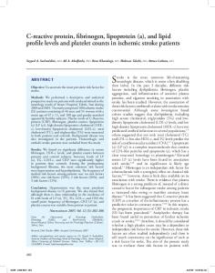
Figure 1 Type of subepidermal lymphoid infiltrate and density of CD8+ and CD1a+ cells in mycosis fungoides. (A,D) Microphotographs taken from two cases with different densities of neoplastic infiltrate and grades of epidermotropism: (A) mild lymphoid infiltrate, superficial, and with mild exocytosis and no tendency to confluence (infiltrate density and epidermotropism grades 1); (D) band-like subepidermal lymphoid infiltrate associated with more than three Pautrier’s microabscesses, one included in this field (infiltrate density and epidermotropism grades 3). (B,E) Immunostaining with anti-CD8 monoclonal antibody in two cases on frozen sections: (B) there are few and isolated dermal reactive lymphocytes in a case with dermal infiltrate grade 1 (CD8 positivity score 1); (E) CD8+ cells are numerous and form small groups in a case with dermal infiltrate grade 3 (CD8 positivity score 3). (C,F) Immunostaining with anti-CD1a monoclonal antibody in two cases on paraffin wax embedded sections: (C) there are few epidermal Langerhan’s cells and dermal dendritic cells, with no tendency to form small groups in a case with dermal infiltrate grade 1 (epidermal and dermal CD1a positivity score 1); (F) there are many Langerhan’s cells in all the epidermal layers with a tendency to cluster (epidermal CD1a positivity score 2), whereas the dermal dendritic cells are particularly numerous and form large aggregates (dermal CD1a positivity score 3): the case shows a dermal infiltrate grade 3.
expression,23 causing TIL apoptosis through the Fas pathway or inducing the expression of molecules such as killing inhibitor receptors on TILs.24 The proportion of TILs in MF tends to reduce with the increase in the lymphoid infiltrate.6 “With prolonged stimulation, subclones might appear that no longer require antigen dependent stimulation to proliferate, but show a tendency to infiltrate and disseminate” Our data on the density of LCs, DDCs, and TILs are consistent with the above immunological model. Our series is mostly made up of early cases, some showing a mild infiltrate. However, we saw a wide spectrum of disease, with the increase of the lymphoid infiltrate being accompanied by a higher clinical stage and a worse response to treatment. Interestingly, a large
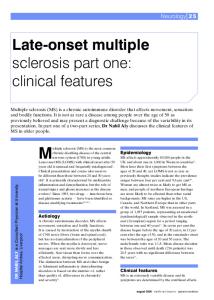
Figure 2 Distribution of CD8 positivity scores in the four groups with different grades of subepidermal lymphoid infiltrate.
www.jclinpath.com
proportion of patients with early disease and a mild lymphoid infiltrate showed a lower number of TILs compared with all the other patients. In daily pathology practice, patients like these represent a diagnostic problem between suspicious early MF or indeterminate lymphoid infiltrate. The diagnostic role of clonality and immunophenotypical analysis is still debated because aberrant phenotype and monoclonality have sometimes been described in bona fide reactive dermatoses.25 The diagnosis should be based on morphological features, such as the presence of lymphocytes with extremely convoluted, medium to large nuclei, single or clustered in the epidermis, and arranged in small sheets in the dermis.25 All our patients with low numbers of TILs showed the typical histological features of MF and were monoclonal with an aberrant phenotype. A possible explanation for low TIL density in these patients might be the absence of activation as a result of low
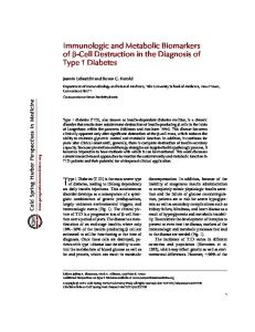
Figure 3 Distribution of dermal CD1a positivity scores in the four groups with different grades of subepidermal lymphoid infiltrate.
CD8+ and CD1a+ cells in MF
Take home messages • CD8+ T cells and CD1a+ dendritic cells appear to be reactive against tumour cells in mycosis fungoides (MF) because their numbers increase as the number of neoplastic cells increases • Our results support the notion that MF originates from the neoplastic transformation of T helper cells involved in a cutaneous immunological response • Thus, the interactions between MF cells and dendritic cells seems to be important both in inducing the proliferation of neoplastic cells and in recruiting reactive CD8+ cells, which are probably activated by the exposure to tumour specific antigens, particularly when the infiltrate increases
tumour specific antigen expression in MF cells. Thus, these cases could represent a very early step in MF; the next step could be the activation of TILs after exposure to a great number of tumour specific antigens. There is controversy on the soundness of treating these patients with early disease in the same way as those with more advanced MF. Large studies with long term follow up of patients with different proportions of CD8+ cells like those in our series are needed to clarify whether such patients exhibit a different clinical behaviour. Moreover, we found that cases with a denser lymphoid infiltrate rarely showed low TIL density, a feature strongly suggesting the activation of TILs against tumorous cells. The lack of evidence of TIL suppression in our four patients with a grade 4 subepidermal infiltrate could be related to the small group size, but may also reflect the inability of the neoplastic cells to downregulate antitumour immunity. A preserved immune reaction against tumour cells could explain the improved survival rate described in advanced MF with high numbers of TILs in some series.13 14 With regard to dendritic cells, we found no significant difference in epidermal LC numbers between groups with different densities of lymphoid infiltrate, suggesting that epidermal LCs could be equally active in all disease phases. Their density was not associated with epidermotropism. In contrast, a higher DDC density was seen in patients with a band-like and nodular dermal infiltrate. Our data are in line with previous observations15 26–28 suggesting that, with the progression of the malignancy, DDCs could present tumour specific antigens, derived from apoptotic MF cells, to other reactive T cells, thereby recruiting them. Under unfavourable circumstances, immature dendritic cells could be exposed to maturational blockers directly produced by neoplastic cells, such as interleukin 10, and would fail to become immunogenic, leading to downregulation of antitumour immunity and promoting tumour progression.29 The possibility of discriminating within the dendritic cell pool between elements at different maturational stages and with different functions could provide interesting information if applied to series with long term follow up. Moreover, the ability to enhance inhibitory dendritic cells and CD8+ TILs at early stages of MF would be an optimal basis for the therapeutic management of these patients, with the aim of suppressing MF cell proliferation and preventing disease progression and dissemination. In conclusion, the interactions between MF cells and dendritic cells seems to be important both in inducing the proliferation of neoplastic cells and in recruiting reactive CD8+ cells, which are probably activated by the exposure to tumour specific antigens, particularly when the infiltrate increases. Our results suggest that in MF, LC numbers are not associated with the density of neoplastic cells because different infiltrate grades show the same distribution, whereas numbers of TILs and DDCs tend to increase with the accumulation of neoplastic cells, suggesting that they are active against tumorous cells.
457
ACKNOWLEDGEMENTS The authors are indebted to all their colleagues at the Multicentric Study Group of Cutaneous Lymphoma of Marche Region (Italy), who offered their cases for the study: Dr V Agostini, Professor I Bearzi, Drs S Barulli, D Brancorsini, G Brandozzi, A Campanati, R Capretti, I Cataldi, A Giacchetti, M Giangiacomi, B Guiducci, S Lanari, L Morresi, G Mozzicafreddo, Professor AM Offidani, Drs M Offidani, S Pulini, R Ranaldi, G Ricotti, S Serresi, A Tassetti, MG Tucci (Ancona); M Ottaviani (Fabriano); G Ciattaglia, L Bugatti, G Filosa (Jesi); A Bettacchi, M Simonacci (Macerata); and E Grilli Cicilioni (San Severino). .....................
Authors’ affiliations G Goteri, A Filosa, B Mannello, D Stramazzotti, G Fabris, Institute of Pathology, University of Ancona, School of Medicine, Azienda Ospedaliera Umberto I-Torrette, 60020 Ancona, Italy S Rupoli, P Leoni, Department of Haematology, University of Ancona, School of Medicine, Azienda Ospedaliera Umberto I-Torrette
REFERENCES 1 Hansen ER. Immunoregulatory events in the skin of patients with cutaneous T-cell lymphoma. Arch Dermatol 1996;132:554–61. 2 Burg G, Dummer R, Haeffner A, et al. From inflammation to neoplasia: mycosis fungoides evolves from reactive inflammatory conditions (lymphoid infiltrates) transforming into neoplastic plaques and tumors. Arch Dermatol 2001;137:949–52. 3 Ralfkiaer E, Wantzin GL, Mason DY, et al. Phenotypic characterization of lymphocyte subsets in mycosis fungoides. Comparison with large plaque parapsoriasis and benign chronic dermatoses. Am J Clin Pathol 1985;84:610–19. 4 Ralfkiaer E. Immunohistological markers for the diagnosis of cutaneous lymphomas. Semin Diagn Pathol 1991;8:62–72. 5 Moll M, Reinhold U, Kukel S, et al. CD7-negative helper T cells accumulate in inflammatory skin lesions. J Invest Dermatol 1994;102:328–32. 6 Bakels V, van Oostveen JW, van der Putte SCJ, et al. Immunophenotyping and gene rearrangement analysis provide additional criteria to differentiate between cutaneous T-cell lymphomas and pseudo T-cell lymphomas. Am J Pathol 1997;150:1941–9. 7 Verga M, Braverman IM. The use of immunohistologic analysis in differentiating cutaneous T-cell lymphoma from psoriasis and dermatitis. Arch Dermatol 1991;127:1503–10. 8 Ashton-Key M, Diss TC, Du MQ, et al. The value of the polymerase chain reaction in the diagnosis of cutaneous T-cell infiltrates. Am J Surg Pathol 1997;21:743–47. 9 Kikuchi A, Naka W, Harada T, et al. Parapsoriasis en plaques: its potential progression to malignant lymphoma. J Am Acad Dermatol 1993;29:419–22. 10 Glusac EJ. Of cells and architecture: new approaches to old criteria in mycosis fungoides. J Cutan Pathol 2001;24:169–74. 11 Guitart J, Kennedy J, Ronan S, et al. Histologic criteria for the diagnosis of mycosis fungoides: proposal for a grading system to standardize pathology reporting. J Cutan Pathol 2001;28:174–83. 12 Vonderheid EC, Tan E, Sobel EL, et al. Clinical implications of immunologic phenotyping in cutaneous T-cell lymphoma. J Am Acad Dermatol 1987;17:40. 13 Hoppe RT, Medeiros J, Warnke RA, et al. CD8-positive tumor-infiltrating lymphocytes influence the long-term survival of patients with mycosis fungoides. J Am Acad Dermatol 1995;32:448–53. 14 Vermeer MH, Van Doorn R, Dukers D, et al. CD8+ T cells in cutaneous T-cell lymphoma: expression of cytotoxic proteins, Fas ligand and killing inhibitory receptors and their relationship with clinical behaviour. J Clin Oncol 2001;19:4322–9. 15 Fujita M, Horiguchi Y, Miyachi Y, et al. A subpopulation of Langerhan’s cells (CD1a+Lag−) increased in the dermis of plaque lesions of mycosis fungoides. J Am Acad Dermatol 1991;25:491–9. 16 Meissner K, Michaelis K, Rehpenning W, et al. Epidermal Langerhan’s cell densities influence survival in mycosis fungoides and Sezary syndrome. Cancer 1990;65:2069–73. 17 Hansen ER, Bang B, Larsen JK, et al. In cutaneous T cell lymphoma, class II MHC molecules on CD1+ antigen-presenting cells are upregulated in involved compared to uninvolved epidermis. Br J Dermatol 1994;131:780–8. 18 Bunn PA, Lamberg SI. Report of the committee on staging and classification of cutaneous T-cell lymphomas. Cancer Treat Rep 1979;63:581. 19 Lamberg S, Green S, Byar D, et al. Clinical staging for cutaneous T-cell lymphoma. Ann Intern Med 1984;100:187. 20 Disse TC, Watts M, Pan LX, et al. The polymerase chain reaction in the demonstration of monoclonality in T-cell lymphomas. J Clin Pathol 1995;48:1045–50. 21 Rupoli S, Barulli S, Guiducci B, et al. Low dose interferon α2b combined with PUVA is an effective treatment of early stage mycosis fungoides: results of a multicentric study. Haematologica 1999;84:809–13. 22 Edelson RL. Cutaneous T cell lymphoma: the helping hand of dendritic cells. Ann N Y Acad Sci 2001;941:1–11.
www.jclinpath.com
458
Goteri, Filosa, Mannella, et al
23 Ni X, Hazarika P, Zhang C, et al. Fas ligand expression by neoplastic T lymphocytes mediates elimination of CD8+ cytotoxic T lymphocytes in mycosis fungoides: a potential mechanism of tumor immune escape? Clin Cancer Res 2001;7:2682–92. 24 Zoi-Toli O, Vermeer MH, De Vries E, et al. Expression of Fas and Fas-ligand in primary cutaneous T-cell lymphoma (CTCL): association between lack of Fas expression and aggressive types of CTCL. Br J Dermatol 2000;143:313–19. 25 Santucci M, Biggeri A, Feller AC, et al. Efficacy of histologic criteria for diagnosing early mycosis fungoides: an EORTC cutaneous lymphoma study group investigation. Am J Surg Pathol 2000;24:40–50.
26 Fivenson DP, Nickoloff BJ. Distinctive dendritic cell subsets expressing factor XIIIa, CD1a, CD1b and CD1c in mycosis fungoides and psoriasis. J Cutan Pathol 1995;22:223–8. 27 Jimbow K, Chiba M, Horikoshi T. Electron microscopic identification of Langerhan’s cells in the dermal infiltrates of mycosis fungoides. J Invest Dermatol 1982;78:102–7. 28 Bagot M, Echchakir H, Mami-Chouaib F, et al. Isolation of tumor-specific cytotoxic CD4+ and CD4+CD8 dim+ T-cell clones infiltrating a cutaneous T cell lymphoma. Blood 1998;11:4331–41. 29 Lüftl M, Feng A, Licha E, et al. Dendritic cells and apoptosis in mycosis fungoides. Br J Dermatol 2002;147:1171–9.
Clinical Evidence—Call for contributors
Clinical Evidence is a regularly updated evidence based journal available worldwide both as a paper version and on the internet. Clinical Evidence needs to recruit a number of new contributors. Contributors are health care professionals or epidemiologists with experience in evidence based medicine and the ability to write in a concise and structured way. Currently, we are interested in finding contributors with an interest in the following clinical areas: Altitude sickness; Autism; Basal cell carcinoma; Breast feeding; Carbon monoxide poisoning; Cervical cancer; Cystic fibrosis; Ectopic pregnancy; Grief/bereavement; Halitosis; Hodgkins disease; Infectious mononucleosis (glandular fever); Kidney stones; Malignant melanoma (metastatic); Mesothelioma; Myeloma; Ovarian cyst; Pancreatitis (acute); Pancreatitis (chronic); Polymyalgia rheumatica; Post-partum haemorrhage; Pulmonary embolism; Recurrent miscarriage; Repetitive strain injury; Scoliosis; Seasonal affective disorder; Squint; Systemic lupus erythematosus; Testicular cancer; Varicocele; Viral meningitis; Vitiligo However, we are always looking for others, so do not let this list discourage you. Being a contributor involves: • Appraising the results of literature searches (performed by our Information Specialists) to identify high quality evidence for inclusion in the journal. • Writing to a highly structured template (about 2000–3000 words), using evidence from selected studies, within 6–8 weeks of receiving the literature search results. • Working with Clinical Evidence Editors to ensure that the text meets rigorous epidemiological and style standards. • Updating the text every eight months to incorporate new evidence. • Expanding the topic to include new questions once every 12–18 months.
If you would like to become a contributor for Clinical Evidence or require more information about what this involves please send your contact details and a copy of your CV, clearly stating the clinical area you are interested in, to Claire Folkes (
[email protected]). Call for peer reviewers
Clinical Evidence also needs to recruit a number of new peer reviewers specifically with an interest in the clinical areas stated above, and also others related to general practice. Peer reviewers are health care professionals or epidemiologists with experience in evidence based medicine. As a peer reviewer you would be asked for your views on the clinical relevance, validity, and accessibility of specific topics within the journal, and their usefulness to the intended audience (international generalists and health care professionals, possibly with limited statistical knowledge). Topics are usually 2000–3000 words in length and we would ask you to review between 2–5 topics per year. The peer review process takes place throughout the year, and our turnaround time for each review is ideally 10–14 days. If you are interested in becoming a peer reviewer for Clinical Evidence, please complete the peer review questionnaire at www.clinicalevidence.com or contact Claire Folkes (
[email protected]).
www.jclinpath.com