J Vasc Access 2010; 11 ( 2 ) : 115 -121
ORIGINAL ARTICLE
Long thoracic vein embolization for the treatment of breast edema associated with central venous occlusion and venous hypertension Gregg Arthur Miller1, Alexander Friedman1, Aleksandr Khariton1, Manish C. Jotwani2, Yevgeny Savransky3 American Access Care of Brooklyn, Brooklyn, NY - USA American Access Care of Queens, Queens, NY - USA 3 American Access Care of Bellmore, Bellmore, NY - USA 1 2
ABSTRACT Purpose: Breast edema is a rare complication in hemodialysis patients with central venous occlusions. The present study sought to determine whether coil embolization of the long thoracic vein is an effective long-term treatment for this pathology. Methods: The study patients were 6 female hemodialysis patients whose primary clinical manifestation of central vein occlusion was breast edema. When conservative treatment (allowing collaterals to dilate over time), as well as recanalization of occlusions through angioplasty with or without stent placement, failed to alleviate symptoms, patients underwent coil embolization of the long (lateral) thoracic vein. Results: In 4 of the 6 cases, the breast edema was completely resolved without recurrence, while the other 2 patients experienced durable symptomatic improvement with only mild residual swelling. Average follow-up was 22 months. There were no adverse sequelae and none of the patients experienced increased swelling elsewhere following the coil embolization procedure. Conclusions: Coil embolization of the long thoracic vein effectively alleviates breast edema in hemodialysis patients with elevated venous hydrostatic pressure due to central venous occlusions. Keywords: Breast edema, Central venous stenosis, ESRD, Hemodialysis, Breast swelling, Venous embolization
INTRODUCTION
rization, unnecessary delays in diagnosis should be avoided by first ruling out central venous disease.
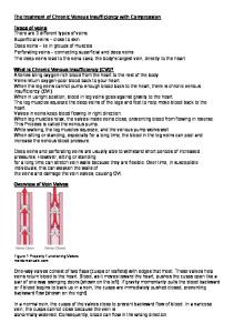
Central venous stenosis (CVS) or occlusion (CVO) refers to the partial or complete obstruction of any central vein (e.g., subclavian, brachiocephalic, superior vena cava) (1). CVS is a common complication of hemodialysis (HD) accesses, occurring in 19-41% of HD patients (2, 3). Endovascular trauma, linked to HD catheters and permanent pacemaker wires, is implicated in the majority of reported cases of CVS (4-8). The symptoms of CVS and CVO include edema of the face, neck, chest, and extremity. Elevated peripheral and central venous pressures are an immediate consequence of arteriovenous HD shunt creation. Severe stenosis or complete occlusion of the central veins leads to recruitment of collateral veins in order to accommodate venous return to the right atrium. When the total volume of flow and pressure exceeds the carrying capacity of collaterals, the result is clinically significant tissue edema of the arm, face, and (infrequently) breast (9, 10) (Fig. 1). Though rare, breast edema secondary to CVS or CVO is an important differential diagnosis to consider in endstage renal disease (ESRD) patients. Clinically expressed as breast pain, engorgement, erythema, and peau d’orange appearance, venous collateral hypertension of the long thoracic vein (LTV) can mimic inflammatory breast cancer (10). In HD patients with a history of central vein cathete-
METHODS From July 2005 through to August 2009, HD patients with breast edema were referred to a freestanding outpatient vascular access center. All relevant demographic information (including the presence of arm edema, access type, and history of HD catheter placement) was recorded for each patient, and all patients provided written informed consent for the procedure. The ethics committee decided approval was not required for this study. All patients had breast enlargement; 5/6 patients were referred to the center with chief complaints of arm, face, and chest edema; 1/6 was a transplant patient with a known central occlusion who was referred specifically for breast edema. Other symptoms included pain (3/6) and skin changes mimicking inflammatory breast carcinoma (3/6). Comprehensive carcinoma work-ups including mammograms (6/6) and needle biopsies (3/6) had been performed prior to presentation to our office; 2/6 patients had a history of severe bleeding following a biopsy which necessitated transfusion.
© 2010 Wichtig Editore - ISSN 1129-7298
115
Long thoracic vein embolization
Fig. 1 - Breast edema, prior to coil embolization.
Procedure Diagnostic angiograms were performed via an ipsilateral functioning HD access when available. Alternatively, the ipsilateral axillary vein was directly cannulated using ultrasound guidance. Attempts to open brachiocephalic and subclavian occlusions were approached via the axillary, femoral, external jugular and internal jugular veins. If symptoms persisted following initial intervention attempts, patients returned after 1 week for embolization of the long thoracic vein. Under ultrasound guidance, access was gained to the axillary vein using a 21-gauge micro-access needle (Cook, Bloomington, IN) which was upsized to a 5F vascular sheath (Terumo, Tokyo, Japan). Injections of intravenous contrast were performed through the 5F sheath and imaging (GE OEC 9800 C-arm) of the axillary vein and collaterals was performed. Central venous imaging was also performed through a 4-French diagnostic Bern catheter (Boston Scientific, Natick, MA).
Detailed imaging of axillary collateral veins was performed and flow from the subclavian vein into a hypertrophied LTV (toward the nipple) was noted. Using a 4F diagnostic catheter with a 180 degree turn and a 0.035 non-stiff Glidewire (Terumo Med Corp, Somerset, NJ), the LTV was selectively cannulated and imaged. Two Nester coils (4-10 mm diameter; Cook, Bloomington, IN) were deployed in the LTV in order to form a hemostatic plug. Selective imaging was again performed confirming the occlusion of the LTV following coil placement (Fig. 2). Alternatively, the LTV was directly cannulated under ultrasound guidance and then embolized. Versed (HoffmanLaRoche Ltd, Mississauga, Ontario, Canada) and fentanyl (Fentanyl-Janssen, Neuss, Germany) were administered during the procedure to achieve conscious sedation. Oxilan (Guerbet, Bloomington, IN) intravenous contrast was used for imaging. RESULTS Six female ESRD patients were treated. Patient ages ranged from 28-79 yrs (mean = 64). Co-morbidities included hypertension (6/6), diabetes mellitus (3/6), coronary artery disease (2/6), and congestive heart failure (1/6). Three patients had a functioning ipsilateral HD access (2/6 graft, 1/6 fistula), two patients had ipsilateral catheters, and one patient had a functional transplant. The transplant patient had one event of an HD catheter (6 months duration) and had never undergone arteriovenous shunt placement. Five patients had a history of ipsilateral HD catheter placement, while the remaining patient had an ipsilateral permanent pacemaker (Tab. I). At presentation, angiography demonstrated complete occlusion of the central veins in all six patients (four brachiocephalic, two subclavian). Central venous flow was fully restored in 3/6 patients
Fig. 2 - Left long thoracic vein showing retrograde flow towards the nipple before embolization and lack of flow following coil placement.
116
Miller et al
through angioplasty (2/6) and stent placement (1/6). Of these patients, restoration of angiographically normal flow through the central veins did not resolve the breast edema in any of the three cases over a period of 1 week. A hypertrophied long thoracic vein (dilated beyond 4 mm) with retrograde flow (away from the subclavian vein and toward the nipple) was observed in all patients. Therefore, coil embolization of the LTV was performed in all six patients. Follow-up time ranged from 18-31 months (mean = 22 months); 4/6 patients reported having no residual swelling (Tab. II); 1/6 patients reported having a venous stasis ulcer which healed following the coil embolization procedure. Although some residual breast swelling remained in patients 3 and 4, a significant reduction in edema was reported following the procedure. No patients developed a symptomatic increase in swelling of the arm or face following the embolization procedure. There were no major complications or adverse events associated with the procedure.
conditions, the direction of flow in the LTV is away from the breast, emptying into the subclavian vein, with alternate drainage into intercostal veins. The presence of a CVO leads to an increase in venous hydrostatic pressure, which is transmitted to these collateral vessels (9, 12, 13). In HD patients with an arteriovenous access, the greater demand placed on these pathways by increases in upper extremity blood flow results in hypertrophy of collateral venous channels and microvascular leak (10), expressed symptomatically as breast edema. The pressure gradient resulting from occluded central veins can be identified angiographically as reversal of flow (toward the peripheral circulation) (14). In the case of the LTV, the direction of flow is reversed, resulting in retrograde flow into the breast (towards the nipple; Fig. 4), rather than antegrade into the subclavian vein. In all six patients that we treated, CVO was associated with breast engorgement. We believe that complete occlusion - rather than merely stenosis - of the central veins is the impetus for the development of breast edema. Treatment options for symptomatic, HD-related CVO range from percutaneous interventions, to surgical bypass procedures, and access ligation. Due to the limited number of alternate access sites, access ligation should only
DISCUSSION The normal venous drainage of the breast is comprised of intercostal veins and deep branches of the axillary vein, including the LTV (11) (Fig. 3). In non-pathologic TABLE I - PATIENT DEMOGRAPHICS Patient #
1
2
3
Access type & location Right TBV fistula IVC Cath, Immature IJ Cath left BCV fistula Hx of cath or PPM Left PermCath Bilateral PermCath Bilateral PermCath Chief complaint Left breast Left breast & arm Bilateral breast edema edema edema Location of occlusion Occlusion left BCV Occlusion left BCV Occlusion bilateral BCV
4
5
None Left forearm (functioning loop graft transplant) Right Left PPM PermCath Right breast & arm Left breast edema edema Occlusion right Occlusion left BCV SCV
6 LUE C-shaped graft Left PermCath Left breast & arm edema Occlusion left SCV
Abbreviations: TBV = transposed basilic vein; IJ = internal jugular; BCV = brachiocephalic vein; IVC = inferior vena cava; SCV = subclavian Vein; Cath = catheter; Hx = history; LTV = long thoracic vein; LUE = left upper extremity; PPM = permanent pacemaker
TABLE II - TREATMENTS, OUTCOMES, AND FOLLOW-UP Patient #
1
2
Angioplasty No No Stent placement & location No No Coil embolization Left LTV Left LTV F/U time 31 months 27 months F/U results, breast No residual No residual edema swelling swelling Arm edema before/after −/− +/+
3
4
5
6
4x: Bilateral BCV, SCV
No
Yes: L SCV
Yes: L SCV
2x: Right BCV and SCV Right LTV 18 months Mild residual swelling +/−
No
No
No
Right LTV 18 months Mild residual swelling +/+
Left LTV 18 months No residual swelling −/−
Left LTV 18 months No residual swelling +/+
Abbreviations: BCV = brachiocephalic vein; SCV = subclavian vein; LTV = long thoracic vein
117
Long thoracic vein embolization
Fig. 3 - Normal blood flow drains from the breast, toward the axillary vein, in the absence of venous obstruction.
Fig. 4 - Left long thoracic vein showing retrograde flow towards the nipple before embolization, and the resultant breast edema.
be a treatment of last resort. Open surgical interventions to bypass the central occlusion have been described, but are complex and rarely reported in the literature (15). Currently, the accepted form of intervention is percutaneous transluminal angioplasty and stent placement, but less than optimal treatment durability makes re-interventions the norm (9). 118
Eighteen cases (in 14 studies) of HD-associated unilateral breast edema and its subsequent treatment have been described in the literature (Tab. III). Treatments have included conservative treatment for persistent but tolerable symptoms (16), angioplasty of the CVO and/or stent placement (9, 10, 17-22), and ligation or excision of the access (14, 20, 23-25). (In two reported cases, the course
Miller et al
Fig. 5 - Post-embolization, breast edema is greatly reduced, and blood flow is redirected towards collaterals.
TABLE III - REPORTED CASES OF BREAST EDEMA IN HEMODIALYSIS PATIENTS Study, Case
History
Presentation
Treatment
Wilson (26) cath SV occlusion N/A Kavallieratos et al, #1 (15) N/A BC vein occlusion axillary to saphenous vein bypass Kavallieratos et al, #2 (15) N/A BC vein occlusion axillary to saphenous vein bypass Phillips et al, #2 (20) HTN, cath graft graft removal Wright et al (25) DM, cath BC AVF kidney transplant, ligation Ruiz-Valverde et al (23) AVF (no stenosis) ligation Topf et al (14) RCAVF, SV occlusion ligation Uzun et al (24) (no prior cath) high flow BC AVF ligation Gadallah et al (16) SV occlusion none Akan et al (27) cath SV stenosis N/A Kim et al (17) HTN BC AVF PTA Kuerer et al (18) cath graft, elevated pressure, BCS PTA Phillips et al, #1 (20) DM, HTN, cath graft, BC occlusion PTA Phillips et al, #3 (20) HTN graft, BC occlusion PTA Phillips et al, #4 (20) DM, HTN graft, SVC occlusion, IJS PTA Youssef et al (22) COPD, HTN, cath graft, Jx stenosis (no central stenoses) PTA Blum et al (10) cath BC stenosis PTA; PTA 6 months later Pacheco et al (19) cath BC occlusion PTA, stent
Outcome N/A Death (unrelated); bridge graft and access patent Thrombectomy of graft-saphenous anastomosis; partial resolution of edema, access in use, bridge graft patent complete relief complete relief complete relief near complete relief complete relief tolerable N/A persistent edema significant relief complete relief complete relief complete relief significant relief partial recurrence; complete relief complete relief
Abbreviations: cath=history of catheterization; SVC=superior vena cava; IJ=internal jugular vein; SV=subclavian vein; PTA=percutaneous transluminal angioplasty; DM=diabetes mellitus; HTN=hypertension; COPD=chronic obstructive pulmonary disease; BC=braciocephalic; AVF=arteriovenous fistula; RC=radiocephalic; ligation=hemodialysis access ligation
of treatment is not described (26, 27).) Treatments for CVO have also included sharp recanalization of the central vein (28) and atrial bypass grafting (29). Kavallieratos et al reported eight HD patients with symptomatic CVO, two of which experienced breast edema. These two patients
were treated with an axillary to saphenous vein bypass. One patient experienced recurrent symptoms with thrombosis of the graft and only partial improvement; the other patient had complete symptomatic improvement (15). To date, no treatments have been described to directly ad119
Long thoracic vein embolization
dress the problem of breast swelling. In rare cases of severe, long-standing CVO, restoration of central venous flow may not completely eliminate breast swelling, as the chronicity of high venous pressure and retrograde flow causes irreversible physiological changes in the venous valves elsewhere in the system. Hypertrophy of the central veins (e.g., subclavian, brachiocephalic) causes the venous valves to become incompetent, continuing to deliver flow into established collaterals (i.e., hypertrophied LTV) despite reopening of the occlusion and reduction of venous hydrostatic pressure. The cutaneous extra-thoracic venous system is especially susceptible to distension due to the lack of venous valves, as the pressure is not attenuated. Additionally, the typical musculature which promotes unidirectional venous flow is lacking in breast tissue. In such cases, coil embolization of the LTV will compensate for the altered physiology and alleviate symptoms by preventing retrograde flow into the LTV. Closing this low-pressure outlet forces the blood to drain through other collaterals. It is observed that following coil embolization, flow is redirected from the LTV into previously underdeveloped collaterals (Fig. 5). Since breast tissue is the most compliant, it is the most susceptible to distension. In our experience, redirecting flow into other, less distensible collateral drainage systems has the desired effect of alleviating symptoms of breast swelling without any symptomatic increase in swelling elsewhere. Female ESRD patients suffering from breast edema should be studied angiographically with significant consideration
given to CVO as an etiology in the differential diagnosis. When reopening the CVO fails to alleviate symptoms, coil embolization of the LTV can be easily and safely performed. In addition, coil embolization alone is an effective, safe and minimally invasive treatment method of achieving symptomatic relief of breast edema. The understanding of venous pathology in this subset of patients can expedite diagnosis and prevent complications associated with performing breast biopsies on tissue with elevated venous pressure.
REFERENCES
permanent cardiac pacemakers and the haemodialysed patient. Nephron 1991; 58: 103-5. 7. Chuang CL, Tarng DC, Yang WC, Huang TP. An occult cause of arteriovenous access failure: central vein stenosis from permanent pacemaker wire. Report of three cases and review of the literature. Am J Nephrol 2001; 21: 406-9. 8. Fourestie V, Godeau B, Lejonc JL, Schaeffer A. Left innominate vein stenosis as a late complication of central vein catheterization. Chest 1985; 88: 636-8. 9. Bakken AM, Protack CD, Saad WE, et al. Long-term outcomes of primary angioplasty and primary stenting of central venous stenosis in hemodialysis patients. J Vasc Surg 2007; 45: 776-83. 10. Blum C, Baker M. Venous congestion of the breast mimicking inflammatory breast cancer: case report and review of literature. Breast J 2008; 14: 97-101. 11. Rosen P. Rosen’s Breast Pathology. Lippincott Williams & Wilkins: New York, 2001.
1.
2.
3.
4.
5. 6.
120
Kwok PC. Endovascular treatment for central venous stenosis due to central vein catheterization for hemodialysis. Saudi J Kidney Dis Transpl 2004; 15: 338-45. Agarwal A, Patel B, Farhan N. Central venous stenosis in hemodialysis patients is a common complication of ipsilateral central vein catheterization. J Am Soc Nephrol 2004; 15: 368A-9A. MacRae JM, Ahmed A, Johnson N, et al. Central vein stenosis: a common problem in patients on hemodialysis. ASAIO J 2005; 51: 77-81. Kwak JY, Kim EK, Chung SY, You JK, et al. Unilateral breast edema: spectrum of etiologies and imaging appearances. Yonsei Med J 2005; 46: 1-7. Agarwal AK, Patel BM, Haddad NJ. Central vein stenosis: a nephrologist’s perspective. Semin Dial 2007; 20: 53-62. Korzets A, Chagnac A, Ori Y, et al. Subclavian vein stenosis,
ACKNOWLEDGEMENTS Artwork for Figures 3, 4, and 5 was created by Carl Boisson. Michael Alesi, RT and John Gravelli, RT provided expert technical advice in order to make these procedures possible and successful. Conflict of interest: None of the authors have a proprietary interest in the products used in this study. No financial support was provided for this study.
Address for correspondence: Gregg Miller, MD Medical Director American Access Care of Brooklyn 577 Prospect Avenue. Brooklyn, NY 11215
[email protected]
Miller et al
12. Criado E, Marston WA, Jaques PF, et al. Proximal venous outflow obstruction in patients with upper extremity arteriovenous dialysis access. Ann Vasc Surg 1994; 8: 530-5. 13. Itkin M, Kraus MJ, Trerotola SO. Extrinsic compression of the left innominate vein in hemodialysis patients. J Vasc Interv Radiol 2004; 15: 51-6. 14. Topf G, Jenkins P, Gutmann FD, et al. Unilateral breast enlargement. A complication of an arteriovenous fistula and coincidental subclavian vein occlusion. JAMA 1977; 237: 571-2. 15. Kavallieratos N, Kokkinos A, Kalocheretis P. Axillary to saphenous vein bypass for treatment of central venous obstruction in patients receiving dialysis. J Vasc Surg 2004; 40: 640-3. 16. Gadallah MF, el-Shahawy MA, Campese VM. Unilateral breast enlargement secondary to hemodialysis arteriovenous fistula and subclavian vein occlusion. Nephron 1993; 63: 351-3. 17. Kim MJ, Kim EK, Oh KK. Unusually asymmetric venous engorgement of the breast after long-term hemodialysis. J Clin Ultrasound 2006; 34: 27-9. 18. Kuerer HM, Wilson MW, Bowersox JC. Innominate vein stenosis mimicking locally advanced breast cancer in a dialysis patient. Breast J 2001; 7: 128. 19. Pacheco D, Polo JR, Lopez-Baena JA, et al. Unilateral breast enlargement secondary to right brachiocephalic vein occlusion. Am J Kidney Dis 2000; 35: E26. 20. Phillips GS, Scheel PJ, Jr, Zeiger MA. Unilateral breast enlargement: four case reports of an “unusual” presentation of central vein stenosis in patients undergoing hemodialysis. Surgery 1998; 123: 699-701.
21. Clark TW, Cohen RA, Kwak A, et al. Salvage of nonmaturing native fistulas by using angioplasty. Radiology 2007; 242: 286-92. 22. Youssef JJ, Brown CD, Friedman EA. Asymmetric breast enlargement minus central venous thrombosis in a hemodialysis patient. Hemodial Int 2008; 12: 30-3. 23. Ruiz-Valverde MP, Fort J, Camps J, et al. Unilateral breast and arm enlargement secondary to haemodialysis arteriovenous fistula without subclavian vein occlusion. Nephrol Dial Transplant 1994; 9: 85-6. 24. Uzun M, Akkan MK, Uzun F, Karaosmanogu D. Asymmetric venous engorgement of the breast due to a high output hemodialysis fistula. J Clin Ultrasound 2009; 37: 163-4. 25. Wright RS, Quinones-Baldrich WJ, Anders AJ, Danovitch GM. Pleural effusion associated with ipsilateral breast and arm edema as a complication of subclavian vein catheterization and arteriovenous fistula formation for hemodialysis. Chest 1994; 106: 950-2. 26. Wilson CM. An unusual cause of asymmetric venous engorgement detected by mammography. Can Assoc Radiol J 1993; 44: 42-4. 27. Akan H, Arik N, Yalin T, Malazgirt Z. Unilateral breast enlargement: a rare complication of subclavian vein catheterization for hemodialysis. Comput Med Imaging Graph 2001; 25: 357-9. 28. Farrell T, Lang EV, Barnhart W. Sharp recanalization of central venous occlusions. J Vasc Interv Radiol 1999; 10: 149-54. 29. El-Sabrout RA, Duncan JM. Right atrial bypass grafting for central venous obstruction associated with dialysis access: another treatment option. J Vasc Surg 1999; 29: 472-8.
121