Osteophytes in cervical vertebrae
Rev Arg de Anat Clin; 2011, 3 (1): 15-21
__________________________________________________________________________________________
Original Communication
LOCATIONS AND LENGTHS OF OSTEOPHYTES IN THE CERVICAL VERTEBRAE Patcharin Chanapa1, Pasuk Mahakkanukrauh 2* 1
Department of Basic Science, Faculty of Science, Payap University, Chiang Mai, Thailand Department of Anatomy, Faculty of Medicine, Chiang Mai University, Chiang Mai, Thailand
2
RESUMEN Muchos pacientes sufren de disfagia, vértigo, dolor en el brazo, entumecimiento o debilidad. Estos problemas pueden ser debidos a la aparición de osteofitos en las vértebras cervicales. El propósito de esta investigación ha sido estudiar las localizaciones y tamaño de los osteofitos en las vértebras cervicales. Se han usado 200 columnas cervicales (139 varones y 61 mujeres) de vértebras secas C3-C7, de un promedio de edad de 71 años (36-98 años). Se han encontrado osteofitos en 184 columnas (92 %), la mayoría en C5, C6, C4, C7 y C3 (83, 77, 74, 65 y 64%, respectivamente). La media del tamaño de los osteofitos en C3 (4.44 ± 1.31 mm) ha sido mayor que los de C4-C7. La mayor cantidad de osteofitos se encontraron en los cuerpos vertebrales, carilla articular y foramen transverso (49,35 y 16%) respectivamente. La mayor longitud de los osteofitos en el cuerpo de las vértebras se encontraron en la vértebra fue 4.28 ± 1.65 mm en C6, en la cara articular fue 5.07 ± 1.57 mm en C5 y en el transverso foramen fue 2.49 ± 1.57 mm en C6. La longitud de los osteofitos del lado anterior superior y de la cara inferior del cuerpo ha sido más larga que la de los lados posterior y lateral. La longitud de los osteofitos muestra una correlación significativa y directa con la edad. Conclusión: Los osteofitos que han aparecido en el cuerpo de las vértebras, la cara y el foramen transverso pueden incidir en las estructuras cercanas. Este estudio puede ayudar a explicar algunos problemas clínicos como la disfagia, insuficiencia vertebrobasilar y braquialgia. Palabras clave: osteofito, disfagia, braquialgia, vértigo
espondilosis
cervical,
ABSTRACT Many patients suffer from dysphagia, vertigo, arm pain, numbness or weakness. These problems may arise
from osteophytes in the cervical vertebrae. The purpose was to study the distribution and lengths of osteophyte in the cervical vertebrae. We used 200 cervical columns (139 male and 61 female) of dry C3C7 vertebrae. Osteophytes were found in 184 columns (92%), mostly at C5, C6, C4, C7 and C3 (83, 77, 74, 65 and 64% respectively) . The average length of osteophytes of C3 (4.44 ± 1.31 mm) was longer than those of C4-C7. The quantity of osteophytes mostly was found at vertebral bodies, articular facets and transverse foramen (49, 35 and 16%) respectively. The greatest osteophyte length of vertebral bodies was at C6 (4.28 ± 1.65 mm.), that of articular facet was at C5 (5.07 ± 1.57 mm.) and that of foramen transversarium was at C6 (2.49 ± 1.57 mm.). The osteophyte length of anterior area of superior and inferior surface of body was longer than posterior and lateral area. The osteophyte length was significantly correlated with age. Conclusion: The osteophytes that occurred at vertebral bodies, facet and transverse foramen may impinge on nearby structures. This study may help in explaining some clinical problems such as dysphagia, vertebrobasilar insufficiency and brachialgia. Keywords: osteophyte, cervical dysphagia, brachialgia, vertigo
spondylosis,
* Correspondence to: Prof. Pasuk Mahakkanukrauh, MD, Department of Anatomy, Faculty of Medicine, Chiang Mai University, Chiang Mai 50200, Thailand.
[email protected] Received: 6 November, 2010. Revised: 18 November, 2010. Accepted: 10 December, 2010.
15 Todos los derechos reservados.
Osteophytes in cervical vertebrae
Rev Arg de Anat Clin; 2011, 3 (1): 15-21
__________________________________________________________________________________________
INTRODUCTION
MATERIALS AND METHODS
Many symptoms may arise from problems caused by osteophytes in the cervical vertebrae. These symptoms include vertebrobasilar insufficiency (VBI) characterized by vertigo, visual disturbances (Bulsara et al., 2006; Bayrak et al., 2009; Takeuchi et al., 2009), degenerative compressive radiculopathy and myelopathy ausing arm pain, numbness or weakness (Harrop et al., 2007; Shedid et al., 2007; White et al., 2007). Some patients have dysphagia caused by ventral osteophytes of the cervical vertebrae (Constantoyannis et al., 2008; Oppenlander et al., 2009; Seidler et al., 2009). The osteophytes occur in various locations along the cervical vertebrae, producing different symptoms. Earlier studies have focused on patients’ symptoms and management. But, there are few studies which research at the locations or lengths of osteophytes in the cervical vertebrae. Therefore, the objectives of this study were to find out the locations and lengths of osteophytes in the cervical vertebrae between C3-C7. As C1-C2 are atypical cervical vertebrae, they were not studied.
Two hundred Thai specimens of dry cervical spines between C3 and C7 which were made at the Department of Anatomy, Faculty of Medicine, Chiang Mai University were used in the present study. Four groups of age were divided into 3550, 51-65, 66-80 and 81-95 years old (8, 46, 119 and 27 cervical spines, respectively). The average age was 71 years old. They were composed of 139 males and 61 females. Osteophytes were observed and measurements of lengths were taken by vernier caliper in the horizontal plane from the edge of the body, articular facet and transverse foramen of the cervical vertebrae to the longest tip of osteophytes. Locations were the superior and inferior surfaces of the body (anterior, posterior and lateral sides), the right and left of superior and inferior articular facets and inside of the transverse foramen. Statistical analysis was performed by the descriptive analysis and Pearson correlation in SPSS version 16.0 for Windows.
Body
Facet
Sup.surface A
B
Ant.
Post
C3
4.44 ±1.31 9.75
3.72 ±1.27 8.5
4.6 (9)
3.0 (3)
C4
4.31 ±1.60 10.50
3.86 ±1.57 10.50
4.5 (10)
C5
4.11 ±1.49 10.00
4.15 ±1.59 12.00
C6
4.08 ±1.22 7.00
C7
4.26 ±1.43 9.00
Inf.surface
Sup.f acet
TF Inf. facet
Ant.
Post.
Lat.
C
3.7 (4)
4.2 (10)
3.5 (4)
3.5 (6)
4.92 ±1.35 9.75
5.29 (10)
4.89 (9.5)
1.83 ±0.75 3.00
2.7 (4)
3.2 (6)
4.5 (12)
2.2 (4)
3.6 (9)
4.99 ±1.74 13.00
5.38 (13)
4.78 (9)
2.07 ±0.89 5.00
4.9 (12)
1.0 (1)
3.5 (6)
4.4 (13)
1.0 (1)
3.6 (6)
5.07 ±1.57 9.00
5.31 (9)
4.99 (9)
2.08 ±0.86 4.00
4.28 ±1.65 12.00
4.6 (11)
1.4 (2)
4.0 (8.5)
4.7 (13)
2.0 (3)
4.2 (9)
4.95 ±1.45 8.00
4.88 (8)
5.11 (7)
2.49 ±0.75 4.50
4.20 ±1.51 9.00
4.5 (12)
3.5 (4)
4.1 (10.5)
4.1 (9)
1.3 (2)
3.7 (5)
4.88 ±1.34 8.00
4.60 (8)
5.04 (8)
2.00 ±0.00 2.00
Lat.
D
Table 1. The Mean length and Maximum Length (Max.) of Osteophytes (mm.) at the Body, Articular Facet and Transverse Foramen. A= Mean±SD, Max. of each level. B = Mean±SD, Max.of Boby. C = Mean±SD, Max.of Facet. D= Mean±SD, Max.of Transverse foramen.
16 Todos los derechos reservados.
Osteophytes in cervical vertebrae
Rev Arg de Anat Clin; 2011, 3 (1): 15-21
__________________________________________________________________________________________
RESULTS Locations of Osteophytes Of the two hundred cervical spines, osteophytes were found in 184 spines (92%), most at C5. The osteophytes in cervical vertebrae were mostly found in the body, articular facet and transverse foramen (49, 35 and 16%, respectively). The percentage growth protruding from the edge of the superior and inferior surfaces of the body to
the anterior side, intervertebral foramen and vertebral canal were 53, 42 and 5%, respectively. The quantity of osteophytes was not related significant to gender. The specimens of housewives and market traders had the most osteophytes at C3 compared with other occupations.
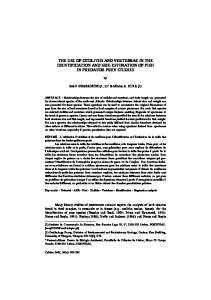
Figure 1 – Osteophytes in the transverse foramen (arrows) at C6.
Lengths of Osteophytes The average length osteophytes was greatest at C3. The maximum length was 10.5 mm at C4 (Table 1). The average lengths of osteophytes in the articular facets between C3 and C7 were longer than those of vertebral bodies and transverse foramens as shown in Table1. The greatest length of osteophyte in the vertebral bodies was 4.28±1.65 mm at C6 and the maximum length was 12 mm. That of the articular facet was 5.07±1.57 mm at C5 and the maximum length was 9 mm. That of the transverse foramen was 2.49±0.75mm at C6 and the maximum length was 4.5 mm. (Table 1 and Fig. 1).The
average length in the anterior side of superior and inferior surfaces in the vertebral bodies was longer than those in the posterior and lateral side (Table 1). The greatest length of osteophytes in the anterior side of the superior surface was 4.9 mm at C5 and the maximum length was 12 mm. That of the posterior side was 3.5mm at C7 and the maximum length was 4 mm. That of the lateral side was 4.1 mm at C7 and the maximum length was 10.5 mm. The greatest length of osteophytes in the anterior side of the inferior surface was 4.68 mm. at C6 and the maximum length was 13 mm. (Fig. 2). That of the posterior
17 Todos los derechos reservados.
Osteophytes in cervical vertebrae
Rev Arg de Anat Clin; 2011, 3 (1): 15-21
__________________________________________________________________________________________ area was 3.5 mm. at C3 and the longest was 4 mm. That of the lateral side was 4.2 mm at C6 and the maximum length was 9 mm (Table 1). The greatest length of superior facet osteophyte was 5.38±1.93mm at C4 and the maximum length was 13mm. That of the inferior facet was
5.11±1.14 mm. at C6 and the maximum length was 13mm (Table 1 and Fig.3). The osteophytes length was correlated significantly with age at the p value of 0.01 level especially at C4, C5, C6 (Table 2) by Pearson correlation in SPSS version 16.0 for Windows.
Figure 2 - The osteophytes in the anterior side of the inferior surface (arrows) at C6.
Level C3 C4 C5 C6 C7 All specimens
Correlation Coefficient age,length
t-value
p-value
.092 .211 .317 .249 .076
1.008 2.608 4.267 3.159 0.838
.157 ** .005 ** .000 ** .001 .203
.377
5.476
.000
**
TABLE 2. The Correlation of Osteophytes Length and Age. ** Significant at the 0.01 level
18 Todos los derechos reservados.
Osteophytes in cervical vertebrae
Rev Arg de Anat Clin; 2011, 3 (1): 15-21
__________________________________________________________________________________________
Figure 3 - The osteophytes in the superior facet (arrows) at C4.
DISCUSSION Locations of Osteophytes This study found more frequently osteophytes at C5 and C6, respectively. This study accords with other similar ones which reported the same level of osteophytes in patients’ cervical vertebrae (Maiuri et al., 2002; Bulsara et al., 2006; Tsutsumi et al., 2008). The first cause of osteophytes was degeneration of intervertebral disc; loss of disc height occurs with subsequent peripheral annular bulging. Proteoglycans and water escape through fissures formed with degeneration of the neucleus pulposus, resulting in further thinning of the disc space. Vertebral sclerosis and osteophytic formation ultimately follow. The inter-vertebral disc absorbs pressure, accommodates movement, provides support, and separates vertebral bodies to lend height to intervertebral foramina. The quantity of intradiscal pressure is different in various neck positions. The supine is 310 kilopascal (kPa), the neutral is 440 kPa, neck flexion is 590 kPa and neck extension is 910 kPa; 1 kPa =weight 10 g. on 2 1cm (Borenstein et al., 2004). The motion of C5 and C6 is mostly flexion and extension. This study found osteophytes mainly at C5 and C6
vertebrae and the reason may result from the intradiscal pressure increases in these positions and excess neck movement maybe the cause of increased disc degeneration. On the other hand, the cause could result from osteoarthritis, increasing obesity and geriatric populations will continue to result in an array of osteoarthritic degenerative changes such as osteophyte formation (Klaassen et al, 2010). This study found high prevalence of osteophtyes (92%), so the research specifically targeting Thais should continue. Relationship Between Osteophytes and the Esophagus The osteophyte length in the anterior side of both superior and inferior surfaces of vertebral bodies was longer than that in the posterior and lateral sides. The maximum length of osteophytes in the anterior side of the inferior surface at C5 and C6 were 13 mm. The esophagus is located in front of C5 and C6 is compressed by these osteophytes, causing dysphagia. There are many reports of patients with dysphagia (Maiuri et al., 2002; Solaroglu et al., 2008; Oppenlander et al., 2009).
19 Todos los derechos reservados.
Osteophytes in cervical vertebrae
Rev Arg de Anat Clin; 2011, 3 (1): 15-21
__________________________________________________________________________________________ Relationship Between Osteophytes and Spinal Nerve Roots The greatest length of osteophytes in the superior facet was at C4 with the longest length of 13mm. Lengths between the uncinate process and superior facets at C3-C7 were 3.7-4.6 mm (Ebraheim et al., 1996). In this study, the average osteophytes length in superior facets at C3-C7 were 4.6-5.38 mm, so the spinal nerve root may be compressed by these osteophytes. The Relationship Between Osteophytes and Vertebral Arteries The greatest length of osteophytes in the trasverse foramen was seen at C5 with the longest of 4.50 mm. There are records of the width of anteroposterior and mediolateral of foramen trasversarium at C3-C7 being 4.76.1mm and 5.0-6.7 mm, respectively and the distance from transverse foramen at medial, lateral, anterior and posterior borders to the vertebral artery were 0.8-2.7 mm. at C3-C6 (Zhao et al., 2008). This study found the average length of osteophytes from the border of transverse foramens at C3-C6 was 1.83-2.49 mm, so the vertebral artery could be compressed by these osteophytes, especially on neck extension. This study supports the previous research on VBI patients (Bulsara et al., 2006; Tsutsumi et al., 2008). This study found that the osteophyte’s length correlated significantly with age. The osteophytes’ lengths are very long at some locations of cervical vertebrae and they may impinge on nearby structures. This study may help in explaining some clinical problems such as dysphagia, vertebrobasilar insufficiency and brachialgia. The high prevalence of osteophtyes indicates that the research specifically targeting Thais should continue.
ACKNOWLEDGMENTS Authors gratefully acknowledge the Department of Anatomy, Faculty of Medicine, Chiang Mai University for samples. Special thanks are expressed to Dr. Pradit Pakerngrangsarit, the President of Payap University for the scholarship and Dr. Chanintr Mahakkanukrauh, the American Board of orthopedic to approve manuscript. The authors have no conflict of interest.
REFERENCES Bayrak IK, Durmus D, Bayrak AO, Diren B, Canturk F. 2009. Effect of cervical spondylosis
on vertebral arterial flow and its association with vertigo. Clin Rheumatol 28: 59-64. Borenstein DG, Wiesel SW, Boden SD. 2004. Anatomy and Mechanics of the Cervical and rd Lumbar Spine. Low Back and Neck Pain. 3 Edition. Philadelphia: Saunders: The McGrawHill companies. Bulsara KR, Velez DA, Villavicencio A. 2006. Rotational vertebral artery insufficiency resulting from cervical spondylosis. Surg Neurol 65: 625-627. Constantoyannis C, Papadas T, Konstantinou D. 2008. Diffuse idiopathic skeletal hyperostosis as a cause of progressive dysphagia: a case report.Cases J 23: 416. Ebraheim NA, An HS, Xu R, Ahmad M, Yeasting R. 1996. The quantitative anatomy of the cervical nerve root groove and the intervertebral foramen. Spine 21: 1619-1623. Harrop JS, Hanna A, Silva MT, Sharan A. 2007. Neurological manifestation of cervical spondylosis: an overview of signs, symptoms and pathophysiology. Neurosurgery 60: S1420. Klaassen Z, Tubbs RS, Apaydin N, Hage R, Jordan R, Loukas M. 2010. Vertebral spinal osteophytes. Anat Sci Int. Apr 10. URL:http://www.ncbi.nlm.nih.gov/pubmed/203 83671 (accessed May2010). Maiuri F, Stella L, Sardo L, Buonamassa S. 2002. Dysphagia and dyspnea due to an anterior cervical osteophyte. Arch Orthop Trauma Surg 122: 245-247. Oppenlander ME, Orringer DA, La Marca F., McGillicuddy JE, Sullivan SE, Chandler WF, Park P. 2009. Dysphagia due to anterior cervical hyperosteophytosis. Surg Neurol 13: 266-70. SeidlerTO, Pérez Alvarez JC, Wonneberger K, Hacki T. 2009. Dysphagia caused by ventral osteophytes of the cervical spine: clinical and radiographic findings. Eur Arch Otorhinolaryngol 266: 285-291. Shedid D, Benzel EC. 2007. Cervical spondylosis anatomy: pathophyology and biomachanics. Neurosurgery 60: S7-13. Solaroglu I, Okutan O, Karakus M, Saygili B, Beskonakli E. 2008. Dysphagia due to diffuse idiopathic skeletal hyperostosis of the cervical spine. Turk Neurosurg 18: 409-411. Takeuchi S, Kawaguchi T, Nakatani M, Isu T. 2009. Hemorrhagic infarction originating from vertebral artery stenosis caused by an osteophyte at the C5 superior articular process. Neurol Med Chir (Tokyo) 49: 114116. Tsutsumi S, Ito M, Yasumoto Y. 2008. Simultaneous bilateral vertebral artery
20 Todos los derechos reservados.
Osteophytes in cervical vertebrae
Rev Arg de Anat Clin; 2011, 3 (1): 15-21
__________________________________________________________________________________________ occlusion in the lower cervical spine manifesting as bow hunter's syndrome. Neurol Med Chir (Tokyo); 48: 90-94. White BD, Buxton N, Fitzgerald JJ. 2007. Anterior cervical foraminotomy for cervical radiculopathy. Br J Neurosurg 21: 370-74.
Zhao L,XuR, HuT,MaW,Xia H, Wang G. 2008. Quantitative evaluation of the location of the vertebral artery in relation to the transverse foramen in the lower cervical spine. Spine 33: 373-378.
21 Todos los derechos reservados.