0022-1554/85/13.30
Vol. 33, No. 7, pp. 665-671,
The Journal of Histochemistry and Cytochemistry Copyright © 1985 by The Histochemical Society,
Printed
Inc.
Original
Department
ofAnatomy
Toronto,
Toronto,
Received
Type
(K.T.;
collagen,
V.1K.),
M55
Division
September
were
ofHistologj,
and
MRC
10, 1984
heparan
localized
and in revised
sulfate
in the
side
proteoglycan,
basement
of
dissected, eye
fixed frozen
were and
mouse
through
IV
sheep
and
anti-mouse
Fluorescein-labeled
appropriate
immunoglobulins
ary
antibodies.
the
BM
of the
staining
for
sulfate
RPE
IV
proteoglycan
laminin,
but
could
all stages
also
was
localized
the
the
sen
in
less
intense
in the
BM
The
and
heparan
than
that
along
the
for basal
basement
membrane
that
separates
epithelial
and
is thought serving
barrier
and
to
as
for
(198
Grant
is an extracellular
have
from
the
tissue
formation
(1981),
functions.
These
of
epithelial
cells,
of macromolecules, and
repair
(for
Farquhar
and
Slavkin
reviews
(198 ( 1980),
see
a
as a Grant
BM
serve
as a useful
KEY
WORDS:
Laminin;
teoglycan;
Fibronectin;
epithelium;
Chick;
Price
BM
Annual 1984.
Meeting
National
account of the
Retinitis
of this work
American
Society
Pigmentosa
cluding and
Rand-Weaver
mm,
(RP)
for Cell
Biology,
Foundation
at the November
BM
24th
for IV
and
have
been
1983). (Ristelhi
considered tion
1983).
is
Three
that (TurkRel
cultures
Res,
can there-
BM
formation.
Heparan
sulfate Retinal
pro-
pigment
thought
to
Yamada,
influence
cell
(1983)).
differentiation
(Rouglahti
1982;
1981;
,
1983;
in the
1981
;
et al., to
and be
Laurie
component
1982;
al.,
1981;
Timpl
Moczar,
with 1983),
involved
Yasome
but
it is
in filtra-
and
of the
and
1982;
associated et
lam-
Yamada,
Martinez-Hernandez
development
(HSPG),
including
et al.,
Robert
appears
Ristelli,
al.
proteoglycan
Kleinman
1982;
Fibronectin
periods
sulfate
mouse molar of BM, in-
glycoproteins,
Yamada,
et
Amenta
and in developing Some constituents
a heparan
isolated
and
and
in epithehial-mesenchymal
development (Bernfield and Banerjee, 1972; as, for example, in kidney tubules (Ekblom,
to be an “extrinsic”
(Amenta
this,
is similar
in vitro
membrane;
ofnon-collagenous
1982;
of
Collagen
studying
implicated
IV collagen,
and
cells
Martinez-Hernandez
been
and
type
of
these
indicate
in vivo
VI:
collagen;
Basement
also
a number
components
in vitro
et al., 1980; Ekblom, 1981) teeth (Thesleff et al., 1981).
1), Heathcote
was presented
model Type
that
results cells
and
stages
Eye.
(1983),
has
the
all the
basic
Kalnins
Hence
concept
by RPE
that
change.
these of RPE
JE,
and
fore
and
the
BM
Sodek
1984)
The
supports important
be-
distribution
fibronectin,
during
deposited
JE,
not
in vivo
Furthermore,
material
4:413-426,
mada, by the
RPE
above
increased the
did
antiserum
photoreceptors.
generally
laminin,
of the
K, Aubin
BM,
9 onward,
of the
components
are
BMs.
region
day
IV collagen, of
composition
Martin,
2A preliminary
of
(4A0209)
the
from
staining
investigated
other,
Hynes
‘Supported of Canada.
BM
during embryonic Hay, 1981, 1983)
inas
and
the
1985
17 of development,
BM
interactions
tis-
of
(1980),
Brownell
(ECM)
connective
support
passage
1), Kefahides
matrix
underlying
a number
a structural
restricting
scaffold
(BM) cells
of
day
of type
in
to the
as second-
Introduction The
University
to staining the
intensity
different
14,
labeling
into
macromolecules
plasma
demonstrated
In addition
4 and
presence
HSPG
and
of development.
the day
development
anti-
of
used
fibronectin,
HSPG)
was
(J.E.A.,].S..
January
a diffuse
extending
the
the
sulfate
fragments
be readily
collagen,
sheep
anti-porcine were
of 20#{176}C
-
heparan
F(ab’)2
during
type
Physiologj’
accepted
RPE. gave
RPE
tween
immuno-
laminin,
(IgGs)
Laminin
in
indirect
anti-mouse
monoclonal
fibronectin.
regions
postfixed by
rabbit
mouse
central
were
immediately
collagen,
proteoglycan,
the
Sections
stained using
type
the
in periodate.-lysine-paraformaldehyde, sections
prepared.
fluorescence
et al.
of the
to HSPG,
membrane
were
6 am
in Periodontal
1 1, 1985;
Whereas
methanol
sue
Group
of chick retinal pigment epithelium (RPE) during various stages of eye development. At different times over a 4-17 day period after fertilization, chick embryo eyes
the
dude
KALNINS
form January
(BM)
and
in
1A8
laminin,
fibronectin
and v.i.
J. SODEK,
AUBIN,
Canada
for publication
IV
and
J.E.
U.S.A.
Articles
Localization of Laminin, Type IV Collagen, Fibronectin, and Heparan Sulfate Proteoglycan Chick Retinal Pigment Epithelium Basement Membrane during Embryonic Development”2 K. TURKSEN,
1985
in
chick
Amenta,
retina 665
666
TURKSEN,
have
been
described
(Rolnik,
1970;
Francois
1982): 1) a period of cell multiplication and the eighth day; 2) a period of eight
and
the
after
the
hum
(RPE)
tenth
tenth
to
be
day
cells
membrane,
which
1982;
The
precise
vivo
has RPE
cells,
to
ponents
of
in
RPE
are
have
in BM that
model
the
of
system
and
Materials
underlying
1984).
the in vivo,
that
all
cells
four
of just
for
in vivo.
Cappel
these
(Turksen
com-
are be
-
in vitro as a
were
at
After
the
30 mm
at room
37#{176}C,and
then
were incubated
were
then
in this buffer
Lot
1982).
Toronto,
(BDH, in Tissue
were Tek
Ontario)
Tek
II (Lab
prepared cryostat
slips
(22
obtained from Glen Fenelon in a humdified atmosphere
rinsed
with
containing
22
-
frozen in Tissue
Tek,
0. 1 M Na2HPO4,
30%
were cases,
in 2-methylbutane Tek
quickly
No.
Naperville,
sucrose
IL).
overnight
1, Fisher)
that
prior
Six
2-methylbutane
and then mounted micrometer
sections
on carbon-coated
had
been
ionized
with antisera were dissected fixation
and
then
Lab
coverjust
prior
as described and quickly
at I : 20.
mouse
monoclonal
of
(EHS)
in
antibodies
PBS
prevent
anti-type
with
30 mm
PBS
and
Chemical,
antibodies
coverslips
On-
(DABCO) (Johnson
dilutions
in PBS:
at 1 : 100,
hybridoma
against
anti-
at 37#{176}C.
Toronto,
photobleaching
IV collagen
room
or goat
for
following
from
at
with FITC-labeled
anti-mouse,
30 mm
con(RIA)
with
mm
incubated
rabbit
at the
Supernatants
rabbit
anti-mouse
FITC
and
cultures
fibronectin
et sheep
rabbit
anti-
containing
were
used
the
undiluted.
IgGs
at
I : 30
(Cappel).
In
controls,
the
filter
set was
used.
Photographs
were
recorded
on Tn-
with Diafine (Acufine Inc.) developer. were recorded with Kodak technical with HC-l 10 (Kodak) developer.
Results We
have
studied
HSPG,
and
vehopment are
three
major
we
have
and
the
in
day
from
tissues
were
in
rather
sections
obtained
antigen distribution, to be similar with
collagen,
chick
5,9,
during
PLP
than of
greater
with
the
1 7 to
quick
freezing
quality
with
ret-
is thought
presented by
the
of the
membrane results
quick former
include
formation
followed
de-
Photomicro-
1 3, and
in the
laminin,
RPE
1 7 of incubation.
stages
prefixation
IV
of
as the stage at which Bruch’s (Figure 1 ). To obtain the
methanol,
because
BM
days
developmental
alone,
of type
the
4 to day
presented
used
then
distribution
fibronectin
from
graphs
here, freezing
and
methanol
less
tearing
approach.
of
In general,
except for HSPG (see below), either preparation technique.
appeared
Laminin Using
Swarm
were
30
5
for 5
in PBS
treated for
as appropriate,
to used
for
(PBS)
incubated
temperature,
for
methanol
saline
first antisera were replaced with PBS. Autofluorescence of the sections was assessed after treatment with PBS alone. A Zeiss photomicroscope II (Zeiss, Oberkochen) equipped with epi-illumination optics and a
the antisera
with
embedded
II.
Antisera Specific
- conpurchased
(FITC)
l,4-diazabicyclo-(2,2,2)-octane
were
at 1 : 200,
HSPG
to be mature
of the eye on an Ames
placed
stained immediately chick embryo eyes
in
nitrogen
regions were
without
frozen
with liquid
the central
20#{176}C.Sections
mm,
to use. Sections below. In some
were
cooled
through at
x
Eyes
been
radioimmunoassay
(St. Lawrence
#4-0706)
Antisera
fixed then
washed
washed
205S
0.25%
(Polysciences al.,
were
in Vinol
containing
ma as well sections.
have
were
buffered
(BSA,
G (IgGs),
sections
mounted
anti-laminin
at 4#{176}C. Frozen
anti-
1983)
FITC-labeled secondary antibodies were diluted with PBS as follows: F(ab’)2 fragments of rabbit anti-sheep IgGs at 1 : 30 (Cappel), F(ab’)2 fragments ofgoat anti-rabbit IgGs at 1 : 30 (Cappel), and F(ab’)2 frag-
eggs
eyes
monoclonal
PA).
were
albumin
film and processed Phase contrast photornicrographs pan film 2415 and developed
fixation,
and
Maryland.
Fluorescein
dried,
Sections
immunoglobulins
selective
and incubated
air
temperature. After that the sections F(ab’ )2 fragments of rabbit anti-sheep, Finally,
Prefixation of frozen sections. At different times from 4 to 17 days after fertilization, chick embryo eyes were dissected and fixed with freshly prepared periodate-lysine-paraformaldehyde (PLP) fixative (McLean and Nakane, 1974; Farr and Nakane, 1981) for 8 hr at 4#{176}C. The 2% paraformaldehyde (Fisher) fixative used contained 0.01 M NaIO4 (Fisher), 0.075 M L-Iysine (Sigma), and 0.0375 M 7.4,
the et al.,
antibodies
in phosphate
serum
for
mm
tario)
used
bovine
Sigma) 30
as above
temperature.
1%
were
components
can
at room
rabbit
Preparation
Na,HPO4.
of
Malvern,
x (Kodak)
pH
and
1984).
Biomedical,
(Hassell
Kleinman,
Bethesda,
(Connor
of secondary
20#{176}C, and rinsed
mm
for
whether
as they
prepared
at
grade;
study, BM
Sections
ments
Tissue
sera
et al.,
fragments
H.K.
ofHealth,
fibronectin
(Cooper
mm
by
Methods
(Gallus gallus) Ontario) and
of
(BM-i)
Hassell,
immunoftuorescence
IV
Embryos Fertilized chick Farms (Toronto, at 37#{176}C.
proteoglycan
JR.
Institutes
plasma
previously
F(ab’)2
taming
differentiation
these
in vitro
in
type
present these
the
RPE
of
cells
determine
in vivo
cultures
RPE
to
during
and Pino
deposited
the of
and
sequence
RPE
In
presence
pig
sulfate Drs.
National
characterization
from
1983).
that are
ofthe
against
jugated
of embryonic
demonstrated
Martin
heparan from
KALNINS
the
1981; RPE
cultures
G.R.
and gifts
SODEK,
staining
Newsome,
fibronectin
et al.,
on
blue,
et al.,
and
1978), were
described
is thought
nonspecific
et al., 1980)
bodies
Bruch’s
Alcian
BM
and
finding
colony
cells,
(Timpl et al., Detailed
epithethe
studies
Crawford
recently
RPE
Our
on
Using
(Turksen
chick
RPE earlier
Robey
of
in a particular
present
good
1975;
demonstrate
cells.
suggests
relied
HSPG,
the
appear
have
4 and
acid-Schiff(PAS), 1983;
we
the
Grun,
the second between the
pigment
day
of the
of
determined.
in vitro
sought
BM
1963;
differentiation
retinal on
Most
(Crawford,
laminin,
RPE cells
they
BM
periodic
composition
collagen,
we
of RPE
been
The
the
Crawford,
not
offinal
pigmented
16.
red
al.,
incubation.
day
using
ruthenium
3) a period
includes
by
techniques,
chick
of
and
become
mature
identification
et
day;
et al.,
between regrouping
AUBIN,
against
tumor,
BM
components
i.e., laminin
(Timplet
of the Engelbreth-HoImal., 1979), type IV collagen
antiserum BM
of
RPE
all four stages tion) (Figure
specific cells;
for the
investigated 2a) up to
laminin,
staining
from day
17
laminin for
was
laminin
day
4 (the
(the
day
day after
localized
in
present
at
of pigmentaformation
of
was
BASEMENT
MEMBRANE
OF
RETINAL
PIGMENT
667
EPITHELIUM
Figure 1 . Phase contrast photomicrograph of the RPE and surrounding tissues in day 5 (a), day 9 (b), day 13 (c), and day 17 (d). Following fixation of eyes in PLP, frozen sections were prepared as described in detail under Materials and Methods. The same fixation and preparation conditions were used for this and all subsequent micrographs.
The
highly
pigmented
layer
of RPE
cells
becomes
with time. The retinal pigment epithelium (RPE) positions of the interphotoreceptor matrix (1PM), brane (BM) are indicated in d. Bar = 10 j.m.
Bruch’s
mm
membrane staining
has been
appeared
completed)
continuous
the RPE sheet and Areas of apparently
increased decreased
discontinuities
probably
or
to
Type
IV
The
eye
are
artifactual
in
The
basal
lam-
surface
of
during development. and regions showing
either
caused
2d).
the
in intensity intensity
prominent
to
the
angle
processing
of
of
section
collagen.
Type
surface pattern
of the similar
IV collagen
2. Immunofluorescence
mouse laminin on 6 pm and day 17 (d). Laminin throughout this period. with age. Bar = 10 m.
labeling sections
at
is associated Note that
with
sheep
day 5 (a), with the the staining
antisera
against
day 9 (b), day 13 (c), BM of the RPE layer appeared to increase
Fibronectin of
studied
Figure
sections.
Collagen
distribution
was
(Figure along
due
tearing
more
and the approximate and basement mem-
type
with IV
IV
sheep
collagen
RPE layer to laminin.
also
collagen
in
antiserum was located from The
increased
the
developing
chick
against mouse type IV exclusively at the basal
day 4 onward (Figure intensity of labeling
with
time
3), in a for type
The
distribution components,
BM time
of
seam
along
of fibronectin with the
development
the
and
basal
was staining the
surface
similar to that of the intensity increasing
labeling
confined
of the
RPE
to
(Figure
other with
a narrow
5).
of development.
Discussion Heparan HSPG
Sulfate was
appearing with
associated
to
intensity
also
to the served
staining diffuse,
apical
surface
day
with
as a continuous antisera
8-9
intense appeared
In this collagen,
Proteoglycan the
ofRPE
of staining,
type
IV
collagen
increased
BM
seam
with
time
and
from
of
at the later to extend
the
RPE
layer,
starting
4b). This apical stages, e.g., day 13-17 into the photoreceptor
(Figure
from
ofchick
staining
also
labeled
the
In addition
that
all
that
seen
we obat the
(Timpl
and The
the BM
BM
fact
that
“authentic”
BM 1982;
antibodies
of the
RPE,
have
that laminin, type IV components of the BM
the
material
of chick
and
Martin,
demonstrated HSPG are
made
murine
supports
a similar
the
chemical
against
EHS
tumor
suggestion composition
Martinez-Hernandez
and
Amenta,
1983).
approximately
was more 4c,d), and
have
in vivo.
from
layer, staining
staining (Figure layer.
RPE
components
The
ofdevelopment. RPE
4 onward, to
laminin.
at the basal surface of the but well above background,
onward
day
similar
study, we fibronectin,
The at
the and
the
presence 16
cell
of stage
laminin (Leivo
in et
BM of embryonic mouse in embryonic mouse tooth
the al.,
BM 1980;
kidney germ
of
the Wu
mouse et
al.,
embryo 1983),
in
(Ekblom et al., 1980), (Lesot et al., 1981) has
TURKSEN,
668
AUBIN,
SODEK,
KALNINS
Figure 3. Immunofluorescence labeling with sheep antisera against mouse type IV cc)llagen on 6 jam sections on day 5 (a), day 9 (b), day 13 (c), and day 17 (d). Type IV collagen is located in the BM of the RPE layer; the intensity ofstaining increased with age. Bar = 10 jtm.
been
reported.
Although
its
clearly
understood,
laminin
diating
the
ofcertain
Engvall cell
adhesion
and
Ruoslahti
polarity
that
laminin
is
is closely
associated
Although al.,
BM, 1982;
that
Mayne
that
sectioned
somewhat also
seen
et
al.,
HSPG
later in the
matrix (1PM) 1PM staining
that
prior
time. zone
the
had fixation,
In
both
corresponding
at the apical ends appeared diffuse,
in
the
the
been
BM
where
epithehial
first
the
of RPE
in vivo.
eye
RPE. as well
dif-
(Fitch
report
of
cells
in many
Our
it was
to the
cells
cells
chick
of fixation,
of cells. whereas
epithelial
RPE
RPE.
prefixed
types
suggestion
BM
of
is the
of
The
located
of the
this BM
is present
in eyes without
BM
discussion,
temporally
the
been
yet
for
of since
and has
i982),
in
1983).
polarization
certain
present
demonstrated
the
(see
polarization
collagen
including
it is also
indicate
IV
not
in me-
establishing
interesting,
membrane
with
type
in et al.,
the
is
to be involved
to ECM
Chung in
of Bruch’s
development
and
is especially
development
ferent
1981;
involved
1981)
in
cells
(1983))
(Hogan,
(Hogan,
role
is thought
to studies
et
show also
It could
be
as in those
detectable
staining
at
was
interphotoreceptor
With prefixation, the without prefixation,
a
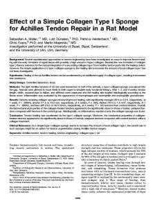
Figure 4. Immunofluorescence labeling with rabbit antisera against mouse HSPG on 6 jim sections on day 5 (a), day 9 (b), day 13 (c), and day 1 (d). HSPG is localized in the BM of RPE cells from day 5 (a) to day 17 (d). In addition, a diffuse staining became detectable, after day 8-9 (b) on the apical surface of the RPE layer. Both the BM and apical surface staining increased with age (b-d). 6 jm sections of day 13 (e) and day 17 (f) eyes labeled with PBS alone, followed by FITC-labeled F(ab’)2 fragments of goat anti-rabbit IgGs. These latter two micrographs have been deliberately underexposed in printing to indicate the low level of fluorescence background due to nonspecific binding of second antibodies. Bar = 10 gm.
BASEMENT
MEMBRANE
OF
RETINAL
PIGMENT
EPITHELIUM
669
the
staining
behing
was
result
masking However, of
of
the
cells
that
in
this
the though
the
known,
various
proteins,
has
teoglycans thus
they
immunolabehing
will
be
esting
with
time
of
layers
and
though
of
our
ofrat
in
BM
Kurkinen
the
the
though
Laurie
nectin only
in
those
through
may
our
serve
strated,
labeling with mouse monoclonal anfibronectin on 6 jam sections at day day 17 (d). Labeling for fibronectin RPE layer. The low level of diffuse apical surface of the RPE layer was from control antibodies. 6 gm section labeled with PBS alone, followed by
Thus,
FITC-labeled
ofrabbit
investigated
fragments
anti-mouse
lgGs.
underexposed background due 10 cm.
These
latter
in printing to nonspecific
to
the
RPE,
must
be
nature
an
increased
our
cells
in
If this
of
the
previously
RPE
demon-
in culture
are
(Turksen
BM
found
function,
BM
also
et al.,
of fibronectin
those
it is
filtered.
the
have
fibronectin
findings
and
during
suggest
fibronectin
all
the
4-17).
component
In
stages,
be
that
general,
of
all four the
of
to increase
accumulation
since
are
stages
appeared
Furthermore, it may
RPE
are
fibro-
(Martinez-
that filtration
that
Al-
that
others
plasma
We
association
function
(day
each
chick
to deposit
HSPG,
for
barrier.
suggested
have
reason clear.
with
that
do
not
that
laminin,
able 1984).
the
BM
contain
it,
further.
summary, cells
of
fibronectin The
is not
argued
suggest
of chick obtained
eye.
of BMs, to
BM
detect
studies
have
amounts
of the
its
with
results
not
have
known
would
that
studied
collagen, RPE
are
large
and
and
In
1983)
results
however,
exclu-
cross-react
chick
two
a variety
as a filtration
to synthesize Figure 5. Immunofluorescence tibodies against porcine plasma 5 (a), day 9 (b), day 13 (c), and is restricted to the BM of the staining occasionally seen at the not above background resulting of day 9 (e) and day 1 7 (1) eyes
1983)
that
which
then
is true
data
located
in the
could
(i982,
Amenta,
BMs
im-
surfaces
All ofthese
the
4-17) the
with
and
com-
comple-
HSPG
is not
from
who (day
between al.
certain
apical
1984).
is present
( 1979),
is associated
Hernandez ,
et
Alclearly
since
may
it differs
developing
discrepancy
least
at the
HSPG
of
series
been
reported
antiserum
fibronectin
al.
at
time
material.
not
Finally,
has
the
1PM
first
increase
HSPG.
since
et
in
of
that
this
the
other
that
to
has
of
inter-
was to
in a complex
1PM
role is
layer
results
et al.,
that that
of
finding
RPE
for
F(ab’)2
or
protein
1PM
antiserum
(Westgate
level
it
appeared
group
same
either
is interesting,
by
two micrographs have been deliberately indicate the low level of fluorescence binding of second antibodies. Bar =
another
before
Nevertheless,
layer.
pro-
biochemical
corresponds
the
RPE
nutrients
and
the
indicate
the
cells
the
core
Our
of
accumulation
by the
suggest
RPE
i.e.
the
of
its
filtration
microscope
intensity
8-9
origin
studies,
for
of pro-
Klucznik, of
distribution
in
which
Day
results
epidermis
sively
staining
retina,
with
together
eye.
the
and virtue
electron
the
are secreted
munoreactivity
the
is un-
and
further
precise
and
some
ponents
the
the
cellular
determined,
at the
8-9,
includes
the
the the
matrix
as a secondary barrier
of the
thereafter.
differentiation
of with
this
by
However,
clarify
that
at day
un-
it is composed
Adler
function
studies
note
view
(GAGs),
1PM,
a selective
regions
detectable
of
that
1981; the
may
in these to
Severin,
suggested
to
HSPG
tion
and
required
in
exclusively
indicated
photoreceptors.
and
from
determined. apical surface
itself
glycosaminoglycans
GAGs,
the
be
at the
composition
have
providing
reach
to in
in la-
or
The 1PM is an ECM located between photoreceptors (Feeney, 1973). Al-
chemical
been
and
matrix,
differences
fixation
is associated
studies
(Adler It
the
is interesting
glycoproteins,
1982).
these of
proteoglycan
precise
teoglycans
in
vivo
(Hassell et al., 1980). RPE layer and the
BM
of
Whether
inadequacies
new antigenic sites remains the mere presence of HSPG
RPE
belief
punctate.
from
these
components four
together
present
in
embryonic the
IV
BM
of
development
intensity with
of
time,
components
were are
type the
in
detectable
fundamental
staining
suggesting the
BM.
at all struc-
670
TURKSEN,
rural
elements
of
be interesting to delineate
the
or simultaneously. be
useful
to
ponents BM
at least
after
investigate as
day
earlier stages four components
Ultrastructural
serve
other
BM
to investigate whether the
4.
It would
ofRPE appear
immunolabehing
whether
one,
nucleation
or
centers
the
of
also
the
addition
of
authors
against
laminin,
invaluable fully
heparan
sections,
Adler
H.K.
proteoglycan
and ofthe
for
type
instruction
]orgensen
Middleton
MD,
and
for
A.O.
Kleinman,
Bethesda,
on the preparation Pat
the
u’ord
and
IV
collagen.
basement
They
on the preparation
Ms.
Shirley
tissues.
Hay
Reimers
The
authors
matrix:
for
of origin.
grate-
processing.
of the bovine
Exp
Res
(1982): matrix:
Eye
32:75
interphotore-
5
Proteins and glycoproteins composition and fractionation.
of the Exp
Amenta PS, Clark CC, Martinez-Hernandez A (1983): Deposition of fibronectin and laminin in the basement membrane of the rat parietal yolk sac: immunohistochemical and biochemical studies. J Cell Biol 96: 104 Bernfield M, Banerjee epithelial-mesenchymal Biol
SD (1972): Acid mucopolysacchanide interface of mouse embryo salivary
glands.
J
52:664
Brownell interactions.
AG, Slavkin HC (1980): Renal Physiol 3:193
Role
Chung AE,Jaffe R, Bender B, Lewis antibodies against the GP-2 subunit Connor NS, Aubin JE, Sodek type I collagen and fibronectin
of basal
lamina
in tissue
J (1983): Independent by normal fibroblast
expression of like cells. J Cell
Sci 63:233
Crawford BJ, Yan AA, MacDonald M (1981): Changes morphology of pigmented retinal cells during differentiation culture. Can J Zool 59:6 1 Crawford BJ (1975): retinal clone. CanJ
The structure Zool 53:560
and development
ofthe
Crawford BJ (1983): Some factors controlling retinal pigment epithelial cells in clonal culture. Ekblom
onic
P (1981):
kidney:
Formation
of basement
an immunohistological
J Cell
pigmented
in the
MG ( 198 1 ): The glomerular
RO,
Johnson Holborow
macromolecular
filter.
by ED Hay.
Plenum
Far AG, Nakane labelled antibodies: Feeney
L (1973):
in mice
and
FitchJM,
rats.
Gibney
In Cell
Press,
Biology
New
embry-
Biol 91:1
membrane:
a selective
interphotoneceptor Biol 32:101
E, Sandenson
RD, Mayne
an-
Edited
p 355-378
PK ( 198 1 ): Immunocytochemistry a brief review. J Immunol Meth The Dev
Matrix.
space
enzyme
47:129
1. Postnatal
R, LinsenrnayerTF
ME
( 198 1 ): The
L.aminin
and
matrix: 2:509
concepts
of
1:819
Retina:
their
molecular
Tissue
epithelial
KM (1982): J Cell
A Com-
In Cell BiPress, New
organization
Kefalides
( 1980):
NA
biosynthesis.
organization
Kleinman matrices
HK, in the
cell attachment.
Fibronectins:
Kurkinen bronectin
and
Chemistry Microcirc
KIebe adhesion
of base-
Nature
290:737
multifunctional
mod-
Biol 95:369
phenomenon
Adv
and
Res 9:191
GD, Davidson RS, McNamee KC, Russel EJ ( 1982): Fading of immunofluorescence of the
G, Goodwin D, during microsJ Immunol Meth
its remedy.
of basement
membranes,
structure
9:295
RJ, Martin and growth
GR (1982): Role of of cells. J Cell Biol
collagenous
88:47
3
M, Alitalo K, Vaheni A, Stenman 5, Saxen L (1979): Fiin the development of embryonic chick eye. Dev Biol 69:589 GW,
Leblond
CP, Martin
GR (1982):
Localization
of type
sulfate proteoglycan membrane. J Cell
Laurie GW, Leblond CP, munolocalization of type teoglycan and fibronectin
Martin GR (1983): Light microscopic imIV collagen, laminin, heparan sulfate proin the basement membranes of a variety of
ratorgans.
AmJ
Anat
and fibronectin Biol 95:340
to the
167:71
Lesot H, Osman M, RuchJV (1981): of collagens fibronectin and laminin odontoblasts. Dev Biol 82:371
Martinez-Hernandez
lagen
A, Amenta
Lab
(1982):
Invest
Immunofluorescent during terminal
PS ( 1983):
isolated
from
1W,
chicken
Nakane
RM,
Essnen of anionic
Cytochem
localization differentiation
The basement
of
membrane
48:656
R, Wiedemann H, Dessau Structural and immunological PK
fixative. A new fixative Cytochem 22:1077 Pino
and Dev
76:100
W, Von den Mark K, Bruckner P characterization of type 1V colEur J Biochem 126:417
tissues. (1974):
Peniodate-lysine-paraformaldehyde
for immunoelectron
E, Pino
LC (1982):
sites
in Bruch’s
microscopy.
Localization membrane
J Histochem
and chemical cornrat. J Histochern
of the
30:245
Rand-Weaver ontogeny
IV
collagen, laminin, heparan basal lamina of basement
position with
NatI
ob-
developing 146:4 15
HJ, Wilczek J, Rennard SI, Martin sulfate containing proteoglycan from Acad Sci USA 77:4494
Int Rev Connect
Yamada
in pathology.
and
of Extracellular
York,
of the
of the Vertebrate Berlin
and extracellular Modern Cell Biol
copy: a study 55:231
Biol
protein binding Res 3:3 59
basement
Cell
ular glycoproteins.
McLean
Fanquhar
Barrach heparan
Proc
Grant
Hynes
Engvall tigenic
Cell adhesive Collagen Rd
G,
B (1981):
Mayne ( 1982):
E (1983): of laminin.
PG, ofa
membranes.
Ekblom P, Alitola K, Vaheni A, Timpl R, Saxen L (1980): Induction of a basement membrane glycoprotein in embryonic kidney: possible role of laminin in morphogenesis. Proc NatI Acad Sci USA 77:485 E, Ruoslahti properties
microscopic
and pecten Ophthalmologica
Leivo I, Vaheri A, Timpl R, Wartiovaara J (1980): Appearance distribution of collagens and laminin in the early mouse embryo.
cell polarity in chick Tissue Cell 15:993
membranes
study.
in surface in clonal
Electron
antibody
p 379-409
Laurie
M, Durkin M (1983): Monoclonal of laminin. Lab Invest 49:5 76
A (1963):
epithelium synthesis.
G, Orkin RW (1981): Current structure and function. Biosci Rep
membrane.
Hogan
and
at the
KALNINS
specificity ofa monoclonal J Cell Biol 95:641
The Development Springer Verlag,
Hay ED ( 1983): mutual dependence.
Proteins
pigment to melanin
SODEK,
ED (1981): Collagen and embryonic development. of Extracellular Matrix. Edited by ED Hay. Plenum
ology York,
ment
KM (1981):
tissues
M, Lagasse
Robey Isolation
Heathcote
Adler AJ, Klucznik KM bovine interphotoreceptor Eye Res 34:423
Cell
in relation
Hassell JR, GR ( 1980):
G.R.
for the antisera
Cited
AJ, Sevenin
ceptor
sulfate to Dr.
suggestions
Literature
Hassell,
ofHealth,
L. Subrahmanyan and
acknowledge
].R.
institutes
to Mrs.
offrozen
to Drs.
National
are grateful
in choroid
Grun G ( 1982): parative Survey.
are indebted
ofthe
J, Babaey
membrane IV collagen.
Grant ME, Heathcote basement membrane
Acknowledgments The
Francois servation
chick
com-
components.
Martin
and basement chicken type
against
might
some,
for
Domain
now
development sequentially
AUBIN,
M, Price
tigenicity
and
variation
Ristelli 59: 185
L, Ristelli
RG ( 1983): Macromolecular in disease. Biosci Rep 3:7 13
J (1981):
Basement
membrane
association
research.
Med
an-
Biol
BASEMENT
Robert 82:839
MEMBRANE
L, Moczar
M ( 1982):
Robey PG, Newsome ent in primate Bruch’s Rolnik VV Translations,
OF
Structural
DA (1983): membrane.
(1970): Bird Jerusalem
Ruoslahti E, Engvall cepts of its structure
RETINAL
glycoproteins.
Embryology.
Israel
Meth
Program
Fibronectin: Rel Res
Enzymol
For
Scientific
current 1:95
con-
Foidart J-M, Vaheni A, Pratt RM, Martin distribution of type IV collagen, laminin, during mouse tooth development. Dev
GR proBiol
81: 182
Timpl Nature EurJ
R, Martin GR, Bruckner of the collagenous protein Biochem 84:43
Timpl (1979): Chem
R, Rohde Laminin-a 254:9933
H,
Timpl
R,
GR
Martin
Robey PG, glycoprotein
In Immunochemistry Furthmayr.
CRC
(1982):
P, Wick G, in a tumor
Rennard from
Components
of the Extracellular Press,
Boca
Raton,
Wiedemann basement
FL,
H (1978): membrane.
SI, Foidart JM, Martin GR basement membranes. J Biol
of basement
Matrix, p 119-150
671
EPITHELIUM
Biosynthesis ofproteoglycans presInvest Ophthal Vision Sci 24:898
E, Hayman G (1981): and functions. Collagen
Thesleff I, Barrach HJ, (198 1): Changes in the teoglycan and fibronectin
PIGMENT
membranes.
vol 2. Edited
by H
Turksen
K, Aubin
tnibution of laminin, proteoglycan during thelial cells in vitro.
JE, Sodek
J, Kalnins VI (1984): Changes in disfibronectin, type IV collagen and heparan sulfate colony formation by chick retinal pigment epiCollagen Rel Res, 4:413
Westgate GE, Shaw DA, Harrap GJ, Couchman nochernical localization of basement membrane hair follicle morphogenesis. J Invest Dermatol Wu TC, Wan ical localization
Dev
Biol
YJ,
Chung ofentactin
AE, Damjanov and laminin
I (1983): in mouse
JR (1984): components 82:259
Immunohistochemembryos and fetuses.
100:496
Yamada KM (1982): Isolation of fibronectin from plasma and In Immunochemistry of the Extracellular Matrix, vol 1. Edited Furthmayr. Boca Raton, FL, p 1 11-123 Yamada KM ( 198 1): Fibronectin Biology of Extracellular Matrix. New York, p 95-114 Yamada terials.
Immuduring
KM Annu
(1983): Cell Rev Biochem
surface 52:761
and other structural proteins. Edited by ED Hay. Plenum
interactions
with
extracellular
cells. by H
In Cell Press,
ma-