Artificial Airways
Lesson Objective The student will be able to: 1. Determine the need to establish an artificial airway and be able to manage and p...
Lesson Objective The student will be able to: 1. Determine the need to establish an artificial airway and be able to manage and provide routine care. 2. List indications, hazards, and physiological effects of intubation and tracheostomy. 3. Describe methods of preventing hazards and complications. 4. Describe proper cuff management. 5. Describe the use of naso and oropharyngeal airways.
Oropharyngeal Airway/ Bite Blocks They come in various sizes. • It should fit into oropharynx area • Used to maintain patent airway • Used often in CPR • Pulls tongue forward to prevent
occlusion of the airway • Tolerated by comatose patient – Conscious person will gag and vomit • Can prevent biting of ETT tube if patient is intubated • Designed to accommodate suction catheter
Sizing and Insertion •Insertion –Place in mouth upside down –Rotate as you insert airway
Other types of “Bite Block”
Nasopharyngeal Airways • Various sizes and materials – Soft rubber or latex • Maintain patent airway • Inserted through nares into oropharyngeal area – Must be lubricated with water soluble gel • Frequently used for NTS
Sizing and Insertion • Tube should be one inch longer than nostril to ear • Easier tolerated than oropharyngeal airway • Must be changed from each nares every eight hours
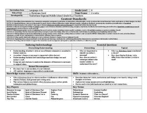
PATIENT POSITIONING A. Neck extension 1. Sniffing position
B. Jaw Thrust
Intubation
What is the need for intubation? Inadequate ventilation/Oxygenation •(over) sedation •neuromuscular paralysis in the operating room, •an obstructed or compromised airway •altered mentation (obtunded) The loss of consciousness, or respiratory failure can lead to brain injury or death within minutes. It is, thus, of great importance to know how to evaluate and address a patient who may require ventilatory support.
Intubation is indicated in order to: 1. Keep a patent airway - (relief of obstruction) 2. Protect airway from aspiration in patients with profound disturbance in consciousness with the inability to protect the airway. 3. Provide bronchial hygiene (suctioning). 4. Provide mechanical ventilation. – severe pulmonary or multi-system injury associated with respiratory failure, such as sepsis, airway obstruction, hypoxemia, and hypercarbia.
Indications for ENDOTRACHEAL INTUBATION in the operating room include: 1. The need to deliver positive pressure ventilation 2. Protection of the respiratory tract from aspiration of gastric contents 3. Surgical procedures involving the head and neck or in non-supine positions that preclude manual airway support 4. Almost all situations involving neuromuscular paralysis 5. Surgical procedures involving the cranium, thorax, or abdomen 6. Procedures that may involve intracranial hypertension
Objective measures may also be used to help determine the need for intubation: • respiratory rate > 35 breaths per minute • vital capacity < 15 ml/kg in adults and 10 ml/kg in children • inability to generate a negative inspiratory force (NIF) of 20 mm Hg • PaO2 (arterial partial pressure of oxygen) < 70n mm Hg • A-a gradient (Alveolar-arterial) > 350 mm Hg on 100% oxygen • PaCO2 (arterial partial pressure of carbon dioxide) > 55 m Hg (except in chronic retainers) • dead space > 0.6 L
Physiological effects of intubation
1. Decreased VD 2. May increase airway resistance 3. May increased WOB
Equipment needed 1. Topical anesthesia • Lidocaine, xylocaine 2. Muscle relaxant (paralytic) • Anectine (Succinylcholine), Pavulon (Pancuronium) • Patient must be sedated 3. Suction equipment 4. Laryngoscope with batteries and functional light • Miller blade – Straight – Directly lifts the epiglottis • MacIntosh blade – Curved – Placed anterior to epiglottis in valecula
5. Endotracheal tubes • various sizes 6. Stylet is a copper wire or plastic used to stiffen tube 7. Magill Forceps • used for nasal intubation • guiding tube anteriorly into trachea 8. Bite Block (oropharyngeal airway) 9. Sponge gauze, Benzoin and tape 10. 10cc or 20 cc syringe for cuff inflation
Equipment needed
Procedure 1. Gather and test equipment – Test laryngoscope light – Check ETT cuff for leak 2. Administer sedation and paralytics 3. Position patient in sniff position 4. Insert laryngoscope 5. Pass tube through vocal cords – (apply cricoid pressure)
Procedure 6. Inflate cuff 7. Remove stylet 8. Connect to Ambu-bag (with O2) and End-Tidal CO2 adaptor and start ventilating 9. Auscultate chest for equal and bilateral breath sounds and note for equal and bilateral chest rise. 10. Secure ET tube 11. Order for Chest x-ray - Tip of tube should be 2-4cm above carina 12. Evaluate cuff inflation 13. May need to insert NG tube
Complications • Immediate complications – Apnea • Due to paralytic
– – – – – –
Laryngospasm Broken teeth Trauma Aspiration Arrythmias Hypotension due to sedation
Hazards of Artificial Airway • Bypasses normal defenses – Can lead to infection
• Removes effectiveness of cough • No ability for verbal communication • Loss of dignity
Extubation Procedure • Hyper-oxygenate patient • Have Ambu-bag and mask at bedside connected to O2 • Suction (Yankaeur and in-line) secretions from above and below cuff • Remove tape • Deflate cuff • Remove tube (on inspiration or during coughing) • Place oxygen on patient • Tell patient to cough and deep breathe • Instruct patient on proper use of IS
Tracheostomy • Indications – Long term CMV (Continuous Mandatory Ventilation) • Usually after 4 – 10 days on a vent – Provide patent airway when intubation is unlikely • Edema • fractured cricoid cartilage • facial trauma
Minimal occluding volume – Cuff is filled until no leakage of air around cuff during inspiration – Auscultated at cuff site Minimal leak technique – Cuff is filled until no leakage of air around cuff during inspiration – Small volume of air withdrawn from cuff – Auscultated at cuff site Cuff pressure – Manometer needed – Cuff pressure should be between 20 – 25mmHg • >30mmHg occludes arterial capillary flow • >18mmHg occludes venous flow • >5mmHg occludes lymphatic flow – The cuff pressure should be lower if the patient has low blood pressure
Tube Markings I.T. – implantation tested Z-79 – committee that sets standards for non-toxicity I.D. – inner diameter O.D. – outer diameter • Radiopaque line – shows up on x-ray
Other Equipment Flexible Fiber Optic Bronchoscope
Fiber Optic Bronschoscopy
Other Equipment • Fome cuff-Kamen Wilkensen & Bivona – Cuff is filled with a soft, spongy foam – Exert low pressure of about 20mmHg • Lanz – External pressure regulator valve & control balloon – Maintain cuff pressure between 19-25mmHg • Trach Talk – Secondary tube above cuff • Allows gas at 4-7lpm pass through vocal cords • Fenestrated trach – Opening in posterior above cuff – Allows patient to breathe through and around trach tube – Used for weaning patient from tracheostomy
Foam filled cuff
Other Equipment • Jackson trach – Silver – Long term
• Easy Cap End Tidal CO2 adapter
• Capnograph – Used to detect CO2 – Indicates proper tube placement
Other Equipment • Trach buttons – Trach button inserted into stoma – Used to wean patient off of a tracheostomy
Other Equipment Passy Muir Valve (PMV) This device that acts as a one-way valve, allowing air in, but not out. Air that’s breathed out has no choice but to go through the vocal chords, which facilitates vocalization.
**It is important to make sure that the cuff is DEFLATED prior to placing the PMV on the trach tube.