Laser-Induced Breakdown Spectroscopy (LIBS) for the Rapid Identification and Classification of Pathogenic Bacteria Steven J. Rehse WSU, Dept. of Physics and Astronomy Qassem Mohaidat WSU, Dept. of Physics and Astronomy Sunil Palchaudhuri WSU, Dept. of Immunology and Microbiology Hossein Salimnia WSU, Dept of Pathology / Detroit Medical Center
Andrzej W. Miziolek US Army Research Laboratory, APG, MD
Leslie M. Collins Duke University, Durham, NC Peter A. Torrione Duke University, Durham, NC
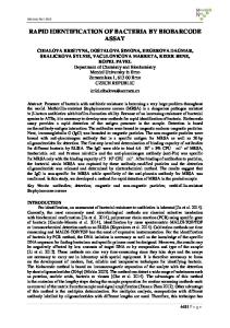
Staph. epidermidis Staph. aureus
there is an urgent need right now in the military, civilian (hospital, food processing, environmental), and first responder communities for a “…rapid point-of-care (multiplex?) diagnostic for disease-causing pathogens.”
E. coli
V. cholerae
How do we identify bacteria? 4 ways • • • •
genetic serological (antigenic) microbiological compositional – LIBS – Raman – MALDI-TOF-MS
things that make a LIBS-based technology powerful • speed / portability / durability (ruggedness) – “rapid point-of-care diagnostic…”
• lack of complicated sample preparation • no expertise required • no genetic or antigenic precursors (consumables) necessary • same technology / hardware useful for explosives, chemical, other threats (CBRNE capable) • capability of sensor fusion
EMMA: Elemental Multivariate Microbiological Analysis • utilizes laser-induced breakdown spectroscopy (LIBS) to measure the unique atomic or elemental composition of bacteria Nd:YAG laser (1064 nm, 8 ns)
Laser-Induced Breakdown Spectroscopy
Echelle spectrometer LIBS Spectrum is like a Spectral Fingerprint: Unique for Each Sample
how we did it… E. coli from liquid specimen. Centrifuged than supernatant removed
about 500-1500 bacteria per sampling location
10 microliter of bacteria pellet
bacto-agar (99% water)
• high signal-to-noise atomic emission lines from inorganic elements allow a classification of the unknown target on the basis of its unique atomic spectrum
E. coli - WSU
H
RDX
RDX - ARL O
Ca CaO
C
CN
C2
CaOH Na N
K
• concentrations of elements (or ratios of concentrations) become independent variables in a chemometric multivariate analysis (e.g. PCA, DFA, LDA, PLS-DA)
Does it work? YES! • “Area under the curve” of 13 emission lines from 6 inorganic elements input as independent variables into a DFA. • This test shows only the first two discriminant function scores for 10 different bacterial types (multiple genera, species, strains)
E. coli
Streptococcus
Staphylococcus
M. smegmatis
Predicted Group Membership (%)
Group 1
2
3
4
5
6
7
8
9
10
1:M. smegmatis (TA)
82.4
17.6
0
0
0
0
0
0
0
0
2:M. smegmatis (WT)
28.0
72.0
0
0
0
0
0
0
0
0
3:E. coli (O157:H7)
0
0
96.0
4.0
0
0
0
0
0
0
4:E. coli (C)
0
0
3.6
96.4
0
0
0
0
0
0
5:E. coli (HF4714)
0
0
0
0
100.0
0
0
0
0
0
6:E. coli (HfrK-12)
0
0
6.7
0
0
93.3
0
0
0
0
7:Staph. saprophyticus
0
0
0
0
0
0
94.1
5.9
0
0
8:Staph. aureus
0
0
0
0
0
0
0
100.0
0
0
9:Strep. mutans
0
0
0
0
0
0
0
0
95.0
5.0
10:Strep. viridans
0
0
0
0
0
0
0
0
0
100.0
The Wayne State Team has already demonstrated… EMMA spectral fingerprint is: – growth-medium independent – independent of state of growth (how “old” the bacteria are) – independent of whether the bacteria are live or dead (or inactivated by UV light) – obtainable even when other types of bacteria or contaminants are present (mixed samples) – capable of strain discrimination – obtainable from about 500 bacteria 6 publications in Applied Physics Letters, Journal of Applied Physics, Applied Optics, and Spectrochimica Acta B
“Mixed” Samples Category 100% M. smegmatis, 0% 90% M. smegmatis, 10% 80% M. smegmatis, 20% 70% M. smegmatis, 40% 50% M. smegmatis, 50% 0% M. smegmatis, 100%
6: pure E. coli
# of Spectra E. coli E. coli E. coli E. coli E. coli E. coli
21 20 16 21 19 25
1: pure M. smegmatis
decreasing M. smegmatis concentration
Classification Results M. smegmatis E. coli S. viridans 100% 0% 0% 100% 0% 0% 100% 0% 0% 76% 24% 0% 47% 53% 0% 0% 100% 0%
• Six separate mixtures of known mixing fraction were prepared from suspensions M. smegmatis and E. coli C. • As long as the majority bacterium comprised 80% of the mixture, we saw 100% identification.
“Dirty” samples • Samples of Staph. epidermidis were prepared in DI water and sterile urine. S. epidermidis: H2O S. viridans S. epidermidis: urine
E. coli
• Samples were collected and tested via LIBS with NO WASHING. • LIBS spectral fingerprint from urineexposed bacteria were identical to water-exposed bacteria. • EMMA correctly classified 100% of the urine-exposed bacteria as being consistent with S. epidermidis
LIBS intensity linearly dependent on number of bacteria • Samples of E. coli with different titer tested on agar.
Total Power of the Spectrum (a.u)
240000 220000 200000
• Each data point is the average of 5 sampling locations.
180000 160000 140000
• As expected, spectra demonstrate a linear dependence with cell number.
120000 100000 80000 60000
0.3
0.4
0.5
0.6
0.7
0.8
0.9
Number of Bacteria × 7500
5 laser sampling locations ~500 bacteria per locations
1.0
1.1
• All spectra were 100% correctly identified (specificity not dependent on number of cells). • Suggests an antibiotic resistance test?
Strain discrimination confirmed by others…
• 100% accuracy exhibited in blind trials of 4 MRSA strains and one E. coli strain • lyophilized (“freeze-dried”) specimens used
We Must Proceed, and Faster… LIBS research must proceed along two equally important avenues: • fundamental research to explore the microbiological diversity that can occur in specimens • specimen preparation and handling protocols and techniques to isolate pathogens from contaminants of biological origin NOTE: we do NOT need to fingerprint hundreds and hundreds of “new” bacteria
what must we do to make LIBS a clinical tool? Develop protocols for clinical sample preparation (blood, urine, sputum) • isolation • concentration under the laser focus
solutions 1. differential centrifugation 2. filtration (sequential?) 3. optical trapping / separation 4. microfluidic separation 5. antibody isolation/phage display technology (consumables!)
Microfluidic separation/concentration (Translume, Inc. Ann Arbor, MI)
monolithically fabricated devices in glass
hydrodynamic (microfluidic) separation of heavier cells from lighter cells
Microfluidic separation/concentration (Translume, Inc. Ann Arbor, MI)
laser trap
bacteria only
optical trap-based separation of heavier cells from lighter cells
Novel substrates 1• 10 mL of a suspended bacterial culture pushed through a 0.22 or 0.44 μm cellulose (carbon) Millipore filter • alternately, bacteria just deposited on filter (wicking) • C line does “contaminate” spectrum, but only at 7% level (same as agar!)
Novel substrates 2 • Acid etched “porous” silicon • Bacteria fixed with polyacrimide 11 mm
• High SNR LIBS spectrum • Si lies do not contaminate spectrum
Conclusions • All EMMA experiments to date have successfully shown the utility of LIBS to identify bacterial samples in a variety of growth conditions, in mixed samples, in dirty samples, etc. • We are ready to move to testing real “clinical” type samples through our in-place organizational structure, which combines expertise in hardware development, software development, microbiological handling, and LIBS development.
My students
Thank you!
genetic • PCR (polymerase chain reaction) • (random primed) RAPID-PCR • FISH (fluorescence in situ hybridization) requires • a priori knowledge of genetic sequence (16s RNA gene is conserved in most) drawbacks • amplification time (multiple generations needed) • nonspecific reactivity • still need to do gel electrophoresis • very contamination sensitive
serological • • • • •
immunoassays microwell devices ELISA (enzyme-linked immunosorbent assay ) fluorescently labeled antibody techniques MEMS requires • a priori knowledge of serology (surface antigens) drawbacks • any mutation (common) undetectable • antibodies are not stable (shelf-life) • consumables • binding affinities may be low
microbiological • • • •
culturing and colony counting phenotyping sensitivity to immunochemicals Gram staining
requires • time • expertise • LOTS of supplies • a priori clinical knowledge (case-history) drawbacks • slow/labor intensive • requires experts