This material is protected by U.S. Copyright law. Unauthorized reproduction is prohibited. For reprints contact:
[email protected]
Leukemias Large Granular Lymphocyte Leukemia Lubomir Sokol,a Thomas P. Loughran, Jr.b a
Department of Interdisciplinary Oncology, University of South Florida and H. Lee Moffitt Cancer Center and Research Institute, Tampa, Florida, USA; b Penn State Cancer Institute, Penn State College of Medicine, Hershey, Pennsylvania, USA Key Words. T-cell LGL leukemia • Aggressive NK-cell leukemia • Cytopenia • Autoimmunity
Learning Objectives After completing this course, the reader will be able to: 1. Discuss the basic principles of molecular and cellular biology of LGL leukemia. 2. Describe distinct clinical entities among disorders of LGLs. 3. Discuss the diagnostic criteria for T-cell LGL leukemia. 4. Discuss the therapeutic algorithm of LGL leukemia. CME
Access and take the CME test online and receive 1 AMA PRA category 1 credit at CME.TheOncologist.com
Abstract Clonal disorders of large granular lymphocytes (LGLs) represent a spectrum of biologically distinct lymphoproliferative diseases originating either from mature T cells (CD3 +) or natural killer (NK) cells (CD3 −). Both subtypes, T-cell and NK-cell LGL leukemia, can manifest as indolent or aggressive disorders. The majority of patients with T-cell LGL leukemia have a clinically indolent course with a median survival time >10 years. Immunosuppressive therapy with low-dose methotrexate, cyclophosphamide, or cyclosporine A can control symptoms and cytopenias in more than 50% of patients, but this approach is not curative. Several cases of an
aggressive variant (CD3 + CD56 +) of T-cell LGL leukemia with a poor prognosis have also been reported. Aggressive NK-cell LGL leukemia is usually a rapidly progressive disorder associated with Epstein-Barr virus (EBV), with a higher prevalence in Asia and South America. This disease is usually refractory to conventional chemotherapy, with a median survival time of 2 months. Chronic NK-cell leukemia/lymphocytosis is a rare EBV-negative disorder with an indolent clinical course. The malignant origin of this subtype is uncertain because clonality is difficult to determine in LGLs of NK-cell origin. The Oncologist 2006;11:263–273
Definition and Classification
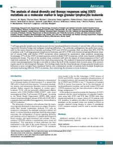
cally, LGLs are medium to large cells with eccentric nuclei and abundant cytoplasm with coarse azurophilic granules (Fig. 1). T-cell LGLs are post-thymic, antigen-primed cytotoxic CD8+ T lymphocytes. NK-cell LGLs belong to the innate immune system with the capability of non-major histocompatibility complex (MHC)–restricted cytotoxicity.
Large granular lymphocytes (LGLs) represent 10%–15% of the total peripheral blood mononuclear cells in normal adults [1]. The majority of these cells (85%) are derived from the CD3− natural killer (NK)–cell lineage, and a minority are derived from the CD3+ T-cell lineage (15%). Cytologi-
Correspondence: Thomas P. Loughran, Jr., M.D., Penn State Cancer Institute, Penn State College of Medicine, 500 University Drive, H072, Hershey, Pennsylvania 17033, USA; Telephone: 717-531-7096; Fax: 717-531-0778; e-mail:
[email protected] Received November 5, 2005; accepted for publication January 3, 2006. ©AlphaMed Press 1083-7159/2006/$20.00/0
The Oncologist 2006;11:263–273 www.TheOncologist.com
LGL Leukemia
264
LGL leukemia was initially described in 1985 as a clonal disorder involving blood, marrow, and spleen [2]. In 1993, we proposed two LGL disorders based on either Tcell or NK-cell lineage, which was subsequently adopted by all pathology classification systems [3]. Clonal disorders of LGLs represent a biologically heterogeneous spectrum of lymphoid malignancies (Fig. 2) [4, 5]. Both the T-cell and NK-cell subtypes can clinically present as an indolent or aggressive diseases (Table 1). World Health Organization (WHO) classification of lymphoid malignancies includes
Figure 1. Peripheral blood film. Large granular lymphocyte (LGL) in T-cell LGL leukemia.
Figure 2. Spectrum of disorders of large granular lymphocytes according to clonality and biological behavior of the disease. Abbreviation: LGL, large granular lymphocyte; NK, natural killer.
Table 1. Clinicopathological characteristics of large granular lymphocyte (LGL) leukemia T-cell LGL leukemia, indolent
T-cell LGL leukemia, aggressive variant
Aggressive NK-cell leukemia
Chronic NK-cell lymphocytosis
Median age (yrs)
60
41
39
60.5
Male:female
1:1
2:1 +
+
+
1:1 +
+
7:1 −
+
+
CD3− CD16+ CD56+
Phenotype
CD3 TCRαβCD8 CD57 CD16+
CD3 TCRαβCD8 CD16+ CD56+
Clonality, TCR gene rearrangement
TCR-β/γ
TCR-β/γ
Germline
Germline
EBV
−
−
+
−
HTLV seroreactivity
+
−
−
+
Clinical presentation
One third asymptomatic; two thirds symptomatic— cytopenias, splenomegaly, rheumatoid arthritis
All patients—B symptoms, organomegaly, lymphadenopathy, cytopenias
All patients—B symptoms, organomegaly, lymphadenopathy, cytopenias
60% asymptomatic; 40% symptomatic—cytopenias, vasculitis, neuropathy, splenomegaly
Therapy
Watch and wait, immunosuppressive
ALL-like induction chemotherapy
ALL-like induction chemotherapy
Watch and wait, immunosuppressive
Prognosis
Good
Poor
Very poor
Good
CD3 CD16 CD56
Abbreviations: ALL, acute lymphoblastic leukemia; EBV, Epstein-Barr virus; HTLV, human T-cell leukemia/lymphoma virus; LGL, large granular lymphocyte; NK, natural killer; TCR, T-cell receptor.
OTncologist he
®
Sokol, Loughran
T-cell LGL leukemia and aggressive NK-cell leukemia as two separate entities among T-cell/NK-cell lymphomas/ leukemias [6]. Since only rare cases of the aggressive variant of T-cell LGL leukemia have been described [7], this subtype was not given separate status in the WHO classification. Transient (6 month) expansions of LGLs are two benign conditions in the spectrum of disorders of LGLs [8]. Transient reactive populations of LGLs have been detected in patients with viral infections, autoimmune diseases, and malignancies, and in patients after solid organ transplantation [9–13]. The reactive LGLs are polyclonal with expression of the T-cell (CD3+) immunophenotype in the majority of cases. LGL count normalizes spontaneously or with therapy of underlying condition, usually within 6 months. Since it is difficult to determine clonality for NK cells, it is uncertain whether chronic NK-cell lymphocytocis represents a chronic NKcell leukemia. In the absence of a clonal marker, clinical presentation is the most important factor available for the differential diagnosis of these two conditions. Patients with systemic symptoms or infiltration of the spleen, liver, or bone marrow could be appropriately classified in the category of chronic NK-cell leukemia. Asymptomatic patients might be better given a diagnosis of benign chronic NK-cell lymphocytosis. In this review we focus mainly on diseases of LGLs included in the WHO classification.
Epidemiology LGL leukemia comprises 2%–5% of all T-cell/NK-cell malignancies, with only 400 cases reported in the literature [8]. Indolent T-cell LGL leukemia is the most common subtype, representing approximately 85% of all cases diagnosed in Western countries. The male-to-female ratio is approximately equal to one. This entity is more frequently diagnosed in older individuals, with a median age at diagnosis of 60 years (Table 1). The aggressive type of NK-cell LGL leukemia typically occurs in younger individuals, with median age at diagnosis of 39 years and with a higher prevalence in Asia and South America [3, 14, 15]. Fewer than 100 cases have been described in literature [16] (Table 1).
Etiopathogenesis The etiology of LGL leukemia is not known. Chronic activation of T cells with autoantigen or viral antigen has been suggested as an initial stimulus leading to an expansion of LGLs [17, 18]. Whether a second molecular event is necessary to establish the full malignant phenotype is not clear. It has also been suggested that T-cell LGL leukemia could represent an autoimmune disorder caused by chronic antigenic stimulation leading to extreme expansion of only one clone of CD8+ cytotoxic T cells [19, 20]. An association of
www.TheOncologist.com
265
T-cell LGL leukemia with several different autoimmune conditions supports this hypothesis. Human T-cell leukemia virus II (HTLV-II) sequences have been detected in two patients with indolent T-cell LGL leukemia [21, 22]. Seroindeterminate reactivity against HTLV-I envelope (env) epitope BA21 has been described in approximately 50% of patients with CD3+ and 73% of patients with CD3− LGL leukemia [22, 23]. Detailed amino acid analysis of BA21 revealed that a 10–amino acid peptide, PP10, was responsible for the seroreactivity [24]. This peptide shared high amino acid homology only with HTLV-I env protein and not with any known human proteins. However, most patients with LGL leukemia are not infected with prototypical members of the HTLV family, including HTLVI, HTLV-II, or bovine leukemia virus (BLV) [25, 26]. Epstein-Barr virus (EBV) is implicated in the pathogenesis of aggressive NK-cell leukemia [27]. It has been suggested that this condition is a leukemic variant of a more common NK-cell/T-cell lymphoma, the nasal type.
Immunophenotype LGL leukemia cells have a mature T- or NK-cell immunophenotype [3]. The most common immunophenotypes for each subtype of LGL leukemia are described in Table 1. CD57 is a 110-kDa glycoprotein found on NK cells and activated, effector CD8+ T cells. It is a characteristic marker for LGL leukemia. It was suggested that LGLs in T-cell LGL leukemia originate in a CD57− memory T-cell compartment that continually produces CD57+ (effector cell) progeny [28]. Rare immunophenotypic variants that are CD3 + T-cell receptor (TCR)-αβCD4 + CD8 +, CD3 +TCRαβCD4 − CD8 −, and CD3 +TCR-γδCD4 − CD8 − have been reported in T-cell LGL leukemia [1, 29]. Aberrant expression of pan T-cell markers, including CD5 and/or CD7, can be useful in differentiating the malignant T-cell LGL population from normal T lymphocytes. The NK-cell marker CD56 is typically detected in aggressive NK-cell LGL leukemia [30]. The immunophenotype of this subtype is similar to that of the nasal type of NK-cell/T-cell lymphoma that is CD2 + CD56 + CD3ε+ [31]. In the differential diagnosis, this aggressive NK-cell malignancy must be distinguished from indolent chronic NK-cell leukemia and benign chronic NK-cell lymphocytosis, which are not associated with EBV (Fig. 2) [3, 32].
Cytogenetics Indolent T-cell LGL leukemia cells most frequently have a normal karyotype. Less than 10% of patients display distinct chromosomal aberrations, including inversion of 12p and 14q, deletion of 5q, and trisomy of 3, 8, and 14 chromosomes [2, 33, 34]. The most frequent clonal chromosomal
LGL Leukemia
266
abnormality in patients with aggressive NK-cell LGL leukemia is the deletion of the 6q chromosome, but cases with complex karyotypes have also been reported [35, 36].
Clonality LGL leukemia is a disorder of mature T or NK cells. T-cell ontogenesis is associated with TCR gene rearrangement resulting in a unique molecular fingerprint for each T lymphocyte. Thus, the malignant LGL population, which arises from a single cell, has the same TCR gene rearrangement pattern. Southern blotting and polymerase chain reaction (PCR) are the two methods most commonly used for confirmation of clonality [37]. Recently, a panel of monoclonal antibodies was used against the variable domain of the β chain as a new clonality technique in T-cell malignancies [38]. Since available antibodies recognize approximately 75% of the Vβ repertoire of T cells, this technique has not yet achieved widespread use in clinical practice. NK cells have TCR genes in a nonrearranged germline position. In the past, cytogenetic abnormalities and clonality studies based on the X-chromosome inactivation pattern (XCIP) were the only methods available for confirmation of the clonal origin of the NK-cell population [36]. Recently, a new class of NK-cell receptors, killer cell immunoglobulinlike receptors (KIRs), has been detected on the surface of NK cells and a subset of T cells [39]. Aberrant expression of these receptors has been reported in some patients with LGL leukemia [39]. In particular, we demonstrated that patients' NK cells had high levels of activating receptors and a loss of inhibitory receptors [40]. These findings suggest a potential use of KIR expression as clonality markers but will require validation in a larger study.
Clinical Presentation Approximately two thirds of the patients with indolent Tcell LGL leukemia develop cytopenias, recurrent bacterial infections, autoimmune disorders, and/or splenomegaly during the course of their disease (Table 1) [1]. Recurrent infections resulting from neutropenia initially manifest in 20%–40% of patients. B symptoms—including fever, night sweats, and weight loss—occur in 20%–40% of patients. Mild-to-moderate splenomegaly is found in 20%–50% of patients, and hepatomegaly is found in 10%–20% of patients. Diffuse infiltration of red splenic pulp with preservation of sinuses and white pulp cords is characteristic of T-cell LGL leukemia [41]. Lymphadenopathy is an uncommon presentation of this disease. Bone marrow biopsy typically reveals discrete interstitial and sinusoidal infiltration with T lymphocytes [8]. Rheumatoid arthritis is the most common autoimmune disorder, manifesting in up to 30% of patients with T-cell LGL leukemia [8].
Clinical manifestations of the aggressive variant of Tcell LGL leukemia closely resemble those of aggressive NK-cell LGL leukemia (Table 1). This condition is very rare, with only several well-described cases in literature [7]. Aggressive NK-cell LGL leukemia typically presents as an acute illness, with B symptoms, lymphocytosis, hepatosplenomegaly, lymphadenopathy, severe anemia and thrombocytopenia, and hemophagocytic syndrome [3, 16]. Chronic NK-cell lymphocytosis usually is an indolent disorder with a good prognosis. Rare cases present with cytopenias, cutaneous vasculitis, peripheral neuropathy, and splenomegaly (Table 1) [42].
Hematologic Features The normal range of LGLs in peripheral blood is 0.2–0.4 ×10 9 /l [1]. Evaluation of peripheral blood film is invaluable if a diagnosis of LGL leukemia is suspected. A previous study suggested that >90% of patients with T-cell LGL leukemia present with an LGL count in excess of 1.0 × 10⁹/l [1]. In our original paper, diagnostic criteria for Tcell LGL leukemia required an absolute LGL count >2 × 10 9 /l [3]. More recent studies revealed that 25%–30% of newly diagnosed patients present with an absolute neutrophil count (ANC)