IMMUNOLOGICAL AND BIOCHEMICAL DIAGNOSIS EJ Thompson min or IgG has not been generally found (Tourtellotte, 1975, pp. 9-26). A strong case can therefore be made for local synthesis of antibody, presumably by lymphocytes, within the central nervous system (CNS). This source has been termed the intrathecal synthesis of antibody.
LABORATORY DIAGNOSIS OF MULTIPLE SCLEROSIS: IMMUNOLOGICAL AND BIOCHEMICAL ASPECTS
1 Comparison of Methods Information from the laboratory investigation of MS can be interpreted from two points of view: that which relates to the general course of the disease, and that which considers a specific patient regarding differential diagnosis from other neurological diseases. The latter is rather more difficult to interpret and will be discussed last (section 3).
E J THOMPSON PhD MD Department of Neurochemistry, Institute of Neurology University ofLondon and National Hospitals for Nervous Diseases, London
a Cerebrospinal Fluid Limited information can be gained from routine examination of the blood from MS patients. Attention has therefore focused on the CSF which is in intimate contact with the brain, particularly with certain areas of myelin. This is especially relevant to MS, for sites of predilection include the periventricular white matter and the surface of the spinal cord.
1 Comparison of methods a Cerebrospinal fluid b Blood 2 Relations of immunoglobulins with the clinical course a Age at onset, duration and severity b Exacerbation versus remission c Steroid therapy d Multiple sclerosis versus optic neuritis 3 Conclusions regarding differential diagnosis References
i Cell Count The cell count is typically normal, or perhaps only slightly elevated. The earlier data of Cumings, drawn from the National Hospital, showed that 86% of 690 MS patients had less than 5 cells/mm3 (Locoge & Cumings, 1958). Examination of the differential cell count showed mainly lymphocytes and rarely polymorphs. As polymorphs adhere more readily to glass, and have a shorter half-life in CSF than have lymphocytes, proper collection and speed of processing are essential. The practice at the National Hospital has been to perform cytology on CSF spun within 5-15 minutes of collection, using a cvto-centrifuge (Thompson et al. 1975). With the use of such methods, a few patients with MS have been noted to have reactive plasma cells. Among the lymphocytes, B cells (which secrete antibody as plasma cells) and T cells (which produce cell-mediated immunity or delayed hypersensitivity) have been enumerated. Some investigators have found that there is a higher percentage of T cells in CSF relative to serum and a relatively lower percentage of B cells (Sandberg-Wollheim & Turesson, 1975). Others have found that at two weeks following an exacerbation the percentage of T cells drops, and at three weeks the percentage of B cells increases (Allen et al. 1975). See section lb(ii) for proportions of T and B cells in the blood.
It has been known for at least 33 years that increased concentrations of antibody are present in the cerebrospinal fluid (CSF) from patients with multiple sclerosis (MS). However, this finding is not diagnostic for MS, as increased CSF antibody levels are found in other diseases. With increased amounts of IgG in the CSF and normal amounts in the plasma, the origin may be: (i) selectively increased permeability to specific IgG in the filtration of plasma proteins during formation of the CSF; 00 generalized synthesis of antibody specifically directed against the brain, this IgG originating from the total-body pool of lymphocytes; and (iii) localized synthesis of IgG by lymphocytes that are situated within the brain. The over-all picture of the data from MS patients will be seen to favour the last interpretation. However, any chronic infection localized within the brain can produce similar results. Methodology is continuously improving, and precise values are related to a given technique. For a general review of earlier progress, see Lumsden (1972, pp. 368-432). During the normal production of the CSF, the plasma is filtered so that only about one molecule in 200 crosses the barrier of the choroid plexus. Within this small population, selection is in favour of the smaller molecules, since these enter into the CSF more readily than do larger molecules (Felgenhauer, 1974). Normal CSF thus contains proportionately more pre-albumin (mol. wt. 61000) and less y-globulin (mol. wt. 155000) than does plasma. As various pathological processes, including perivascular cuffing or general tissue destruction, can cause leakage of plasma proteins through blood-vessels, so the normally low percentage of immunoglobulin in CSF (3-5 %) can approach that normally found in the plasma (15-18 %). Yet the amounts of IgG in CSF from patients with MS can easily exceed values that would be achieved by the entry of plasma protein into the CSF. Selective permeability to specific proteins such as albu-
ii Total Protein Estimation of the total protein concentration in the CSF indicates that it is usually normal or only slightly elevated. Cumings found that 51 % of patients at the National Hospital had less than 40mg/100ml and 96% had less than 100mg/ 100ml (Locoge & Cumings, 1958). In the determination of total protein, it is important that a method should not be disproportionately sensitive to albumin versus globulin. The trichloroacetic acid precipitation method is quite satisfactory in this regard. The sulphosalicylic acid method is rather less satisfactory (Meulemans, 1960). Values found with use of the Lowry method are intermediate between those with methods using trichloroacetic acid and sulphosalicylic acid; but one must be careful, since phenolic drugs (e.g. aspirin) can yield a false elevation of protein value. 28 Br. lied. Bull. 1977
1MMUNOLOGICAL AND BIOCHEMICAL DIAGNOSIS E J Thompson Methods involving electrophoretic separation of proteins must allow careful distinction of other proteins, e.g. haptoglobin polymers (Giebel & Saechtling, 1973), which can also migrate in the Y-globulin region when total protein values are increased in the CSF. Normally, these higher-molecularweight haptoglobins are relatively excluded from the CSF, as are the immunoglobulins (Blau et al. 1963). It is also important to know that results of the dye binding show the same extinction coefficient for albumin and globulin as discussed above (section la(ii)). Amido Black (AmidoSchwarz or Naphthalene Black) satisfies this criterion. A good correlation has been found for the Laurell electroimmunoassay results compared with the results obtained by scanning the Y-globulin region on cellulose acetate strips when stained with Amido Black (Schuller & Tompe, 1973; Ansari et al. 1975). Results for the amounts of IgG are best expressed in relation to other proteins, since the total amount of protein can vary, depending upon the state of the blood-CSF barrier. Most frequently the IgG values are expressed as a percentage of the total CSF protein. Alternatively, they can be given as ratios with albumin (Tourtellotte et al. 1971), transferrin, i.e., the (3-globulin : y-globulin ratio (Cumings et al. 1970), or a2macroglobulin (Schliep & Felgenhauer, 1974). The lastnamed was chosen to reflect the relatively larger size of IgG, i.e., as being more than twice as large as albumin. Most formulae which propose correction factors for any break-down of the blood-CSF barrier are based upon albumin (Delpech & Lichtblau, 1972; Tourtellotte, 1975, pp. 9-26). Relative values for detection of abnormalities in MS and other neurological diseases (of non-infectious aetiology) are contrasted in Table I. The most commonly used method for determination of IgG as a percentage of total CSF protein is that of radial immunodiffusion. In normal subjects IgG forms up to 12% of total CSF protein, and values greater than 15% (i.e., the serum level) strengthen confidence in the diagnosis of MS. Although values in excess of 50% have been reported, this may be the result of measurement of fragments of IgG (Link & MUller, 1971). In our experience, values of the percentage of IgG in CSF obtained by immunodiffusion can be much higher than values from the same sample when estimated by electrophoresis, e.g. 33% for immunoprecipitation and 14% by electrophoresis. Rabat's originalfindingsof increased IgG were based upon electrophoresis (Kabat et al. 1942).
iii Qualitative Tests
Two qualitative tests for Y-globulin which are often very useful are the Pandy and the Lange methods. The Pandy technique requires one drop of CSF to be pipetted into concentrated phenol. It is principally sensitive to the globulins and these may be the only abnormality of the CSF found on routine examination. The Lange colloidal gold reaction can be most informative, but depends upon satisfactory preparation of an appropriately reactive gold solution. The method requires only 0.1ml of CSF and the results can be read the following morning. Previous experience with the Lange method at the National Hospital has shown that there were abnormalities in 50% of MS patients, i.e., in 17% the curves were of the paretic type and in 32 % of the luetic type. It can be seen that the method yields results rather similar to the method of immunodiffusion for IgG. In particular, we have found that the first-zone (paretic or left-sided) curve predicts a large increase in the slow Y-globulin region, as detected by acrylamide-gel electrophoresis (Thompson et at. 1975). If the total protein is increased, the curve will be shifted more toward the mid-zone type. Bauer (1975, p. 29) reported a higher incidence of abnormalities (81 % of MS patients), using the colloidal mastix reaction. iv IgG
The quantitative determination of immunoglobulin has been performed for many years. Two general types of reaction are utilized: (i) the proteins are fractionated electrophoretically and the amount of protein in the Y-globulin zone is referred to the other constituent proteins, e.g. albumin or transfenin; and (ii) an antiserum of presumed specificity is used to develop a precipitate under denned conditions. Each method has its advantages and limitations, some of which are given below. Methods based upon differential salting out of globulin fractions have generally yielded less satisfactory results. Almost all methods utilizing antisera rely upon commercial sources for the reference or calibrated antigen, as well as for the rabbit or goat antiserum. The best calibrated antigens will be known to be electrophoretically pure. An error of some 200 % can be made, depending upon the specificity of the antiserum (Grubb, 1973), hence antiserum raised against the Fc fragment of immunoglobulins is to be preferred. The Mancini type of radial immunodiffusion gives results similar to the Laurell type of "rocket" or electroimmunoassay (Perry et al. 1974; Ansari et al. 1975). Since H Link has published the most extensive clinical correlations with the percentage of IgG, his results are discussed later in this paper.
v Electrophoresis and the " Oligoclonal Pattern"
Certainly the highest incidence of abnormal findings is detected by visualization of specific bands in the Y-globulin region, i.e., the oligoclonal pattern. Although this abnormality was seen by TABLE I. Percentage of abnormal immunoglobulin findings in Lowenthal (1964) on agar-gel electrophorethe cerebrospinal fluid in multiple sclerosis (MS) and other sis, this finding has also been confirmed neurological diseases ( O N D ) (Laterre, 1965) and may be detected even when the percentage of IgG is normal MS Immunoglobulin determination OND References (Laterre & Heulle, 1972). Careful visual inspection of the starch-gel electrophoreto2 Oligoclonal bands on agar 95 Link, 1973 gram may also reveal the oligoclonal patOllgoclonal bands on agar 79 A Uterreetol. 1970 tern in MS patients (Kutt et al. 1960). This Oligoclonal bands on agarose 98 16 Link, 1973 oligoclonal abnormality is also found in Oligoclonal bands on cellulose 41 15 Castaigne et al. 1972 well-recognized inflammatory diseases of IgG quotient (with albumin) 88 18 Olsson & Pettenson, 1976 lgG:p-globulin ratio 93 18 Vandvik & Skrede, 1973 the CNS, such as neurosyphilis, subacute Percentage of IgG In total protein 73 16 Link & Mailer, 1971 sclerosing panencephalitis, meningitis, Percentage of IgG in total protein 80 41 Castaigne et al. 1972 encephalitis, Guillain-Barre syndrome 29
Vol. 33 No. 1
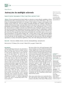
IMMUNOLOGICAL AND BIOCHEMICAL DIAGNOSIS EJ Thompson FIG. I. Oligoclonal bands in the Y-globulin region of cerebrospinal fluid (CSF)
and toxoplasmosis. However, it may occasionally be found in other conditions such as motor neurone disease, hydrocephalus, tumour and stroke (Laterre, 1965). Oligoclonal bands can also be detected electrophoretically using agarose, but in all methods serum from the patient should be examined by electrophoresis in parallel, since slight damage to the blood-CSF barrier may allow serum changes (e.g. the appearance of oligoclonal bands in subacute sclerosing pancncephalitis) to appear also within the CSF (Link, 1973). It is critical that the amount of IgG applied in agar- or agarose-gel electrophoresis should be carefully controlled (Link, 1967). Electrophoresis on cellulose acetate is a less discriminating method for the detection of the oligoclonal bands than that on agarose (Vandvik & Skrede, 1973). It has been argued that the oligoclonal pattern on electrophoresis is not as objective as the IgG determination. Hence the amount of IgG in CSF has been related to the amount of albumin and corrected for the respective serum values to generate a quotient. But values so produced remain less discriminating than the oligoclonal pattern (see Table I). However, improved gels and/or densitometers may allow better separation and quantitative analysis of these oligoclonal bands (see fig. la). Isoelectric focusing of CSF proteins permits demonstration of the oligoclonal pattern (Kjellin & Vesterberg, 1974), as does acrylamide-gel electrophoresis (fig. lb). The diffuse Y-globulin bands are easily distinguished from the much sharper haptoglobin bands which may occur with high total protein values. Some workers have felt that an acrylamide gel was not suitable for demonstration of oligoclonal bands. As the stacking (upper) gel can often contain IgG (Felgenhauer, 1971, p. 38), use of the appropriate buffer conditions allows visualization of the abnormal diffuse bands in the stacking gel with CSF from the MS patient (Felgenhauer, 1972). However, it is simpler to omit the upper gel, as appreciated by Cumings et al. (1970). An additional advantage of acrylamide-gel electrophoresis is that concentration of the CSF prior to electrophoresis is not required. This concentration procedure can lead to alteration in CSF proteins (Cunningham, 1964). Acrylamide-gel electrophoresis is also advantageous in that the precise amount of protein to be applied is not as critical as in agar- or agarose-gel electrophoresis (Tourtellotte, 1975, P. 35). The oligoclonal pattern found in CSF from MS patients cannot usually be absorbed by measles antigen, although this antigen regularly removes the oligoclonal bands from the CSF of patients with subacute sclerosing panencephalitis (Norrby & Vandvik, 1975). An intriguing recent finding in patients presenting with optic neuritis (Stendahl et al. 1976) has been that the oligoclonal pattern may be associated with the histocompatibility antigen LD-7a (HLA-Dw2). If this work is confirmed, the oligoclonal pattern might have prognostic significance in this context.
Densitometrlc tcan (ISCO) of acrylamlde gel of CSF from a MS patient. Arrows point to the oligoclonal bands. Gel Is 11 cm long and the origin Is on the left Acrylamide gels of CSF from another MS patient (left) and from a normal adult (right). Arrows point to the oligoclonal bands. Gels are 11 cm long and the origin is at the top
1975, p. 28) it is likely that free light chains exist in CSF (Bollengier et al. 1976). When the B lymphocytes from CSF were typed, they showed the same elevated «:X ratio (approximately 2.0) for light chains (Sandberg-Wollheim & Turesson, 1975). Although the (J trace protein has been isolated in significant amounts from CSF, the increase seen in MS is also found in many other neurological diseases and probably reflects destruction of CNS tissue (Olsson et al. 1976). Abnormalities of other specific proteins have been extensively documented in patients with MS and with other neurological diseases (Schuller et al. 1970; Schuller et al. 1971; Schuller et al. 1972a; Schuller et al. 1972b). Although fragments of myelin have been found in CSF of MS patients (Herndon & Johnson, 1970), the immunoassay of myelin basic protein has been complicated by its high affinity for a2-macroglobulin (Lennon & Mackay, 1972) as well as by the fact that myelin basic protein may be lost during concentration of the CSF (see section la(vii)).
vi Other Cerebrospinal Fluid Proteins Amongst the other proteins of interest, the light chains of immunoglobulins are of prime concern. It is a consistent finding that in about half the MS patients the *:X ratio for light chains in serum (i.e., 1.0) is normal, but the ratio is higher (approximately 2.0) in CSF. This alteration is not to be found among patients with known CNS infections (link & Mflller, 1971). Because of the occurrence of double pretiprtin lines on immunoelectrophoresis and immunodiffusion (Bauer,
vii Unconcentrated versus Concentrated Cerebrospinal Fluid As many studies have been performed with concentrated CSF, a number of caveats should be raised. The protein concentration should always be checked after filtration in order to correct for any losses of protein. Adsorption, especially of the more basic proteins (e.g. myelin basic protein or immunoglobulins), to glass as well as 30 Br. Med. Butt. 1977
IMMUNOLOGICAL AND BIOCHEMICAL DIAGNOSIS EJ Thompson to collodion bags (Link, 1967) or to other filters should be carefully considered (Cunningham, 1964). Proteolysis may occur not only in the CSF in vivo (link & Mttller, 1971) but also after collection in vitro. This process of protein hydrolysis as well as that of aggregation may produce loss of antibody during storage at 4°C or in the freezer (Felgenhauer, 1971, p. 51). All filters have a nominal pore size and hence cut off in Gaussian fashion at given molecular weights, e.g. collodion bags retain molecules of weights greater than 20000, conical centrifuge filters trap those larger than 50000. However, when filters are operated under different positive or negative pressures, the pore size may change with the stretch of the fabric, e.g. Visking dialysis tubing may pass light chains (23000 molecular weight) under the negative pressure of a water pump. Myelin basic protein has a molecular weight of 18500 and therefore may be readily lost.
2% of MS patients with the use of agar-gel electrophoresis, and in 29 % of MS patients with the use of agarose-gel electrophoresis (Link, 1973). Glial cytotoxic factors and demyelinating activities have been found in the serum of MS patients, but have also been noted in several other neurological diseases, and thus are not diagnostic (see papers by Davison & Cuzner, pp. 60-66, Haire, pp. 40^44, and Mertin & Meade, pp. 67-71, in this Bulletin for further specific details). ii Leucocytes Because of the ability to distinguish between B and T lymphocytes, which mediate immediate and delayed hypersensitivity, respectively, enumeration of these cell types has been attempted. Because of difficulties in identification of each cell type various results have been reported. Oger et al. (1975) found an increased proportion of only B cells, while Lisak et al. (1975) found an increased proportion of T cells and Sandberg-Wollheim & Turesson (1975) found no increase in either T or B cells. The function of B cells, namely to secrete circulating antibody, has been discussed in section la(i). Tests of T-cell function, both general responses as well as responses to CNS specific antigens, e.g. myelin basic protein (Sheremata et al. 1974), are discussed by Knight, pp. 45-50, Davison & Cuzner, pp. 60-66, and Fraser, pp. 34-39, in this Bulletin. Leucocyte neutral proteinase activity has been studied in patients followed longitudinally and the results are described by Davison & Cuzner. , . -
viii Antibody Synthesis The synthesis of antibody within the CNS has been estimated by Tourtellotte (1970a). However, lymphocytes recovered from the CSF and subsequently cultured in vitro have continued to synthesize immunoglobulins (Cohen & Bannister, 1967). This work has been confirmed and extended. It has been shown that CSF lymphocytes synthesized oligoclonal IgG, while serum lymphocytes from the same patients synthesized only homogeneously distributed Y-globulins. The amount of antibody synthesized by CSF lymphocytes was greater in exacerbation than in remission (Sandberg-Wollheim, 1974).
iii Platelets The platelets have been considered as part of a theory of micro-infarction related to perivascular cuffing. The methods for measurement of platelet adhesion are quite delicate and hence their application has been limited. Although increased platelet stickiness is found in chronic cases of MS it is probably a secondary event, rather than a primary cause (Lumsden, 1972, p. 492).
ix Myelin Lipids There are still problems associated with the methods used for the determination of CSF lipids. Lumsden (1972, pp. 498-511) has reviewed the earlier work. Recent evidence shows that the changes in total and individual phospholipids, as well as in free and esterified cholesterol, are not sufficiently large to be of diagnostic use in MS. Disagreements with earlier work may have been due to less discriminating methods (Pedersen, 1974). Since cerebrosides are an important part of the myelin sheath it would be expected that this lipid would be released into the CSF of MS patients during an exacerbation. Unfortunately, current analytical techniques are not sufficiently sensitive to assess this possibility (Tourtellotte, 1975, p. 36).
2 Relations of Immimoglobulins with the Clinical Course With different clinical courses to the disease, one must beware of generalizations, particularly when referring to the age of the patient, i.e., the age at onset and the duration of the disease may be quite different for several patients of the same age. Correlations of IgG concentrations in CSF with clinical status have not been clearly demonstrated nor agreed upon by various investigators. This may be owing in part to the manner in which the clinical material is divided.
b Blood
The blood is generally analysed because of its close relation to the CSF. Peripheral lymphocytes are examined, since these cells, as well as circulating metabolites, may find their way into the brain via the perivascular cuffs and eventually may enter the CSF. The implied question is: do circulating factors seek out the CNS to produce primary damage to this tissue only ?
a Age at Onset, Duration and Severity The distinction between age at onset and duration has been carefully noted and compared with the degree of disability by Olsson et al. (1976). They found that the highest concentrations of CSF IgG were in the more disabled patients, who had either a shorter duration of the disease (