The Hematology Journal (2001) 2, 103 ± 107 ã 2001 The European Haematology Association All rights reserved 1466 ± 4680/01 $15.00 www.nature.com/thj
Deregulated expression of prostate apoptosis response gene-4 in less dierentiated lymphocytes and inverse expressional patterns of par-4 and bcl-2 in acute lymphocytic leukemia Simone Boehrer1, Kai U Chow1, Elena Puccetti1, Martin Ruthardt1, Shahrzad Godzisard1, Andrea Krapohl1, Bernd Schneider1, Dieter Hoelzer1, Paris S Mitrou1, Vivek M Rangnekar2 and Eckhart Weidmann*,1 1
Department of Medicine III, Johann Wolfgang Goethe-University Hospital, Frankfurt am Main, Germany; 2Department of Radiation Medicine, University of Kentucky, Lexington, KY, USA
Introduction: Prostate apoptosis response gene-4, known as par-4, is a new proapoptotic factor functionally required but not sucient for apoptosis. Since there is evidence from prostate cancer cells that par-4 is involved in regulation of bcl-2 we assessed expression of par-4 and bcl-2 in dierent populations of normal and neoplastic lymphocytes. Materials and methods: Expression of par-4 mRNA and protein in dierent subpopulations of normal and neoplastic lymphocytes was assessed by reverse transcription polymerase chain reaction and Western blot. Results: Par-4 mRNA was not detectable in lymphocytes of healthy volunteers (n=10), but was present in the majority of samples of chronic lymphocytic leukemia (n=30), chronic lymphocytic leukemia/prolymphocytic leukemia (n=6) and acute lymphocytic leukemia (n=10). Par-4 protein was expressed unanimously in samples of mononuclear cells from healthy volunteers and patients with CLL, but less frequently in immature lymphocytes, including neoplastic cells of CLL/PLL and ALL. The decreased frequency of par-4 expression in immature subpopulations was con®rmed by results on lymphocytic cell lines at various stages of maturation. Comparing the expressional patterns of par-4 and bcl-2 there was an inverse relationship of both proteins in ALL and dierent lymphocytic cell lines, indicating a functional relationship of par-4 and bcl-2. Conclusions: This study establishes par-4 as a factor expressed in the majority of normal and neoplastic lymphocytic cells, demonstrating a decreased frequency of protein expression in less dierentiated lymphocytes and an inverse expressional pattern of par-4 and bcl-2 in lymphocytic cell lines and ALL. The Hematology Journal (2001) 2, 103 ± 107 Keywords:
par-4; bcl-2; apoptosis; ALL; CLL; prostate cancer
Introduction Prostate apoptosis response gene-4 (par-4) is a newly described protein with a proapoptotic function.1,2 Transfection experiments with cell lines derived from prostate cancer, renal cell carcinoma and malignant melanoma show that par-4 overexpression increases the sensitivity of cells to apoptotic stimuli, such as chemotherapeutic agents, UV-radiation and intracellular calcium elevation.1 ± 4 Furthermore par-4 expression is induced exclusively by proapoptotic stimuli and not *Correspondence: E Weidmann, Department of Medicine III, Johann Wolfgang Goethe-University, Theodor-Stern-Kai-7, 60590 Frankfurt, Germany; Tel: +49 69 6301 5779; Fax: +49 69 6301 7373; E-mail:
[email protected] Received 12 July 2000; accepted 31 October 2000
by growth arresting, growth promoting or necrotic stimuli.1,5 Reduction of bcl-2 protein levels by overexpression of par-4 protein in ®broblasts and prostate cancer cells is followed by an increase in the susceptibility of cells to apoptotic stimuli.6 The antiapoptotic protein bcl-2 is expressed in many dierent cell types, including acute (ALL) and chronic (CLL) lymphocytic leukemia cells.7 ± 9 Although many studies tried to de®ne the role of bcl-2 in these entities, taken by itself bcl-2 fails to be of reliable prognostic signi®cance.9 ± 11 Nevertheless bcl-2 has been suggested to be at least partly responsible for the resistance of CLL cells to proapoptotic stimuli.12 Chronic lymphocytic leukemia is the most common form of leukemia in the western world.13 In contrast to other forms of leukemia the disease is thought to
Par-4 expression in normal and neoplastic lymphocytes S Boehrer et al
104
arise mainly from a defect in the apoptotic pathway rather than from overproliferation of leukemic cells.11,14 Closely related to typical CLL but clinically more aggressive is the chronic lymphocytic leukemia with an increased number of prolymphocytes (between 11% and 55% of lymphocytes), called CLL/PLL (chronic lymphocytic leukemia/prolymphocytic leukemia). The cell population is more heterogenous, due to the increased portion of less mature prolymphocytes.15,16 In contrast to CLL and CLL/PLL, acute lymphocytic leukemia (ALL) exhibits a highly aggressive clinical course, although it also harbors possible curability due to the high chemosensitivity of its malignant lymphocytes.17 In vitro studies demonstrate that the expression level of bcl-2 in primary leukemic cells from patients with ALL is correlated with a cell's ability to survive in vitro,18 and, furthermore, that human ALL cell lines transfected with bcl-2 exhibit not only prolonged survival, but also increased chemoresistance towards vincristine and dexamethasone, both components of standard induction chemotherapy regimens.19,20 Regardless of all previous eorts, the molecular mechanisms governing the expression level of bcl-2 are still poorly understood.21 Since the malignant cells of CLL, CLL/PLL and ALL all arise from a lymphocytic origin but constitute diseases diering widely in pathogenesis, clinical appearance and prognosis, it seems feasible to search for molecular reasons explaining these dierences. Because there is no knowledge about par-4 expression in lymphocytic cells, we ®rst assessed the frequency of par-4 expression in dierent normal and neoplastic lymphocytic populations. Since there is evidence from other cell types that par-4 regulates the expression of bcl-2,6 we also compared the expression patterns of both proteins.
Materials and methods Patients and cell lines After informed consent, peripheral blood (PB) from 10 healthy volunteers, 30 patients with CLL, six patients with CLL/PLL, and 10 patients with ALL was collected, using standard procedures. Strict criteria were applied to de®ne diagnosis, and entities were classi®ed according to surface immunophenotype and morphology. Nine lymphocytic cell lines were examined: four ALL cell lines: Nalm-6, SEM, SD1 (Ph+), TOM (Ph+), one cell line derived from a patient in lymphocytic blast crisis of chronic myelocytic leukemia (BV 173), one human leukemic T-lymphoblastic lymphoma (Jurkat), one cutaneous T-cell lymphoma (HUT 78), and two human Burkitt lymphomas (Daudi, Raji). Mononuclear cells (MNC) were isolated by Ficoll-Hypaque density-gradient centrifugation followThe Hematology Journal
ing the manufacturer's instructions (Biochrom, Berlin, Germany).
Cell sorting Samples of three healthy individuals were sorted into B cells and T cells using FACScalibur System (Becton Dickinson, Heidelberg, Germany) equipped with a sorting option. Cells were sorted after labeling with a PE-conjugated monoclonal antibody anti-CD19 or anti-CD3, respectively (both antibodies purchased from Becton Dickinson). Purity of sorted cells exceeded 98%.
Extraction of RNA, reverse transcription and polymerase chain reaction RNA was extracted using the RNAzol B method following the manufacturer's instructions (WAK Chemie, Bad Nauheim, Germany). Total RNA (4 mg) from each sample was reverse transcribed into cDNA, subsequently reverse transcription-PCR was performed according to the previously published protocol.22 All reagents were purchased from Gibco, Berlin, Germany. To assess integrity of RNA for preparation of all cDNAs b-actin was ampli®ed as an internal control. Primers were used as follows: 5'-CCGCTACCGCCGCGACTTC-3' and 5'-AAACAGAGGCCGCATGCTG-3' for bcl-2, 5'-CCTAGATATAACAGGGATGC-3' and 5'-TTTATTTTCCTGCTTTAGCTG-3' for par-4. Annealing temperature for bcl-2 was 708C, for par-4 548C.23 Ampli®cation products were separated by electrophoresis on an 1% agarose gel and visualized by ethidium bromide staining.
Western blot A total of 16106 cells were pelleted and fractionated by SDS-page (12 ± 15% gradient gels) and proteins were transferred to a nitrocellulose membrane using an electroblotting apparatus (Bio-Rad, Hercules, CA, USA) using standard protocols. The loading of equal amounts of protein was veri®ed by Ponceau staining of the nitrocellulose membranes and by Coomassie staining of the polyacrylamide gels. The membrane was blocked with a 5% nonfat, dry milk for 1 h and subsequently incubated with the primary antibody at a dilution of 1 : 1000 (bcl-2: DAKO, Glostrup, Denmark, par-4: Santa Cruz Biotechnology, Santa Cruz, CA, USA) for 2 h at room temperature. Unbound antibody was removed by washing with Tris buered saline (pH 7,2) containing 0.5% Tween20. The membrane was then incubated with the secondary antibody (alkaline-phosphatase-conjugated antibody, Sigma, Deisenhofen, Germany) for 2 h at room temperature. After extensive washing with Tris-buered saline proteins were detected upon addition of the staining substrates (BCIP: 5-Bromo-4-chloro-3-indolyl-phosphate, Boehringer Mannheim, Mannheim, Germany; NBT: 4-Nitro-blue-tetrazolium-chloride, Boehringer Mannheim, Indianapolis, USA).
Par-4 expression in normal and neoplastic lymphocytes S Boehrer et al
105
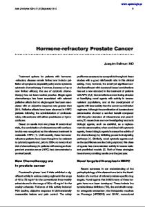
Results In samples of MNC from healthy volunteers par-4 mRNA was not detectable, but in all 10 samples of normal MNC par-4 protein was present with little inter-individual variability (Table 1). In sorted MNC of healthy individuals two of three samples of T cells as well as of B cells contained par-4 protein (data not shown). In contrast to normal MNC, 19 of 30 CLL samples contained detectable amounts of par-4 mRNA. All 30 samples of patients with CLL expressed high amounts of par-4 protein with little inter-patient variability (Table 1). Three of six samples of CLL/PLL assessed for par-4 expression contained detectable amounts of par-4 protein. These three par-4 protein positive samples were also par-4 mRNA positive (Table 1). Because of the lower frequency of par-4 in CLL/ PLL (an entity harboring an increased number of more immature prolymphocytes) than in CLL, we hypothesized that expression of par-4 is down-regulated in less mature cells. We therefore studied a possible association of par-4 expression with the level of lymphocytic dierentiation by testing 10 samples of ACL. Par-4 protein was present in seven of 10 samples of ALL patients, exhibiting considerable inter-patient variability in the level of expression (Figure 1). Six of 10 samples contained mRNA (in accordance with the protein positive samples). Furthermore, of nine lymphocytic cell lines tested for par-4 expression, three (Nalm-6, SEM, SD 1) of four ACL cell lines had no detectable amounts of par-
4, and only one Philadelphia chromosome positive cell line (TOM) expressed par-4 protein. One cell line derived from a patient suering from CML in lymphocytic blast crisis (BV 173) was also par-4 protein negative. In contrast the more mature T-cell lymphomas (Jurkat, HUT 78) and Burkitt-lymphomas (Raji, Daudi) contained par-4 protein (Figure 2). All 10 samples of normal MNC contained bcl-2 protein. Three of 10 samples were bcl-2 mRNA positive. Also all samples of CLL were bcl-2 protein positive with high inter-individual dierences in the amount expressed, 25 of 30 samples of CLL contained bcl-2 mRNA. In CLL/PLL, four of six samples expressed bcl-2 protein as well as bcl-2 mRNA (data not shown). Of the 10 patients with ALL nine samples were bcl-2 protein positive, all 10 samples containing bcl-2 mRNA. With the exception of Daudi and HUT 78, all cell lines expressed bcl-2 protein. A noteworthy observation is that when comparing protein levels, only samples of ALL with high amounts of par-4 protein had low levels of bcl-2 and vice versa (Figure 1), demonstrating an inverse expressional pattern of the two proteins in ALL. This pattern was also seen in cell lines originating from Burkitt- and Tcell lymphomas (Figure 2), but not in samples of normal MNC, CLL or CLL/PLL.
Discussion
MNC=mononuclear cells of healthy volunteers; CLL=chronic lymphocytic leukemia; CLL/PLL=chronic lymphocytic leukemia/ prolymphocytic leukemia; ALL=acute lymphocytic leukemia.
Originally the par-4 gene was isolated by dierential screening for genes upregulated when prostate cancer cells were induced to undergo apoptosis.1,2 Subsequent experiments demonstrated that an elevated par-4 protein expression increases the sensitivity of these cells to apoptotic stimuli by down-regulating the level of bcl-2.6 Bcl-2 is a well-described protooncogene in normal and malignant hematopoetic cells confering longevity.24 It is of particular interest for two reasons. Firstly, overexpression of bcl-2 prolongs cell survival, a decisive step in leukemogenesis/lymphomagenesis, since longer living cells are subjected to more oncogenic events promoting the emergence of malignancy.25 Secondly, bcl-2 is described as a novel multidrug resistance protein, delaying or preventing apoptosis induced essentially by all currently used chemotherapeutic agents.26,27
Figure 1 Protein expression of par-4 (38 kDa) and bcl-2 (25 kDa) in leukemic cells of seven patients with acute lymphocytic leukemia (ALL, lanes 1 ± 7), demonstrating an inverse expression pattern of both proteins.
Figure 2 Protein expression of par-4 (38 kDa) and bcl-2 (25 kDa) in dierent lymphocytic cell lines detected by Western blot, demonstrating an inverse expression pattern of both proteins.
Table 1 Frequency of mRNA and protein expression of par-4 in dierent populations of normal and neoplastic lymphocytes
Entity MNC CLL CLL/PLL ALL
Number of tested samples
Number/ percentage of par-4 mRNA positive samples
Number/ percentage of par-4 protein positive samples
10 30 6 10
0/0 19/63 3/50 6/60
10/100 30/100 3/50 7/70
The Hematology Journal
Par-4 expression in normal and neoplastic lymphocytes S Boehrer et al
106
Compared with CLL a deregulated expression of bcl-2 in ALL is well documented, although the majority of correlative clinical/laboratory studies fail to establish a clear association between bcl-2 expression and patient characteristics or treatment outcome.8,25 Despite the abundance of studies conducted there is limited knowledge about factors in¯uencing the level of bcl-2 expression.21 Therefore, the documented role of bcl-2 in lymphocytic neoplasms warrants further study to investigate potential regulators of its expression. Since a recent study showed regulation of bcl-2 by upstream par-4 in prostate cancer cells,6 we tested normal and neoplastic cells of lymphocytic origin for the presence of proapoptotic par-4 protein and mRNA and their potential associations with the expression level of antiapoptotic bcl-2. Our study establishes par-4 as a molecule expressed in dierent populations of normal and neoplastic lymphocytes. Noteworthy, are the distinct patterns of expression of protein and mRNA observed in dierent lymphocytic subpopulations. In contrast to immature neoplastic lymphocytic cells, all samples of unsorted MNC from healthy individuals showed expression of par-4 protein with little inter-individual variability in the amount present. Furthermore none of the samples of normal MNC had detectable levels of mRNA. Possibly, par-4 mRNA in these cells is only expressed during a short period of time thus evading detection by PCR. Nevertheless a more reasonable explanation might be that normal MNC contain lower amounts of the par-4 transcript than their neoplastic counterparts, and that the PCR applied is not suciently sensitive to detect these smaller amounts. Despite the fact that par4 mRNA was not detectable in lymphocytes of healthy individuals, par-4 protein was present in all samples of normal MNC, as well as in the sorted subpopulations of normal B and T lymphocytes. Compared with normal lymphocytes all samples from patients with CLL expressed par-4 protein, but the majority of these patients also had detectable amounts of par-4 mRNA, demonstrating a dierent regulation of par-4 in CLL. In samples of CLL there was no obvious relationship between the protein levels of par-4 and bcl-2.
Contrary to the situation in CLL, where all samples were par-4 protein positive, three of six patients with CLL/PLL were par-4 negative (protein as well as mRNA). Although this population is too small to draw substantial conclusions, the results indicate that par-4 is less frequently expressed in populations comprising less mature lymphocytes. This hypothesis is corroborated by par-4 expression in immature lymphocytic cell lines. The decreased frequency of expression in less mature subpopulations is of particular interest since a previous study assessing par-4 expression in dierent normal rat tissues by immunohistochemistry5 detected more intensive staining for par-4 in the stem cell compartments of the skin and the mucosa of the duodenum. Due to the staining for par-4 being less pronounced in the more mature layers of the skin and the duodenum, it was concluded that par-4 has to be down-regulated for successful dierentiation.5 The deregulated expression of par-4 in more immature/less dierentiated cells is further con®rmed by the decreased frequency of protein expression in samples of patients suering from ALL. Furthermore, the inverse expressional pattern of par-4 and bcl-2 suggests a functional interaction of the two proteins, possibly a regulation of bcl-2 by upstream par-4 as described most recently in prostate cancer cells.6 In conclusion our study establishes par-4 as a factor widely expressed in dierent populations of normal and neoplastic lymphocytes. We also describe a deregulated expression of par-4 in neoplastic lymphocytes, in particular a decreased frequency of par-4 protein expression in populations of less mature lymphocytes. The inverse expressional pattern of par4 and bcl-2 protein in ALL and dierent lymphocytic cell lines demonstrated here, suggests that in lymphoproliferative diseases par-4 interacts with bcl-2 and may have a functional role in pathogenesis of these diseases.
Acknowledgements This work was supported in part by the Adolf-MesserStiftung, Koenigstein, Germany.
References 1 Sells SF, Wood Jr DP, Joshi-Barve SS, Muthukumar S, Jacob RJ, Crist SA, Humphreys S, Rangnekar VM. Commonality of the gene programs induced by eectors of apoptosis in androgen-dependent and -independent prostate cells. Cell Growth and Dierentiation 5: 457, 1994. 2 Sells SF, Han SS, Muthukkumar S, Maddiwar N, Johnstone R, Boghaert E, Gillis D, Liu G, Nair P, Monnig S, Collini P, Mattson MP, Sukhatme VP, Zimmer SG, Wood Jr DP, McRoberts JW, Shi Y, Rangnekar VM. Expression and function of the leucine zipper protein Par-4 in apoptosis. Molecular and Cellular Biology 17: 3823, 1997. The Hematology Journal
3 Diaz-Meco MT, Municio MM, Frutos S, Sanchez P, Lozano J, Sanz L, Moscat J. The product of par-4, a gene induced during apoptosis, interacts selectively with the atypical isoforms of protein kinase C. Cell 86: 777, 1996. 4 Cook J, Krishnan S, Ananth S, Sells SF, Shi Y, Walther MM, Linehan WM, Sukhatme VP, Weinstein MH, Rangnekar VM. Decreased expression of the proapoptotic protein Par-4 in renal cell carcinoma. Oncogene 18: 1205, 1999.
Par-4 expression in normal and neoplastic lymphocytes S Boehrer et al
107
5 Boghaert ER, Sells SF, Walid AJ, Malone P, Williams NM, Weinstein MH, Strange R, Rangnekar, VM. Immunohistochemical analysis of the proapoptotic protein Par-4 in normal rat tissues. Cell Growth and Dierentiation 8: 881, 1997. 6 Qiu G, Ahmed M, Sells SF, Mohiuddin M, Weinstein MH, Rangnekar VM. Mutually exclusive expression patterns of Bcl-2 and Par-4 in human prostate tumors consistent with down-regulation of Bcl-2 by Par-4. Oncogene 18: 623, 1999. 7 Hermine O, Haioun C, Lepage E, d'Agay MF, Briere J, Lavignac C, Fillet G, Salles G, Marolleau JP, Diebold J, Reyas F, Gaulard P. Prognostic signi®cance of bcl-2 protein expression in aggressive non- Hodgkin's lymphoma. Groupe d'Etude des Lymphomes de l'Adulte (GELA). Blood 87: 265, 1996. 8 Maung ZT, MacLean FR, Reid MM, Pearson AD, Proctor SJ, Hamilton PJ, Hall AG. The relationship between bcl-2 expression and response to chemotherapy in acute leukaemia. British Journal of Haematology 88: 105, 1994. 9 Uckun FM, Yang Z, Sather H, Steinherz P, Nachman J, Bostrom B, Crotty L, Sarquis M, Ek O, Zeren T, Tubergen D, Reaman G, Gaynon P. Cellular expression of antiapoptotic BCL-2 oncoprotein in newly diagnosed childhood acute lymphoblastic leukemia: a Children's Cancer Group Study. Blood 89: 3769, 1997. 10 McConkey DJ, Chandra J, Wright S, Plunkett W, McDonnell TJ, Reed JC, Keating M. Apoptosis sensitivity in chronic lymphocytic leukemia is determined by endogenous endonuclease content and relative expression of BCL-2 and BAX. Journal of Immunology 156: 2624, 1996. 11 Pepper C, Hoy T, Bentley DP. Bcl-2/Bax ratios in chronic lymphocytic leukaemia and their correlation with in vitro apoptosis and clinical resistance [see comments]. British Journal of Cancer 76: 935, 1997. 12 Gottardi D, Alfarano A, De Leo AM, Stacchini A, Bergui L, Caligaris-Cappio F. Defective apoptosis due to Bcl-2 overexpression may explain why B-CLL cells accumulate in G0. Current Topics in Microbiology and Immunology 194: 307, 1995. 13 Crossen PE. Genes and chromosomes in chronic B-cell leukemia. Cancer Genetics and Cytogenetics 94: 44, 1997. 14 Reed JC. Bcl-2 family proteins: regulators of apoptosis and chemoresistance in hematologic malignancies. Seminars in Hematology 34: 9, 1997. 15 Lens D, Dyer MJ, Garcia-Marco JM, De Schouwer PJ, Hamoudi RA, Jones D, Farahat N, Matutes E, Catovsky D. p53 abnormalities in Cll are associated with excess of prolymphocytes and poor prognosis. British Journal of Haematology 9: 848, 1997.
16 Oscier DG, Matutes E, Copplestone A, Pickering RM, Chapman R, Gillingham R, Catovsky D, Hamblin, TJ. Atypical lymphocyte morphology: an adverse prognostic factor for disease progression in stage A CLL independent of trisomy 12. British Journal of Haematology 98: 934, 1997. 17 Crump M, Keating A. Acute leukemia in adults. Current Opinion in Hematology 2: 247, 1995. 18 Campana D, Coustan-Smith E, Manabe A, Buschle M, Raimondi SC, Behm FG, Ashmun R, Arico M, Biondi A, Pui CH. Prolonged survival of B-lineage acute lymphoblastic leukemia cells is accompanied by overexpression of bcl-2 protein. Blood 81: 1025, 1993. 19 Coustan-Smith E, Kitanaka A, Pui CH, McNinch L, Evans WE, Raimondi SC, Behm FG, Arico M, Campana D. Clinical relevance of BCL-2 overexpression in childhood acute lymphoblastic leukemia. Blood 87: 1140, 1996. 20 Tang C, Willingham MC, Reed JC, Miyashita T, Ray S, Ponnathpur V, Huang Y, Mahoney ME, Bullock G, Bhalla K. High levels of p26BCL-2 oncoprotein retard taxol-induced apoptosis in human pre-B leukemia cells. Leukemia 8: 1960, 1996. 21 Reed JC. Dysregulation of apoptosis in cancer. Journal of Clinical Oncology 17: 2941, 1999. 22 Karakas T, Maurer U, Weidmann E, Miething CC, Hoelzer D, Bergmann L. High expression of bcl-2 mRNA as a determinant of poor prognosis in acute myeloid leukemia [see comments]. Annals of Oncology 9: 159, 1998. 23 Johnstone RW, See RH, Sells S F, Wang J, Muthukkumar S, Englert C, Haber DA, Licht JD, Sugrue SP, Roberts T, Rangnekar VM, Shi Y. A novel repressor, par-4, modulates transcription and growth suppression functions of the Wilms' tumor suppressor WT1. Molecular and Cellular Biology 16: 6945, 1996. 24 Korsmeyer SJ. Bcl-2 initiates a new category of oncogenes: regulators of cell death. Blood 80: 879, 1992. 25 Yang T, Buchan HL, Townsend KJ, Craig RW. MCL-1, a member of the BLC-2 family, is induced rapidly in response to signals for cell dierentiation or death, but not to signals for cell proliferation. Journal of Cellular Physiology 166: 523, 1996. 26 Reed JC, Kitada S, Takayama S, Miyashita T. Regulation of chemoresistance by the bcl-2 oncoprotein in non-Hodgkin's lymphoma and lymphocytic leukemia cell lines. Annals of Oncology 5 (Suppl. 1): 61, 1994. 27 Thomas A, El Rouby S, Reed JC, Krajewski S, Silber R, Potmesil M, Newcomb EW. Drug-induced apoptosis in B-cell chronic lymphocytic leukemia: relationship between p53 gene mutation and bcl-2/bax proteins in drug resistance. Oncogene 12: 1055, 1996.
The Hematology Journal