Br. J. Anaath. (1984), 56,1351
INTRATHECAL ADMINISTRATION OF HYPERBARIC MORPHINE FOR THE RELIEF OF PAIN IN LABOUR T. K. ABBOUD, S. M. SHNIDER, P. A. DAILEY, J. A. RAYA, F. SARKIS, N. M. GROBLER, S. SADRI, S.-S. KHOO, B. DESOUSA, C. L. BAYSINGER AND F. MILLER
SUMMARY
Thirty healthy women in active labour received an intrathecal injection of morphine 0.5 mg (n = 12) or 1 mg (n - 18) in 7.5% dextrose. Both doses provided excellent analgesia for labour, 93% of patients obtaining at least 50% pain relief. Analgesia began 15-60min after injection and did not decrease until 6 - 8 h after injection. Analgesia was satisfactory until distension of the perineum, either by forceps or the infant's head. The intrathecal injection of morphine did not adversely affect the condition of the infant. Eighty per cent of patients developed pruritus; 53%, nausea or vomiting, or both; 43%, urinary retention; and 43%, drowsiness. These side effects were decreased by naloxone, which did not affect the degree of analgesia. There was no significant depression of ventilation in any patient. These results suggest that morphine 0.5 rag or 1 mg, administered intrathecally, effectively decreases the pain of labour, and that i.v. administration of naloxone can alleviate the common side effects.
Interest has focused recently on the efficacy of extradural and intrathecal opiates in relieving the pain of labour. For example, Hughes and colleagues (1984) found that 2, 5 or 7.5mg of extradurally administered morphine did not abolish the pain of labour and their findings confirmed earlier observations (Husemeyer, O'Connor and Davenport, 1980; Writer, James and Wheeler, 1981). However, in contrast, the intrathecal injection of morphine sulphate 1.5 mg in normal saline eliminated the pain of the first stage of labour in 12 patients (Scott et al., 1980). Similar success was reported using morphine 1 or 2 mg (in normal saline) (Baraka, Noueihid and Haji, 1981). Nevertheless, with both extradural (Reiz and Westberg, 1980; Bromage et al., 1982) and intrathecal (Baraka, Noueihid and Haji, 1981) administration (of morphine) the incidence of pruritus, nausea, vomiting and somnolence was high. The present study investigated whether smaller doses of morphine would effectively relieve the pain of labour, and whether a hyperbaric solution of T. K. ABBOUD, M.D.;J. A. RAYA, M . D . ; F . SARJUS, M.D.; N. M. GROBLER, M.D.J S. SADRI, M.D. ; S.-S. KHOO, M.D. ; B. DESOUSA,
M.D.; Department of Anesthesia, Los Angeles County-University of Southern California Medical Center, 1200 North State Street, Box 12, Los Angeles, California 90033, U.S.A. S. M. SHNIDER, M.D.; P. A. DAILEY, M.D. ; C. L. BAYSINGER, M.D. ; Department
of Anesthesia, University of California, San Francisco, California 94143, U.S.A. F. MILLER, M.D.; Department of Obstetrics and Gynecology, Los Angeles County-University of Southern California Medical Center, 1200 North State Street, Los Angeles, California 90033, U.S.A.
morphine would decrease the incidence of side effects. PATIENTS AND METHODS
Thirty healthy parturients having no signs of fetal distress were randomly assigned to receive an intrathecal injection of 0.5 ing (n= 12) or 1 mg (n=18) of preservative-free morphine sulphate lmgml" 1 in 7.5% dextrose. Approval from the Committee on Human Research and informed consent were obtained. The morphine was prepared by the pharmacy of the University of California, San Francisco. No patient received sedatives or narcotics within 4 h of the administration of morphine. Morphine was injected to the intrathecal space when the patient was in active labour and cervical dilatation was 4-8 cm. The parturient was positioned on her side and the head was elevated 30°. Lumbar puncture was performed at the L3-4 space using a 25-gauge spinal needle. After injection, the patient was turned supine and the uterus displaced to avoid aorto-caval compression. The patient's head was kept elevated for 24 h. After analgesia was established, six patients for whom midforceps delivery was anticipated (because of malposition of the fetal head, such as occiput posterior or transverse) were again placed on their side. Using an 18-gauge Crawford needle, a catheter was inserted to the extradural space above or below the site of the intrathecal injection—this catheter to be used if the analgesia from the intrathecal injection of morphine proved inadequate for labour and delivery. © The Macmillan Press Ltd 1984
1352 Using internal electrodes and catheters, fetal heart rate and uterine activity were monitored continuously from at least 30 min before the administration of morphine until delivery. During labour, the patient assessed the intensity of pain by marking a point on a "Visual Linear Analogue Scale" that represented, on one end, "no pain", and on the other end, the "worst pain that I have ever experienced." An independent investigator assessed the intensity of pain using the following scale: 0 = no pain; 1 = mild pain (easily tolerated); 2 = moderate pain (some discomfort and continuous awareness of pain); and 3 = severe pain (considerable pain that causes marked and continuous discomfort). The independent investigator also evaluated the quality of pain relief, using the following rating scale and criteria: 0 = pain worse (pain worse than before anaesthesia was given); 1 = no relief (pain unchanged); 2 = fair relief (pain less than 50% relieved; some movement or complaint with contraction); 3 = good relief (pain more than 50% relieved; slight grimace with contraction); and 4 = excellent pain relief (pain completely relieved). These assessments were made immediately after a uterine contraction just before the injection of morphine and after the injection (that is, approximately every 15 min for 1 h, every 30 min for the next 7 h and then every 1 h until delivery). Maternal heart rate, arterial pressure and respiratory rate were observed at these times, and for 24 h after the injection of morphine. Samples of maternal venous blood were obtained before the injection, at delivery and every 4 h after the injection of morphine for 24 h for analyses of blood-gas tensions. Patients were observed and directly questioned for 24 h for adverse side-effects, especially nausea, vomiting, pruritus, drowsiness, headache, urinary retention and respiratory depression (defined as being a respiratory rate of 10 b.p.m. or less). Initially, nausea and vomiting were treated with promethazine, and pruritus with diphenhydramine. Later in the study, after several anecdotal reports of the reversal of side effects with naloxone came to our attention (Forster et al., 1972; Glynn et al., 1979; Scott and McClure, 1979; Jones and Jones, 1980), naloxone was used to treat nausea, vomiting, pruritus and drowsiness. Urinary retention was relieved by catheterization of the bladder. If additional analgesia was required at delivery, the patients received local infiltration, pudendal block, inhalation analgesia or extradural anaesthesia, or a combination of these. Patients who
BRITISH JOURNAL OF ANAESTHESIA required Caesarean section received either general anaesthesia or extradural blockade. After delivery, patients were allowed to sit up in bed to eat or to care for their babies, and to walk to the bathroom, unless they complained of headache. Patients were not allowed to lie flat until 24 h after the intrathecal administration of morphine. "Slow slope active phase," a classification of abnormal progress of labour, was defined as being cervical dilatation of less than 0.7 cm h"1 in nulliparous patients and 1.1 cm h"1 in multiparous patients (Heffron and Laros, 1978). "Active phase arrest" was defined as being no change in cervical dilatation for 2h. "Prolonged second stage" occurred when the second stage of labour lasted for more than 2 h. Augmentation of labour with oxytocin was initiated at the request of the obstetrician. The condition of the infant was evaluated as follows: Apgar scores at 1 and 5 min; the times to sustained respiration; analysis of blood-gas tensions in venous and arterial blood obtained from a doublyclamped segment of umbilical cord; and the Neurologic and Adaptive Capacity Scores (NACS) (Amiel-Tison et al., 1982) at 15 min, 2 h and 24h. For purposes of comparison, each patient's assessment of pain was standardized to her initial evaluation of pain using the data from the Visual Linear Analogue Scale and the equation shown in figure 1. Mean values and standard errors of the mean are provided. Data were compared using Student's rtest for unpaired data and the Fisher exact test. Differences were considered significant when P < 0.05. RESULTS
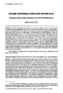
Obstetric analgesia The intrathecal administration of morphine 0.5 and 1 mg provided excellent relief of the pain of labour. Analgesia began 15-60 min after injection ANALOGUE LINE REPRESENTNG MORPHINE (rm\)
ANALOGUE UNE REPRESENTING " PAIN AFTER MORPHINE (mm)
ANALOGUE LWE REPRESENTING PAM BEFORE MORPHINE (mm)
PERCENTAGE X 1 0 0 = OF MATERNAL PAIN RELIEF
FIG. 1. Equation used to calculate the parturient's assessment of relief of labour pain (as a percentage) after intrathecal injection of morphine. Data from the "Visual Linear Analogue Scale" were used. Maternal pain relief was the percentage of change from her initial assessment of pain, that is, before morphine was administered.
INTRATHECAL INJECTION OF MORPHINE FOR LABOUR
1353
100 y
'Morphine 0.5 mg Morphine 1.0 mg
80
c 0) CO
a. 60
I 40
£. 20
15
30
45 60
90
120
150
180
Time after intrathecal injection of morphine (mm) FIG. 2. Onset (min) of painrelief( 50% or greater) after intrathecal administration of morphine 0.5 mg or 1 mg.
(fig. 2); and by 1 h, 58% and 76% of patients receiving 0.5 and 1 mg, respectively, reported at least 50% pain relief (n.s.). Based on data from the Visual Linear Analogue Scale, all patients given morphine 0.5mg and 88% of those given 1 mg obtained at least 50% pain relief; 42% and 59% of patients given 0.5 and 1.0 mg, respectively, achieved 100% pain relief (n.s.). Maximum pain relief occurred approximately 90-120 min after the administration of morphine and did not decrease until 6-8 h later (fig. 3). At 3 h, two-thirds of patients who had not delivered (n = 26) reported more than 80% pain relief. Two of the three patients who had not delivered by 12 h after the injection still reported more than 50% pain relief. The investigator also rated pain relief as good-to-excellent (fig. 4). Four hours after the intrathecal injection of morphine, 91% of the patients who had not delivered (n = 23) were judged by the independent investigator to have good-to-excellent pain relief. Two patients (both given 1 mg) requested further analgesia before delivery. Progress of labour
Seventy-seven per cent of patients were primiparous (table I). The mean cervical dilatation immediately before receiving morphine was approxi-
mately 6 cm. Forty-three per cent of patients had augmentation of labour with oxytocin; in 40% of these, oxytocin was commenced before or at the time of the administration of morphine. For all patients as a group, the mean interval between the injection of morphine and delivery was 7.4 h. Three patients required Caesarean section, all for unsuspected cephalopelvic disproportion first manifested by active phase arrest; one patient had received morphine 0.5 mg and two 1 mg. Of the patients who delivered per vaginam, the mean rate of cervical dilatation after intrathecal morphine was 1.35cmh"': 1.30cmh"' for primiparous patients and 1.49cmh"' for multiparae. Seventy-four per cent of these patients had normal progress of the first stage of labour, and 59% had a normal duration of the second stage of labour (table II). Fifty-five per cent of the primiparous patients had a prolonged second stage, while none of the multiparous patients had a second stage lasting longer than 120 min. Delivery Although the intrathecal injection of morphine relieved the pain of the first stage of labour, it did not adequately relieve pain secondary to distension of the perineum, application of forceps or episiotomy (table HI). The frequency of midforceps
BRITISH JOURNAL OF ANAESTHESIA
1354
TABLE I. Conditions of labour for 30 parturients receiving intrathecal administration of morphine 0.5mg or 1 mg for relief of pain. Values are mean ± SEM. Then were no significant differences between the two groups Morphine
Primiparae (no. of patients) Cervical dilatation before intrathecal injection of morphine (cm) Augmentation of labour with oxytocin (no. of patients) Duration of analgi-mn before delivery (h)
0.5mg
1 mg (n=18)
Combined data (n = 30)
10
13
23
6.1
5.6
±0.4
±0.3
5.8 ±0.3
4
9
13
7.1 ±0.8
7.4 ±0.7
7.8
±1.1
100
90
I