Critical Care
October 2004 Vol 8 No 5
Gattinoni et al.
Review
Bench-to-bedside review: Chest wall elastance in acute lung injury/acute respiratory distress syndrome patients Luciano Gattinoni, Davide Chiumello, Eleondra Carlesso and Franco Valenza Institute of Anesthesia and Critical Care, University of Milan, Policlinico – IRCCS Hospital, Milan, Italy
Corresponding author: Luciano Gattinoni,
[email protected]
Published online: 7 May 2004 This article is online at http://ccforum.com/content/8/5/350 © 2004 BioMed Central Ltd
Critical Care 2004, 8:350-355 (DOI 10.1186/cc2854)
Abstract The importance of chest wall elastance in characterizing acute lung injury/acute respiratory distress syndrome patients and in setting mechanical ventilation is increasingly recognized. Nearly 30% of patients admitted to a general intensive care unit have an abnormal high intra-abdominal pressure (due to ascites, bowel edema, ileus), which leads to an increase in the chest wall elastance. At a given applied airway pressure, the pleural pressure increases according to (in the static condition) the equation: pleural pressure = airway pressure × (chest wall elastance / total respiratory system elastance). Consequently, for a given applied pressure, the increase in pleural pressure implies a decrease in transpulmonary pressure (airway pressure – pleural pressure), which is the distending force of the lung, implies a decrease of the strain and of ventilator-induced lung injury, implies the need to use a higher airway pressure during the recruitment maneuvers to reach a sufficient transpulmonary opening pressure, implies hemodynamic risk due to the reductions in venous return and heart size, and implies a possible increase of lung edema, partially due to the reduced edema clearance. It is always important in the most critically ill patients to assess the intra-abdominal pressure and the chest wall elastance. Keywords acute respiratory distress syndrome, chest wall elastance, intra-abdominal pressure, pleural pressure, ventilator-induced lung injury
350
Introduction
Respiratory mechanics: general concepts
The respiratory system includes the lung and the chest wall, in series, and the overall mechanical behavior depends on the mechanical characteristics of its components and their interactions [1]. The common increase in the elastance (decrease in compliance) of the whole respiratory system in acute lung injury (ALI) and in acute respiratory distress syndrome (ARDS) has traditionally been attributed to the lung component. It has long been reported, however, that the chest wall elastance was also altered in many cases [2–5]. Recently, mainly due to the increased concern for the abdominal pressure, more attention has been paid to the chest wall mechanics in critically ill patients [6,7]. The problems of the mechanical impairment of the chest wall and its consequences are now widely recognized. The present review will focus on the dimension of these problems and their consequences in the critically ill patient.
When partitioning the respiratory mechanics into its lung and chest wall components, it is convenient to refer to elastance instead of compliance. The total elastance of the respiratory system is the pressure required to inflate it 1 l above its resting position. This is, the applied airway pressure is spent in part to inflate the lung and in part to inflate the chest wall. The chest wall comprises the anterior and posterior thoracic cage walls and the diaphragm, which is the ‘abdominal component’. Indeed, in static conditions, when the airway resistance is nil: Paw = Pl + Ppl (1) and Etot = El + Ecw (2)
ALI = acute lung injury; ARDS = acute respiratory distress syndrome; Ecw = chest wall elastance; El = lung elastance; Etot = total respiratory system elastance; Paw = airway pressure; Pl = transpulmonary pressure; Ppl = pleural pressure; VILI = ventilator-induced lung injury.
Available online http://ccforum.com/content/8/5/350
where Paw is the (static) airway pressure, Pl is the transpulmonary pressure, Ppl is the pleural pressure, Etot is the total respiratory system elastance, El is the lung elastance, and Ecw is the chest wall elastance. On the basis of these classical equations it is easy to grasp the mechanical interaction between the lung and the chest wall. First, however, it is important to recall that the concept of ‘transmission’ of alveolar pressure to the thoracic cavity is misleading [8]. Let us assume that we inflate an isolated lung at an alveolar pressure of 10, 15 or 20 cmH2O. The pressure measured at the pleural surface will always be 0 cmH2O (i.e. atmospheric pressure) because the alveolar pressure is not ‘transmitted’. When the thoracic cage surrounds the lungs as they are inflated, however, the cage has to change its volume. The lungs ‘push’ the thoracic cage, and the pressure generated by the interaction between the lung and the chest wall, which may have different elasticities, is the pleural pressure. If we consider that the pressure is ‘transmitted’, then the pleural pressure would depend on the airway pressure and the lung elasticity (the stiffer the lung, the lower the transmission). However, this approach ignores the contribution of the chest wall. If the thoracic cage is ‘soft’ the pleural pressure generated to drive it will be low, but if the thoracic cage is ‘stiff’ then a higher pleural pressure will be needed (Fig. 1). The distending force of the lung is the pressure difference between the alveoli and pleural pressure (the transpulmonary pressure), while the distending force of the thoracic cage is the pleural pressure to which all of the intrathoracic structures, such as the heart and the intrathoracic vessels, are subjected. In mathematical terms, because of and by rearranging equations 1 and 2, it follows that: Ppl = Paw × Ecw / Etot (3) and Pl = Paw × El / Etot (4) The pleural pressure depends on the pressure applied to the airways, and on the ratio between the chest wall elastance and the total elastance of the respiratory system, which is the sum of the chest wall elastance and the lung elastance (see equation 2). In normal conditions this ratio is about 0.5 at functional residual capacity, because the chest wall elastance and the lung elastance are similar. In ARDS, however, the elastance ratio may vary from 0.2 to 0.8 [6,8,9]. It is thus clear that, for the same applied airway pressure (let us say 30 cmH2O) and with a chest wall elastance/total respiratory system elastance ratio of 0.5, the transpulmonary pressure will be 15 cmH2O and the pleural pressure will be the same. If the ratio is 0.2, however, the transpulmonary pressure will be 24 cmH2O but the pleural pressure will be 6 cmH2O, while if the ratio is 0.8 the transpulmonary pressure will only be 6 cmH2O and the pleural pressure will be 24 cmH2O.
These simple calculations illustrate the importance of knowing the mechanical characteristics of both the lung and the chest wall. The same airway pressure may generate dramatically different transpulmonary pressures and pleural pressures, with marked consequences on lung distension (mainly a function of transpulmonary pressure) and on hemodynamics (partly a function of pleural pressure). We shall now look at the available tools to measure the pleural pressure, the causes of pleural pressure increases and the clinical consequences of elevated pleural pressure.
Measuring pleural pressure and intra-abdominal pressure The only method available in clinical practice to measure the pleural pressure is the measurement of the changes of esophageal pressure as detected by an esophageal balloon [10,11]. Unfortunately, the pressure measured in the esophageal balloon does not reflect the absolute value of the pleural pressure. In animal experiments in which we measured the pleural pressures directly by wafers in nondependent lung regions, in middle lung regions and in dependent lung regions in the supine position, in both healthy lungs and in edematous lungs, we found that esophageal pressure was a good estimate of the real pleural pressure in the middle lung but that it overestimated the nondependent pleural pressure and underestimated the dependent pleural pressure [12]. It is worth pointing out, however, that the differences in pressure recorded in the esophageal balloon closely match the differences in pleural pressure [13]. Although it must always be remembered that the esophageal pressure is only an estimate of the real pleural pressure, which varies in the different lung regions, we strongly believe that esophageal pressure measurement is sufficiently informative in the clinical scenario (i.e. to estimate the change in pressure). We may also wonder when this measurement is indicated in ALI/ARDS patients. As the chest wall impairment in these patients is usually due to an abnormal increase in intraabdominal pressure [6,14], in clinical practice we measure the esophageal pressure when the intra-abdominal pressure is altered. We in fact found that chest wall elastance increases linearly with the intra-abdominal pressure according to the following equation: Ecw = 0.47 × intraabdominal pressure (cmH2O) + 1.43 [6]. Accordingly, the pleural pressure/intra-abdominal pressure relationship may be estimated as: Ppl = Paw [(0.47 × intra-abdominal pressure + 1.43) / (0.47 × intra-abdominal pressure + 1.43 + lung elastance)] It is perhaps appropriate at this stage to briefly discuss the relationship between the intra-abdominal pressure and the pleural pressure. It is important to understand what the
351
Critical Care
October 2004 Vol 8 No 5
Gattinoni et al.
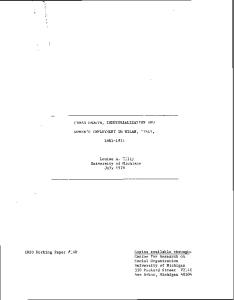
Figure 1
Effect of different lung elastance (EL) and chest wall elastance (Ew) on the total elastance (Etot) of the respiratory system. An equal total elastance of the respiratory system may arise (a) from a high lung elastance and a low chest wall elastance or (b) from identical lung elastance and chest wall elastance.
independent variable is, because the relationship is changed when the independent variable is the intra-abdominal pressure or when it is the intrathoracic pressure (pleural pressure). In addition, the diaphragm elastance plays a key role in this relationship. We routinely measure intra-abdominal pressure in critically ill patients admitted to the intensive care unit, because the incidence of an abnormal increase in intra-abdominal pressure occurs in 24–30% of these patients [7,15]. As the intraabdominal pressure levels that start to impair the chest wall elastance cannot be clinically assessed [16], we estimate the intra-abdominal pressure by measuring the bladder pressure [7,17,18], which is considered the best approach in clinical practice. The ‘normal’ intra-abdominal pressure during spontaneous breathing in healthy subjects is approximately 0 mmHg, while in mechanically ventilated patients this pressure is higher (range, 5–8 mmHg) [7,19].
Causes of chest wall impairment Several factors may affect the chest wall mechanics. The anatomical configuration of the thoracic cage, being overweight or pleural effusion can all increase, to various degrees, the chest wall elastance in sedated and paralyzed patients [19–23]. However, the most common causes of increased chest wall elastance in ALI/ARDS patients are abdominal diseases (such as bowel distension, ascites, hemoperitoneum).
352
Along with other workers [14], we have found striking differences in the chest wall mechanics between patients
with pulmonary ARDS, usually due to diffuse pneumonia, and those with extrapulmonary ARDS, usually due to abdominal diseases [6]. Although the total respiratory system elastance was similar in both groups, the pleural pressure was normal in pulmonary ARDS patients but abnormally high in extrapulmonary ARDS patients. This was linearly correlated with the increase of intra-abdominal pressure [2]. The presence of abdominal diseases (as well as obesity) in critically ill patients with ALI/ARDS should be a drive for careful investigation of their respiratory mechanics. Moreover, the differences we found in chest wall elastance in pulmonary ARDS patients and in extrapulmonary ARDS patients do represent a general trend. An individual patient with pulmonary ARDS may have a concomitant increase in intra-abdominal pressure.
Physiopathological consequences For a given applied airway pressure, as previously mentioned, the pleural pressure increases when the chest wall elastance is elevated. Accordingly, the transpulmonary pressure (the distending force of the lung) drops. We shall now discuss the respiratory and hemodynamic consequences of high pleural pressure. Respiratory system The key point is that, for a given applied pressure, the transpulmonary pressure falls when the pleural pressure rises (see equation 1). This may have important implications in understanding some of the differences in the presentation of ALI/ARDS and in setting the ventilator in these patients.
Available online http://ccforum.com/content/8/5/350
Patients with pulmonary ARDS and patients with extrapulmonary ARDS have different mechanical behavior, different lung morphology and different positive end-expiratory pressure response [6,14,2,24–26]. The chest wall elastance differences explain most of these different behavioral patterns. Extrapulmonary ARDS patients have diffuse lung edema due to inflammatory mediators originating in extrapulmonary foci [24]. The increase in lung weight causes compression atelectasis of the dependent lung regions [27]. Pulmonary ARDS patients, on the contrary, tend to have less homogeneous lung alteration. The main feature in these patients is the consolidation of some lung regions instead of lung collapse. As an example, the ‘safe’ airway plateau pressure between 30 and 35 cmH2O may give widely differing transpulmonary pressures [28–30] in patients with normal or increased chest wall elastance. In the extrapulmonary ARDS patients, the elevated pleural pressure caused by increased chest wall elastance will cause the transpulmonary pressure to be far lower than in pulmonary ARDS patients with normal elastance. Two factors contribute to the lung collapse in extrapulmonary ARDS patients. The first is due to the nature of the main pathological alteration (interstitial edema [31]), and the second factor is due to the high chest wall elastance, leading to a lower transpulmonary pressure. This diffuse collapse associated with interstitial edema and lower transpulmonary pressure leads to a different morphological pattern (with the prevalence of ground glass opacification) in extrapulmonary ARDS patients compared with the consolidation usually prevalent in pulmonary ARDS patients [25,26]. The potential for recruitment is also greater in extrapulmonary ARDS patients than in pulmonary ARDS patients [6,32,33]. The increased chest wall elastance may also be important in the pathogenesis of ventilator-induced lung injury (VILI). As this is probably due to the excessive and unphysiological strain on the lung structures, which in turn depends on the applied transpulmonary pressure [34], we may expect more VILI for a given applied pressure when the chest wall elastance is normal. VILI is in fact worse with an open chest (zero chest wall elastance [35]) than in the conditions in which the chest wall elastance is increased (as in experiments in which the thoracic cage was artificially constrained) [36]. It is possible, however, that the collapsed tissue would also be less prevalent in the presence of high transpulmonary pressure. Consequently, the average strain might be the same or lower. As the transpulmonary pressure is the trigger of VILI, if we take into account the chest wall elastance then the differences between barotrauma and volotrauma vanish. The barotrauma was in fact attributed to the applied airway pressure. What is important, however, is not this pressure but
the transpulmonary pressure applied to the lung structures (Paw – Pl), which in turn causes the strain. Some contradictory data arising from randomized studies with different tidal volumes [29,37–39] can be explained if we take into account the transpulmonary pressure. The same tidal volume, depending on the total elastance of the respiratory system and on the chest wall elastance/total respiratory system elastance ratio, might result in a completely different transpulmonary pressure [34], and consequently result in different VILI. The chest wall elastance has to be taken into account when performing recruitment maneuvers. What is also important for lung opening in this case is the transpulmonary pressure and not the airway pressure. If the opening pressure of some lung regions are of the order of 25–30 cmH2O transpulmonary pressure (sticky atelectasis [25,33]), then the airway pressure applied to reach this target will be completely different in patients with normal or abnormal chest wall elastance [8,32,33]. Changes in chest wall elastance also dictate the oxygenation response to the prone position. Pelosi and colleagues [40] and Guerin and colleagues [41] showed that the greater the decrease in chest wall compliance during the prone position, the greater the increase in oxygenation. Moreover, it has been shown that extrapulmonary ARDS patients have a greater potential for oxygenation improvement in the prone position than do pulmonary ARDS patients [42]. These findings are plausible in the light of the chest wall elastance changes; an increase of chest wall elastance in the prone position presumably leads to a more even distribution of the ventilation in pulmonary ARDS patients, while the changes in regional transpulmonary pressure in extrapulmonary ARDS patients explain the lung density redistributions and the better oxygenation in the prone position [43]. Hemodynamics and lung edema The increased pleural pressure may lower the cardiac output by reducing the venous return and the cardiac volume [44]. The evaluation of hemodynamics therefore calls for special care in cases of increased intra-abdominal pressure [7,45]. Moreover, both the central venous pressure and the wedge pressure may appear ‘falsely’ elevated in the presence of increased pleural pressure [7]. In a recent series of experiments in pigs in which edema was induced by oleic acid and the pleural pressure was changed by pneumoperitoneum, we found a decrease in gas volume due to a decreased transpulmonary pressure [46]. However, this was associated with an almost 100% increase of pulmonary edema. This effect may possibly be due to the blood shift induced by the abdominal pressure increase, which in turn may favor edema formation in a ‘leaking’ lung. A decrease of the edema clearance due to the increased pleural pressure is another coexisting possibility.
353
Critical Care
October 2004 Vol 8 No 5
Gattinoni et al.
Conclusion We suggest that a rational approach to the treatment of ALI/ARDS requires the knowledge of both lung elastance and chest wall elastance. Although not routinely carried out, we firmly believe that the measurement of the intra-abdominal pressure, the leading cause of chest wall impairment, should be performed.
Competing interests The authors declare that they have no competing interests.
References 1.
2.
3. 4. 5.
6.
7. 8.
9.
10. 11. 12.
13. 14.
15. 16.
17. 354
Agostoni E, Hyatt RE: Static behaviour of the respiratory system. In Handbook of Physiology, Section 3: The Respiratory System, Volume III, Part 1. Edited by Geiger SR. Bethesda, MD: American Physiological Society; 1986:113-130. Pelosi P, Cereda M, Foti G, Giacomini M, Pesenti A: Alterations of lung and chest wall mechanics in patients with acute lung injury: effects of positive end-expiratory pressure. Am J Respir Crit Care Med 1995, 152:531-537. Suter PM, Fairley HB, Isenberg MD: Effect of tidal volume and positive end-expiratory pressure on compliance during mechanical ventilation. Chest 1978, 73:158-162. Katz JA, Zinn SE, Ozanne GM, Fairley HB: Pulmonary, chest wall, and lung-thorax elastances in acute respiratory failure. Chest 1981, 80:304-311. Jardin F, Genevray B, Brun-Ney D, Bourdarias JP: Influence of lung and chest wall compliances on transmission of airway pressure to the pleural space in critically ill patients. Chest 1985, 88:653-658. Gattinoni L, Pelosi P, Suter PM, Pedoto A, Vercesi P, Lissoni A: Acute respiratory distress syndrome caused by pulmonary and extrapulmonary disease. Am J Respir Crit Care Med 1998, 158:3-11. Malbrain M: Abdominal pressure in the critically ill. Curr Opin Crit Care 2000, 6:1-21. Gattinoni L, Vagginelli F, Chiumello D, Taccone P, Carlesso E: Physiologic rationale for ventilator setting in acute lung injury–acute respiratory distress syndrome patients. Crit Care Med 2003, 31:300s-304s. Gattinoni L, Carlesso E, Cadringher P, Valenza F, Vagginelli F, Chiumello D: Physical and biological triggers of ventilator induced lung injury and its prevention. Eur Respir J 2003, 47: 15s-25s. Milic-Emili J, Mead J, Turner JM: Improved technique for estimating pressure from esophageal balloons. J Appl Physiol 1964, 19:207-211. Maxted KJ, Shaw A, Macdonald TH: Choosing a catheter system for measuring intra-oesophageal pressure. Med Biol Eng Comput 1977, 15:398-401. Pelosi P, Goldner M, McKibben A, Eccher G, Caironi P, Losappio S, Gattinoni L, Marini JJ: Recruitment and derecruitment during acute respiratory failure. An experimental study. Am J Respir Crit Care Med 2001, 164:122-130. Baydur A, Behrakis PK, Zin WA, Jaegr M, Milic-Emili L: A simple method for assessing the validity of the oesophageal balloon technique. Am Rev Respir Dis 1983, 126:788-791. Ranieri VM, Brienza N, Santostasi S, Puntillo F, Mascia L, Vitale N, Giuliani R, Memeo V, Bruno F, Fiore T, Brienza A, Slutusky AS: Impairment of lung and chest wall mechanics in patients with acute respiratory distress syndrome. Am J Respir Crit Care Med 1997, 156:1082-1091. Sugrue M, Hilman KM: Intra-abdominal hypertension and intensive care. In Yearbook of Intensive Care and Emergency Medicine. Edited by Vincent JL. Berlin: Springer-Verlag; 1998:667-676. Sugrue M, Bauman A, Jones F, Bishop G, Flabouris A, Parr M, Stewart A, Hillman K, Deane SA: Clinical examination is an inaccurate predictor of intra-abdominal pressure. World J Surg 2002, 26:1428-1431. Iberti TJ, Lieber CE, Benjamin E: Determination of intraabdominal pressure using a transurethral bladder catheter: clinical validation of the technique. Anaesthesiology 1989, 70:47-50.
18. Kron JL, Harman PK, Nolan SP: The measurement of intraabdominal pressure as a criterion for abdominal reexploration. Ann Surg 1984, 199:28-30. 19. Pelosi P, Ravagnan I, Giurati G, Panigada M, Bottino N, Tredici S, Eccher G, Gattinoni L: Positive end expiratory pressure improves respiratory function in obese patients but not in normal subjects during anesthesia and paralysis. Anesthesiology 1999, 91:1221-1231. 20. Krell WS, Rodarte JR: Effects of acute pleural effusion on respiratory system mechanics in dog. J Appl Physiol 1985, 59: 1458-1463. 21. Sousa AS, Moll RJ, Pontes CF, Saldiva PH, Zin WA: Mechanical and morphometrical changes in progressive bilateral pneumothorax and pleural effusion in normal rats. Eur Respir J 1995, 8:99-104. 22. Pelosi P, Croci M, Ravagnan I, Tredici S, Pedoto A, Lissoni A, Gattinoni L: The effects of body mass on lung volumes, respiratory mechanics, and gas exchange during general anesthesia. Anesth Analg 1998, 87:654-660. 23. Ladosky W, Botelho MA, Albuquerque JP, Jr: Chest mechanics in morbidly obese non-hypoventilated patients. Respir Med 2001, 95:281-286. 24. Pelosi P, D’Onofrio D, Chiumello D, Paolo S, Chiara G, Capelozzi VL, Barbas CSV, Chiaranda M, Gattinoni L: Pulmonary and extrapulmonary acute respiratory di stress sindrome are different. Eur Respir J 2003, 22:48s-56s. 25. Gattinoni L, Caironi P, Pelosi P, Goodman LR: What has computed tomography taught us about the acute respiratory distress syndrome? Am J Respir Crit Care Med 2001, 164: 1701-1711. 26. Goodman LR, Fumagalli R, Tagliabue M, Ferrario M, Gattinoni L, Pesenti A: Adult respiratory distress syndrome due to pulmonary and extrapulmonary causes: CT, clinical and functional correlations. Radiology 1999, 213:545-552. 27. Brismar B, Hedenstierna G, Lundquist H, Strandberg A, Svesson L, Tockics L: Pulmonary densities during anesthesia with muscular relaxation. Anesthesiology 1985, 62:422-428. 28. Slutsky AS: Consensus conference on mechanical ventilation — January 28–30, 1993 at Northbrook, Illinois, USA. Part 2. Intensive Care Med 1994, 20:150-162. 29. The Acute Respiratory Distress Syndrome Network: Ventilation with lower tidal volumes as compared with traditional tidal volumes for acute lung injury and the acute respiratory distress syndrome. N Engl J Med 2000; 342:1301-1308. 30. Tobin MJ: Culmination of an era in research on the acute respiratory distress syndrome. N Engl J Med 2000, 342:13601361. 31. Bone RC: The ARDS lung. New insights from computed tomography. JAMA 1993, 269:2122-2127. 32. Grasso S, Mascia L, Del Turco M, Malacarne P, Giunta F, Brochard L, Slutsky AS, Ranieri VM: Effects of recruting maneuvers in patients with acute respiratory disress sindrome ventilated with protective ventilatory strategy. Anesthesiology 2002, 96:795-802. 33. Pelosi P, Cadringher P, Bottino N, Panigada M, Carrieri F, Riva E, Lissoni A, Gattinoni L: Sigh in acute respiratory distress syndrome. Am J Respir Crit Care Med 1999, 159:872-880. 34. Gattinoni L, Carlesso E, Cadringher P, Valenza F, Vagginelli F, Chiumello D: Physical and biological triggers of ventilator induced lung injury and its prevention. Eur Respir J 2003, 47: 15s-25s. 35. Woo SW, Hedley-Whyte J: Macrophage accumulation and pulmonary edema due to thoracotomy and lung over inflation. J Appl Physiol 1972, 33:14-21. 36. Dreyfuss D, Soler P, Basset G, Saumon G: High inflation pressure pulmonary edema. Respective effects of high airway pressure, high tidal volume, and positive end-expiratory pressure. Am Rev Respir Dis 1988, 137:1159-1164. 37. Amato MB, Barbas CS, Medeiros DM, Magaldi RB, Schettino GP, Lorenzi-Filho G, Kairalla RA, Deheinzelin D, Munoz C, Oliveira R, Takagaki TY, Carvalho CR: Effect of a protective-ventilation strategy on mortality in the acute respiratory distress syndrome. N Engl J Med 1998, 338:347-354. 38. Brochard L, Roudot-Thoraval F, Roupie E, Delclaux C, Chastre J, Fernandez-Mondejar E, Clementi E, Mancebo J, Factor P, Matamis D, Ranieri M, Blanch L, Rodi G, Mentec H, Dreyfuss D, Ferrer M, Brun-Buisson C, Tobin M, Lemaire F: Tidal volume reduction for
Available online http://ccforum.com/content/8/5/350
39.
40.
41.
42.
43.
44. 45.
46.
prevention of ventilator-induced lung injury in acute respiratory distress syndrome. The Multicenter Trail Group on Tidal Volume reduction in ARDS. Am J Respir Crit Care Med 1998, 158:1831-1838. Brower RG, Shanholtz CB, Fessler HE, Shade DM, White P, Jr, Wiener CM, Teeter JG, Dodd Almog Y, Piantadosi S: Prospective, randomized, controlled clinical trial comparing traditional versus reduced tidal volume ventilation in acute respiratory distress syndrome patients. Crit Care Med 1999, 27:14921498. Pelosi P, Tubiolo D, Mascheroni D, Vicardi L, Crotti S, Valenza F, Gattinoni L: Effects of the prone position on respiratory mechanics and gas exchange during acute lung injury. Am J Respir Crit Care Med 1998, 157:387-393. Guerin C, Baudet M, Rosselli S, Heyer L, Sab JM, Langevin B, Philit F, Fournier G, Robert D: Effects of prone position on alveolar recruitment and oxygenation in acute lung injury. Intensive Care Med 1999, 25:1222-1230. Lim CM, Kim EK, Lee JS, Shim TS, Lee SD, Koh Y, Kim WS, Kim DS, Kim WD: Comparison of the response to the prone position between pulmonary and extrapulmonary acute respiratory distress syndrome. Intensive Care Med 2001, 27:477-485. Gattinoni L, Pelosi P, Valenza F, Mascheroni D: Patient positioning in acute respiratory failure. In Principles and Practice of Mechanical Ventilation. Edited by Tobin M. New York: McGraw Hill; 1994:1067-1077 Pinsky M, Desmet JM, Vincent JL: Effect of positive end expiratory pressure on right ventricular function in humans. Am Rev Respir Dis 1991, 143:25-31 Malbrain MLNG. Intra-abdominal pressure in the intensive care unit: clinical tool or toy? In Yearbook of Intensive Care and Emergency Medicine. Edited by Vincent JL. Berlin: SpringerVerlag; 2001:547-585. Quintel M, Pelosi P, Caironi P, Meinhardt JP, Luecke T, Herrmann P, Taccone P, Rylander C, Valenza F, Carlesso E, Gattinoni L: An increase of abdominal pressure increases pulmonary edema in oleic-acid induced lung injury. Am J Respir Crit Care Med 2003 (epub ahead of print), in press.
355