GENERAL ARTICLES
Inhibition of growth of urinary type calcium hydrogen phosphate dihydrate crystals by tartaric acid and tamarind K. C. Joseph, Bharat B. Parekh and M. J. Joshi* Urinary stone is one of the oldest and the most common afflictions in humans, animals and birds. A number of people suffer from urinary stones. Brushite or calcium hydrogen phosphate dihydrate (CHPD) crystals are frequently found in urinary stones. Many fruits contain high amount of tartaric acid. Therefore, the effect of tartaric acid on the growth of CHPD crystals is presented in this article. Moreover, tamarind is widely used in Indian cooking, and contains high amount of tartaric acid. The effect of tartaric acid and tamarind is investigated in terms of the number of crystals grown, their morphologies and their average size under in vitro conditions. The growth of CHPD crystals is carried out using single-diffusion gel growth technique in silica gel medium. This study incorporates multidisciplinary interests and may be used in formulating a strategy for the prevention or dissolution of urinary stones. A number of people suffer from problems due to urinary stones (calculi). Areas of high incidence of urinary calculi, include the British Isles, Scandinavian countries, northern Australia, Central Europe, northern India, Pakistan and Mediterranean countries1. Saurashtra region, Gujarat has higher prevalence of urinary stones. According to an estimate, every year 600,000 Americans suffer from urinary stones. In India, 12% of the population is expected to have urinary stones, out of which 50% may end up with loss of kidneys or renal damage. Also, nearly 15% of the population of northern India suffers from kidney stones2. Fewer occurrences of urinary calculi are found in southern India, which may be due to regular dietary intake of tamarind3. As tamarind contains tartaric acid, in the present investigation, the effect of tartaric acid and tamarind is studied on one of the urinary type crystals, calcium hydrogen phosphate dihydrate (CHPD) or brushite, under in vitro conditions. Calcium stones are most common, comprising 75% of all urinary calculi. Majority of them are calcium oxalate monohydrate (COM) whewellite or calcium oxalate dihydrate (COD) weddelite, which may be pure stones of calcium oxalate (50%) and calcium phosphate (5%), or a mixture of calcium oxalate and calcium phosphate (45%). In general, oxalate, phosphate, uric acid and urate crystals are found in urinary calculi1 (Table 1). The urinary calculi are composed mainly of crystalline components. Nucleation, growth and aggregation are involved in the formation of the crystals. Nucleation, the initiation process, may be induced by a variety of substances, including proThe authors are in the Crystal Growth Laboratory, Department of Physics, Saurashtra University, Rajkot 360 005, India *For correspondence. (e-mail:
[email protected]) 1232
teinaceous matrix, crystals, foreign bodies and other particulate matter. Heterogeneous nucleation is the process by which crystal nuclei form on existing surfaces, such as epithelial cells, cell debris, urinary casts, and other crystals as well as red-blood cells in the urine. Nuclei form in a pure solution by the process of homogeneous nucleation. Stone formation requires super-saturated urine. Super-saturation depends on urinary pH, ionic strength, solute concentration and complexation1.
A simple model for in vitro studies The growth of crystals in a static solution is generally explained with the help of standard laws of physical chemistry. However, normal urine in the human body is not a static solution; new solutes are added and subtracted from the solution. Moreover, it is difficult to mimic the urinary tract in vitro, but the growth of crystals in a gel medium under static environment helps explain the growth of urinary calculi in the body to a certain extent. Usually, silica hydrogel is used as the medium for crystal growth. The gel is prepared by mixing sodium meta-silicate solution of desired specific gravity with a diluted weak acid, so that appropriate pH value is obtained. Monosalicylic acid is formed, which polymerizes with liberation of water. This process repeats itself and a three-dimensional network of Si–O links is established. The pores in the gel are separated by a solid film of 2 × 10⁻⁵ cm thickness, and the dimensions of the pores depend on concentration of the gel. The effective pore diameter of silica gel is of the order of 50 to 100 Å. CURRENT SCIENCE, VOL. 88, NO. 8, 25 APRIL 2005
GENERAL ARTICLES Table 1.
Crystalline components of urinary calculi1
Compound Oxalates Phosphates
Uric acid Urates
Calcium oxalate monohydrate Calcium oxalate dihydrate Hydroxyapatite Calcium hydrogen phosphate dihydrate Tricalcium phosphate Magnesium ammonium phosphate hexahydrate Magnesium hydrogen phosphate trihydrate Carbonate apatite Octacalcium phosphate Uric acid anhydrous Uric acid dihydrate Ammonium acid urate Sodium acid urate monohydrate
Diffusing two types of reactants leads to a critical concentration at some places in the gel and a few nuclei are formed. Further, the supply of nutrients to the nuclei helps them grow into crystals. Usually glass test tubes or U-shaped tubes are preferred as crystallization apparatus4–6. The gel framework, which is chemically inert, acts like a three-dimensional matrix or a container in which the crystal nuclei are delicately held and supplied with different nutrients for growth. This method can be considered as a simplified model to study the growth of urinary calculi in the human body under in vitro conditions.
CHPD crystals Phosphates of calcium are present in urinary calculi as either apatite [Ca10(PO4)6(OH)2] or brushite (CaHPO4⋅2H2O)1. Brushite was earlier thought of as isomorphous with gypsum. However, it was found7 that it has structure of the lower symmetry space group Cs4. The phosphate tetrahedra are not exactly regular; the P–O separation is between 1.34 and 1.69 Å. Each calcium atom has around it six phosphate oxygens and together with the PO4 groups, they form corrugated sheets between which water molecules are present. The lower symmetry of brushite indicates piezoelectricity, which is not found in gypsum crystals. The lattice parameters of brushite are: a = 9.973 Å, b = 7.288 Å, c = 6.293 Å and β = 106.87°, indicating monoclinic type structure of crystal7. According to an assumption, in aqueous solutions the bi-layers of water molecules are for most of the time exposed at surface of the (010) face. On the other hand, the latter faces have a mixed ionic character with intercalated water molecules8. Because of the typical ionic structure, CHPD serves as a good model crystal for studying the several aspects of interactions between additives and crystals. Skiric et al.9 have summarized them as follows: (i)
Importance of molecular size and structure of the additive, i.e. small or macromolecules, number of func-
CURRENT SCIENCE, VOL. 88, NO. 8, 25 APRIL 2005
Mineralogic name Whewellite Weddellite Hydroxyapatite Brushite Whitlockite Struvite Newberyite Carbonate apatite – – – – –
Formula CaC2O4⋅H2O CaC2O4⋅2H2O Ca10(PO4)6(OH)2 CaHPO4⋅2H2O Ca3(PO4)2 MgNH4PO4⋅6H2O MgHPO4⋅3H2O Ca10(PO4, CO3, OH)6(OH)2 Ca4H(PO4)3⋅2.5H2O C 5 H4 N4 O3 C5H4N4O3⋅2H2O C5H3N4O3NH4 C5H3N4O3Na⋅H2O
tional groups in the molecule and its overall charge in the growth of CHPD and other related crystals. This may be useful in the selection of inhibiting molecule. For example, glutamate and aspartate ions have only one overall negative charge, produce no significant effect on the CHPD crystal faces, which can be increased by attaching more negative groups like OH–. (ii) Importance of a structural fit between the organic molecule and the ionic structure of a particular crystal face. This decides the order of inhibition or reaction at a particular crystalline face. It may affect various crystalline faces differently depending upon the crystalline face exposed to the solution. As a result, it may change the morphology of the growing crystal. Small molecules with several negatively charged groups, such as citrate ions, do interact with the lateral faces of CHPD crystals. They slowdown crystallization and invite changes in the crystal morphology. (iii) Influence of the hydration layer exposed on the surface of the crystals. For polyaspartic acid and CHPD crystals, such structural fit exists between distances of carboxylic groups in the polyaspartic β-sheet, and distances of neighbouring calcium ions from two adjacent layers constitute one Ca-HPO4 bi-layer lying beneath the hydrated layer parallel to the (010) plane. The typical growth morphology of CHPD reflects the (010) crystallographic plane as the prominent crystal face.
Effect of tartaric acid and tamarind solution on CHPD crystals In the present study CHPD crystals are grown by singlediffusion gel growth technique. The effect of tartaric acid and tamarind solution on growth and dissolution of crystals is investigated. For growth of CHPD crystals, sodium metasilicate solution of desired specific gravity was mixed with appropriate molar concentration solution of orthophosphoric acid and 1233
GENERAL ARTICLES the mixture was stirred well. Thereafter, the mixture was transferred into test tubes of 25 mm diameter and 140 mm length, and allowed to set in a gel form. Then different supernatant solutions were poured along with calcium chloride solution, without disturbing the set gel. Nucleation of crystals took place in a day or two, and complete growth of CHPD crystals was obtained within a month, due to reaction between the two reactants in the gel and continuous supply of reactants to the growth sites. Different types of crystal morphologies were observed, i.e. platelet, needle-type and star-type. Maximum average length was estimated to be 0.4–0.5 cm in various experiments with the help of a travelling microscope with a least count of 0.001 cm. The test tubes were placed in the water bath at constant temperature of 37 ± 1°C. Table 2 gives details of different growth condition parameters. Figure 1 shows the growth of CHPD crystals in the gel medium. Crystal growth occurs due to the following reaction. CaCl2 + H3PO4 → CaHPO4 + 2HCl. Molar concentrations of calcium chloride as well as orthophosphoric acid solutions were varied in equal amounts and the effects were studied on the number and size of crystals10. As the molar concentration increases for both solutions, the number of crystals also increases. The crystals grown were characterized by different techniques, such as FTIR, TGA and SEM and the results were in agreement with those reported by Joshi and Joshi11. The FTIR spectrum revealed the presence of the water of hydration, PO–2 4 ion and metal oxygen bonds. The TGA results suggested that the crystals decomposed upon heating through stages such as dehydration of crystal, and finally turned into calcium pyrophosphate. The SEM study revealed the growth of the crystal in the form of a platelet having prominent crystallographic face (010). The structure of the crystal was confirmed through X-ray powder diffraction and the values of the cell parameters were matching with those available in the literature. These characterization results confirmed the formation of CHPD crystals.
solution and the average length, and type of morphology of the grown crystal were measured. Figure 2 a shows the variation in the number of crystals grown with molar concentration of tartaric acid in the supernatant solution. It is noticed that as the molar concentration of tartaric acid increases, the number of grown crystals drastically decreases and becomes nil at a concentration of 0.5 M tartaric acid. Similar results were obtained for the variation of average crystal length with the molar concentration of tartaric acid, which is exhibited in Figure 2 b. It is interesting to note that at 0.1 M concentration of tartaric acid the average crystal length reduces to 0.21 cm, which is lower than the maximum length of crystal or stone that can be flushed out though the urinary tract.
Effect of tartaric acid on grown crystals To check the effect of tartaric acid on the already grown crystals, the crystals were first allowed to grow up to the maximum size and then tartaric acid solutions of different molar concentrations were added to calcium chloride supernatant solution in the respective test tubes. From the measurement of apparent length of growing crystals with time, it was observed that the average crystal length reached its saturation level on the fifth day from pouring the supernatant solution. In other words, the crystals achieved maximum average length on the fifth day and then the growth was slow. Molar concentrations of tartaric acid such as 0.2, 0.4, 0.6, 0.8 and 1.0 M were selected.
Effect of tartaric acid on growth of CHPD crystals To study the effect of tartaric acid on the growth of crystals, 0.01, 0.05, 0.1, 0.5 and 1 M solutions of tartaric acid were added in equal amounts to calcium chloride in supernatant
Table 2. Gel growth parameters for growing CHPD crystals to study inhibiting effect of tartaric acid and tamarind solutions under in vitro conditions Specific gravity of sodium-meta-silicate solution pH of mixture of sodium meta-silicate and orthophosphoric acid Molarity of orthophosphoric acid Temperature 1234
1.05 to 1.08 3.8 to 6.0 1 to 1.5 M 37 ± 1°C
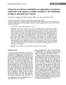
Figure 1.
CHPD crystals in gel medium in a test tube.
CURRENT SCIENCE, VOL. 88, NO. 8, 25 APRIL 2005
GENERAL ARTICLES
Figure 2. a, Plot of number of grown crystals versus molar concentration of tartaric acid in supernatant solution. b, Plot of average apparent length of crystals versus molar concentration of tartaric acid in supernatant solution.
Figure 3 a–f. Plot of apparent length of growing crystals versus time for 0.01, 0.2, 0.4, 0.6, 0.8 and 1.0 M concentration of tartaric acid.
The nature of crystal growth patterns is exhibited in Figure 3 a–f. Figure 3 a depicts the growth of CHPD crystals without tartaric acid. When 0.2 M tartaric acid is added, the inhibition of growth and slow dissolution of crystals is observed, as shown in Figure 3 b. It can be observed from Figure 3 c–f that as the concentration of tartaric acid CURRENT SCIENCE, VOL. 88, NO. 8, 25 APRIL 2005
increases, dissolution of crystals becomes rapid and for 1 M concentration, it is the highest. For higher concentration of tartaric acid, growth of calcium tartrate is initiated at the same time when dissolution of CHPD crystals occurs. Therefore, it is important to arrive at a proper molar concentration of tartaric acid which dissolves CHPD crystals 1235
GENERAL ARTICLES but does not initiate the growth of calcium tartrate crystals. This had tempted the authors to verify the solubility of CHPD crystals in tartaric acid. A small CHPD crystal was selected, put in a 1 M tartaric acid solution and observed under the microscope. Initially, the surface of the selected crystal was heavily etched and got eroded due to tartaric acid. It was observed that appreciable amount of dissolution of the crystal was seen within a few hours and within a day, the crystal was completely dissolved.
Effect of tamarind solution on growth of CHPD crystals Since tamarind contains a high amount of tartaric acid, the effect of a solution of tamarind on growth of CHPD crystal was studied. A solution of tamarind was prepared by boiling 150 g of tamarind, obtained from Aleppey district, Kerala, in 1000 ml of distilled water for 15 min and then doublefiltered with Whatman filter paper. The following supernatant solutions were selected to be poured on the set gels; 25 ml CaCl2 (solution A); 24 ml CaCl2 + 1 ml tamarind solution (solution B); 23 ml CaCl2 + 2 ml tamarind solution (solution C); 22 ml CaCl2 + 3 ml tamarind solution (solution D) and 21 ml CaCl2 + 4 ml tamarind solution (solution E). Needle-shaped and star-shaped crystals were grown in the gel. Figure 4 depicts a plot of apparent length of growing crystal versus time. The apparent length of growing crystals is much less in the case of different volumes of tamarind solution than that for pure CaCl2-containing supernatant solution. This suggests that the presence of tamarind solution inhibits the growth of CHPD crystals. It is important to note that crystals of this average length can be flushed out through the urinary tract, since stones up to 5 mm can pass through the urethra.
Figure 4. Plot of average apparent length of growing crystals versus time for different volumes of tamarind solution. â, X, p, ˜, ¢ indicate supernatant solution containing solutions A, B, C, D and E. 1236
Discussion The growth of urinary calculi is a complex process involving various parameters. When the nucleation process occurs in pure solution it is known as homogeneous nucleation12. Apart from this, the concept of aggregation is also important. Crystal nuclei cannot grow large enough to attach to and occlude on renal tubular lumens within 5 to 7 min and pass through tubules and enter the renal pelvis. Nevertheless, they can aggregate into large clumps within an order of a minute13. It is, therefore, required to incorporate the growth and aggregation phenomenon to explain the occurrence of stones. Many chemicals are found as important inhibitors of crystal aggregation. For example, nephrocalcin, a type of acidic glycoprotein of renal origin, is found to be responsible for inhibition of calcium oxalate nucleation, growth and aggregation14,15. Also magnesium and citrate inhibit crystal aggregation. It is interesting to note that the interference with crystal growth and aggregation is a possible therapeutic strategy for prevention of recurrent stone diseases1,12,16. The present article is based on the interference of tartaric acid and tamarind in the nucleation and growth of CHPD urinary type crystals, which may have possible therapeutic applications. Thermodynamic solubility product, saturation and supersaturation are important parameters from the physical chemistry point of view. The point at which saturation is reached and crystallization begins is commonly known as thermodynamic solubility product KSp, which is a constant. Urine contains inhibitors and other molecules and can hold large amount of solute as in a meta-stable state. If the concentration of solute increases and a point is reached where it cannot be held in solution, this concentration is known as Kfp, which is the formation of product in urine. The supersaturation state of solution is critical for formation of the crystal, which depends on the solute concentration, pH, ionic strength and existence of soluble complexes1. Pak et al.17 and Brown et al.18 have developed methods to estimate calcium oxalate and calcium phosphate saturations. Tamarind (Tamarindus indica L.) is largely used in different Indian cuisines. The pulp of tamarind contains nearly about 10% tartaric acid, citric acid and malic acid and their salts, as well as about 8% potassium hydrogen tartrate. Tamarind also contains polysaccharides, balsamine, catechin, nasturtium, tamarin, phosphatidic acid and its choline, ethanolamine, serine, inositol and alkaloids. Variations in composition of commercial varieties of tamarind are19,20 in citric acid from 4 to 6%, tartaric acid from 5.3 to 5.8%, potassium bi-tartrate from 4.6 to 4.7%, malic acid from 10 to 15% and reducing sugar and insoluble matter from 12 to 20%. However, malic acid, reducing sugar and insoluble matter are comparatively lesser in amount. A critical survey of tamarind chemistry, technology and uses has been carried out by Shankaracharya21. Sur et al.3 have found that the lower incidence of urinary calculi in South India compared to that in North India is CURRENT SCIENCE, VOL. 88, NO. 8, 25 APRIL 2005
GENERAL ARTICLES probably due to regular dietary intake of tamarind in the south, although there are some other dietary differences, including a lower animal protein intake in the south22. It has been claimed23,24 that the daily intake of 10 g of tamarind pulp significantly increases the inhibition of calcium oxalate crystallization, which is due to the high tartaric acid content of tamarind. It has been earlier established that between 14 and 20% of injected tartrate is excreted in the urine. This increases tartrate level in the urine. Tartrates are expected to form metal ion complexes with calcium25. The presence of tartrate in urine may decrease the amount of ionized calcium available for calcium oxalate precipitation. Tartrates bind with the cations needed for crystal formation and subsequent growth, and also function as a crystal growth inhibitor of calcium oxalate by chemical adsorption on the crystallization sites at a growing interface26. An evaluation of tamarind and tartaric acid as inhibitor of calcium oxalate crystallization in urine is reported by Croft et al.27. Singh et al.28 have earlier reported the inhibitory effect of tamarind on urolith in urine. Thomas et al.29 also attempted to explore the possibility of using tamarind and tomatoes in controlling crystalluria. Moreover, Anasuya and Sasikala30 have studied lithogenic properties of urine before and after ingestion of 10 g tamarind daily. They found complete disappearance of calcium oxalate crystal aggregates, fall in density in crystalluria, reduction in crystal size, decrease in excretion of oxalic acid, increase in excretion of phosphorus and increased inhibitory activity of urine towards crystal growth. A joint report of the Food and Agriculture Organization (FAO) of the United Nations and World Health Organization (WHO) has evaluated tartaric acid and its potassium– sodium and sodium salts for daily intake31. When taken by mouth, only about 20% of ingested tartaric acid is eliminated in the urine, inasmuch as it is destroyed in the intestinal tract by bacterial action. Finkle24 suggested that L (+)-tartaric acid is inert in the human body. A comparative study was reported, in which the percentages of both L (+) and D (–) tartaric acids, eliminated in urine after intramuscular injection to a man, were found only slightly greater than those after oral adminsitration32. The excretion of L (+)-tartaric acid in urine after postoperative administration was investigated by Underhill et al.33 in rat, guinea pig, dog and rabbit. Sodium tartrate in daily doses of up to 10 to 20 g is usually preferred in medical practice as a laxative. Gold and Zahm34 found that daily doses of 10 g of sodium tartrate when administered to 26 patients for an average of 11.8 doses, gave laxative responses in 66% of the patients. The only side effects noticed were nausea/vomiting (1.6%) and abdominal cramps (2.1%). Following a single oral dose of 400 mg/kg, monosodium (14C) L (+)-tartrate to rats, 70% of the radioactivity was excreted in urine, 16% in expired air and 14% in the faeces within 48 h. Altogether, CHPD crystal growth and inhibition in the presence of tartaric acid exhibits interesting results, no CURRENT SCIENCE, VOL. 88, NO. 8, 25 APRIL 2005
major in vitro study on the growth and inhibition of these crystals under the influence of tartaric acid and tamarind has been reported. The number of crystals and their average length decreases rapidly with the concentration of tartaric acid. This further confirms that tartaric acid inhibits the growth of CHPD crystals. Inhibition of CHPD crystal growth is due to the formation of a complex of calcium tartrate, which can be confirmed by the high concentration of tartaric acid leading to the growth of calcium tartrate crystals on the gel–liquid interface. The growth of calcium tartrate crystals was confirmed and characterized by Joshi and Joshi35. However, such a high concentration of tartaric acid is not recommended by the WHO report from animal studies from a toxicity point of view31. The same results were obtained in the already grown crystals. The higher concentration solutions of tartaric acid dissolve the grown CHPD crystals. The dissolution time and nature depend on the concentration of tartaric acid. Here also, higher concentration solution of tartaric acid leads to the growth of calcium tartrate crystals. Both these reports, nevertheless, are important for in vitro studies to formulate strategies for preventing the growth of calculi or dissolving them. Figure 4 indicates the inhibition of growth of CHPD crystals in the presence of tamarind solutions of different volumes. In the present study, mainly two types of CHPD crystal morphologies are observed: (i) platelet or needletype with prominent (010) face and (ii) a star shape in which all needles or platelets come out from a single point or from a common centre. A similar study was reported by Joshi and Joshi36 on CHPD crystals in the presence of citric acid and lemon juice, which also showed strong inhibition of growth of CHPD crystals. The present and earlier studies together establish tartrate and citrate inhibitions in the case of CHPD crystals, which were earlier established for calcium oxalate crystals. This study covers multifaceted interests and multifarious subjects such as crystal growth, crystallography, physical chemistry, pharmacology, ayurveda, and food and nutrition. It may help identify the potential inhibitors of growth of urinary crystals and their concentrations or doses as well as their actions as new guidelines for in vivo studies.
Conclusion The inhibition of CHPD crystals increases as the concentration of tartaric acid increases; consequently, the number of grown crystals and their average size decreases. Tartaric acid inhibits the growth of CHPD crystals in vitro. The dissolution time of grown CHPD crystals depends on the concentration of tartaric acid added in the supernatant solution; as the concentration of tartaric acid increases, dissolution occurs faster. This also confirms that the tartrate readily forms complexes with calcium and inhibits the growth of CHPD crystals. Nevertheless, it grows calcium tartrate crystals at 1237
GENERAL ARTICLES very high concentrations of tartaric acid which is not used as per WHO recommendations. 1. Menon, M., Parulkar, B. G. and Drach, G. W., Urinary lithiasis, etiology, diagnosis, and medical management. Campbell’s Urology, W.B. Saunders Co, London, 1998, 7th edn, pp. 2661–2733. 2. http://www.Tribuneindia.com/2000/Nov., 2000. 3. Sur, B. K., Pandey, H. N., Deshpande, S., Pahiva, R., Singh, R. K. and Tarachandra, Future of tamarind and tartrate in preventing reoccurrence of renal calculi. Urolithiasis: Clinical and Basic Research (eds Smit, L. H. et al.), Plenum Press, New York, 1973, pp. 333–336. 4. Henisch, H. K., Crystal Growth in Gels, The Pennsylvania State University Press, University Park, 1973, pp. 41–51. 5. Henisch, H. K., Dennis, J. and Hanoka, J. J., Crystal growth in gels. J. Phys. Chem. Solids, 1965, 26, 493–500. 6. Patel, A. R. and Rao, A. V., Crystal growth in gel media. Bull. Mater. Sci., 1982, 4, 527–548. 7. Wyckoff, W. G., Crystal Structure, John Wiley, 1965, p. 644. 8. Haninen, D., Geiger, B. and Addadi, L., Fibronectin adsorption to surfaces of hydrated crystals. An analysis of the importance of bound water in protein–substrate interactions. Langmuir, 1993, 9, 1058–1065. 9. Skiric, M., Sarig, S. and Füredi-Milhofer, H., The interaction of small and macromolecules with growing calcium hydrogen phosphate dihydrate crystals. Prog. Colloid Polym. Sci., 1998, 110, 300–304. 10. Joseph, K. C. and Joshi, M. J., The study of different parameters affecting Liesegang ring formation during the growth of calcium hydrogen phosphate dihydrate crystals. Indian J. Phys. A, 2002, 76, 159–163. 11. Joshi, V. S. and Joshi, M. J., FTIR spectroscopic, thermal and growth morphological studies of calcium hydrogen phosphate dihydrate crystals. Cryst. Res. Technol., 2003, 38, 817–821. 12. Brown, C. M. and Purich, D. L., Physical chemical process in kidney stone formation. In Disorders of Bone and Mineral Metabolism (eds Coe, F. L. and Flavuj, M. J.), Raven Press, New York, 1992, pp. 613–624. 13. Kok, D. J., Papapolous, S. E. and Bijovet, O. L., Crystal agglomeration is a major element in calcium oxalate urinary stone formation. Kidney Int., 1990, 37, 51–56. 14. Nakagawa, Y. et al., Isolation from human calcium oxalate renal stone of nephrocalcin, a glycoprotein inhibitor of calcium oxalate crystal growth; Evidence that nephrocalcin from patients with calcium oxalate nephrolithiasis is deficient in gamma carboxy glucotamic acid. J. Clin. Invest., 1987, 79, 1782–1787. 15. Asplin, J., DeGannelli, S., Nakagawa, Y. and Coe, F. L., Evidence that nephrocalcin and urine inhibit nucleation of calcium oxalate nephrolithiasis. Am. J. Physiol., 1991, 260, F569–F578. 16. Shiraga, H., Min, W. and Van Dusen, W. J., Inhibition of calcium oxalate crystal growth in vitro by uropontin: Another member of the aspartic acid-rich protein superfamily. Proc. Natl. Acad. Sci. USA, 1992, 89, 426–430. 17. Pak, C. Y. C., Hayashi, Y., Finlayson, B. and Chu, S., Estimation of the state of saturation of brushite and calcium oxalate in urine: A comparison of three methods. J. Lab. Clin. Med., 1977, 89, 891– 901. 18. Brown, C. M., Ackerman, D. K. and Purich, D. L., Equil 93: A tool for experimental and clinical urolithiasis. Urol. Res., 1994, 22, 119–126.
1238
19. Iyengar, A. V. V., Tartaric acid from tamarind fruit. Curr. Sci., 1938, 6, 610. 20. Gogte, V. M., Ayurvedic Pharmacology and Therapeutic Uses of Medicinal Plants, Bharatiya Vidya Bhavan’s SPARC, Mumbai. 21. Shankaracharya, N. B., Tamarind – Chemistry, technology and uses: A critical appraisal. J. Food Sci. Technol., 1998, 35, 193–208. 22. Anderson, D. A., Environmental factors in the etiology of urolithiasis. Proceedings of the International Symposium on Renal Stone Research, Madrid, 1972, pp. 130–144. 23. Chadwick, V. S., Vince, A., Killingley, M. and Wrong, O. M., The metabolism of tartrate in men and rat. Clin. Sci. Mol. Med., 1978, 54, 272–281. 24. Finkle, P., The fate of tartaric acid in human body. J. Biol. Chem., 1933, 100, 349–355. 25. Sillen, L. G. and Martell, A. E. (eds), Stability Constants of Metal Ion Complexes, Suppl. No. 1, Chemical Society of London, Burlington House, 1971. 26. Fleisch, H., Inhibitors and promoters of stone formation. Kidney Int., 1978, 13, 361–371. 27. Croft, K., Adair, J. H., Bowyer, R. and Brockis, J. G., An evaluation of tamarind and tartaric acid as inhibitors of calcium oxalate crystallization in urine. Urinary Stones. Proceedings of the Second International Urinary Stone Conference, Singapore, 1983, Churchill Livingstone, Melbourne, 1984, pp. 189–197. 28. Singh, P. P., Hada, P., Narula, I. M. S. and Gupta, S. K., In vivo effect of tamarind on urolith inhibitory activity in urine. Indian J. Exp. Biol., 1987, 25, 863–865. 29. Thomas, J., Sachidev, K., Sindhu, S., Vathsala, R. and Marickar, Y. M. F., The role of tamarind and tomatoes in controlling crystalluria. Urol. Res., 1988, 16, 242–243. 30. Anasuya, A. and Sasikala, M., Tamarind ingestion and lithogenic properties of urine in men. Nutr. Res., 1990, 10, 1109–1117. 31. Tweny-first Report of the Joint FAO/WHO Experts Committee on Food Additives, WHO Technical Report Series No. 617, Geneva, 1977. 32. Bauer, C. W. and Pearson, R. W., A comparative study of the metabolism in human body of some isomers of tartaric acids. J. Am. Pharm. Assoc. Sci. Ed., 1957, 46, 575–578. 33. Underhill, F. P., Leonard, C. S., Gross, E. G. and Joleski, T. C., Studies on metabolism of tartrates. (II) The behavior of tartrates in the organism of the rabbit, dog, rat and guinea pig. J. Pharmacol., 1931, 43, 359. 34. Gold, H. and Zahm, W., Method for the evaluation of laxative agents in constituted human subjects with a study of the comparative laxative potencies of fumarates, sodium tartrate and magnesium acid citrate. J. Am. Pharm. Assoc., 1943, 32, 173. 35. Joshi, V. S. and Joshi, M. J., FTIR spectroscopic and thermal studies of calcium tartrate trihydrate crystals grown by gel assistance. Indian J. Phys. A, 2001, 75, 159–163. 36. Joshi, V. S. and Joshi, M. J., Influence of inhibition of citric acid and lemon juice to the growth of calcium hydrogen phosphate dihydrate urinary crystals. Indian J. Pure Appl. Phys., 2003, 41, 183–192.
ACKNOWLEDGEMENTS. M.J.J. and B.B.P. thank the Department of Biotechnology, New Delhi for financial assistance, and Prof. K. N. Iyer and Dr A. B. Vaidya, SPARC, Mumbai, for their keen interest.
Received 26 April 2004; revised accepted 8 December 2004
CURRENT SCIENCE, VOL. 88, NO. 8, 25 APRIL 2005