Inflammatory Localized and Generalized Bone Loss in Recent-onset Rheumatoid Arthritis
Melek Güler-Yüksel
VRIJE UNIVERSITEIT
Inflammatory Localized and Generalized Bone Loss in Recent-onset Rheumatoid Arthritis
ACADEMISCH PROEFSCHRIFT ter verkrijging van de graad Doctor aan de Vrije Universiteit Amsterdam, op gezag van de rector magnificus prof.dr. L.M. Bouter, in het openbaar te verdedigen ten overstaan van de promotiecommissie van de faculteit der Geneeskunde op woensdag 12 januari 2011 om 15.45 uur in de aula van de universiteit, De Boelelaan 1105
door Melek Güler-Yüksel geboren te Rijssen in 1981
Promotores
prof.dr. W.F. Lems prof.dr. T.W.J. Huizinga Leids universitair medisch centrum, Leiden prof.dr. B.A.C. Dijkmans
Copromotor
dr. C.F. Allaart Leids universitair medisch centrum, Leiden
Referent
dr. D. Baeten Academisch medisch centrum, Amsterdam
The research presented in this thesis was financially supported by the Dutch College of Health Insurances, with additional funding by Schering-Plough B.V. and Centocor Inc.. The publication of this thesis was financially supported by ABBOTT B.V., Amgen B.V., AstraZeneca B.V., Merck Sharp & Dohme B.V., Pfizer B.V., Roche Nederland B.V., Schering Plough B.V., Sectra dxr-online, Teva Pharma NL, UCB Pharma B.V., and WillPharma B.V.. © Melek Güler-Yüksel, 2010. All rights reserved. No part of this thesis may be reproduced in any form without written permission from the author. Cover design Printing
Daniela Dimitrova Ipskamp Drukkers B.V., Enschede
ISBN
978-90-9025936-9
Voor Ufuk Voor mijn ouders
Voorwoord Het proefschrift dat voor u ligt is het resultaat van ruim 3 jaar onderzoek naar ontstekingsgerelateerde, gelokaliseerde en gegeneraliseerde osteoporose en botverlies in recent gediagnosticeerde reumatoïde artritis en in hand artrose. Een actueel en boeiend probleem vanwege het feit dat meer kennis over de mechanismen van botverlies kan en zal leiden tot betere en specifiekere behandelingsopties in beide aandoeningen; aandoeningen die het leven van vele patiënten negatief beïnvloeden en beperken. Zonder enige twijfel hebben veel mensen door hun harde, en vooral passievol, werk een significante bijdrage geleverd aan de totstandkoming van dit proefschrift. Woorden schieten mij tekort om hen te bedanken, maar hierbij toch een poging. En zoals u al gemerkt heeft, niet zoals gebruikelijk achter in het proefschrift, maar juist helemaal voor in, want zonder hen zou dit proefschrift er niet zijn geweest. Allereerst dank ik drs. J.M. de Jonge-Bok. Han, jij bent degene die me geïntroduceerd heeft in de wereld van de reumatologie, ik ben je dankbaar hiervoor. De mensen achter de BeSt studie, die eind jaren negentig de missie hadden om een goed uitgedachte studie te lanceren om daarmee de wetenschap en de individuele patiënt een grote dienst te bewijzen, ben ik erkenning verschuldigd: prof.dr. F.C. Breedveld, prof.dr. B.A.C. Dijkmans, dr. D. van Zeben, en de overige leden van de STRO groep, dank voor het opzetten en voortzetten van deze unieke studie. Mede dankzij de gezamenlijke en dynamische aanpak is de BeSt studie zo een succes geworden en gebleven! Alle deelnemende reumatologen, reumatologen in opleiding, onderzoeksverpleegkundigen, en reumaconsulenten, dank voor het includeren en blijven motiveren van de patiënten, en de enthousiaste betrokkenheid. Het secretariaat, Jacomien, Hanny, Joyce en Hughine, en het datamanegement, Jozé, dank voor alle ondersteuning. Al deze logistieke inspanningen om alle benodigde onderzoeksdata van alle partijen te verzamelen resulteerde in de immense, en tot vandaag de dag blijvend groeiende, prachtige database, die op vele onderzoeksvragen een licht geworpen heeft; en het einde lijkt nog niet in zicht. En uiteraard, achter alle getallen en nummers schuilen de mensen om wie het allemaal draait, de patiënten. Bovenal wil ik hen bedanken voor hun inzet, motivatie en loyaliteit. Ik dank prof.dr. W.F. Lems voor zijn begeleiding gedurende mijn promotieonderzoek. Beste Willem, al was ik helaas niet vaak in de VU, telefonisch discussieerden we lang over de gevonden resultaten, en jouw kennis met betrekking tot osteoporose was van onschatbare waarde bij de interpretatie en presentatie van de data. Dat ik de eerste ben die de eer heeft om bij jou te mogen promoveren! Ik wil prof.dr. T.W.J. Huizinga bedanken voor zijn waardevolle commentaar op de artikelen. Dr. C.F. Allaart, lieve Renée, jij pakte mijn hand, en sleepte mij mee de, als een speer gaande, BeSt studie in. Je liet me niet los. De lessen die ik van jou heb geleerd zijn ontelbaar en van
grote waarde; door jou ben ik gegroeid als wetenschapper. Dr. M. Kloppenburg, beste Margreet, ik heb veel geleerd van je epidemiologische visie en interpretatief vermogen, mijn oprechte dank hiervoor. Ook ben ik je erkentelijk voor het beschikbaar stellen van de GARP data; het is een fraai stuk geworden. Yvonne en Jeska, jullie hebben veel bijgedragen aan de opzet en het doen (hard)lopen van de studie, hierbij alle kinderziektes te lijf gaand, waardoor ik in 2006 belandde in een strak lopende studie. Mijn grote vriend Sjoerd, jij hebt me wegwijs gemaakt in het zogenaamde leven van een BeSt promovendus, dankjewel voor alles. Naomi, het was geweldig om met jou uitkomsten van lastige statistische analyses te bediscussiëren, lang leve de GEE analyse. Linda, bedankt voor het versterken van het BeSt team. Onze samenwerking krijgt nog hopelijk een vervolg met andere botgerelateerde projecten. Jessica, de osteoporose database die je had aangemaakt tijdens je studententijd, kwam me enorm van pas. Het was erg leuk, en vooral leerzaam met jouw epidemiologische kennis, om later samen met je onderzoek te doen naar hand artrose. Onno, jij was met je harde werken en grote ambities een goed voorbeeld, maar bovenal was je een gezellige overbuurman voor me. Karen, jij was vaak mijn inspiratiebron, ik heb altijd respect gehad voor jouw manier van aanpak van alle facetten van wetenschappelijk onderzoek. Daarnaast ben je ook een geweldige vriendin, ik kijk uit naar de dag dat we weer samen werken onder één dak (terug op de afdeling reumatologie). Ook Mohamed, Emalie, Diane, Rosanne, Wing-Yee, Angga, Rachel en Rosaline wil ik bedanken voor de zinnige en onzinnige (Angga) discussies, het uitwisselen van kennis en ervaring, het teamgevoel, de geweldige sfeer, en vooral voor jullie vriendschap. Mede dankzij jullie ben ik de reumatologie afdeling als mijn tweede thuis gaan beschouwen. Tot slot wil ik mijn dank betuigen aan mijn vrienden en familie. In het bijzonder Őzlem, voor haar stimulatie om de wetenschappelijke wereld te ontdekken, en Selma, mijn zuster, via wie ik lang geleden al enthousiast raakte voor het medisch onderzoek. Ik voel me vereerd dat jullie beiden mijn paranimfen zijn. Annem ve babam, sizin hayatımdaki őneminizi hiç bir sőzle ifade edemem. Çok sağolun, tűkenmez sevginiz, desteğiniz, gűveniniz ve dualarınız için. Size layık bir evlat olabiliyorsam ne mutlu bana. Sizleri seviyorum. Liefste Ufuk, met jouw oprechte interesse in de medische wetenschap, stimulatie tot kritisch denken, interpreteren en relativeren, en onvoorwaardelijke steun en begrip, vorm jij de kalme kracht achter mij. Met de ware loyaliteit en liefde waarmee je mij omhelst ben jij de verpersoonlijking van het beste wat het leven mij te bieden heeft. Samen gaan wij een mooie toekomst tegemoet waarin we, naast deze, vele mijlpalen zullen behalen.
Contents Chapter 1
General introduction -- 13
Chapter 2
Bone mineral density in patients with recent-onset, active rheumatoid arthritis: cross-sectional exploratory analyses from the BeSt study -- 29
Chapter 3
Changes in generalized bone mineral density in patients with recent-onset rheumatoid arthritis: 1-year follow-up analyses from the BeSt study -- 41
Chapter 4
Changes in localized and generalized bone mineral density in patients with recent-onset rheumatoid arthritis: 2-year follow-up analyses from the BeSt study -- 55
Chapter 5
Changes in localized bone mineral density are associated with the level of disease activity in patients with rheumatoid arthritis -- 71
Chapter 6
Accelerated localized bone mineral density loss is associated with progressive joint damage in hands and feet in recent-onset rheumatoid arthritis -- 85
Chapter 7
Accelerated localized bone mineral density loss is associated with radiographic progressive hand osteoarthritis -- 103
Chapter 8
Treatment with TNF-α inhibitor infliximab might reduce hand osteoarthritis in patients with rheumatoid arthritis -- 115
Chapter 9
Summary and general discussion -- 131 Nederlandse samenvatting en bespreking -- 142 List of publications -- 150
General introduction
1
Rheumatoid arthritis Rheumatoid arthritis (RA) is a systemic auto-immune inflammatory disease characterized by arthritis of particularly the smaller joints of the hands and feet, and to a lesser extent the larger joints.[1] Extra-articular manifestations, such as nodules, vasculitis, and pulmonary and ocular inflammation are seldom seen. The pathogenesis of RA is incompletely understood, but thought to be multifactorial involving multiple genes and environmental factors.[2] Worldwide, patients are classified as RA according to the in 1987 revised American College of Rheumatology (ACR) criteria when at least four of the seven following criteria are met (criteria 1-4 at least 6 weeks present): 1. morning stiffness lasting at least 1 hour, 2. swelling of at least three joint areas, 3. swelling of a hand joint area (wrist, metacarpalphalangeal, proximal interphalangeal), 4. symmetrical joint swelling, 5. subcutaneous nodules, 6. positive rheumatoid factor, and 7. erosions or juxta-articular osteoporosis on hand and wrist X-rays.[3] New classification criteria, including the (high) presence of anticitrullinated peptide antibodies (ACPA) and acute phase reactants, and excluding the earlier for diagnosis obligated disease duration of six weeks, and the presence of subcutaneous nodules and bone damage on X-rays, are developed to identify more early stages of the disease, in order that patients can benefit more from early institution of anti-rheumatic treatment to stop and prevent development of the disease.[4] Based on the 1987 criteria, the prevalence of RA is approximately 0.5 to 1.0 percent of the general population, and the incidence varies between 20 and 50 per 100.000 new patients per year.[5] Both prevalence and incidence of RA increases with age, and it affects more women by a ratio of three. The natural course of RA results in chronic inflammation leading to longstanding synovial hyperplasia, and subsequently articular damage, consisting of cartilage loss, subchondral cysts, and erosive damage. Both active inflammation and bone destruction lead to significant functional impairment and disability. Therefore, uncontrolled RA places a substantial burden on the patient, by reduced quality of life and life expectation, in particular by higher prevalence of cardiovascular disease and higher fracture risks due to osteoporosis, and on the society, by high health care costs and work loss.[6-8] Early and effective treatment of RA is necessary to stop the inflammatory process, and to prevent bone and cartilage destruction. Anti-rheumatic treatment The last decade considerable improvements were made with regard to treatment of RA, both on use of (combinations of) conventional disease-modifying anti-rheumatic drugs (DMARDs) with or without corticosteroids, and new biological drugs. Furthermore, target goals are set to improve disease outcomes.
14
Targeted therapy Several tools have been developed to measure disease activity in RA.[9] Objectifying disease activity has not only been important for the design and execution of clinical trials to test new drugs and drug combinations, it has also provided clinicians with means to follow up the effect of their therapeutic interventions in daily practice, and set themselves treatment goals. Several studies have shown that regular treatment adjustments aimed at achieving a predefined low level of disease activity result in better clinical and radiological outcomes of RA than non-target-steered treatment.[10-12] Conventional DMARDs Currently, methotrexate (MTX) is still regarded as the anchor drug in RA treatment due to high efficacy and low toxicity, well-reported in several randomized clinical trials.[13,14] The exact working mechanism remains unclear, however it is believed that MTX has immunosuppressive actions via various routes.[15] MTX is currently recommended as first choice in the treatment of RA by European league against rheumatism (EULAR), as monotherapy or in combination with other drugs.[16] MTX therapy combined with the systematic temporary use of corticosteroids adequately suppresses inflammatory activity and radiological damage progression in patients with early RA.[17] Other widely used DMARDs are sulfasalazine, leflunomide, and antimalarial drugs such as hydroxychloroquine, and, to a lesser extent, due to an unfavorable toxicity profile and the superiority of other drugs, azathioprine, cyclosporine, and gold salts. Biologicals Since the introduction in the 1990s of biologicals, drugs produced by molecular biology techniques targeting specific cells or cytokines in the pathophysiological pathways of RA, exciting improvements in the management of RA have been made. The first biologicals are the inhibitors of tumor necrosis factor alpha (TNFα), a pro-inflammatory cytokine playing a central role in inflammatory processes: 1. infliximab, a chimeric anti-TNFα, 2. etanercept, a recombinant human soluble anti-TNFα receptor, and 3. adalimumab, a recombinant humanized anti-TNFα.[18] The efficacy of these three agents seem comparable, but no head-to-head trials have been performed.[19] Two new TNF-blockers, certolizumab pegol and golimumab, and other mode of action biologicals, such as rituximab, a B-cell depletor, abatacept, an inhibitor of T-cell co-stimulatory pathways, anakinra, an interleukine (IL)-1 receptor antagonist, and tocilizumab, an IL-6 receptor antagonist, are also nowadays available in the Netherlands. For most of these therapies it is not yet fully clear at which stage of the disease course they would be most beneficial to the patients, in terms of rapid symptom relief and inhibition of damage progression in relation to possible toxicity risks and side effects, compared to other drugs. Dynamic treatment strategy trials rather than comparative drug trials should provide us the answers. 15
The BeSt study The BeSt study, Dutch acronym for Behandel Strategieën, i.e. treatment strategies, is a randomized clinical trial comparing the efficacy of the following four treatment strategies in a tight control setting aiming low disease activity in recent-onset, active RA patients: group 1. sequential monotherapy starting with MTX, group 2. step-up combination therapy starting also with MTX, group 3. initial combination therapy with MTX, sulphasalazine and a quickly tapered high dose of prednisone, and group 4. initial combination therapy with MTX and infliximab. Three-monthly evaluations of disease activity based on the validated disease activity score (DAS) were performed by for treatment blinded experienced research nurses. If the aim of low disease activity, defined as DAS≤ 2.4, was not achieved after 3 months, treatment was immediately adjusted by proceeding to the next step in the allocated treatment group. If after at least six months the DAS remained ≤2.4, medication was gradually tapered until one drug remained in a maintenance dose. In case of sustained clinical remission, defined as DAS< 1.6 during six consecutive months, the last drug was finally tapered until zero. The tight control setting resulted in a remarkable clinical improvement and reduction in joint damage progression.[11] In the initial combination groups, group 3 and 4, low disease activity was reached earlier, and therefore a greater reduction of joint damage progression was seen compared to the initial monotherapy groups, group 1 and 2. After 6 years, 51% of the at baseline highly active RA patients were in clinical remission, and 17% of the patients in prolonged remission without any anti-rheumatic treatment and with no radiographic progression, suggesting that drug free remission is a achievable goal.[20] Bone destruction in RA Bone is a dynamic organ continuously formed and resorbed by osteoblasts and osteoclasts, respectively, along the surface. How the mechanisms between bone formation and bone resorption exactly are coupled is not completely understood, however it is believed that locally cytokines are playing a crucial role in this process.[21] In RA there is a clear imbalance with high bone resorption and low bone formation, resulting in structural joint damage, and bone mineral density (BMD) loss. Joint damage In RA, the hyperplastic inflammatory synovial tissue, the so-called pannus, invades, and thereby destroys the neighboring cartilage and subchondral bone by activated osteoclasts, while at the same time bone formation by osteoblasts is also impaired. Osteoclasts require essential signals which promote their differentiation from monocytic precursor cells, and activation.[22] Several inflammatory cytokines and mediators such as TNFα, IL-1, IL-6, IL-7, IL-17, and prostaglandin
16
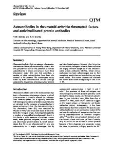
E2 (PGE2) which are produced by macrophages, T cells, or fibroblasts stimulate osteoclast formation and activation by inducing the expression of receptor activator of nuclear factor κB ligand (RANKL) and macrophage colony stimulating factor (MCSF), expressed by synovial fibroblasts, activated T cells, or macrophages (see figure).[23] A decoy receptor, the osteoprotegerin (OPG), controls the amount of osteoclastogenesis. RANKL expression is increased in the synovial membrane, and the OPG expression is insufficient to counteract RANKL effects in RA, leading to subsequent bone loss. Moreover the activation of wingless and Int-1 (Wnt) genes, that play an important role in differentiation of osteoblasts from mesenchymal precursors, is strongly inhibited by expression of the Wnt-signaling antagonist Dickkopf-1 (Dkk-1), induced by TNFα and produced by synovial fibroblasts.[24,25] Furthermore, TNFα also induces the expression of sclerostin in osteocytes, which is a potent downregulator of bone formation.[26] To date, treatment of patients with RA is not merely aimed at relief of current symptoms of pain and limitations in daily functioning due to inflammation of joints, it is as important to suppress damage progression, since this determines functional ability in the long term. It is clearly proven that suppression of disease activity in RA, by intensive anti-inflammatory treatment with DMARDs, glucocorticoids, and biologicals results in effective suppression of progression of radiographic joint damage.[27-30] Figure. Influence of inflammation on bone remodeling.
TNF, tumour necrosis factor; IL, interleukin; PGE2, prostaglandin E2; RANKL, receptor activator of nuclear factor κB ligand; MCSF, macrophage colony stimulating factor; Dkk-1, Dickkopf 1; OPG, osteoprotegerin.
17
Assessment of joint damage Plain radiographs of hands and feet still form the gold standard of assessing joint damage (progression) in RA. Probably the most widely used method is the by van der Heijde modified Sharp scoring method quantifying joint space narrowing and erosions in 44 joints locations in hands, wrists, and feet with a maximum score of 448 points.[31] The percentage of patients with joint damage progression above the measurement error can be calculated by the smallest detectable change (SDC).[32] Osteoporosis and BMD loss in RA Osteoporosis is the most common metabolic bone disease, resulting in micro-architectural deterioration and decreased mechanical strength of bone, leading to increased incidence of fractures.[33,34] Due to this higher fracture risk, it has a major impact both on patient and society by increased morbidity, mortality, and health care costs.[35] Bone strength is dependent on its structure, the material properties, and bone turnover, and can unfortunately not be tested in vivo.[36] Also assessment of bone quality is hampered by the inaccessibility of bone biopsies for investigation. At present, the assessment of BMD is the only aspect that can be easily and reliable be measured in clinical practice, and it forms the cornerstone for the diagnosis and general management of osteoporosis. In RA, osteoporosis or BMD loss occurs in two forms: 1. generalized BMD loss with axial distribution including the spine, pelvis, hips, ribs, and sternum, and 2. peri-articular or localized BMD loss, extra-articular bone loss seen in the proximity of the inflamed joints.[37] With regard to generalized BMD loss, the hip and spine are clinically the most relevant locations due to the highest fracture rates. In established RA, generalized osteoporosis is a well-known complication with overall occurrence of 7% to 26% in the hip, and 19% to 32% in the spine.[3843] In a population-based study of patients with established RA, a two-fold increase in generalized osteoporosis was seen in women, and a two-fold increase in reduced BMD in men compared with control groups.[41,44] Furthermore, a two-fold increase in risk of hip and vertebral fractures is seen in RA.[45,46] Over 1 year, BMD loss of 4.3% and 2.4% was observed in the hip and spine, respectively, in patients already fulfilling the in 1987 revised ACR criteria for RA, however not receiving any kind of anti-rheumatic therapy with DMARDs and/or corticosteroids, in a study that has been performed over 15 years ago.[47] There are several explanations for the increased prevalence of osteoporosis and accelerated BMD loss in established RA. Firstly, there is a significant overlap in demographics of patients with RA and patients with osteoporosis: both diseases are occurring predominantly in middle-, and highaged patients, and, especially postmenopausal, women. Secondly, advanced RA can lead to low
18
body mass index, a well-known risk factor of BMD loss. Thirdly, in established RA both active disease and severe structural damage might lead to reduced physical activity, and even immobility, and therefore to osteoporosis. Fourthly, the use and non-use of anti-inflammatory drugs, especially corticosteroids, might result in accelerated BMD loss. Lastly, the inflammatory disease itself might result in high BMD loss as a part of the development and progression of the disease, with the, by inflammation driven, osteoclast playing a crucial role in this process.[48-50] With regard to peri-articular or localized BMD loss, the hand and foot are clinically the most relevant locations due to involvement of particular the smaller joints in the hands and feet in RA. At present, the assessment of localized BMD can be readily measured only in the hands. In RA, a mean loss in hand BMD was 4.3% of baseline BMD in 1 year.[51] The earlier mentioned explanations for accelerated generalized BMD loss in RA are also applicable for localized BMD loss. However, it is strongly thought that localized BMD loss is more influenced by inflammatory activity in the proximity of the inflamed joints. Whereas accelerated generalized BMD loss seems to occur in a later phase of RA, localized BMD loss is found in the early phase of RA, and even in the undifferentiated phase of the RA process, probably due to a more direct and local effect of the inflammatory activity in the nearby joints.[51-54] When inflammation plays a crucial role in localized BMD loss in RA, the question is raised whether long-term absence of the inflammation will result in repair of bone destruction. It is known that repair of erosive damage is rarely seen, takes months to years to detect on plain radiographs, and is mostly seen in established, very destructive disease.[55-57] On the other hand gain in localized BMD in the hands might be earlier detectable in the disease process and during a shorter follow-up period due to the process itself, and the more sensitive way of measuring bone involvement.[52] Hence, in contrast to established RA, little is known about localized and generalized BMD loss in recent-onset RA with modern, tight controlled treatment. Knowledge about this is crucial to explore the direct effect of inflammation on BMD, both local and general, without the disturbance of factors due to advanced disease, such as reduced functional ability due to high structural damage, and long term use of corticosteroids. Furthermore, when localized BMD loss is likely in presence of inflammatory activity and BMD gain in absence of inflammatory activity, this indicates that changes in localized BMD over time might be an accurate and dynamic outcome measure for disease activity and bone involvement in RA. Localized BMD loss and prediction of destructive RA Since localized BMD loss is found in the early phase of RA or even in the undifferentiated phase of the disease, mostly before the stage of joint destruction, changes in localized BMD might be used as a predictor for subsequent joint destruction.[51-54,58] Two previous clinical studies showed that localized hand BMD loss predicted progressive joint damage in RA.[59,60] To incorporate hand 19
BMD loss as predictor for severe, destructive RA in prediction models, it is necessary to investigate the predictive value of hand BMD loss in comparison with other well-known predictors of destructive RA. Anti-rheumatic treatment and BMD Treatment with corticosteroids has a catabolic effect on generalized BMD by increased bone resorption, mediated by increased osteoclast formation and activity, and decreased bone formation, mediated particularly by osteoblast and osteocyte apoptosis.[25] However, more recent studies showed that this catabolic effect might be neutralized by the suppression of inflammatory activity.[61-64] Little is known about the effect of glucocorticoids on localized BMD.[65] Treatment with anti-TNFα might protect against generalized BMD loss, and might even result in BMD increase, however the results are inconclusive.[66-68] Again, even less is known about the effect of anti-TNFα on localized BMD.[69] In general, conventional DMARDs seem not to have a positive nor negative effect on generalized BMD.[70-73] Besides the effect of single drugs on localized and generalized BMD, it is extremely important to explore the effect of dynamic treatment strategies on BMD, since this is mimicking daily clinical practice in which anti-rheumatic treatment adjustments are made continuously in order to resolve symptoms, and stop bone destruction. Furthermore, if necessary, it is essential to start antiresorptive treatment to reduce or prevent BMD loss. Anti-resorptive treatment and BMD While the efficacy of only calcium and vitamin D supplements remains inconclusive, use of bisphosphonates does protect against generalized BMD loss, which is particularly important in patients who are treated with corticosteroids.[74-76] In contrast, the influence of anti-resorptive treatment on localized BMD loss remains unclear.[77] Assessment of generalized BMD
a
The gold standard for assessment of BMD is the dual energy X-ray radiogrammetry (DXA) calculating total BMD, both cortical and trabecular, by measuring bone mineral content and bone area. The accuracy and precision is good, and the radiation exposure is very low.[78,79] The BMD is expressed in different ways: 1. the absolute value expressed in gram/cm2, and 2. the relative values expressed in T-scores and Z-scores. T-score is calculated by subtracting the mean BMD of a youngadult reference population from the patient’s BMD, and dividing by the standard deviation (SD) of young-adult population. T-score ≤-2.5 SD is defined as osteoporosis. Z-score is calculated by subtracting the mean BMD of an age-, ethnicity-, and sex-matched reference population from the 20
patient’s BMD, and dividing by the SD of the reference population. Z-score is used to compare the patient’s BMD to a population of peers especially in case of secondary osteoporosis or BMD loss. Zscore ≤-1.0 SD is defined as reduced BMD. Changes in BMD are expressed in changes in gram/cm2 between two time points or in percentages compared to baseline BMD. Assessment of localized BMD Digital X-ray radiogrammetry (DXR) is a computerized version of the traditional technique of radiogrammetry originally proposed by Barnett and Nordin.[80] The DXR technique uses plain hand radiographs to estimate the BMD in the metacarpals. On the radiograph the computer automatically identifies regions of interest around the narrowest part of the second, third, and fourth metacarpal bone.[81] A mean surrogate BMD, based on the mean volume per area, is calculated in gram/cm2 with correction for the estimated porosity. BMD measured by DXR is highly correlated with DXA measurements, and has even better reproducibility and higher sensitivity for detecting changes in BMD.[81-83] Furthermore, DXR measurements only require plain radiographs of the hands, which are used in routine clinical practice at present, and gives therefore no extra radiation exposure. Common pathways between RA and osteoarthritis Another common type of arthritis is osteoarthritis (OA), a heterogenous group of conditions with alterations in articular cartilage, subchondral bone, and synovium.[84] OA leads to as much loss in function and quality of life as RA.[85] While effective, remission-induction therapies are available in RA, at present, no disease modifying therapies are available in OA. Nevertheless, an increasing body of evidence supports a common inflammatory pathway between RA and hand OA, signaling potential new targets for the treatment of hand OA.[86] Inflammation in hand OA Both local and low-grade systemic inflammation is associated with hand OA. High resolution magnetic resonance imaging demonstrated subchondral bone edema, synovial enhancement, and bone erosions in interphalangeal joints in the majority of OA patients.[87,88] A two- to threefold increase in high sensitive C-reactive protein levels is seen in OA patients.[89-92] Pro-inflammatory cytokines are found in increased levels in synovial fluid of OA joints, and heritable differences in cytokine production are associated with the development and progression of OA.[93-97] Increased TNFα production and increased p55 TNFα receptor expression on chondrocytes imply the relevance of TNFα on joint destruction in OA.[98-100] It is shown that TNFα inhibitors are able to suppress nitric oxide production in human cartilage.[101] 21
If, as in RA,[102] accelerated localized BMD loss would be found to be associated with progressive osteoarthritic joint destruction in adjacent joints, this might indicate the presence of inflammatory activity in hand OA. At present, drug therapies used in OA are limited to symptomatic treatment. When inflammatory processes play a significant role in the development and progression of hand OA this might be a target for treatment. In theory TNFα inhibitors might be effective against hand OA. Two pilot studies using anti-TNF-α therapy in erosive hand OA reported some improvement in clinical efficacy measures.[103,104] State of the art: anti-TNFα against secondary OA in RA Since in RA simultaneous development and progression of secondary hand OA exists, the effect of local and systemic inflammation, and the effect of anti-TNFα treatment on secondary hand OA can be explored in RA. These observations will increase our knowledge about the role of inflammation and anti-TNFα treatment in secondary hand OA, and might lead to further research to new disease modifying treatment targets, such as TNFα inhibitors, in primary hand OA. Assessment of hand OA The most widely used method for scoring the presence and progression of hand OA is the KellgrenLawrence overall scale based on plain radiographs.[105] OA damage is graded from 0 to 4 points per joint, defining OA by the presence of a definite osteophyte, and more severe grades by the presumed successive appearance of joint space narrowing, sclerosis, cysts, and deformity in the hand joints. In our studies, we have defined hand OA as K-L score of ≥2 points in at least two hand joints. The Osteoarthritis Research Society International (OARSI) atlas is used worldwide to assess joint damage progression in hands over time, quantifying changes in joint space narrowing, and osteophytes in the hand joints.[106] The percentage of patients with joint damage progression above the measurement error can be calculated by the SDC.
22
Outline of thesis This thesis evaluates the effect of inflammation and anti-inflammatory treatment on generalized and localized osteoporosis and accelerated BMD loss in recent-onset, active RA, and in hand OA. Chapter 2 evaluates the extent of generalized osteoporosis and reduced BMD in the hip and spine cross-sectionally, and the effect of high inflammatory activity on osteoporosis and reduced BMD in patients with recent-onset, untreated RA. Chapter 3 investigates generalized BMD loss in the first year of RA treated with intensive, tight-controlled, modern treatment strategies, including high dose of prednisone and anti-TNFα, and the effect of inflammatory variables on BMD loss. Chapter 4 discusses the differences in generalized and localized BMD loss, and the effect of anti-rheumatic and anti-resorptive treatment on generalized and localized BMD loss in the first two years of RA treated with various therapeutic strategies. In chapter 5 changes in localized hand BMD, especially BMD gain, over 1 year in patients with RA in sustained clinical remission compared to patients with high or low disease activity are analyzed. In chapter 6 the value of localized hand BMD loss by DXR as potential predictor of subsequent destructive RA is studied. To increase knowledge in the role of inflammation in hand OA, localized hand BMD loss, as marker for inflammation, in progressive hand OA is investigated in chapter 7. Chapter 8 addresses the role of high inflammatory activity, and the effect of treatment with anti-TNFα on incident and progressive secondary hand OA in patients with RA. Finally, the findings in this thesis are summarized and discussed in chapter 9.
23
References 1. 2. 3. 4. 5. 6. 7. 8. 9. 10. 11. 12.
13. 14. 15. 16. 17. 18. 19. 20. 21. 22. 23. 24. 25. 26. 27.
24
Klareskog L, Catrina AI, Paget S. Rheumatoid arthritis. Lancet 2009;373:659-72. Plenge RM. Rheumatoid arthritis genetics: 2009 update. Curr Rheumatol Rep 2009;11:351-6. Arnett FC, Edworthy SM, Bloch DA, et al. The American Rheumatism Association 1987 revised criteria for the classification of rheumatoid arthritis. Arthritis Rheum 1988;31:315-24. Aletaha D, Neogi T, Silman AJ, et al. 2010 rheumatoid arthritis classification criteria. Arthritis Rheum 2010;62:2569-81. Alamanos Y, Voulgari PV, Drosos AA. Incidence and prevalence of rheumatoid arthritis, based on the 1987 American College of Rheumatology criteria: a systematic review. Semin Arthritis Rheum 2006;36:182-8. M Kosinski, SC Kujawski, R Martin, et al. Health-related quality of life in early rheumatoid arthritis: impact of disease and treatment response. Am J Manag Care 2002;8:231-40. Sokka T, Abelson B, Pincus T. Mortality in rheumatoid arthritis: 2008 update. Clin Exp Rheumatol 2008;26:S35-S61. Allaire S, Wolfe F, Niu J, et al. Contemporary prevalence and incidence of work disability associated with rheumatoid arthritis in the US. Arthritis Rheum 2008;59:474-80. Schoels M, Knevel R, Aletaha D, et al. Evidence for treating rheumatoid arthritis to target: results of a systematic literature search. Ann Rheum Dis 2010;69:638-43. Grigor C, Capell H, Stirling A, et al. Effect of a treatment strategy of tight control for rheumatoid arthritis (the TICORA study): a single-blind randomised controlled trial. Lancet 2004;364:263-9. Goekoop-Ruiterman YP, de Vries-Bouwstra JK, Allaart CF, et al. Comparison of treatment strategies in early rheumatoid arthritis: a randomized trial. Ann Intern Med 2007;146:406-15. Verstappen SM, Jacobs JW, van der Veen MJ, et al. Intensive treatment with methotrexate in early rheumatoid arthritis: aiming for remission. Computer Assisted Management in Early Rheumatoid Arthritis (CAMERA, an open-label strategy trial). Ann Rheum Dis 2007;66:1443-9. Hoffmeister RT. Methotrexate therapy in rheumatoid arthritis: 15 years experience. Am J Med 1983;75:69-73. Weinblatt ME, Kaplan H, Germain BF, et al. Methotrexate in rheumatoid arthritis. A five-year prospective multicenter study. Arthritis Rheum 1994;37:1492-8. Wessels JA, Huizinga TW, Guchelaar HJ. Recent insights in the pharmacological actions of methotrexate in the treatment of rheumatoid arthritis. Rheumatology 2008;47:249-55. Smolen JS, Landewe R, Breedveld FC, et al. EULAR recommendations for the management of rheumatoid arthritis with the synthetic and biological disease-modifying antirheumatic drugs. Ann Rheum Dis 2010;69:964-75. Choy EH, Smith CM, Farewell V, et al. Factorial randomised controlled trial of glucocorticoids and combination disease modifying drugs in early rheumatoid arthritis. Ann Rheum Dis 2008;67:656-63. Brennan FM, Chantry D, Jackson A, et al. Inhibitory effect of TNF alpha antibodies on synovial cell interleukin-1 production in rheumatoid arthritis. Lancet 1989;2:244-7. Kievit W, Adang EM, Fransen J, et al. The effectiveness and medication costs of three anti-tumour necrosis factor alpha agents in the treatment of rheumatoid arthritis from prospective clinical practice data. Ann Rheum Dis 2008;67:1229-34. Klarenbeek NB, Dirven L, Güler-Yüksel M, et al. The outcomes of six years DAS steered treatment in four dynamic treatment strategies in patients with recent onset rheumatoid arthritis. Ann Rheum Dis 2010;69(Suppl3):205. Matsuo K, Irie N. Osteoclast-osteoblast communication. Arch Biochem Biophys 2008;473:201-9. Teitelbaum SL. Bone resorption by osteoclasts. Science 2000;289:1504-8. Schett G. Review: Immune cells and mediators of inflammatory arthritis. Autoimmunity 2008;41:224-9. Diarra D, Stolina M, Polzer K, et al. Dickkopf-1 is a master regulator of joint remodeling. Nat Med 2007;13:156-63. Schett G, Saag KG, Bijlsma JWJ. From bone biology to clinical outcome: state of the art and future perspectives. Ann Rheum Dis 2010;69:1415-9. Lam J, Takeshita S, Barker JE, et al. TNF-alpha induces osteoclastogenesis by direct stimulation of macrophages exposed to permissive levels of RANK ligand. J Clin Invest 2000;106:1481-8. Strand V, Cohen S, Schiff M, et al. Treatment of active rheumatoid arthritis with leflunomide compared with placebo and methotrexate. Leflunomide Rheumatoid Arthritis Investigators Group. Arch Intern Med 1999;159:2542-50.
28. 29.
30.
31. 32. 33. 34. 35. 36. 37. 38.
39. 40. 41.
42. 43. 44.
45. 46. 47. 48. 49. 50. 51. 52. 53.
Kirwan JR, Bijlsma JW, Boers M, et al. Effects of glucocorticoids on radiological progression in rheumatoid arthritis. Cochrane Database Syst Rev 2007;CD006356. Breedveld FC, Weisman MH, Kavanaugh AF, et al. The PREMIER study: A multicenter, randomized, double-blind clinical trial of combination therapy with adalimumab plus methotrexate versus methotrexate alone or adalimumab alone in patients with early, aggressive rheumatoid arthritis who had not had previous methotrexate treatment. Arthritis Rheum 2006;54:2637. Emery P, Breedveld FC, Hall S, et al. Comparison of methotrexate monotherapy with a combination of methotrexate and etanercept in active, early, moderate to severe rheumatoid arthritis (COMET): a randomised, double-blind, parallel treatment trial. Lancet 2008;372:375-82. van der Heijde D. How to read radiographs according to the Sharp/van der Heijde method. J Rheumatol 2000;27:261-3. Bruynesteyn K, Boers M, Kostense P, et al. Deciding on progression of joint damage in paired films of individual patients: smallest detectable difference or change. Ann Rheum Dis 2005;64:179-82. Lotz JC, Cheal EJ, Hayes WC. Fracture prediction for the proximal femur using finite element models: Part I--Linear analysis. J Biomech Eng 1991;11:353-60. Marshall D, Johnell O, Wedel H. Meta-analysis of how well measures of bone mineral density predict occurrence of osteoporotic fractures. BMJ 1996;312:1254-9. PM Brooks. The burden of musculoskeletal disease--a global perspective. Clin Rheumatol 2006;25:778-81. Lems WF. Bisphosphonates and glucocorticoids: effects on bone quality. Arthritis Rheum 2007;56:3518-22. Roux C. Osteoporosis in inflammatory joint diseases. Osteoporos Int, 2010 Jun 15. Lodder MC, Haugeberg G, Lems WF, et al. Radiographic damage is associated with low BMD and vertebral deformities in rheumatoid arthritis. The Oslo-Truro-Amsterdam (OSTRA) collaborative study. Arthritis Rheum Arthritis Care Res 2003;49:209-15. Forsblad DH, Larsen A, Waltbrand E, et al. Radiographic joint destruction in postmenopausal rheumatoid arthritis is strongly associated with generalised osteoporosis. Ann Rheum Dis 2003;62:617-23. Lodder MC, de Jong Z, Kostense PJ, et al. Bone mineral density in patients with rheumatoid arthritis: relation between disease severity and low bone mineral density. Ann Rheum Dis 2004;63:1576-1580. Haugeberg G, Uhlig T, Falch JA, et al. Bone mineral density and frequency of osteoporosis in female patients with rheumatoid arthritis: results from 394 patients in the Oslo County Rheumatoid Arthritis register. Arthritis Rheum 2000;43:522-30. Sinigaglia L, Nervetti A, Mela Q, et al. A multicenter cross sectional study on bone mineral density in rheumatoid arthritis. Italian Study Group on Bone Mass in Rheumatoid Arthritis. J Rheumatol 2000;27:2582-9. Tengstrand B, Hafstrom I. Bone mineral density in men with rheumatoid arthritis is associated with erosive disease and sulfasalazine treatment but not with sex hormones. J Rheumatol 2002;29:2299-305. Haugeberg G, Uhlig T, Falch JA, et al. Reduced bone mineral density in male rheumatoid arthritis patients: frequencies and associations with demographic and disease variables in ninety-four patients in the Oslo County Rheumatoid Arthritis Register. Arthritis Rheum 2000;43:2776-84. Orstavik RE, Haugeberg G, Mowinckel P, et al. Vertebral deformities in rheumatoid arthritis: a comparison with populationbased controls. Arch Intern Med 2004;164:420-5. van Staa TP, Geusesn P, Bijlsma JW, et al. Clinical assessment of the long-term risk of fracture in patients with rheumatoid arthritis, Arthritis Rheum 2006;54:3104-12. Gough AK, Lilley J, Eyre S, et al. Generalised bone loss in patients with early rheumatoid arthritis. Lancet 1994;344:23-7. Sambrook PN. The skeleton in rheumatoid arthritis: common mechanisms for bone erosion and osteoporosis? J Rheumatol 2000;27:2541-2. Green MJ, Deodhar AA. Bone changes in early rheumatoid arthritis. Best Pract Clin Rheumatol 2001;15:105-23. Haugeberg G, Orstavik RE, Kvien TK. Effects of rheumatoid arthritis on bone. Curr Opin Rheumatol 2003;15:469-75. Haugeberg G, Green MJ, Quinn MA, et al. Hand bone loss in early undifferentiated arthritis: evaluating bone mineral density loss before the development of rheumatoid arthritis. Ann Rheum Dis 2006;65:736-740. Deodhar AA, Brabyn J, Pande I, et al. Hand bone densitometry in rheumatoid arthritis, a five year longitudinal study: an outcome measure and a prognostic marker. Ann Rheum Dis 2003;62:767-770. Brook A, Corbett M. Radiographic changes in early rheumatoid disease. Ann Rheum Dis 1977;36:71-3.
25
54.
Jensen T, Klarlund M, Hansen M, et al. Bone loss in unclassified polyarthritis and early rheumatoid arthritis is better detected by digital x ray radiogrammetry than dual x ray absorptiometry: relationship with disease activity and radiographic outcome. Ann Rheum Dis 2004;63:15-22. 55. van der Linden MP, Boja R, Klarenbeek NB, et al. Repair of joint erosions in rheumatoid arthritis: prevalence and patient characteristics in a large inception cohort. Ann Rheum Dis 2010;69:727-9. 56. Rau R, Herborn G, Wassenberg S. Healing of erosive changes in rheumatoid arthritis. Clin Exp Rheumatol 2004;22:S44-9. 57. Klareskog L, van der Heijde D, de Jager JP, et al. Therapeutic effect of the combination of etanercept and methotrexate compared with each treatment alone in patients with rheumatoid arthritis: double-blind randomised controlled trial. Lancet 2004;363:675-81. 58. Hoff M, Haugeberg G, Kvien TK. Hand bone loss as an outcome measure in established rheumatoid arthritis: 2-year observational study comparing cortical and total bone loss. Arthritis Res Ther 2007;9:R81. 59. Stewart A, Mackenzie LM, Black AJ, et al. Predicting erosive disease in rheumatoid arthritis. A longitudinal study of changes in bone density using digital X-ray radiogrammetry: a pilot study. Rheumatology 2004;43:1561-4. 60. Hoff M, Haugeberg G, Odegard S, et al. Cortical hand bone loss after 1 year in early rheumatoid arthritis predicts radiographic hand joint damage at 5-year and 10-year follow-up. Ann Rheum Dis 2009;68:324-9. 61. Sambrook PN, Cohen ML, Eisman JA, et al. Effects of low dose corticosteroids on bone mass in rheumatoid arthritis: a longitudinal study. Ann Rheum Dis 1989;48:535-8. 62. Verhoeven AC, Boers M, te Koppele JM, et al. Bone turnover, joint damage and bone mineral density in early rheumatoid arthritis treated with combination therapy including high-dose prednisone. Rheumatol 2001;40:1231-7. 63. Verhoeven AC, Boers M. Limited bone loss due to corticosteroids; a systematic review of prospective studies in rheumatoid arthritis and other diseases. J Rheumatol 1997;24:1495-503. 64. Hall GM, Spector TD, Griffin AJ, et al. The effect of rheumatoid arthritis and steroid therapy on bone density in postmenopausal women. Arthritis Rheum 1993;36:1510-6. 65. Haugeberg G, Strand A, Kvien TK, et al. Reduced loss of hand bone density with prednisolone in early rheumatoid arthritis: results from a randomized placebo-controlled trial. Arch Intern Med 2005;165:1293-7. 66. Barnabe C, Hanley DA. Effect of tumor necrosis factor alpha inhibition on bone density and turnover markers in patients with rheumatoid arthritis and spondyloarthropathy. Semin Arthritis Rheum 2009;39:116-22. 67. Lange U, Teichmann J, Muller-ladner U, et al. Increase in bone mineral density of patients with rheumatoid arthritis with anti-TNF-α antibody: a prospective open-label pilot study. Rheumatol 2005;44:1546-8. 68. Seriolo B, Paolino S, Sulli A, et al. Bone metabolism changes during anti-TNF-α therapy in patients with active rheumatoid arthritis. Ann N Y Acad Sci 2006;1069:420-7. 69. Vis M, Haavardsholm EA, Haugeberg G, et al. Evaluation of bone mineral density, bone metabolism, osteoprotegerin and RANKL serum levels during treatment with infliximab in patients with rheumatoid arthritis. Ann Rheum Dis 2006;65:14959. 70. Buckley LM, Leib ES, Cartularo KS, et al. Effects of low dose methotrexate on the bone mineral density of patients with rheumatoid arthritis. J Rheumatol 1997;24:1489-94. 71. Minaur NJ, Kounali D, Vedi S, et al. Methotrexate in the treatment of rheumatoid arthritis. II. In vivo effects of bone mineral density. Rheumatol 2002;41:741-9. 72. Di Munno O, Mazzantini M, Sinigaglia L, et al. Effect of low dose methotrexate on bone density in women with rheumatoid arthritis: results from a multicenter cross-sectional study. J Rheumatol 2004;31:1305-9. 73. Dolan AL, Moniz C, Abraha H, et al. Does active treatment of rheumatoid arthritis limit disease-associated bone loss? Rheumatol 2002;41:1047-51. 74. Sambrook PN. How to prevent steroid induced osteoporosis. Ann Rheum Dis 2005;64:179-8. 75. Adachi JD, Ioannidis G. Calcium and vitamin D therapy in corticosteroid-induced bone loss: what is the evidence? Calcif Tissue Int 1999;65:332-6. 76. de Nijs RN, Jacobs JW, Lems WF, et al. Alendronate or alfacalcidol in glucocorticoid-induced osteoporosis. N Engl J Med 2006;355:675-84. 77. Hyldstrup L, Jorgensen JT, Sorensen TK, et al. Response of cortical bone to antiresorptive treatment. Calcif Tissue Int 2001;68:135-9.
26
78. 79. 80. 81. 82. 83. 84. 85. 86. 87. 88. 89. 90. 91. 92. 93. 94. 95. 96.
97. 98. 99.
100. 101. 102. 103.
Mazess R, Chesnut CH 3rd, McClung M, et al. Enhanced precision with dual-energy X-ray absorptiometry. Calcif Tissue Int 1992;51:14-7. Njeh CF, Fuerst T, Hans D, et al. Radiation exposure in bone mineral density assessment. Appl Radiat Isot 1999;50:215-36. Barnett E, Nordin BE. The radiological diagnosis of osteoporosis: a new approach. Clin Radiol 1960;11:166-74. Rosholm A, Hyldstrup L, Backsgaard L, et al. Estimation of bone mineral density by digital X-ray radiogrammetry: theoretical background and clinical testing. Osteoporos Int 2001;12:961-9. Elliot JR, Fenton AJ, Young T, et al. The precision of digital X-ray radiogrammetry compared with DXA in subjects with normal bone density or osteoporosis. J Clin Densitom 2005;8:187-90. Hoff M, Dhainaut A, Kvien TK, et al. Short-time in vitro and in vivo precision of direct digital X-ray radiogrammetry. J Clin Densitom 2009;12:17-21. Samuels J, Krasnokutsky S, Abramson SB. Osteoarthritis: a tale of three tissues. Bull NYU Hosp Jt Dis 2008;66:244-50. Ay S, Tur BS, Kucukdevi, A. Evaluation of diasability in patients with degenerative and inflammatory arthritis. Int J Rehabil Res 2008;31:159-63. Rogers J, Shepstone L, Dieppe P. Is osteoarthritis a systemic disorder of bone? Arthritis Rheum 2004;50:452-7. Tan AL, Grainger AJ, Tanner SF, et al. High-resolution magnetic resonance imaging for the assessment of hand osteoarthritis. Arthritis Rheum 2005;52:2355-65. Grainger AJ, Farrant JM, Connor PJ, et al. MR imaging of erosions in interphalangeal joint osteoarthritis: is all osteoarthritis erosive? Skeletal Radiol 2007;36:737-45. Spector TD, Hart DJ, Nandra D, et al. Low-level increases in serum C-reactive protein are present in early osteoarthritis of the knee and predict progressive disease. Arhrtitis Rheum 1997;40:723-7. Sharif M, Shepstone L, Elson CJ, et al. Increased serum C reactive protein may reflect events precede radiographic progression in osteoarthritis of the knee. Ann Rheum Dis 2000;59:71-4. Pearle AD, Scanzello CR, George S, et al. Elevated high-sensitivity C-reactive protein levels are associated with local inflammatory findings in patients with osteoarthritis. Osteoarthritis Cartilage 2007;15:516-23. Bos SD, Suchiman HE, Kloppenburg M, et al. Allelic variation at the C-reactive protein gene associates to both hand osteoarthritis severity and serum high sensitive C-reactive protein levels in the GARP study. Ann Rheum Dis 2008;67:877-9. Goldring MB. The role of cytokines as inflammatory mediators in osteoarthritis: lessons from animal models. Connect Tissue Res 1999;40:1-11. Fernandes JC, Martel-Pelletier J, Pelletier JP. The role of cytokines in osteoarthritis pathophysiology. Biorheology 2002;39:237-46. Smith MD, Triantafillou S, Parker A, et al. Synovial membrane inflammation and cytokine production in patients with early osteoarthritis. J Rheumatol 1997;24:365-71. Riyazi N, Slagboom E, de Craen AJ, et al. Association of the risk of osteoarthritis with high innate production of interleukin1ß and low innate production of interleukin-10 ex vivo upon lipopolysaccharide stimulation. Arthritis Rheum 2005;52:144350. Botha-Scheepers S, Watt I, Slagboom E, et al. Innate production of tumour necrosis factor alpha and interleukin 10 is associated with radiological progression of knee osteoarthritis. Ann Rheum Dis 2008;67:1165-9. Haraoui B, Pelletier JP, Cloutier JM, et al. Synovium membrane histology and immunopathology in rheumatoid arthritis and osteoarthritis. In vivo effects of anti-rheumatic drugs. Arhritis Rheum 1991;34:153-63. Kammermann JR, Kincaid SA, Rumph PF, et al. Tumor necrosis factor-alpha (TNF-alpha) in canine osteoarthritis: Immunolocalization of TNF-alpha, stromelysin and TNF receptors in canine osteoarthritic cartilage. Osteoarthritis Cartilage 1996;4:23-34. Venn G, Nietfeld JJ, Duits AJ, et al. Elevated synovial fluid levels of interleukin-6 and tumor necrosis factor associated with early experimental canine osteoarthritis. Arthritis Rheum 1993;36:819-26. Vuolteenaho K, Moilanen T, Hamalainen M, et al. Effects of TNF-α antagonists on nitric oxide production in human cartilage. Osteoarthritis Cartilage 2002;10:327-332. Geusens PP, Lems WF. Measuring metacarpal cortical bone by digital x-ray radiogrammetry: a step forward? Arthritis Res Ther 2009;11:127. Magnano MD, Chakravarty EF, Broudy C, et al. A pilot study of tumor necrosis factor inhibition in erosive/inflammatory osteoarthritis of the hands. J Rheumatol 2007;34:1323-7.
27
104. Fioravanti A, Fabbroni M, Cerase A, et al. Treatment of erosive osteoarthritis of the hands by intra-articular infliximab injections: a pilot study. Rheumatol Int 2009 Feb 6.[Epub ahead of print] 105. Kellgren JH, Lawrence JS. Epidemiology of Chronic Rheumatism. Philadelphia: F.A. Davis 1963. 106. Altman RD, Gold GE. Atlas of individual radiographic features in osteoarthritis, revised. Osteoarthritis Cartilage 2007;15:S156.
28
Bone mineral density in patients with recent-onset, active rheumatoid arthritis: cross-sectional exploratory analyses from the BeSt study
2
Bone mineral density in patients with recently diagnosed, active rheumatoid arthritis: results from 381 patients in the BeSt study M. Güler-Yüksel, J. Bijsterbosch, Y.P.M. Goekoop-Ruiterman, J.K. de Vries-Bouwstra, H.K. Ronday, A.J. Peeters, J. M. de Jonge-Bok, F.C. Breedveld, B.A.C. Dijkmans, C.F. Allaart, and W.F. Lems Annals of the Rheumatic Diseases 2007;66:1508-1512. Abstract Objectives Osteoporosis is a well known extra-articular phenomenon in patients with uncontrolled, longstanding rheumatoid arthritis (RA). In the present study, the extent of osteoporosis and reduced bone mineral density (BMD) and the disease-related and demographic factors that are associated with osteoporosis and reduced BMD are examined in patients with recently diagnosed, active RA. Methods BMD of the total hip and the lumbar spine was measured in 381 DMARD and corticosteroid-naïve patients with recently diagnosed, active RA using dual energy x-ray absorptiometry. Osteoporosis was defined as T-score ≤-2.5 SD and reduced BMD as Z-score ≤-1 SD. Multivariable logistic regression analyses were performed to detect associations of osteoporosis and reduced BMD with disease activity, functional disability and joint damage (Sharp/vanderHeijde Score), as well as with demographic factors. Results Osteoporosis and reduced BMD were found in 11% and 25%, respectively, of the patients in the spine and/or the hip. Longer symptom duration and presence of rheumatoid factor (RF) were the only RA-specific markers for osteoporosis and reduced BMD. Further, postmenopausal status in females, a low BMI and familial osteoporosis and, remarkably, male gender were independently associated with osteoporosis and reduced BMD. Conclusion In DMARD and corticosteroid-naïve patients with recently diagnosed active RA, BMD seems to be well preserved and predominantly related to demographic factors. Longer symptom duration and a positive RF, but not higher disease activity or more joint damage, were related to osteoporosis and reduced BMD.
30
Generalized osteoporosis is a well known extra-articular complication in uncontrolled, longstanding rheumatoid arthritis (RA).[1-3] Underlining the clinical significance of low bone mineral density (BMD) in RA, the risk of hip [4-6] and vertebral fractures [6-8] and associated morbidity, mortality and health care costs are increased in RA patients. It is thought that the pathogenesis of both peri-articular and generalized osteoporosis and local bone erosions share common pathways.[9] This hypothesis has been strengthened by the discovery that osteoclasts, stimulated mostly by the receptor activator of nuclear factor kappaB ligand (RANKL) pathway, play a central role in all of these processes.[10-12] A clinical study with early untreated RA patients showed that baseline RANKL:osteoprotegerin (natural decoy receptor of RANKL) ratio is independently predictive for joint damage progression after five years of follow-up.[13] Over the past few years, the extent of osteoporosis in patients with established RA became more clear. The overall occurrence of generalized osteoporosis in patients with longstanding RA patients ranges from 7% to 26% in the hip and 19% to 32% in the spine.[14-19] In a population-based study of patients with established RA, a 2-fold increase of osteoporosis was seen in 394 women [19] and a 2-fold increase of reduced BMD in men [20] compared with control groups. Previous studies, performed with patients with established RA, reported disease-related factors, such as long disease duration, high disease activity, joint damage, functional disability and corticosteroids use, as determinants of osteoporosis or reduced BMD.[14-22] Hence, patients with longstanding RA with destructive disease, functional disability or immobilisation or long-term corticosteroids use are at high risk for osteoporosis.[23] In the past, only a few studies focusing on BMD in patients with early RA were performed, however the disease duration in some were up to five years.[24-26] Very little is known about the extent of osteoporosis and the influence of disease-associated factors on BMD in patients with recently diagnosed RA.[27,28] These data are required in order to unravel the common mechanisms between generalized osteoporosis and RA. In this paper we present data from the BeSt study, a multicenter, randomized clinical trial with a large cohort of patients with recently diagnosed, active, DMARD and corticosteroid-naïve RA.[29] We evaluated the frequency of osteoporosis in the lumbar spine and total hip and studied the influence of disease and demographic factors on osteoporosis and reduced BMD in recent onset RA that might indicate a common pathway between rheumatoid inflammation and generalized osteoporosis. Patients and Methods Study design All measurements were performed in the setting of the BeSt study.[29] This was a multicenter, randomized clinical trial designed to compare the effectiveness of four different treatment strategies in patients with recently diagnosed, active RA. The BeSt study was 31
conducted by rheumatologists participating in the Foundation for Applied Rheumatology Research in 18 peripheral and two university hospitals in the Western part of the Netherlands. The medical ethics committee at each participating center approved the study protocol and all patients gave written informed consent prior to participation in the study. A total of 508 patients, who met the definition of RA as defined by the American College of Rheumatology 1987 revised criteria, were included in the trial. Other inclusion criteria were symptom duration less than 2 years, at least 18 years of age and active disease with ≥6 of 66 swollen joints, ≥6 of 68 tender joints and either an erythrocyte sedimentation rate (ESR) ≥28 mm/hour or a Visual Analogue Scale (VAS) global health, reported by the patient, of ≥20 mm (on a scale of 0-100 mm, 0=best and 100=worst). Exclusion criteria were previous treatment with DMARDs other than antimalarials, concomitant treatment with an experimental drug, a malignancy within the previous five years, bone marrow hypoplasia, a serum liver enzymes (ALAT/ASAT) more than three times of the upper limit of normal, serum creatinine of more than 150 mmol/L or an estimated creatinine clearance of less than 75%, diabetes mellitus, alcohol and/or drug abuse, pregnancy, planning to conceive during the study period or inadequate contraception. BMD measurements In 381 patients, BMD of the lumbar spine, L2-L4 antero-posterior view, and the total hip was measured at baseline in 14 out of 20 centers participating in the BeSt trial where dual energy x-ray absorptiometry was available. The BMD measurements were carried out with a Hologic 4500 QDR (Hologic, Waltham, MA, USA) in eight centers and with a Lunar DPX-L (Lunar, Madison, WI, USA) in six centers. Osteoporosis, defined as T-score (number of standard deviations [SD] from the mean of young, healthy persons) ≤-2.5 SD and reduced BMD, defined as Z-score (number of SD from the mean of healthy age- and sex-matched persons) ≤-1 SD were determined according to locally used reference populations provided by the manufacturers. In the centers with Lunar equipment the United Kingdom or the United States spine and hip references were used and in centers with Hologic equipment Hologic’s spine reference group and the NHANES (National Health and Examination Survey) femur reference population were used. Absolute values of BMD, measured with the two densitometers, are not comparable due to calibration differences.[30] However, if there are no differences in the mean T- and the Z-scores between the different equipments and reference populations, the two measurement methods are probably reliable and interchangeable.[31] Demographic and clinical variables Socio-demographic and clinical data were obtained partly by selfreport questionnaires and partly by interview and clinical examinations that were performed by specially trained research nurses. The following variables were collected: age, gender, race, height, weight, symptom duration, body mass index (BMI), menopausal status, age at menopause and premature menopause (0.5. Statistical Analysis The demographic and disease-related variables in relation to the occurrence of osteoporosis and reduced BMD in the different measurement sites were analyzed by regression analyses adjusted for possible confounders. Potential contribution of the variables, as independent risk factors of osteoporosis or reduced BMD was evaluated by stepwise multivariable regression analyses, performed as forward (conditional) procedures. All results were adjusted for age, gender, race, menopausal status, current smoking and alcohol status, except adjustment for themselves. Additionally, symptom duration, joint damage, RF status, DAS and HAQ were adjusted for each other. The adjusted odds ratios obtained by regression analyses were corrected into relative risks with the formula of Zhang et al. to interpret the magnitude of the associations more appropriately.[32] All tests were two tailed and p-values ≤0.05 were considered statistically significant. Results Patient characteristics 381 patients received BMD measurements: 378 patients of the lumbar spine, 329 patients of the left total hip and 30 patients of the right total hip. For 22 patients no total hip measurement was performed, two of these due to bilateral hip prosthesis and others due to logistic reasons. Three patients did not receive spine measurements. Table 1 shows the characteristics of the 381 patients with BMD measurements. There were no significant differences in the demographic and disease variables between the group of patients with BMD measurements and the group without BMD measurements (n=127) (data not shown). There were no significant differences in the mean T- and Z-scores and the frequencies of osteoporosis and reduced BMD as a result of measurements with different equipments and references (data not shown). The study population consisted mainly of postmenopausal female 33
Table 1. Demographic and clinical characteristics of the 381 patients who received BMD measurement. Demographic variables Age, years, † (n=381) BMI, kg/m2, † (n=378) Women, % (n=381) Postmenopausal, % (n=269) Age at menopause, years, † Premature menopause, % Surgical ovariectomy, % Premature ovarian failure, % Caucasian race, % (n=381) Current smoker, % (n=381) Cigarettes/day, ‡ Current alcohol user, % (n=381) Glasses alcohol/day, ‡ Previous clinical fractures >30 years, % n=381) Postmenopausal fractures, % (n=381) Familial (first degree) osteoporosis, % (n=379)
55 (13) 26 (4) 71 68 47 (5) 7 33 67 93 36 13 (8-20) 52 7 (3-14) 14 4 15
Disease related variables Symptom duration, weeks, ‡ (n=381) Positive IgM RF, % (n=381) DAS 44, † (n=381) HAQ score, 0-3 scale, † (n=370) Total SHS, 0-448 scale, ‡ (n=375)
23 (13-53) 64 4.4 (0.9) 1.4 (0.7) 4.0 (1.58.5) 72 37 (42) 924 (356) 2 51 (30) 1 21 5 (2-14) 1
Erosive disease, % (n=375) CRP, mg/L, † (n=352) Calcium intake, mg/day, † (n=378) Calcium suppletion, % (n=381) 25(OH) vitamin D, nmol/L, † (n=323) Vitamin D suppletion, % (n=381) HRT, % (n=271) No. of years used, ‡ Bisphosphonates use, % (n=381) Spine L2-4 (n=378) T-score, SD, † -0.44 (1.5) Z-score, SD, † 0.29 (1.5) Total hip (n=359) T-score, SD, † -0.43 (1.3) Z-score, SD, † 0.26 (1.2) † mean (standard deviation); ‡ median (interquartile range); BMI (body mass index); RF (rheumatoid factor); DAS (disease activity score); HAQ (health assessment questionnaire); SHS (Sharp-van der Heijde Score); CRP (C-reactive protein); HRT (hormone replacement therapy).
patients, aged 55, with recent onset RA with median symptom duration of 23 weeks. All patients had active disease with a mean (SD) DAS of 4.4 (0.9) and 72% of the patients had erosive disease. The mean calcium intake per day was 926 mg, 7% and 2% of the patients received calcium and vitamin D supplements, respectively. Five patients (1%) used bisphosphonates and 21% of the women (had) used HRT. The mean T-score was -0.4 SD and the mean Z-score was 0.3 SD in both the hip and the spine. Prevalence of osteoporosis/reduced BMD The overall frequency of osteoporosis was 9% in the spine, 4% in the total hip and 11% in either the spine or the hip. The proportion of all patients having reduced BMD was 19% in the spine, 14% in the hip and 25% in either the spine or the hip (table 2). More men than women had reduced BMD and more postmenopausal women than premenopausal women had osteoporosis in the measured sites. With increasing age, the frequency of osteoporosis and reduced BMD increased as well, except for the highest age group.
34
Table 2. Prevalence of osteoporosis and reduced BMD in the spine, total left hip or either. All (n=381) Men (n=109) Women All women (n=272) Premenopausal (n=83) Postmenopausal (n=187) Age, years 20-49 (n=126) 50-59 (n=118) 60-69 (n=80) 70-89 (n=54) BMD (bone mineral density).
Spine 9.0 9.3
Osteoporosis Total hip 4.0 3.3
Either 11.3 11.0
Spine 19.1 31.0
Reduced BMD Total hip 14.2 19.0
Either 24.7 32.1
8.9 2.3 12.0
4.3 0 6.3
11.5 1.3 16.5
14.2 9.9 16.4
12.3 12.5 12.1
21.8 16.7 24.5
2.4 11.0 13.8 13.0
0 4.8 9.0 4.1
2.4 15.2 17.9 14.3
16.4 24.0 23.0 8.7
15.5 15.8 15.9 4.9
21.6 29.5 30.6 12.2
Determinants of osteoporosis/reduced BMD All variables, listed in table 1, were entered in regression analyses adjusted for possible confounders. Regarding the disease variables, longer symptom duration before inclusion was associated with osteoporosis and reduced BMD in the hip and the presence of RF was associated with reduced BMD in the spine. DAS, HAQ and SHS were not related to osteoporosis or reduced BMD in the spine or the hip (table 3). Table 3. Regression analysis of osteoporosis and reduced BMD in the spine and the hip (dependent variables) and disease variables (independent variables). Osteoporosis Reduced BMD RR (95% CI) Spine Total hip Spine Total hip Symptom duration, 1.001 (0.99-1.002) 1.004 (1.001-1.006) 1.001 (0.99-1.003) 1.002 (1.001weeks 1.004) Positive RF 1.17 (0.87-1.38) 1.24 (0.64-1.48) 1.31 (1.09-1.45) 1.07 (0.83-1.43) DAS 0.89 (0.64-1.15) 0.89 (0.48-1.36) 0.89 (0.70-1.08) 1.10 (0.85-1.34) HAQ 1.02 (0.70-1.36) 0.94 (0.43-1.54) 1.12 (0.85-1.39) 0.93 (0.63-1.27) Total SHS 1.00 (0.98-1.02) 0.96 (0.91-1.02) 1.00 (0.98-1.02) 1.00 (0.98-1.03) All variables are adjusted for age, sex, menopausal status, race, smoking and alcohol status, except for themselves. Symptom duration, RF (rheumatoid factor) status, DAS (disease activity score), HAQ (health assessment questionnaire) and total SHS (Sharp-van der Heijde Score) were additionally adjusted for each other. RR (95% CI) (relative risk (95% confidence interval)).
35
Table 4. Multivariable regression analysis of osteoporosis and reduced BMD in the spine and the hip (dependent variables) and demographic and disease variables (independent variables). Osteoporosis Reduced BMD RR (95% CI) Spine Total hip Spine Total hip Male gender 2.54 (0.88-6.16) 2.05 (0.73-4.56) 1.80 (1.23-2.41) 1.43 (0.74-2.47) Postmenopausal women 1.93 (1.17-3.07) 1.60 (0.78-3.85) 2.05 (0.99-4.55) 1.45 (0.66-3.15) 0.90 (0.84-0.96) 0.74 (0.58-0.90) 0.99 (0.94-1.04) 0.92 (0.87-0.97) BMI, kg/m2 Familial osteoporosis 1.54 0.93-2.02 1.15 (0.44-3.20) 2.16 (1.23-3.39) 2.47 (1.30-4.00) Symptom duration, 1.001 (0.99-1.002) 1.004 (1.001-1.006) 1.001 (0.99-1.003) 1.002 (1.001weeks 1.004) Positive RF 1.17 (0.87-1.38) 1.24 (0.64-2.48) 1.31 (1.09-1.45) 1.07 (0.83-1.43) All variables are adjusted for age, sex, menopausal status, race, smoking and alcohol status, except for themselves. Symptom duration, RF (rheumatoid factor) status, disease activity score, health assessment questionnaire and total Sharpvan der Heijde Score were additionally adjusted for each other. BMI (body nass index); RR (95% CI) (relative risk (95% confidence interval)).
Regarding the demographic variables, postmenopausal women, a low BMI and current smokers were associated with osteoporosis and male gender, a low BMI, familial (first degree family) osteoporosis and current smokers were associated with reduced BMD in the measured sites (data not shown). Variables that showed significant associations in the regression analyses, adjusted for possible confounders, were entered in a multiple regression analyses. Longer symptom duration and positive RF were independently associated with osteoporosis and reduced BMD in the hip and reduced BMD in the spine, respectively. Postmenopausal women and low BMI remained related with osteoporosis in the spine or the hip. Male gender, low BMI and familial osteoporosis were independently associated with reduced BMD in the spine or the hip (table 4). Discussion In this large cross-sectional study we examined BMD in a large group of patients with recent onset, active, DMARD and corticosteroid-naïve RA. We found that approximately 11% of both men and women with RA have osteoporosis (T-score ≤-2.5 SD) and 32% of the male RA patients and 22% of the female patients have reduced BMD (Z-score ≤-1 SD). The independent disease-related determinants of osteoporosis and reduced BMD in these patients were longer symptom duration and the presence of RF. Since the prevalence of osteoporosis is dependent of many population-specific factors, such as genetic factors, race, age and gender, it is important to compare our data on the prevalence of osteoporosis with data in the general Dutch population. Versluis et al. showed in a cohort of 449 Dutch postmenopausal females, with a mean age of 67 years, that osteoporosis was present in 7% in 36
the femoral neck and this is in accordance with our results (6% osteoporosis in the hip in postmenopausal women, mean age 61 years).[33] In addition, Smeets-Goevaers showed the presence of osteoporosis in the lumbar spine in 13% of 1079 Dutch postmenopausal females with a mean age of 51 years and this matches the prevalence of osteoporosis in the spine in our postmenopausal females (12%).[34] These agreements might suggest that the prevalence of osteoporosis in our patients with recently diagnosed, untreated RA is not increased compared to the general Dutch population, however comparison of these populations should be done with caution due to differences in demographic characteristics, especially in age. In accordance with our results, Keller et al. showed in 227 DMARD-naïve RA patients with a mean disease duration of 6 months that BMD values did not differ significantly between RA patients and controls.[27] However, the proportion of patients with reduced BMD (Z-score ≤-1 SD; 45% of women and 51% of men) was higher compared to the reference population (16%), even in this early stage of the disease. Unfortunately, no data exists on the prevalence of reduced BMD in the general Dutch population, however compared to the population of Keller et al. reduced BMD in our population seems to be less frequent (32% of men and 22% of women). In addition, Forslind et al. showed in 134 DMARD-naïve female RA patients with mean disease duration of 6 months BMD similar to that of age-matched controls, whereas the BMD in the 70 male RA patients was lower than the controls.[28] Hence, despite the bad prognostic markers in our study, with RF present in 64% and erosions in 72% of the patients after median symptom duration of 23 weeks, BMD is fairly well preserved in the very early stage of the disease. This could be explained by the fact that generalized osteoporosis is more associated with longstanding, destructive and disabling RA [35], whereas early RA is associated with peri-articular osteoporosis.[3] This is further underlined by the fact that longer symptom duration is independently associated with more generalized osteoporosis in studies, including the present one.[14] On the other hand, other studies have found lower BMD in early RA patients than in the reference population.[24-26] However, in these studies methodological aspects varied, such as the sample size, the selection of patients (with disease duration up to 5 years) and the (non-)treatment of RA and osteoporosis. In our study, none of the patients was treated for RA (at baseline) and only five of the 381 patients were using bisphosphonates for a few months and that did not disturb the analyses. Symptom duration and the presence of RF were the only RA-specific markers for osteoporosis and reduced BMD in this study. It is known that seropositive RA is associated with more aggressive joint disease and is more commonly complicated by extra-articular manifestations than seronegative RA.[36-40] In accordance, previous studies showed an independent association between the presence of RF and osteoporosis or reduced BMD in established and recent onset RA.[20,27] This suggest that there might be a common pathway between (future) local bone involvement in RA and 37
generalized osteoporosis with osteoclasts, stimulated mostly by the RANKL pathway, playing a crucial role in the pathogenesis.[11-13] However, although joint damage, disease activity and functional disability were related to lower BMD in several studies with established [14,15,17-19] and early [24-26] RA patients, these were not associated with osteoporosis or reduced BMD in the present study with recently diagnosed RA. This could be explained by the very short disease duration of our patients and follow-up might reveal associations between these disease-related variables and BMD. In agreement with previous studies, osteoporosis and reduced BMD were independently associated with the well-known risk factors of low BMI, postmenopausal women and familial osteoporosis.[14,16-19,41] In this study, we also, remarkably, found more reduced BMD in the spine in men than in women. After adjustment was made for confounders, such as menopausal status for females, age was not associated with osteoporosis and reduced BMD in both women and men. However, it was remarkable that the highest age group had lower prevalence of reduced BMD than the younger groups. This could be due to degenerative changes in the lumbar spine, the smaller number of patients and selection of the fittest patients. In conclusion, in our large cohort of DMARD and corticosteroid-naïve patients with early, active and mostly erosive RA, we found that a longer symptom duration and the presence of RF, predictive for aggressive joint disease, was associated with osteoporosis and reduced BMD, suggesting that there might be a common pathway between these events. Nevertheless, a comparison with Dutch population-based cohorts suggests that the prevalence of osteoporosis in our patients overlaps with that of the Dutch population, indicating that generalized osteoporosis in recently diagnosed RA is predominantly related to the well-known risk factors unrelated to disease, such as being postmenopausal, having familial osteoporosis and a low BMI. Further studies will be done in the BeSt cohort to evaluate the effect of disease activity, joint damage progression and antirheumatic medication, including the use of corticosteroids and TNF inhibitors, on BMD loss.
38
References 1. 2. 3. 4. 5. 6. 7. 8. 9. 10. 11.
12. 13.
14.
15. 16.
17. 18. 19.
20.
21.
Deodhar AA, Woolf AD. Bone mass measurement and bone metabolism in rheumatoid arthritis: a review. Br J Rheumatol 1996;35:309-22. Westhovens R, Dequeker J. Rheumatoid arthritis and osteoporosis. Z Rheumatol 2000;59 (suppl 1):33-8. Green MJ, Deodhar AA. Bone changes in early rheumatoid arthritis. Best Practice & Research Clinical Rheumatology 2001;15:105-23. Cooper C, Coupland C, Mitchell M. Rheumatoid arthritis, corticosteroid therapy and hip fracture. Ann Rheum Dis 1995;54:49-52. Huusko TM, Korpela M, Karppi P, Avikainen V, Kautiainen H, Sulkava R. Threefold increased risk of hip fractures with rheumatoid arthritis in Central Finland. Ann Rheum Dis 2001;60:521-2. Van Staa TP, Geusens P, Bijlsma JWJ, Leufkens HGM, Cooper C. Clinical assessment of the long-term risk of fracture in patients with rheumatoid arthritis. Arthritis Rheum 2006;54:3104-12. Spector TD, Hall GM, McCloskey EV, Kanis JA. Risk of vertebral fracture in women with rheumatoid arthritis. BMJ 1993;306:558. Peel NF, Moore DJ, Barrington NA, Bax DE, Eastell R. Risk of vertebral fracture and relationship to bone mineral density in steroid treated rheumatoid arthritis. Ann Rheum Dis 1995;54:801-6. Sambrook PN. The skeleton in rheumatoid arthritis: common mechanisms for bone erosion and osteoporosis? J Rheumatol 2000;27:2541-2. Gravallese EM, Manning C, Tsay A, Naito A, Pan C, Amento E et al. Synovial tissue in rheumatoid arthritis is a source of osteoclast differentiation factor. Arthritis Rheum 2000;43:250-8. Takayanagi H, Iizuka H, Juji T, Nakagawa T, Yamamoto A, Miyazaki T et al. Involvement of receptor activator of nuclear factor kappaB ligand/osteoclast differentiation factor in osteoclastogenesis from synoviocytes in rheumatoid arthritis. Arthritis Rheum 2000;43:259-69. Hofbauer LC, Khosla S, Dunstan CR, Lacey DL, Boyle WJ, Riggs BL. The roles of osteoprotegerin and osteoprotegerin ligand in the paracrine regulation of bone resorption. J Bone Miner Res 2000;15:2-12. Geusens PP, LAndewe RBM, Garnero P, Chen D, Dunstan CR, Lems WF, Stinissen P, van der Heijde DMFM, van der Linden S, Boers M. The ratio of circulating osteoprotegerin to RANKL in early rheumatoid arthritis predicts later joint destruction. Arthritis Rheum 2006;54:1772-7. Sinigaglia L, Nervetti A, Mela Q, Bianchi G, Del Puente A, Di Munno O et al. A multicenter cross sectional study on bone mineral density in rheumatoid arthritis. Italian Study Group on Bone Mass in Rheumatoid Arthritis. J Rheumatol 2000;27:2582-9. Tengstrand B, Hafstrom I. Bone mineral density in men with rheumatoid arthritis is associated with erosive disease and sulfasalazine treatment but not with sex hormones. J Rheumatol 2002;29:2299-305. Lodder MC, Haugeberg G, Lems WF, Uhlig T, Orstavik RE, Kostense PJ et al. Radiographic damage associated with low bone mineral density and vertebral deformities in rheumatoid arthritis: the Oslo-Truro-Amsterdam (OSTRA) collaborative study. Arthritis Rheum 2003;49:209-15. Forsblad DH, Larsen A, Waltbrand E, Kvist G, Mellstrom D, Saxne T et al. Radiographic joint destruction in postmenopausal rheumatoid arthritis is strongly associated with generalised osteoporosis. Ann Rheum Dis 2003;62:617-23. Lodder MC, de Jong Z, Kostense PJ, Molenaar ET, Staal K, Voskuyl AE et al. Bone mineral density in patients with rheumatoid arthritis: relation between disease severity and low bone mineral density. Ann Rheum Dis 2004;63:1576-80. Haugeberg G, Uhlig T, Falch JA, Halse JI, Kvien TK. Bone mineral density and frequency of osteoporosis in female patients with rheumatoid arthritis: results from 394 patients in the Oslo County Rheumatoid Arthritis register. Arthritis Rheum 2000;43:522-30. Haugeberg G, Uhlig T, Falch JA, Halse JI, Kvien TK. Reduced bone mineral density in male rheumatoid arthritis patients: frequencies and associations with demographic and disease variables in ninety-four patients in the Oslo County Rheumatoid Arthritis Register. Arthritis Rheum 2000;43:2776-84. Kroot EJ, Nieuwenhuizen MG, Waal Malefijt MC, van Riel PL, Pasker-de Jong PC, Laan RF. Change in bone mineral density in patients with rheumatoid arthritis during the first decade of the disease. Arthritis Rheum 2001;44:1254-60.
39
22. 23. 24. 25. 26. 27. 28. 29.
30. 31. 32. 33. 34.
35. 36. 37. 38. 39.
40. 41.
40
Haugeberg G, Orstavik RE, Uhlig T, Falch JA, Halse JI, Kvien TK. Bone loss in patients with rheumatoid arthritis: results from a population-based cohort of 366 patients followed up for two years. Arthritis Rheum 2002;46:1720-8. Lems WF, Dijkmans BAC. Should we look for osteoporosis in patients with rheumatoid arthritis? Ann Rheum Dis 1998;57:325-7 Laan RF, Buijs WC, Verbeek AL, Draad MP, Corstens FH, van de Putte LB et al. Bone mineral density in patients with recent onset rheumatoid arthritis: influence of disease activity and functional capacity. Ann Rheum Dis 1993;52:21-6. Shenstone BD, Mahmoud A, Woodward R, Elvins D, Palmer R, Ring F et al. Bone mineral density in nonsteroid treated early rheumatoid arthritis. Ann Rheum Dis 1994;53:681-4. Gough AK, Lilley J, Eyre S, Holder RL, Emery P. Generalised bone loss in patients with early rheumatoid arthritis. Lancet 1994;344:23-7. Keller C, Hafstrom I, Svensson B. Bone mineral density in women and men with early rheumatoid arthritis. Scand J Rheumatol 2001;30:213-20. Forslind K, Keller C, Svensson B, Hafstrom I. Reduced bone mineral density in early rheumatoid arthritis is associated with radiological joint damage at baseline and after 2 years in women. J Rheumatol 2003;30:2590-6. Goekoop-Ruiterman YP, Vries-Bouwstra JK, Allaart CF, van Zeben D, Kerstens PJ, Hazes JM et al. Clinical and radiographic outcomes of four different treatment strategies in patients with early rheumatoid arthritis (the BeSt study): a randomized, controlled trial. Arthritis Rheum 2005;52:3381-90. Kolta S, Ravaud P, Fechtenbaum M, Dougados M, Roux C. Accuracy and Precision of 62 densitometerd using a European Spine Phantom. Osteoporos Int 1999;10:14-9. Tothill P, Hannan WJ, Wilkinson S. Comparisons between a pencil beam and two fan beam dual energy X-ray absorptiometers used for measuring total body bone and soft tissue. British Journal of Radiology 2001;74:166-76. Jun Zhang MB, Kai F Yu. What’s the Relative Risk? JAMA 1998;280:1690-1. Versluis RGJA. Petri H, van de Ven CM, Scholtes ABJ, Papapoulos SE, Springer MP. Prevalentie van osteoporose bij postmenopauzale vrouwen in een huisartspraktijk. Ned Tijdschr Geneeskd 1999;143:20-4. Smeets-Goevaers CG, Lesusink GL, Papapoulos SE, Maartens LW, Keyzer JJ, Weerdenburg JP, Beijers LM, Zwinderman AH, Knottnerus JA, Pols HA, Pop VJ. The prevalence of Low Bone Mineral Density in Dutch Perimenopausal Women: The Eindhoven Perimenopausal Osteoporosis Study. Osteoporos Int 1998;8:404-9. Als OS, Gotfredsen A, Riis BJ, Christiansen C. Are disease duration and degree of functional impairment determinants of bone loss in rheumatoid arthritis? Ann Rheum Dis 1985;44:406-11. Egeland, T, Munthe, E. The role of the laboratory in rheumatology. Rheumatoid factors. Clin Rheum Dis 1983;9:135. Jacoby, RK, Jayson, MI, Cosh, JA. Onset, early stages, and prognosis of rheumatoid arthritis: a clinical study of 100 patients with 11-year follow-up. Br Med J 1973;2:96. Westedt, ML, Herbrink, P, Molenaar, JL, et al. Rheumatoid factors in rheumatoid arthritis and vasculitis. Rheumatol Int 1985;5:209. Rheumatoid factor is the strongest predictor of radiological progression of rheumatoid arthritis in a three-year prospective study in community-recruited patients. Vittecoq O, Pouplin S, Krzanowska K, Jouen-Beades F, Menard JF, Gayet A, Daragon A, Tron F, Le Loet X. Rheumatology 2003 8;42:939-46. Clinical significance of rheumatoid factors in early rheumatoid arthritis: results of a follow up study. Van Zeben D, Hazes JM, Zwinderman AH, Cats A, van der Voort EA, Breedveld FC. Ann Rheum Dis 1992 9;51:1029-35. Henderson LB, Adams JS, Goldstein DR, Braunstein GD, Rotter JI, Scheuner MT. A familial risk profile for osteoporosis. Genet Med 2000:2:222-5.
Changes in generalized bone mineral density in patients with recent-onset rheumatoid arthritis: 1-year follow-up analyses from the BeSt study
3
Changes in bone mineral density in patients with recent onset, active rheumatoid arthritis: 1-year follow-up of the BeSt study M. Güler-Yüksel, J. Bijsterbosch, Y.P.M. Goekoop-Ruiterman, J.K. de Vries-Bouwstra, H.M.J. Hulsmans, W.M. de Beus, K.H. Han, F.C. Breedveld, B.A.C. Dijkmans, C.F. Allaart, and W.F. Lems Annals of the Rheumatic Diseases 2008;67:823-828. Abstract Objectives We examined the effects of four different treatment strategies on bone mineral density (BMD) in patients with recently diagnosed, active rheumatoid arthritis (RA) and the influence of disease-related and demographic factors on BMD loss after 1 year of follow-up in the BeSt trial. Methods BMD measurements of the lumbar spine and total hip were performed in 342 recently diagnosed RA patients at baseline and after 1 year. Multivariable regression analyses were performed to determine independent associations between disease and demographic parameters and BMD loss after 1 year. Results Median BMD loss after 1 year was 0.8% and 1.0% of baseline in the spine and the hip, respectively. No significant differences between treatment groups, including corticosteroids and the anti-TNFα infliximab, were observed with regard to BMD loss after 1 year of treatment. Joint damage at baseline and joint damage progression according to the Sharp-van der Heijde score were independently associated with more BMD loss after 1 year. The use of bisphosphonates independently protected against BMD loss. Conclusion After 1 year of follow-up in the BeSt study, we did not find differences in BMD loss between the four treatment strategies, including high dose of corticosteroids and anti-TNFα. Joint damage and joint damage progression are predictors of high BMD loss, which emphasizes that BMD loss and erosive RA have common pathways in their pathogenesis.
42
Accelerated generalized bone loss is regarded as a complication in rheumatoid arthritis (RA) [1,2] and an increased risk of hip [3-5] and vertebral fractures [5-7] has been demonstrated in patients with RA. Previous studies showed that treatment of RA with disease-modifying antirheumatic drugs (DMARDs) generally do not have any effect on bone mineral density (BMD).[8-11] On the other hand, it is well known that treatment with corticosteroids may negatively influence the rate of BMD loss in RA patients, however this effect might be neutralized by the suppression of inflammatory activity, an important risk factor for high BMD loss in RA.[12-17] In contrast, treatment with tumor necrosis factor alpha antagonist (anti-TNFα) might protect against BMD loss.[18-20] It is thought that the pathogenesis of generalized osteoporosis and local bone erosions share common pathways.[21] This hypothesis is strengthened by the finding that osteoclasts, stimulated mainly by the receptor activator of nuclear factor kappaB ligand (RANKL), play a central role in both pathophysiological mechanisms.[22-25] In accordance with this, high BMD loss in RA patients was associated with joint damage progression, disease activity, functional disability and immobility in previous longitudinal studies, even in early RA.[26-29] While the efficacy of calcium and vitamin D supplements is not very clear, the use of bisphosphonates is protective against, especially corticosteroid induced, BMD loss.[30-34] The extent and the risk factors of BMD loss have not been studied in recently diagnosed RA patients while treated aggressively with DMARDs, corticosteroids or anti-TNFα. In this longitudinal study with a large cohort of patients with recently diagnosed, active RA, we evaluated the changes in BMD in the lumbar spine and the total hip after 1 year of treatment in the BeSt study and we studied the influence of disease-related and demographic factors on BMD loss in recent onset RA that might indicate a common pathway between rheumatoid inflammation and generalized osteoporosis. Patients and methods Patients All measures were performed in the setting of the BeSt study.[35] 508 patients, who met the definition of RA as defined by the ACR 1987 revised criteria, were included in the trial. Other inclusion criteria were symptom duration 150 mmol/L) were excluded. The inclusion and exclusion criteria were expanded reported previously.[35] The BeSt study was conducted by rheumatologists participating in the Foundation for Applied Rheumatology Research (FARR), in 18 peripheral and 2 university hospitals in the Western part of the Netherlands. The medical ethics committee at each
43
participating center approved the study protocol and all patients gave written informed consent prior to participation in the study. Study design The BeSt study is a multicenter, randomized clinical trial designed to compare the effectiveness of four different treatment strategies in patients with recently diagnosed, active RA. In case of insufficient response to therapy, based on the disease activity score (DAS) in 44 joints >2.4, the medication was immediately adjusted by proceeding to the next step according to the pharmacoprotocol. The patients assigned to sequential monotherapy (group 1) started with 15 mg/week methotrexate (MTX), which could be increased to 25-30 mg/week and subsequent steps were sulphasalazine (SSZ) and leflunomide monotherapies. The patients assigned to step-up combination therapy (group 2) also started with 15 mg/week MTX, which could be increased to 25-30 mg/week and in the next steps SSZ and hydrochloroquine could be added. The patients assigned to initial combination therapy with prednisone (group 3) started with the combination 7.5 mg/week MTX, 2000 mg/day SSZ and 60 mg/day prednisone quickly tapered to 7.5 mg/day in 7 weeks and MTX could be increased to 25-30 mg. The patients assigned to initial combination therapy with infliximab started with infliximab (group 4) 3 mg/kg at weeks t=0, 2, 6 and every 8 weeks thereafter and MTX 25-30 mg/week, the dose of infliximab could be increased step by step to 6 mg/kg, 7.5 mg/kg and finally 10 mg/kg. If the clinical response was consistently adequate (DAS ≤2.4 for at least 6 months), medication was tapered until 1 drug remained in maintenance dose, which was MTX 10 mg/week, SSZ 2000 mg/day or leflunomide 10 mg every other day. The treatment protocol is previously described more expanded.[35] In case of calcium intake 0.5. Progression of joint damage after 1 year was defined as SHS progression greater than the smallest detectable change (SDC). Statistical analysis Changes in BMD between the four treatment strategies were compared by regression analyses adjusted for use of bisphosphonates, vitamin D and calcium supplements, hormone replacement therapy (HRT) and intra-articular steroids. Potential contribution of the demographic and disease-related variables as independent predictors of BMD loss was evaluated by multivariable regression analyses performed as forward (conditional) procedures and adjusted for age, gender, menopausal status, race, smoking and alcohol status. Additionally, symptom duration, joint damage, RF status, DAS and HAQ were adjusted for each other and disease-related variables after 1 year were corrected for their baseline values. Further, use of bisphosphonates, vitamin D, calcium, HRT and intra-articular steroids were adjusted for each other. The adjusted odds ratios obtained by regression analyses were corrected into relative risks with the formula of Zhang et al. to interpret the magnitude of the associations more appropriately.[37] All tests were two tailed and pvalues ≤0.05 were considered statistically significant. Results Patient characteristics 342 patients received BMD measurements in the lumbar spine and the total hip at baseline and after 1 year. In 16 patients no hip measurement was performed at baseline and/or after 1 year, 2 of these due to bilateral hip prosthesis, others due to logistic reasons. Two patients didn’t receive spine measurements at both time points. Table 1 shows the baseline characteristics of the 342 patients. The group of patients in whom BMD
45
was measured did not differ significantly from the patients in whom BMD was not measured (n=166) with regard to the distribution in treatment strategies, demographic and disease variables (data not shown). The baseline demographic and disease variables were not significantly different between the four treatment groups (table 1). In table 2, the treatment with anti-resorptive agents and intra-articular steroid injections during follow-up is listed. Patients in group 3 used more bisphosphonates, calcium and vitamin D supplements than patients in the other groups (p 30 years, % (n=341) Postmenopausal fractures, % 0 5 7 5 Familial osteoporosis, % 11 13 17 22 (n=341) Disease related variables Symptom duration, weeks, ‡ 23 (14-58) 27 (15-54) 23 (14-58) 22 (13-38) (n=342) Positive IgM RF, % (n=342) 67 61 66 64 DAS, † (n=342) 4.5 (0.9) 4.4 (0.8) 4.3 (0.8) 4.3 (0.9) HAQ score, 0-3 scale, † 1.4 (0.7) 1.4 (0.6) 1.4 (0.7) 1.4 (0.7) (n=333) Total SHS, 0-448 scale, ‡ 4 (2-9) 5 (2-8) 3 (1-7) 4 (2-9) (n=337) Erosive disease, % (n=337) 73 68 67 76 Calcium intake, mg/day, † 906 (360) 935 (351) 938 (350) 932 (363) (n=340) † mean (standard deviation); ‡ median (interquartile range); BMI (body mass index); RF (rheumatoid factor); DAS (disease activity score); HAQ (health assessment questionnaire); SHS (Sharp-van der Heijde score).
46
Table 2. Treatment with anti-resorptive agents and intra-articular steroids injections during follow-up. Sequential monotherapy n=81 9
Treatment Group Step-up Initial combination combination with therapy n=84 prednisone n=89 8 27
Bisphosphonates use, % (n=342) No. of months used 9 (6-9) 8 (3-9) during follow-up, ‡ Calcium suppl., 21 18 % (n=342) No. of months used 9 (6-12) 9 (3-10) during follow-up, ‡ Vit D suppl., 9 12 % (n=342) No. of months used 9 (6-9) 9 (5-9) during follow-up, ‡ Calcium and/or vitamin 26 24 D, % (n=342) HRT use, 14 13 % (n=342) No. of years used, ‡ 10 (3-17) 9 (2-15) 25 25 Intra-articular steroids injections (min 1-max 6), % ‡ median (interquartile range); HRT (hormone replacement therapy).
Initial combination with infliximab n=88 6
9 (6-9)
6 (5-11)
39
21
9 (9-12)
9 (8-12)
23
8
9 (7-9)
9 (6-9)
43
21
16
17
5 (3-11) 9
5 (1-10) 14
More patients received at least once intra-articular steroid injection in groups 1 and 2 than in groups 3 and 4 (p=0.01). Patients who used bisphosphonates were more often present in group 3 and postmenopausal women, older, had a lower BMI and used more often calcium and vitamin D suppletion (data not shown). There were no significantly differences in disease-related factors between bisphosphonates-users and non-users (data not shown). Effect of treatment strategies on BMD change After 1 year of treatment, the median (IQR) BMD change was -0.8% (-3.0 to 1.5) of baseline in the spine and -1.0% (-3.3 to 1.4) in the hip. Patients who received bisphosphonates and/or HRT had less BMD loss after 1 year in the spine (-0.9% [-3.1 to 1.1] versus 0.0% [-2.3 to 2.3], p=0.012), but not in the hip (-1.0% [-3.5 to 1.2] versus -0.9% [-3.0 to 2.1], p=0.4), than patients who did not receive these agents. There were no statistically significant differences in the BMD changes between the four treatment groups (table 3, overall p=0.9).
47
Table 3. Median delta BMD (interquartile range) after 1 year of follow-up, in percentages of baseline, at spine and hip in the 4 treatment groups. Sequential monotherapy Step-up therapy Combination therapy with prednisone Combination therapy with infliximab
Spine -0.2 (-2.7-1.7) -1.3 (-2.9-1.4) -1.2 (-3.0-2.0)
Total hip -1.2 (-3.2-1.1) -1.0 (-3.7-1.3) -0.9 (-4.7-1.6)
-0.6 (-3.3-1.3)
-0.8 (-3.0-1.8)
After adjustments were made for use of bisphosphonates and HRT, calcium and vitamin D supplements and intra-articular steroids during follow-up, still no differences in changes in BMD were seen between the 4 groups (overall p=0.8, data not shown). In group 3 the mean (SD) cumulative dose prednisone was 2.5 (0.5) g/patient during follow-up, equivalent to 9.3 mg/day. In group 4, the mean cumulative dose of infliximab was 34.5 (10.9) mg/kg/patient, equivalent to approximately 4.5 mg/kg/gift. Subanalyses between current prednisone-users and non-prednisone-users and current infliximab-users and non-infliximab-users showed no significant differences in BMD changes after 1 year (data not shown). Determinants of BMD loss We divided the changes in BMD after 1 year in tertiles and the third tertile, with no BMD loss, was used as reference. Univariable regression analyses showed that SHS at baseline and SHS progression >SDC (4.18 points) after 1 year were associated with high BMD loss in the hip and the spine, respectively. Other disease-related factors, such as high DAS or HAQ or a positive RF, were not associated with more BMD loss (table 4, data of the hip are not shown). Of the demographic variables listed in table 1, higher age and smoking were associated with BMD loss in the hip and patients with BMD loss in the spine included more men than women and more postmenopausal than premenopausal women. Osteoporosis treatment Of the patients who were advised to use bisphosphonates, based on T-score ≤2.5 SD in non-corticosteroids-users or T-score ≤-1.0 SD in corticosteroids-users, 43% were actually prescribed oral bisphosphonates (67% alendronate and 33% risendronate). Thirty-five percent of the patients with low calcium intake (SDC, † 1.99 (1.13-3.84) 2.41 (1.19-4.40) ref DAS baseline, † 1.01 (0.86-1.17) 1.02 (0.87-1.18) ref ∆DAS after 6 months, † 1.16 (0.92-1.47) 1.06 (0.84-1.34) ref HAQ baseline, † 1.17 (0.97-1.36) 1.07 (0.87-1.27) ref ∆HAQ after 6 months, † 1.03 (0.69-1.54) 1.31 (0.86-1.98) ref Positive RF, † 1.03 (0.82-1.20) 0.96 (0.75-1.15) ref Bisphosphonates-users, † 0.34 (0.13-0.77) 0.44 (0.20-0.93) ref Vitamin D and/or calcium, † 0.66 (0.36-1.15) 0.71 (0.39-1.20) ref The tertile with no BMD loss is used as reference population. † at spine; ‡ at total hip; RR (95% CI) (relative risk (95% confidence interval)); BMI (body mass index); RF (rheumatoid factor); DAS (disease activity score); HAQ (health assessment questionnaire); total SHS (Sharp-van der Heijde score); SDC (smallest detectable change).
Table 5. Multivariable regression analysis of ∆BMD after 1 year of follow-up, in tertiles with high, low and no BMD loss, at the spine and the hip (dependent variables) and disease and demographic variables (independent variables). RR (95% CI) High BMD loss Low BMD loss No BMD loss SHS baseline, ‡ 1.03 (1.01-1.05) 0.99 (0.98-1.02) ref SHS progression >SDC, † 1.95 (1.06-3.80) 1.94 (1.07-3.10) ref Bisphosphonates-users, † 0.44 (0.23-0.87) 0.54 (0.32-0.96) ref The tertile with no BMD loss is used as reference population. † at spine, ‡ at total hip. All variables are adjusted for age, sex, menopausal status, race, smoking status and alcohol status; symptom duration and RF (rheumatoid factor) status are additionally adjusted for symptom duration, RF status, disease activity score, health assessment questionnaire and total SHS (Sharp-van der Heijde score), except for themselves. Further, use of bisphosphonates was adjusted for use of vitamin D, calcium, hormone replacement therapy and intra-articular steroids. RR (95% CI) (relative risk (95% confidence interval)); SDC (smallest detectable change).
Multivariable analyses Variables that showed significant associations with BMD loss in the univariable analyses where entered in a multiple regression analyses, adjusted for possible confounders. Total SHS at baseline and SHS >SDC were independently associated with BMD loss in the hip and spine, respectively (table 5). Bisphosphonates, adjusted for other anti-resorptive treatment, independently protected against BMD loss in the spine.
49
Discussion In this large longitudinal study we examined the changes in BMD in 342 patients with recent onset, active RA after 1 year of treatment in the BeSt study. The main finding of this study is that there are no differences between the treatment groups, including treatment strategies using quickly tapered high-dose of corticosteroids and the anti-TNFα infliximab, considering changes in BMD after 1 year of follow-up. The independent disease-related determinants of BMD loss in these patients were joint damage at baseline, joint damage progression and non-use of bisphosphonates. Since BMD change is dependent of many population-specific factors, such as genetic factors, race, environmental and dietary influences, age and gender, it is important to compare our data on BMD loss with data in the general Dutch population. The Rotterdam study, a population-based longitudinal cohort study of 4333 men and women, aged ≥55 years, with BMD measured in the femoral neck at baseline and after 2 years, showed a BMD change of -0.4% in men and -0.6% in women.[38] In our population, patients aged ≥55 years showed a BMD change of -1.4% in men and -1.5% in women in the hip after 1 year. This suggests that the BMD loss in our patients with recently diagnosed RA patients treated for 1 year in the BeSt trial is higher compared to the general Dutch population, however comparisons should be done with caution due to demographic differences between the two populations. In previous longitudinal studies, annual BMD changes ranged from -0.3% to -2.4% in the lumbar spine and from -0.1% to -4.3% in the hip.[27-29,39-41] The magnitude of BMD loss demonstrated in the present study was generally less. An explanation for the relatively small BMD loss in our population is the strict monitoring of the disease activity with adequate adjustments in treatment during disease flares, DAS >2.4, to suppress the inflammation, which is a risk factor for bone loss, immediately. Further, the use of anti-resorptive agents was not allowed in some of the previous studies. Numerous previous studies had shown glucocorticoid-induced high bone loss in RA patients.[16,39] In contrast we did not find more BMD loss in the initial combination group with high dose of prednisone compared to the other treatment groups. In line with our results, Verhoeven et al. did not find differences in BMD loss between patients treated with SSZ monotherapy and combination therapy with high dose prednisone, almost similar to group 3 in our study, in patients with early (disease duration