METHODS: A Companion to Methods in Enzymology 18, 281–285 (1999) Article ID meth.1999.0785, available online at http://www.idealibrary.com on
In Situ Reverse Transcription for Detection of Hybridization between Oligonucleotides and Their Intracellular Targets Joan C. Politz* ,1 and Robert H. Singer† *Department of Biochemistry and Molecular Biology, University of Massachusetts Medical School, Worcester, Massachusetts 01605; and †Department of Anatomy and Structural Biology, Albert Einstein College of Medicine, Bronx, New York 10461
It is often important to know that a phenotypic change caused by antisense treatment has occurred because the antisense molecule has specifically hybridized to its intracellular target, rather than by some nonspecific, indirect route. We describe here a method that can be used to detect hybridization of an antisense oligodeoxynucleotide to its intracellular target RNA and, furthermore, to identify the sites at which hybrids are located in situ. Oligodeoxynucleotides are first taken up by the live cell and then cells are fixed and subjected to an in situ reverse transcription reaction. The reverse transcription assay exploits the fact that only oligonucleotides that are hybridized to RNA will act as primers for reverse transcriptase and allow incorporation of labeled nucleotide into cDNA; unhybridized oligonucleotides will not prime reverse transcription. We illustrate this approach by comparing the levels of oligo(dT) hybridized to poly(A) RNA in cells that have taken up the oligo(dT) with and without cationic lipid in the medium. © 1999 Academic Press
The use of antisense molecules to arrest translation of a target mRNA, both for application in cell biological research and for possible therapeutic use, has been studied in some depth over the last 20 years [see (1–3) for reviews]. Although hybridization of an antisense molecule (often oligodeoxynucleotides, or oligos) to its target can be easily demonstrated in in vitro experiments, such hybridization after oligo treatment of whole cells or tissues is more difficult to detect and measure. Therefore, the antisense effect often is eval1 To whom correspondence should be addressed at University of Massachusetts Medical School, Room 337 Four Biotech, 377 Plantation Street, Worcester, MA 01605. Fax: (508) 856 – 8668. E-mail:
[email protected].
1046-2023/99 $30.00 Copyright © 1999 by Academic Press All rights of reproduction in any form reserved.
uated indirectly, by demonstrating a decrease in the amount of the targeted protein present, for example, or, in some cases, simply by showing a phenotypic change in the treated cells. As has become more clear over the years, both phenotypic effects and changes in the amount of protein synthesized can be induced by oligos through routes other than the expected hybridization to the target mRNA (1–3). Unfortunately, such nonspecific effects can seriously compromise experiments in which oligos are used to specifically inhibit the expression of a particular gene. Antisense molecules can also be used as hybridization tags to track the movement of target RNA molecules in live cells (4, 5). Oligonucleotides covalently labeled with fluorochromes can be introduced into cells and allowed to hybridize to target mRNA. The intracellular movement of the tagged mRNA can then be followed over time. Because the oligo is being used as a hybridization tag, these experiments again require that an antisense molecule hybridize specifically to the target mRNA. In all of these cases, it would be helpful to detect and optimize the amount of antisense molecule actually hybridized to the target mRNA in the cell. To this end, we have developed a modified in situ reverse transcription method that allows the visualization of the intracellular location of hybrids between oligos and their endogenous RNA targets (6). In situ reverse transcription has previously been described by Eberwine et al. (7) as IST (in situ transcription) and by Mogensen et al. (8) as PRINS (primed in situ labeling of DNA). The basic principle employs the in situ detection of a specific target RNA using an oligo that hybridizes to the RNA and then primes cDNA synthesis by reverse transcriptase. In the originally described procedures (7, 8), the priming oligo281
282
POLITZ AND SINGER
search, Inc., Sterling, VA) present every 10 nucleotides. The oligo(dT) and oligo(dA) used in these experiments were 43 nucleotides long with a modified thymidine at positions 2, 12, 22, 32, and 42 (6). After gel purification of the oligo, 6 nmol of oligo (;30 nmol of modified thymidine) was incubated with 1.5 mmol of fluorescein isothiocyanate (Molecular Probes, Eugene, OR) or ;100 nmol of the succinimidyl ester of the cyanine dye, cy-3 (1 vial of Cy3 monoreactive dye pack, Amersham Pharmacia, Arlington Heights, IL), in 200 ml 0.1 M sodium carbonate– bicarbonate, pH 9 (9). If a precipitate immediately appeared in the reaction mixture, up to 0.5 vol dimethyl sulfoxide was added. The reaction was incubated overnight at room temperature and unreacted fluorochrome was removed by Sephadex G-50 chromatography in 10 mM triethylammonium bicarbonate. Fractions containing labeled oligo were lyophilized, resuspended in water, and stored at 220°C. Introduction of Oligonucleotide into Cultured Cells
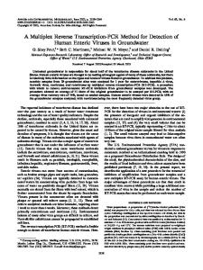
FIG. 1. Diagrammatic representation of in situ reverse transcription method.
nucleotide was added to fixed cells in a hybridization mixture and then the hybrids were detected after reverse transcription. In the procedure described here (Fig. 1), the priming oligonucleotide is the antisense oligo that hybridizes to the target RNA in live cells. Because unhybridized oligo in the cell will not prime reverse trancription, only hybridized antisense oligo is detected. The reverse transcripts are synthesized in the presence of labeled nucleotide, e.g., digoxigenin-11-dUTP, and incorporated label is detected with antibodies directed against the label using standard techniques. To illustrate the technique and one application, we show experiments that detect hybridization of oligo(dT) to poly(A) RNA in cells that were allowed to passively take up (antisense) oligo(dT), and then compare this with the amount of hybridization in cells that took up oligo(dT) complexed with cationic lipid. The oligo(dT) used is a phosphodiester backbone 43-mer that is fluorescently labeled at every tenth nucleotide with an aminohexyl-linked fluorochrome. We have found that poly(A) RNA hybrids formed with oligo(dT) labeled in this way are stable in live cells for at least 24 h (6).
DESCRIPTION OF METHOD Synthesis and Labeling of Oligonucleotides Oligonucleotides were automatically synthesized with aminohexyl-modified thymidine residues (Glen Re-
L6 rat myoblasts were plated onto glass coverslips [typically 12,500 cells per 12-mm round coverslip (Fisher, Pittsburgh, PA) that had been placed in a 24-well dish (VWR)] and allowed to grow in Dulbecco’s minimum essential medium (DMEM) with 10% fetal bovine serum at 37°C, 5% CO 2, until they reached about 60% confluence (usually around 24 h). For passive uptake of oligo, L6 cells were rinsed with DMEM without serum and incubated with 0.1–1 mM oligo in DMEM for 2 h at 37°C, 5% CO 2, in the dark. Cells were then rinsed three times with DMEM with serum and then incubated at least one more hour in DMEM with serum before extraction and fixation (see below). For uptake of oligo complexed with cationic lipid, the cationic lipid solution Tfx-50 (Promega, Madison, WI) or Pfx-4 or Pfx-6 (Invitrogen, San Diego, CA) was mixed with oligo at a ratio of 3 mg oligo:1 ml lipid according to manufacturer’s instructions to give a final oligo concentration of 0.1– 0.3 mM in DMEM without serum. L6 rat myoblasts were incubated for 2 h with the oligo/cationic lipid mixture, rinsed three times in DMEM with serum, and then allowed to grow for 1–2 h in DMEM with serum at 37°C, 5% CO 2. Live cells were visualized using an inverted fluorescence microscope modified to allow cells to be maintained at 37°C, 5% CO 2 during viewing (10). Other cationic reagents tested [DMRIE-C (GibcoBRL, Gaithersburg, MD), DOTAP (Boehringer-Mannheim, Indianapolis, IN), Pfx 1-3, 5, 7, 8 (Invitrogen), and Superfect (Qiagen, Santa Clarita, CA)] did not increase oligo uptake substantially (data not shown). Cell Permeabilization and Fixation Treated cells were either rinsed with 1 mM KH 2PO 4, 10 mM Na 2 HPO 4 , 0.137 M NaCl, 2.7 mM KCl, pH 7.0
283
IN SITU TRANSCRIPTION TO DETECT ANTISENSE HYBRIDIZATION
(K-PBS), and fixed directly in 4% formaldehyde in K-PBS, 5 mM MgCl 2 , or permeabilized with Triton X-100 and then fixed. To permeabilize with Triton, cells were rinsed with cytoskeletal extraction buffer [CSK buffer (11), see below] on ice, extracted with Triton/CSK buffer for 90 s on ice, and rinsed with CSK buffer again on ice. The permeabilized cells were then fixed for 15 min at room temperature in 4% formaldehyde, 0.1% glutaraldehyde in K-PBS, 5 mM MgCl 2 , rinsed with 70% ethanol three times, and stored in 70% ethanol at 4°C [see also Ref. (12)]. CSK Buffer Stock Sucrose KCl PIPES, pH 6.9 EGTA MgCl 2 Distilled deionized H 2O Total volume Chill to 4°C and add: Leupeptin Trypsin Optional: Phenylmethylsulfonyl fluoride Vanadyl ribonucleoside complex
Stock Final concentration Addition concentration — 1M 0.1 M 50 mM 1M
5.13 g 5.0 ml 5.0 ml 2.0 ml 250 ml 37.75 ml 50 ml
300 100 10 2 5
mM mM mM mM mM
20 mg/ml 20 mg/ml
2.5 ml 2.5 ml
1 mg/ml 1 mg/ml
0.2 M
300 ml
1.2 mM
0.2 M
500 ml
2.0 mM
Stock solutions should be autoclaved and/or made with diethyl pyrocarbonate-treated water to avoid RNase contamination. Five hundred microliters of 10% (v/v) Triton X-100 is added to 9.5 ml CSK buffer to give Triton/CSK buffer. In Situ Reverse Transcription Cells stored in 70% ethanol were washed first in K-PBS, 5 mM MgCl 2, and then twice in 13 SSC (150 mM NaCl, 15 mM sodium citrate, pH 7.0) at room temperature for 10 min each time, by slowly swirling the 24-well plate on a shaker. (Wash solutions should be poured or pipetted gently onto and from the coverslips so as not to dislodge the cells.) A 10-ml drop of reverse transcription (RT) mixture (below) is then pipetted onto a smooth sheet of Parafilm wrapped on a glass plate and a coverslip containing cells is inverted onto the drop, taking care to avoid bubbles. If one side of the coverslip is allowed to touch the Parafilm near the drop first and then the coverslip is slowly lowered onto the drop, few bubbles form. A second layer of Parafilm is overlaid on the glass plate and the reactions are incubated in a humidified chamber for 1 h at 37°C.
Reverse Transcription Mixture Stock Avian myeloblastosis virus (AMV) RT RNasin dNTPs Digoxigenin-11-dUTP AMV buffer Distilled deionized H 2O Total volume
Stock concentration
Volume added
Final concentration
8 U/ml
1.5 ml
1.2 U/ml
ml ml ml ml ml ml
0.4 U/ml 250 mM 50 mM 31
40 U/ml 5 mM 1 mM 35
0.1 0.5 0.5 2.0 5.4 10.0
AMV reverse transcriptase and AMV buffer were purchased from Promega (Madison, WI). RNAsin and digoxigenin-11-dUTP were from Boehringer-Mannheim and dNTPs were from Pharmacia (Piscataway, NJ). Biotin-14-dATP (Gibco-BRL) can also be used in this reaction. (All four unlabeled dNTPs are used at 250 mM regardless of the labeled dNTP used.) After the reaction, coverslips were returned to the 24-well dish and washed twice for 30 min at 37°C in 15% formamide, 23 SSC, followed by one 15-min wash in 13 SSC at room temperature. Antibody Detection of Digoxigenin-Labeled Reverse Transcripts Cells on coverslips were washed with 1% bovine serum albumin (BSA) in SSC for 10 min at room temperature to block sites that would bind nonspecifically to antidigoxigenin antibodies. Coverslips were then inverted as described above onto 10-ml drops of a 1:10 dilution (in SSC, 1% BSA) of either sheep antidigoxigenin (or goat antibiotin) antibodies linked to 1-nm gold particles (Goldmark Laboratories, Phillipsburg, NJ) or a 1:250 dilution of antidigoxigenin Fab fragments linked to alkaline phosphatase (BoehringerMannheim). After a 0.5-1-h incubation in a humidified chamber at 37°C, cells were washed three times with 1% BSA in SSC at room temperature for 10 min each time. Gold particles were enhanced using silver staining performed according to manufacturer’s instructions (Amersham). Alkaline phosphatase (AP) was detected using bromochloroindolyl phosphate/nitro blue tetrazolium as described (13). Coverslips were mounted in Vectashield (Vector Labs, Burlingame, CA). Comparison of IST Signals after Passive and Cationic Lipid-Mediated Uptake of Fluorescein-Labeled Oligo(dT) IST signal detected after passive uptake of fluoresceinlabeled oligo(dT) by L6 cells is shown in Fig. 2c. Detector precipitate is present in the cytoplasm of almost all cells and the distribution of this signal closely resembles the pattern seen when oligo(dT) is hybridized to poly(A) RNA in situ. The proportion of cells exhibiting nuclear signal in a given population varies from 10 to 90%. No signal is detected when cells take up control oligo(dA) instead (Fig. 2d). Oligo(dA) would not be expected to give detectable
284
POLITZ AND SINGER
FIG. 2. Hybridization of antisense oligo(dT) to poly(A) RNA in L6 myoblasts detected using in situ reverse transcription. Cells growing on glass coverslips were incubated with 0.1 mM modified oligo(dT) [or control oligo(dA)] in the presence or absence of Tfx-50 and then cells were permeabilized, fixed, and subjected to in situ reverse transcription using digoxigenin-11-dUTP. Incorporated label was detected using anti-digoxigenin antibodies linked to alkaline phosphatase. (a) Cells incubated with oligo(dT) plus Tfx-50; (b) cells incubated with oligo(dA) plus Tfx-50; (c) cells incubated with oligo(dT) alone; (d) cells incubated with oligo(dA) alone. Phase contrast micrographs. 1503.
IN SITU TRANSCRIPTION TO DETECT ANTISENSE HYBRIDIZATION
hybridization signal in this assay because there are few oligo(U) sequences in cellular RNA. The intensity of hybridization signal increased if oligo(dT) was taken up in the presence of Tfx-50 (Fig. 2a) or Pfx-4 (not shown). When an automated imaging system was used to quantitate signal intensity (6) and compare it with the intensity of IST signal in cells that were allowed to take up oligo(dT) passively, the mean average intensity of hybridization signal in cells incubated with oligo(dT) alone was 0.082 6 0.027, while the mean average intensity in cells that had taken up oligo(dT) complexed with Tfx-50 was 0.301 6 0.069. This three- to fourfold increase in hybridization was also obtained in the absence of cationic lipid if higher concentrations of oligo were used [;1 mM; see Ref. (6)].
CONCLUDING REMARKS Poly(A) RNA, typically present at 10 4–10 5 copies/cell in mammalian cells, is a very abundant intracellular hybridization target for antisense oligo(dT). We have found that the modified in situ reverse transcription technique as described here is also sensitive enough to detect oligos hybridized to a specific mRNA present at moderate abundance in the cell, viz., the hybridization of phosphorothioate antisense oligos to actin mRNA in chick embryo fibroblasts (6). Actin RNA is present at about 1000 copies per cell; therefore, other transcripts present at similar levels, including transcripts from transfected plasmids, should be detectable using this assay. An additional consideration should also be mentioned. Because a goal of this work has been to use antisense oligos as hybridization tags for RNA in vivo, an antisense oligo that did not appear to elicit RNase H activity was used. This oligo, a phosphodiester oligo(dT) that was labeled every 10 bases with a fluorochrome, was found to form hybrids that were stable for as long as 24 h after oligo treatment of L6 cells (6). We think it likely that the aminohexyl-linked fluorescein or cy-3 interferes with the binding of RNase H to the oligo/RNA hybrid, as Ueno et al. have suggested for an oligo modified in a similar fashion with 5-(N-aminohexyl)carbamoyl-29-deoxyuridine (14). As would be expected, cells that were incubated with unmodified phosphodiester oligo(dT) showed no IST hybridization signal, presumably because the oligos were degraded soon after addition to the cells and/or because the oligos that did hybridize induced rapid RNase H cleavage of the poly(A). In contrast, in experiments using unlabeled phosphorothioate oligo(dT), an internucleoside linkage that is more resistant to intracellular DNases, hybridization signal was detected using the IST assay. However, the hybridization of the phosphorothioate oligo(dT) did not appear as stable as that of the labeled phosphodiester oligo(dT) because signal was detected un-
285
til only about 4 h after oligo treatment (6). This may be because the phosphorothioate oligo(dT)/poly(A) RNA hybrid has a lower T m than phosphodiester oligo(dT)/ poly(A) RNA hybrids or because the phosphorothioate oligo(dT)/poly(A) RNA hybrid is less sensitive to digestion by RNase H [see Ref. (15)]. In support of the latter interpretation, IST signal was also only detectable early after treatment with antiactin phosphothioate oligos (6; unpublished results). Therefore, if one wishes to test for hybridization of an antisense oligo that elicits RNase H activity, it would be best to use the IST assay soon after oligo addition to cells. In closing, the IST method described here is useful for detecting hybridization of antisense oligos to intracellular RNAs of moderate abundance. The assay additionally allows one to identify the intracellular sites of hybridization, therefore indicating the intracellular location of the target RNA.
ACKNOWLEDGMENTS We thank Dr. Thoru Pederson for helpful conversations and a thorough reading of the manuscript. This work was supported by National Institutes of Health Grant HD18066 to R.H.S. and by National Institutes of Health National Research Service Award Postdoctoral Fellowship AR-08361 to J.C.P.
REFERENCES 1. 2. 3. 4. 5. 6. 7. 8.
9. 10. 11. 12. 13. 14. 15.
Branch, A. D. (1998) Trends Biochem. Sci. 23, 45–50. Stein, C. A. (1995) Nature Med. 1, 1119 –1121. Goodchild, J. (1990) Bioconj. Chem. 2, 166 –187. Politz, J. C., Browne, E. S., Wolf, D. E., and Pederson, T. (1998) Proc. Natl. Acad. Sci. USA 95, 6043– 6048. Politz, J. C., Tuft, R. A., Pederson, T., and Singer, R. H. (1999) Curr. Biol. 9, 285–291. Politz, J. C., Taneja, K. L., and Singer, R. H. (1995) Nucleic Acids Res. 23, 4946 – 4953. Eberwine, J., Spencer, C., Miyashiro, K., Mackler, S., and Finnell, R. (1988) Methods Enzymol. 216, 80 –100. Mogensen, J., Kolvraa, S., Hindkjaer, J., Petersen, S., Koch, J., Nygard, M., Jensen, T., Gregersen, N., Junker, S., and Bolund, L. (1991) Exp. Cell Res. 196, 92–98. Agrawal, S., Christodoulou, S., and Gait, M. J. (1986) Nucleic Acids Res. 14, 6227– 6245. Jacobson, M. R., and Pederson, T. (1997) in mRNA Formation and Function (Richter, J., Ed.), pp. 341–359, Academic Press, New York. Lenk, R., Ransom, L., Kaufmann, Y., and Penman, S. (1977) Cell 10, 67–78. Bassell, G. J., Powers, C. M., Taneja, K. L., and Singer, R. H. (1994) J. Cell Biol. 126, 863– 876. Singer, R. H., Lawrence, J. B., and Villnave, C. (1986) Biotechniques 4, 230 –249. Ueno, Y., Kumagai, N., and Matsuda, A. (1997) Nucleic Acids Res. 25, 3777–3782. Agrawal, S., Mayrand, S. H., Zamecnik, P. C., and Pederson, T. (1990) Proc. Natl. Acad. Sci. USA 87, 1401–1405.