CASE REPORT
ACTA RADIOLOGICA
In Situ Central Pulmonary Artery Thrombosis in Primary Pulmonary Hypertension P. P. AGARWAL, A. L. WOLFSOHN, F. R. MATZINGER, J. M. SEELY, R. A. PETERSON & C. DENNIE Department of Diagnostic Imaging and Department of Pathology, The Ottawa Hospital, Ottawa, Ontario, Canada Agarwal PP, Wolfsohn AL, Matzinger FR, Seely JM, Peterson RA, Dennie C. In situ central pulmonary artery thrombosis in primary pulmonary hypertension. Acta Radiol 2005;46:696–700. A rare case of extensive in situ central pulmonary artery thrombosis in primary pulmonary hypertension (PPH) is presented. The differentiation from chronic thromboembolic pulmonary arterial hypertension (CTEPH) is of paramount importance because of different therapeutic strategies. In this case, the presence of mural thrombus in the central pulmonary arteries on computed tomography made the distinction difficult. However, the possibility of in situ thrombosis was suggested on the basis of absence of other findings of CTEPH (abrupt narrowing/truncation of segmental arteries, variation in size of segmental vessels, arterial webs, mosaic attenuation, pulmonary infarcts, and dilated bronchial arteries), and this was confirmed on final pathology. Key words: In situ; primary pulmonary hypertension; thrombosis Carole Dennie, M.D., The Ottawa Hospital, Civic Campus, 1053 Carling Avenue, Ottawa, Ontario, Canada K1Y 4E9 (fax. +1 613 737 8957, e-mail.
[email protected])
We present a rare occurrence of extensive in situ central pulmonary artery thrombosis in primary pulmonary hypertension (PPH) detected on multidetector helical computed tomography (MDCT). The differentiation from chronic thromboembolic pulmonary arterial hypertension (CTEPH) is of paramount importance in determining the appropriate therapeutic strategy. While CTEPH can be potentially cured by surgical thrombendarterectomy, the treatment for PPH primarily consists of oxygen, vasodilators, anticoagulation, and lung transplantation (9). In this case, differentiation between the two conditions was difficult because of the presence of extensive mural thrombus in central pulmonary arteries. In situ thrombosis was favored because of the absence of other findings of CTEPH, i.e. mosaic perfusion, marked variation in size of segmental vessels, abrupt cut-offs, peripheral parenchymal densities/parenchymal bands, and enlarged bronchial arteries. None of these features was seen in this case, making CTEPH less likely. Case Report In 1994, a 28-year-old female presented with increasing shortness of breath on exertion. She
had no history of deep venous thrombosis or pulmonary embolism. However, she had recently been diagnosed with liver cirrhosis and portal hypertension secondary to autoimmune hepatitis. At that time, her presenting symptom was jaundice, and serological tests showed her disease to be related to autoimmunity. She was being treated with prednisolone for the same. On physical examination, a decrescendo diastolic murmur was detected. An echocardiogram revealed tricuspid insufficiency and pulmonary arterial hypertension. The right ventricle was hypertrophic and dilated and showed severe systolic dysfunction. In addition, there was right atrial dilatation. The pulmonary arterial pressure was estimated at 116 mmHg, based on a right atrial pressure estimate of 14 mmHg. Neither pulmonary angiography nor a CT of the thorax was performed at the time. A presumptive diagnosis of PPH was made. The condition slowly progressed over the initial 6 years, but deteriorated more rapidly over the last 2 years prior to admission, with development of New York Class IV heart failure. She was on home oxygen therapy and could carry out most activities of daily living at a modified pace. The patient was put on the heart/lung transplant waiting list in 2001. DOI 10.1080/02841850500215501
# 2005 Taylor & Francis
697
P. P. Agarwal et al.
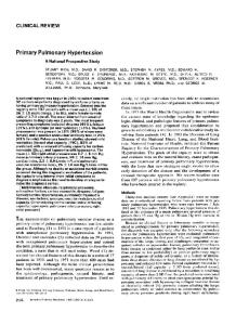
In August 2002, at age 36, there was considerable worsening of symptoms for 2 weeks and she was admitted to hospital with profound hypoxia. Oxygen saturation ranged from 70% to 80%. Her respiratory distress was presumed to be on the basis of either right heart failure or pulmonary thromboembolism. Chest radiography (Fig. 1) demonstrated cardiomegaly, mainly right-sided, and enlarged central pulmonary arteries with rapid tapering of peripheral branches. An ECG revealed right axis deviation, right ventricular hypertrophy, enlargement, and strain. A multi-slice CT (Lightspeed Plus; G.E. Medical Systems, Milwaukee, Wisc., USA) with contrast injection (150 ml of Omnipaque 300; Amersham Health, Oakville, Ontario, Canada) at a rate of 5 cc per second was performed using 2.5-mm thickness reconstructed at 1-mm intervals. This revealed a large non-occlusive, eccentric, or mural thrombus in the main pulmonary trunk extending into both proximal pulmonary arteries (Fig. 2). On the right, it extended into the proximal aspect of the upper lobe pulmonary artery and into the lateral basal segmental artery to the lower lobe. On the left, it extended into the lower lobe pulmonary artery. The distal pulmonary arteries were normal with no beading, abrupt cut-off, or webs. The pulmonary parenchyma was normal (Fig. 3). Specifically, there was no mosaic pattern of lung attenuation, variation in size of segmental vessels, peripheral
Fig. 2. Contrast-enhanced CT scan reveals a large non-occlusive mural thrombus involving the enlarged main pulmonary trunk and extending into the right proximal pulmonary artery. The thrombus also extended into the proximal left pulmonary artery (not shown).
Fig. 3. The lung parenchyma is normal without any evidence of mosaic perfusion.
Fig. 1. Chest X-ray demonstrates cardiomegaly with enlarged central pulmonary arteries and rapidly tapering peripheral pulmonary arteries. Acta Radiol 2005 (7)
parenchymal densities/parenchymal bands, or enlarged bronchial arteries. Acute pulmonary thromboembolism was unlikely, because the thrombus was mural rather than intraluminal. Furthermore, Doppler examination of the legs showed no evidence of venous thrombi. The authors favored in situ thrombosis of the pulmonary arteries over CTEPH because of the absence of indirect lung findings (2). Ventilation/perfusion scintigraphy and pulmonary angiography were planned to exclude chronic thromboembolic disease, but the patient deteriorated and had a cardio-respiratory arrest, from which she could not be resuscitated. At autopsy, there was in situ thrombosis of the central pulmonary arteries (Fig. 4) associated
In Situ Central Pulmonary Artery Thrombosis
2.
3.
4. 5.
Fig. 4. Autopsy specimen reveals an adherent, laminated thrombus beginning just above the pulmonary valve, partially occluding the dilated main pulmonary trunk and proximal left and right arteries.
with intimal atherosclerosis and medial degeneration. The thrombus was mostly old and organized, with some superimposed recent non-occlusive thrombus centrally (Fig. 4). There were features of a plexogenic arteriopathy in the peripheral pulmonary arteries/-oles, consistent with PPH. There were no peripheral pulmonary thromboemboli, lung infarcts, or systemic venous thrombi, thereby ruling out CTEPH. Features of right heart failure were seen as the immediate cause of death through either pump failure or a ventricular arrhythmia. There was established liver cirrhosis without active hepatitis. Discussion PPH is a rare disease characterized by elevated pulmonary arterial pressure with no demonstrable cause. The incidence of this disorder is approximately one to two cases per million people in the Western population, with a female/male ratio of 1.7:1.0. It is a progressive disease, and if untreated has a mean survival of less than 2 years from the time of diagnosis (11). According to the classification proposed by the World Health Organization in 1998, PHT is divided into five main categories (11) as follows: 1.
Pulmonary arterial hypertension (including PPH and PHT with similar clinical and pathological
698
features occurring in association with collagen vascular diseases, congenital systemic to pulmonary shunts, portal hypertension, HIV infection, or use of anorexigens). Pulmonary venous hypertension (e.g. left-sided atrial/ventricular heart disease, left-sided valvular heart disease, extrinsic compression of central pulmonary veins, pulmonary veno-occlusive disease). PHT associated with disorders of the respiratory system and/or hypoxaemia (e.g. chronic obstructive pulomonary disease, interstitial lung disease, sleep-disordered breathing, alveolar hypoventilation disorders, chronic exposure to high altitude, neonatal lung disease, alveolar capillary dysplasia). PHT due to chronic thrombotic and/or embolic disease. PHT due to disorders directly affecting the pulmonary vasculature (e.g. schistosomiasis, sarcoidosis) (11).
Pulmonary hypertension in combination with portal hypertension, also known as portopulmonary hypertension (PPHT) was initially described in 1951 by MANTZ & CRAIG. There are various theories of pathogenesis for this disease, and the suggested initiating factors are (3): (i) an increased flow phenomenon secondary to the hyperdynamic circulation; (ii) an increased volume phenomenon secondary to increased pulmonary vascular volume, which may or may not be associated with a specific cardiac complication of cirrhosis; and (iii) vasoconstriction due to an increase in vasoconstrictive substances entering the pulmonary circulation from the hepato-splanchnic vascular bed. Clinically, this ‘‘portopulmonary hypertension’’ resembles PPH, with dyspnoea and fatigue as the main symptoms, histopathology and response to prostacyclin therapy (10). The pathological findings of PPH in the distal pulmonary arterial circulation range from intimal fibrosis and thickening, medial hypertrophy and concentric fibroelastosis, fibrinoid necrosis and arteritis, plexiform lesions to angiomatoid/dilatation lesions (4). The last-mentioned is found in many cases of PPH, but is not pathognomonic (1), as it can be found in other causes of severe pulmonary hypertension, such as left-to-right cardiac shunts. It has been proposed that plexiform lesions of PPH may represent a neoplastic lesion due to dysregulation of endothelial growth or, alternatively, an angiogenic response to local ischemia or hypoxia with creation of collateral vessels due to obstructed arteries in other vascular beds (1). Acta Radiol 2005 (7)
699
P. P. Agarwal et al.
In situ thrombosis of the peripheral pulmonary arterioles is occasionally encountered in PPH and is likely due to endothelial cell injury creating a procoagulant environment (1). Development of large thrombi in the main, right, or left pulmonary arteries, however, is extremely rare in PPH and has been described in only one report (9). A variety of investigations can be used for assessment of pulmonary hypertension, including echocardiography, ventilation/perfusion scintigraphy, chest CT, and pulmonary arteriography. The relative lack of radiological literature on the CT appearance of PPH can probably be explained by the rarity of the disease. This case is even more unusual, as there is superimposed in situ thrombosis of the central pulmonary arteries. The indirect signs of pulmonary hypertension include dilatation of the main pulmonary arteries and pruning of peripheral pulmonary vasculature with right ventricular hypertrophy and enlargement. PPH causes a relatively uniform involvement of small muscular pulmonary arteries that manifests as loss of normal gravity dependent attenuation gradient (5). This is in contrast to the patchy embolic occlusion of larger generation pulmonary arteries seen in CTEPH, which is responsible for the conspicuous mosaic pattern of lung attenuation (7). Other findings described in CTEPH include abrupt truncation or narrowing of pulmonary arteries, webs, disparity in size of segmental arteries, pulmonary infarcts, and bronchial collateral vessels. Cylindrical bronchial dilatation has also been described in CTEPH adjacent to stenotic pulmonary arterial segments in two-thirds of patients (8). BERGIN et al. (2) studied the reliability of HRCT appearance of lung (mosaic attenuation, irregular vessel size, and peripheral densities) in distinguishing patients with CTEPH from those with other pulmonary abnormalities including PPH. In the 67 patients included in the study, all patients with CTEPH (17/17) showed a mosaic pattern of variable attenuation compatible with irregular pulmonary perfusion, while none (0/ 6) of the patients with PPH had regional inhomogeneity. Peripheral densities that result from infarcts were likewise absent in the PPH group. Marked variation in segmental vessel size was found to be a more specific feature of the CTEPH group. SHERRICK et al. (12), however, showed a lower diagnostic accuracy for mosaic attenuation in distinguishing various causes of pulmonary arterial hypertension. In their study of 64 patients with PHT, they found mosaic attenuation in 80% (12/15) of patients with CTEPH, in 50% (2/4) of patients with PPH, in 100% (2/2) of patients with Acta Radiol 2005 (7)
veno-occlusive disease, in 5% (1/21) of patients with underlying lung disease, and in 12% (2/17) of patients with cardiac disease. They concluded that mosaic attenuation is seen significantly more in PHT due to vascular diseases than secondary to lung/cardiac disease. Ventilation/perfusion scintigraphy is thought to be the key test in differentiating CTEPH from PPH (9). In CTEPH, the scan shows segmental or lobar defects, whereas in PPH it is either normal or shows mottling (9). Some patients with severe PPH may show reverse mismatch with the areas of ventilatory defect corresponding to areas of engorged pulmonary vessels (6). In conclusion, it is crucial to identify and differentiate PPH with in situ pulmonary artery thrombosis from CTEPH, as this requires a different therapeutic approach. Surgical thromboendarterectomy is potentially curative in CTEPH, while oxygen, vasodilators, anticoagulation, and heart-lung transplantation are the treatment options for PPH (9). In the current case, the presence of non-occlusive filling defects as opposed to central filling defects in the pulmonary arteries and no leg vein thrombosis differentiated the process from acute thromboembolic disease, while the lack of mosaic lung attenuation, variation in size of segmental vessels and peripheral parenchymal densities/parenchymal bands favored in situ thrombosis in the setting of long-standing PPH. References 1. Archer S, Rich S. Primary pulmonary hypertension: a vascular biology and translational research ‘‘Work in progress’’. Circulation 2000;02:2781–91. 2. Bergin CJ, Rios G, King MA, Belezzouli E, Luna J, Auger WR. Accuracy of high-resolution CT in identifying chronic pulmonary thromboembolic disease. Am J Roentgenol 1996;166:1371–7. 3. Blendis L, Wong F. Portopulmonary hypertension: an increasingly important complication of cirrhosis. Gastroenterology 2003;125:622–4. 4. Burke AP, Farb A, Virmani R. The pathology of primary pulmonary hypertension. Mod Pathol 1991; 4:269–82. 5. Cailes JB, Du Bois RM, Hansell DM. Density gradient of the lung parenchyma on CT in patients with lone pulmonary hypertension and systemic sclerosis. Acad Radiol 1996;3:724–30. 6. Engeler CE, Kuni CC, Tashjian JH, Engeler CM, du Cret RP. Regional alterations in lung ventilation in endstage primary pulmonary hypertension: correlation between CT and scintigraphy. Am J Roentgenol 1995; 164:831–5. 7. Hansell DM. Small vessel disease of the lung: CTPathologic correlates. Radiology 2002;225:639–53.
In Situ Central Pulmonary Artery Thrombosis 8. Kauczor HU, Schwickert HC, Mayer E, Schweden F, Schild HH, Thelen M. Spiral CT of bronchial arteries in chronic thromboembolism. J Comput Assist Tomogr 1994;18:855–61. 9. Moser KM, Fedullo PF, Finkbeiner WE, Golden J. Do patients with primary pulmonary hypertension develop extensive central thrombi? Circulation 1995;91:741–5.
700
10. Naeije R. Hepatopulmonary syndrome and portopulmonary hypertension. Swiss Med Wkly 2003;133:163–9. 11. Peacock AJ. Primary pulmonary hypertension. Thorax 1999;54:1107–18. 12. Sherrick AD, Swensen SJ, Hartman TE. Mosaic pattern of lung attenuation on CT scans: frequency among patients with pulmonary artery hypertension of different causes. Am J Roentgenol 1997;169:79–82.
Acta Radiol 2005 (7)