Implementation and Evaluation of an Augmented Reality System Supporting Minimal Invasive Interventions Michael Schnaider1, Sandra Röddiger2, Helmut Seibert1, Bernd Schwald1 1
ZGDV – Computer Graphics Center Department Visual Computing Fraunhoferstr.5 D-64283 Darmstadt
2
Klinikum Offenbach Strahlenklinik Starkenburgring 66 D-63069 Offenbach am Main
Abstract Minimal invasive surgery and interventions promise a number of advantages for the patient. From the medical point of view, the biggest advantage is a reduced trauma for the patient, however at the cost of a limited view for the physician. The project MEDARPA addresses this limitation by applying Augmented Reality (AR) technology to enhance the real view of the surgeon with preoperatively acquired medical data from CT or any other 3D imaging modality. In the last three years, a group of clinical, medical engineering and research partners, has designed and implemented a prototype of an AR-enhanced navigation support system for minimal invasive interventions, almost ready to be used in the operation theatre. It comprises of e.g. a transparent display device, called the ‘AR window’, a hybrid tracking system, simultaneously determining orientation and position of the display, the physician’s head and instruments that are used during the intervention, an audio-visual navigation support, real-time volume rendering of medical 3Dimage modalities in a compact PC-based system. The MEDARPA system has been designed to be applicable to a variety of medical scenarios. For the proof of concept cardiosurgery, radiooncology and pneumology scenarios were considered. Furthermore ergonomic constraints played a decisive role in the design of the system. The system is easy to handle, movable and easy to displace, if not needed and can be integrated smoothly into the medical intervention process. This paper gives an overview of the design of the MEDARPA system and its evaluation.
1. Introduction Minimal invasive operation techniques have become more important and more accepted in the last 15 years. While offering obvious advantages for the patient such as reduced access trauma, faster postoperative recovery and better cosmetic result, these techniques confront the surgeon with a restricted view of the region of intervention. The application of Augmented Reality (AR) techniques is most promising and it offers a solution to minimize or even overcome the problem of the physician’s limited visual perception. However, many AR approaches are using Head Mounted Displays (HMD) for the fusion of the real and the virtual scene, either as variants with video see-through or as optical seethrough. Although HMDs can be an appropriate choice as a display system in many application areas like maintenance, training etc., most surgeons tend not to accept these devices. A likely cause is correlated to the reduced flexibility in movements and to ergonomic constraints. The project MEDARPA (Medical Augmented Reality for Patients) described here
addresses these issues by assembling a novel HMD-less AR System using a transparent display, here called an AR window, for Computer Aided Surgery (CAS) and minimalinvasive interventions in general. As a major field of activity for AR applications, a lot of work has been invested in the medical domain already. Early approaches that provided an "x-ray view" to the physician can be found among others in [Mellor95], [Lorensen93], [Grimson95], [Betting95]. Some of the early applications date back until fifteen years ago, but a number of new AR navigation prototypes for CAS were developed in the recent years. The tracking systems used for these applications cover electromagnetic trackers as well as optical systems [Seibert98], [Tonet00], [Birkfellner00]. Trials similar to our AR window approach were made using a beam splitter (a half-silvered mirror), which is both transparent and reflective [Blackwell98], [Blackwell00], [Nakajima01], the later providing the user with a stereoscopic view using shutter glasses. An alternative technique for the generation of stereoscopic displays has been proposed in [Sandin01] but this auto stereoscopic technique is not applicable for transparent displays. Other approaches such as proposed in [King00] use a surgical microscope for augmentation or make investigations to integrate a holographic screen [Dohi00], [Lievin01].
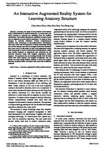
2. System and Components This section describes the hardware set-up of the MEDARPA system and its integration into the clinical environment. Figure 1 depicts different aspects of the MEDARPA system.
Figure 1: System overview including components (boxes), workflow (horizontal) and technical layers (vertical). Using the system requires some preparatory steps in the image data acquisition phase. In order to enable to perform a patient registration in the operational phase, markers are attached to the patient. These are detectable e.g. in the CT-images. Following the data acquisition phase, the pre-processing of the image data is performed. This includes identification of the markers, definition of the area of treatment and/or target regions and the creation of the 3D patient model that is used for the augmentation. The third and final step is the operational use of the
system functionality for the treatment, including patient registration, augmentation and navigation support. Figure 1 also shows the technological layers of the system. The tracking and input layer contains the components for the determination of poses of the instruments, the physician, the display and the patient, i.e. the two different tracking systems, as well as the input device controller for the supported types of interaction. In the layer called registration and controlling the functionality to process all 3D data and provide the registration procedures are enclosed. The visualization and modeling layer includes components for the pre-processing of the acquired medical image data (see above) and for their visualisation in the correct perspective for the physician performing the medical treatment.
2.1.
Hardware Set-up
All components of the Medarpa system are embedded in or attached to a trolley. On top of the trolley a swivel arm holds the transparent display, the main component of the system. Figure 2 shows a test set-up of the system in an operation theatre. The swivel arm enables the physician to easily move the transparent display to the desired place such that the patient can be observed in the usual manner. The physician’s view to the patient is enhanced with the superimposed virtual information prepared during the pre-processing phase. The positions and orientations of the patient, the instrument, the display and the surgeon's viewpoint are continuously determined in order to provide the correct virtual overlay. This is performed by a hybrid tracking system combining an optical and an electromagnetic tracking system. This approach allows the utilization of the specific strengths of the two tracking systems with respect to task that needs to be performed.
Figure 2: Set-up of the Medarpa system in an operation theatre. For the optical tracking, the EOS system [EOS], developed at the ZGDV, is used. It consists of a stereo camera system equipped with infrared filters. Similar video-based tracking systems have been introduced in [Madritsch96], [Dorfmüller98], [Ribo01] and few are also available on the market [ARTtrack], [NDI]. These two cameras are mounted on a stand attached to the trolley. Due to EOS’ flexibility in camera placement, an optimised placement was determined to minimize the amount of occlusions while tracking display and surgeon. Tests of the optical
tracking system showed statistical position accuracy of approx. 1mm for the MEDARPA setup, covering an interaction volume of 1.5m by 0.8m by 1m (width, depth, height). EOS simultaneously tracks several six degrees of freedom sensors in form of rigid bodies based on infrared markers. Active infrared landmarks are attached to the display and the physician's glasses. They build up the sensors for the optical position and orientation tracking. In favour of the optical solution, an electromagnetic (EM) tracking system [Ascension] has been chosen to track the medical instrument used for the intervention. This approach avoids any problems with potential occlusions when used below the display.
Figure 3: The AR window, manufactured by GfM. The user navigates inside an anthropomorphic phantom to the red target region. The instrument has a green overlay. GfM [GfM] has designed and manufactured the display, see Figure 3, and its swivel arm especially for the MEDARPA project. It is based on a modified 17'' TFT screen allowing a resolution of 1024 x 768 at 75Hz. Due to the available display technology the transparency is reasonable, but can be improved by sufficiently lightening the observed scene. In the current implementation the display can be moved freely within a working volume of at least 2 m3.
2.2.
Typical Scenario
Three clinical scenarios are included in this project. The first is a cardio-surgical scenario, in which the Medarpa system helps to find the optimal position for ports needed for robotassisted coronary surgery. In the second scenario, the Medarpa system is used for bronchoscopic puncture of suspicious regions. In the last scenario, the Medarpa system offers assistance for the navigation of brachytherapy catheters within the patient’s body. Brachytherapy means radiotherapy very close to a tumour area or inside a tumour. For the latter, catheters, e.g. metal needles or plastic tubes need to be implanted. Prior to this procedure, nine computer tomography (CT)-markers are fixed on the patient’s skin and a CT
scan of the region of interest is done. During the preplanning process, organs at risk are defined as well as the tumour area. Target regions within the tumour can be defined for optimal catheter navigation. As soon as the patient is in place and sedated, the system’s set-up can be started. The patient is registered to the system by the CT-markers. After disinfection of the skin, the display can be placed above the region of interest. The first catheter is fixed on the handle with the electromagnetic (EM) tracker. The display and the physician are tracked by the optical tracking system, while the patient and the handle are tracked by the EM system. Guided by the AR view through the display, the physician starts the needle implantation.
2.3.
Navigation support
Due to the lack of available display technology, the transparent MEDARPA display does not offer a stereoscopic view for the surgeon. However, to compensate this lack of stereoscopic depth perception head movement and principles like occlusion or collision between virtual objects are utilized. To ease up the task of navigation, a special colour feedback is provided; the colour of the virtual instrument changes depending on its position and direction to the target regions. Figure 4 shows three steps of navigation in the volume data set of a virtual patient. The small blue spheres are the virtual image of the markers, needed for registration, the larger red spheres, only visible in the third step, are the defined target regions. In the first step the instrument is pointing into an arbitrary direction and therefore coloured in red. The instrument changes colour to yellow, when it is pointing into one of the target directions. This allows moving the instrument straight into the direction of target region by taking care, that the instrument is always displayed in yellow. If the target region is reached, the virtual instrument changes colour to green.
Figure 4: The virtual overlay of the instrument changes colours: red for non-correct direction to target region, yellow for correct direction to target region and green, if the instrument’s tip hits the target region. An additional optional navigation support is a triangle that is spanned between the virtual instrument’s tip, the target region and a point on the virtual elongation of the instrument. If the instrument is pointing in direction of the target region, the triangle is reduced to a line and barely visible, the more it is pointing into a wrong direction, the larger the triangle gets.
3. Evaluation and Test Reports Among other properties, the achievable overall accuracy is one of the most important ones for a medical system. In fact, specify an overall accuracy value for the complete system is not an easy task, but accuracies can be given for special aspects of the system as for navigation or the quality of the overlay in the view of the surgeon.
Figure 5: Errors, induced by the different tracking systems and registration procedures. Arrow direction means ‘depends on’. As mentioned in section 2.1 a hybrid tracking system combining an optical and an electromagnetic tracking system determines poses of the display, the physician's glasses, instruments and the patient. Figure 5 gives an overview how the accuracies depend on the tracking systems and registration procedures. When an object is tracked, normally offsets between sensors and e.g. the tip of an instrument need to be calculated, inducing a new source of errors. Additionally the rotation error of the tracking system increases. Practically speaking, the larger the distance between an instrument’s sensor and the instrument’s tip is, the larger will be the position error at the tip of the instrument. In Figure 5 this additional error is included in ‘Accuracy of Electromagnetic Tracking’. In the following, we focus on giving a value for the navigation accuracy, depending on the patient registration and the accuracy of the instrument, as introduced above. The accuracy or quality of the overlays is difficult to measure in an objective way and differs for every person. However, in case of the MEDARPA system, the overlay quality is a less critical issue in contrast to the navigation accuracy.
3.1.
Registration and Positioning of the Patient
Before the Medarpa system is ready to be used in the medical treatment procedure, the patient is registered by moving the registration device to the spherical markers attached to the patient’s body. These markers were attached before the patient is scanned and their position in the virtual patient model is identified in the pre-processing step (see [Wesarg03]). Figure 6 depicts the registration of one marker with the registration device.
Figure 6: Patient registration by moving the concave tip of the registration device to spherical markers on the patient’s body. After all marker positions are recorded, the virtual patient can be registered to the physical patient by registering two 3D point sets. This procedure is described in more detail in [Schwald04]. Table 1 shows representative results from this registration procedure under laboratory conditions. Test
Mean (mm)
Max (mm)
1 2 3 4 5 6 7 8 9 10 Mean Stddev Max
1.72 0.86 0.86 1.42 0.76 1.51 1.46 0.88 1.48 1.11 1.21 0.349 1.72
3.30 1.97 1.61 2.65 1.34 3.18 3.04 1.32 2.58 1.90 2.29 0.756 3.30
Translation (mm) x y z 277.8 190.4 497.8 276.7 189.8 499.0 276.7 189.4 499.8 277.5 190.2 497.5 277.3 189.0 499.4 276.9 190.6 497.2 277.8 190.1 497.8 276.7 189.3 499.0 277.4 190.1 497.7 277.0 189.2 499.3 277.18 189.81 498.3 1.174 1.505
Table 1: Results from ten registrations in the same set-up with an anthropomorphic phantom with nine markers. In this test series nine markers were attached to an anthropomorphic phantom, which was placed in a distance of approximately 300mm from the electromagnetic emitter. Ten
registrations were performed. The mean and max errors are results, calculated after each test as quality value. The translations are together with the rotations (not shown here) the results, which are then used in the Medarpa system. The mean error of the ten tests is with 1.21mm very close to the standard deviation of the translations with 1.17mm, such that the mean error is seen as a reliable quality value. Furthermore this error corresponds to the accuracy of the electromagnetic tracking system and is therefore within our expectations of a good patient registration. From a practical point of view, the number of the spherical markers should be minimized in order to make the registration as comfortable as possible for the surgeon. Several test in the clinical environment showed, that the accuracy of the registration decreases strongly, if less than six markers are used. In general eight to ten markers are a good basis for the registration procedure.
3.2.
Navigation error
To evaluate the accuracy of the navigation needles are inserted into a patient under Medarpa AR navigation support and their positions are stored by the system. After the insertion, a second control CT scan is recorded. Registering the two CT scans to each other now allows a very precise evaluation, how close the needles were inserted to the defined target regions. Table 2 shows the results of such tests, performed in two different hospitals. The distance between instrument’s sensor and its needle tip was in the first test approx. 150mm and in the second test approx. 225mm. The mean error of the measurements is 6.3mm, respectively 9.8mm, which we denote the overall navigation error of the Medarpa system. Test1, hospital Frankfurt
Test2, hospital Offenbach
Needle length
~ 150mm
~ 225mm
Number of measurements
15
16
Mean error at tip
6.3mm
8.9mm
Minimal error
4.7mm
3.4mm
Maximum error
9.3mm
12.8mm
Standard deviation of errors
1.3mm
2.5mm
Table 2: Most recent results of navigation tests by evaluating a CT Scan with implanted needles. Not taking major disturbances (see e.g. section 3.3) of the EM tracking system into account, the navigation error is basically influenced by the accuracy of the registration, the accuracy of the calibration of the instrument (distance sensor to needle’s tip), the position accuracy of the sensor and the rotation accuracy of the sensor. The registration accuracy and the position accuracy of the sensor are both independent of the type of instrument and we expect approx. 1.5mm for both (see section 3.1). In contrast, the error induced by the rotation accuracy of the sensor and the accuracy of the calibration of the instrument both depend on the length of the needle to be used. The longer a needle is, the higher is the loss of accuracy. The rotation error
of the EM tracking system is specified with 0.5°. Therefore the position error at the tip of a needle, which is 150mm remote from the sensor, is increased by approx. 1.3mm, for a needle with 225mm by approx. 2.0mm. Similar values are supposed for the accuracy of the calibration of the instrument. Additional problems arise, if the instrument is very flexible. Summing up these theoretical values the navigation error at the tip of a 150mm instrument would be 5.6mm and at the tip of a 225mm instrument 7.0mm.
3.3.
General Experiences in Hospital
Major disturbances of the EM tracking system could be observed when using the Medarpa system next to a computer tomograph or close to high voltage cables. Minor disturbances were seen in the operation room. These findings led to a modification of the brachytherapy scenario. Initially, it was planned to use the Medarpa system and the CT in parallel during the implantation procedure for the verification of the catheter positions. In an undisturbed environment the systems is easy to handle and needs - after installation - 6 minutes (3.5-10.0) for the set-up (calibration of tracking systems and registration of the phantom/patient). The MEDARPA-prototype enables the physician to implant brachytherapycatheters guided by Augmented Reality view. Target regions can be reached either freehand or guided by the navigation support tools. The puncture of organs at risk can be avoided. The Medarpa system is furthermore useful for the training of young physicians.
4. Realization Potential, Outlook The first tests show that the concept of the system appears to be useful to support planning of interventions and training purposes. The AR system itself is not only designed for the planning of minimal invasive cardiac interventions or needle implantation in brachytherapy, but as a generic system for several medical domains, including navigation support. The prototype of a transparent display developed in the MEDARPA project is due to available display hardware applicable, but upcoming display technologies should allow improving the display quality in near future. Alternatives like projecting the virtual augmentations directly on the skin of a patient is one of the visions for a future system, making AR even more user friendly and seamlessly integrated into the real environment. The accuracy of the navigation of about 6.3mm for a needle with approx. 150mm is not fully satisfying yet, but has already been considered a useful ‘add-on’ for existing interventional procedures. We expect to be able to reduce the navigation error to around 4mm within the existing set-up and a needle of approx. 150mm length. Furthermore the flexible software architecture of the Medarpa system allows easy integration of new tracking technologies, as soon as they are available and we then expect increased accuracy of the overall system.
Acknowledgements The project MEDARPA (www.medarpa.de) has been funded by research grants of the German Federal Ministry of Education and Research (BMBF), research grant 01IRA09B. We want to thank all our project partners for their cooperation and encouraging input.
References [ARTtrack] [Ascension] [Betting95] [Birkfellner00] [Blackwell00] [Blackwell98] [Dohi00] [Dorfmüller98] [EOS] [GfM] [Grimson95] [King00] [Lievin01] [Lorensen93] [Madritsch96] [Mellor95] [Nakajima01] [NDI] [Ribo01] [Sandin01] [Schwald04] [Seibert98] [Tonet00]
[Wesarg03]
“ARTtrack”, http://www.ar-tracking.de, 2003. “Ascension”, http://www.ascension-tech.com/products, 2003. F. Betting, J. Feldmar, et al. A New Framework for Fusing Stereo Images with Volumetric Medical Images. Conf Proc., on Computer Vision and Pattern Recognition, pp 30–39, 1995. W. Birkfellner, K. Huber, F. Watzinger, M. Figl, F. Wanschitz, R. Hanel, D. Rafolt, R. Ewers, and H. Bergmann. Development of the Varioscope AR: A See-through HMD for ComputerAided Surgery. In Proc. Int. Symposium Augmented Reality - ISAR, pages 47–53, 2000. M. Blackwell, C. Nikou, A. DiGioia, and T. Kanade. An Image Overlay system for medical visualization. Transactions on Medical Image Analysis, 4:67–72, 2000. M. Blackwell, A. DiGioia, and C. Nikou. Image Overlay Techniques for Surgery. Transactions on Medical Image Analysis, June 1998. T. Dohi. Surgical Robotics and Three Dimensional Display for Computer Aided Surgery. in Proc. of Computer Aided Radiology and Surgery, CARS 2000, San Francisco, U.S.A., pp 715– 719, June 2000. K. Dorfmüller, “An Optical Tracking System for VR/AR-Applications“, in Virtual Environments 99, Proceedings of the Eurographics Workshop, A. H. M. Gervautz and D. Schmalstieg, eds., Springer Computer-Science, Vienna, Austria, 1999. “Optical Tracking System EOS”, http://www.zgdv.de/zgdv/departments/z2/Z2Projects/EOS Gesellschaft für Medizintechnik mbH, http://www.gfmmbh.de/ W. E. L. Grimson, G. J. Ettinger, et al. Evaluating and validating an automated registration system for enhanced reality visualization in surgery. Proceedings of Computer Vision, Virtual Reality, and Robotics in Medicine ’95, pages 3–12,1995. A. P. King, P. Edwards, C. Maurer, D. de Cunha, R. P. Gaston, M. Clarkson, D. Hill, D. Hawkes, M. Fenlon, A. Strong, T. Cox, and M. Gleeson. Stereo Augmented Reality in the Surgical Microscope. Presence: Teleoperators and Virtual Environments, 9(4), 2000. M. Lievin and E. Keeve. Stereoscopic Augmented Reality System for Computer Assisted Surgery. In: Proc. Computer Assisted Radiology and Surgery, CARS’01, pages 27–30, 2001. W. Lorensen, H. Cline, et al. Enhancing Reality in the Operating Room. Proceedings of the 1993 IEEE Visualization Conference, pages 410–415, 1993. F. Madritsch and M. Gervautz, “CCD-Camera Based Optical Beacon Tracking for Virtual and Augmented Reality”, Eurographics (15(3)), 1996. J. P. Mellor. Enhanced Reality Visualization in a Surgical Environment. AI Lab . Cambridge, MA, Massachusetts Institute of Technology, page 102, 1995. S. Nakajima, K. Nakamura, K. Masamune, I. Sakuma, and T.Dohi. Three-dimensional medical imaging display with computer-generated integral photography. Computer Med Imaging Graph, (25(3)):235–241, 2001. “Northern Digital”, http://www.ndigital.com/products.html, 2003. M. Ribo, A. Prinz and A.L. Fuhrmann, “A new Optical TrackingSystem for Virtual and Augmented Reality”, IEEE Instrumentation and Measurement Technology Conference, Budapest, Hungary, May 21-23, 2001. M. Sandin, B. Bruegge, G. Klinker, A. MacWilliams, T. Reicher, S. Riß, C. Sandor, and M. Wagner. Design of a Component-Based Augmented Reality Framework. IEEE and ACM International Symposium on Augmented Reality (ISAR),New York,NY, October 29-30 2001. B. Schwald and H. Seibert, “Registration Tasks for a Hybrid Tracking System for Medical Augmented Reality”, to be published in the Proc. of the Int. Conf. on Computer Graphics, Visualization and Computer Vision (WSCG 2004), 2004. F. Seibert. Stereo-based Augmented Reality in Medicine.3. German-Korea Joint Conference on Advanced Medical Image Processing, EWHA University, Seoul, Korea, August 1998. O. Tonet, G. Megali, S. D’Attanasio, P.Dario, M. Carrozza, M. Maracci, S. Martelli, and P. L. Palombara. An Augmented Reality Navigation System for Computer Assisted Arthroscopic Surgery of the Knee. Lecture Notes in Computer Science, S.L. Delp, A.M. DiGioia and B. Jaramaz Eds., Springer, 1935:1158–1162 (MICCAI 2000), October 2000. S. Wesarg, T. H. Lauer, E. A. Firle and C. Dold, “Several Marker Segmentation Techniques for Use with a Medical AR System – a Comparison” in Computer Assisted Radiology and Surgery, H. U. Lemke, et al., eds., Proc. Of the 17th CARS 2003, p.1303, Elsevier, 2003.