Molecular Human Reproduction Vol.8, No.6 pp. 546–551, 2002
Immunohistochemical localization of glutaredoxin and thioredoxin in human endometrium: a possible association with pinopodes A.Stavre´us-Evers1, B.Masironi1, B.-M.Landgren2, A.Holmgren3, H.Eriksson1 and L.Sahlin1,4 1Division
for Reproductive Endocrinology, Department of Woman and Child Health, Karolinska Hospital, Stockholm, 2Division of Obstetrics and Gynecology, Department of Clinical Sciences, Huddinge University Hospital, Huddinge and 3Medical Nobel Institute for Biochemistry, Department of Biochemistry and Biophysics, Karolinska Institutet, Stockholm, Sweden 4To
whom correspondence should be addressed at: Division for Reproductive Endocrinology, Karolinska Hospital, L5:01, S-171 76 Stockholm, Sweden. E-mail:
[email protected] The human endometrium is only receptive for blastocyst implantation during a short period of the menstrual cycle. Pinopodes have been suggested to be markers of uterine receptivity, but little is known about their function and the biochemical processes taking place in them. In this study, we have examined the presence of glutaredoxin (Grx) and thioredoxin (Trx) and their colocalization with pinopodes in the normal human endometrium. Endometrial biopsies were obtained from fertile women with normal menstrual cycles. The biopsies were examined by scanning electron microscopy for detection of pinopodes and by immunohistochemistry for the expression of Grx and Trx. The pinopodes showed strong immunostaining for Grx. Increasing levels of Grx immunoreactivity were seen in the luminal and glandular epithelial cells concomitant with pinopode formation. Trx immunostaining was most intense in the ciliated cells of the luminal and glandular epithelium, while the staining was moderate to strong in a majority of the other cells, both epithelial and stromal. Trx levels did not change during the secretory phase of the cycle. The intense immunostaining concomitant with the presence of pinopodes suggests that Grx plays an important role during implantation, possibly by protecting the epithelial cells from apoptotic actions of the trophoblast cells. Key words: endometrium/glutaredoxin/pinopodes/redox regulation/thioredoxin
Introduction The human endometrium is only receptive for blastocyst implantation during a short period of the menstrual cycle, the so-called implantation window. During this period, pinopodes appear on the endometrial surface. The pinopodes have been suggested as structural markers of the implantation window (Martel et al., 1981; Nikas et al., 1995) since they arise, in the shape of a flower or mushroom, from the apical surface of the epithelial cells during this time period. Their function has been discussed. In rodents, where pinopodes were first discovered (Nilsson, 1958; Warren and Enders, 1964), they have been shown to be responsible for the uptake of uterine secretion (Enders and Nelson, 1973; Parr and Parr, 1974). Uterine secretion is also reduced in humans at the time of implantation (Gemzell-Danielsson and Hamberg, 1994), although it has not been shown that pinopodes are responsible for this reduction. The formation of pinopodes would, by preventing cilia from ‘sweeping material’ across the epithelial surface, help the blastocyst to get in closer physical contact with the epithelial surface. Adhesion molecules, such as integrins, increase in the endometrium at the time of implantation (Lessey et al., 1992). Thus, it is possible that adhesion molecules are present on the pinopode surface. Glutaredoxin (Grx) and thioredoxin (Trx) are thioldisulphide oxidoreductases originally discovered as dithiol hydrogen donors for ribonucleotide reductase (Holmgren, 1985, 1989). Glutaredoxins,
546
which catalyse GSH–disulphide oxidoreductions and thioredoxins, which are reduced by NADPH and the selenoenzyme thioredoxinreductase, are important for antioxidant defence and for the regulation of the cellular redox state (Holmgren and Åslund, 1995; Holmgren et al., 1998; Arne´r and Holmgren, 2000). Grx and Trx have also been shown to be involved in regulation of other activities, e.g. transcription, cell signalling and cell growth and death (Rhee, 1999; Finkel, 2000). There is little data on the expression of Grx in the reproductive tract. However, it has been shown that Grx is increased in the cervix of term pregnant women compared with non-pregnant controls (Sahlin et al., 2000a). In addition, Grx has been immunolocalized to the human corpus luteum (Garcia-Pardo et al., 1999) and the human placenta (Sahlin et al., 2000b). Trx has been shown to be present in the rat and human reproductive tissues including endometrium, (Rozell et al., 1985; Fujii et al., 1991; Maruyama et al., 1997), cervix (Sahlin et al., 1997a), ovary (Iwai et al., 1992), pregnancy deciduas (Kobayashi et al., 1995) and placenta (Padilla et al., 1995; Sahlin et al., 2000b). Trx and Trx mRNA in the endometrium are cycle dependent, with the highest expression in glandular and stromal cells during the early secretory phase (Maruyama et al., 1997). Trx has been shown to play a role in the development of the embryo (Natsuyama et al., 1992) and, in Trx knock-out mice, it was shown that the early embryos die shortly after implantation (Matsui et al., 1996). The Trx/Grx system in the endometrium has not been extensively © European Society of Human Reproduction and Embryology
Glutaredoxin and thioredoxin in human endometrium
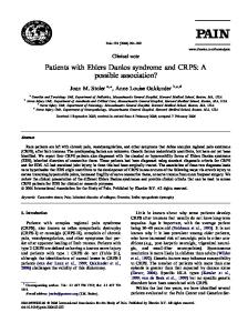
Figure 1. Representative images of the endometrial epithelial surface are shown. (a) Endometrial surface, day LH ⫹2. The ciliated and microvillous cells are clearly seen and the surface is bulging. A few droplets, normally seen after ovulation, are also seen on the epithelial surface. (b) Receptive endometrium, day LH ⫹7. Numerous pinopodes are present on the surface. (c) Endometrial surface in the late secretory phase, day LH ⫹11. The microvillous cells exhibit a polygonal shape.
studied. The aim of this study was therefore to examine the possible correlation between the presence of pinopodes on the endometrial surface and the expression of Trx and Grx in the endometrium at the time around the implantation window.
Materials and methods Endometrial tissue Endometrial biopsies were obtained from 22 women. Thirteen women were normal healthy volunteers, and nine volunteered for the study in connection with a sterilization operation by laparoscopy. All women were healthy with a history of regular menstrual periods (25–35 days) and with proven fertility. The mean age was 37 years (range 25–44). None of the women had used steroidal contraceptives or an intrauterine device for at least 3 months before the study. None had had a pregnancy or pelvic inflammatory disease within 1 year prior to the study. The day of the LH surge was determined in the morning using a self-test (Clearplan; Unipath Ltd, Bedford, UK). The biopsies were randomly selected in the interval between days LH ⫹0 and ⫹11, with the day of the LH surge indicated as day 0. The biopsies were taken from the anterior wall of the uterine cavity, without dilatation of the cervix, using a Randall curette (Stille Werner AB, Stockholm, Sweden). Each biopsy was divided in two; one of the pieces was processed for scanning electron microscopy and the other for immunohistochemistry. Samples for scanning electron microscopy were fixed in a solution containing 2.5% (w/v) glutaraldehyde (Sigma-Aldrich, Stockholm, Sweden), 0.5% paraformaldehyde, 0.1 mol/l sucrose, 0.1 mol/l sodiumcacodylate and 3 mmol/l calcium chloride (all Merck Eurolab, Stockholm, Sweden) (pH 7.4). Samples for immunohistochemistry were fixed in 4% phosphate-buffered formaldehyde (Apoteket, Gothenburg, Sweden) for a maximum of 24 h and thereafter stored in 70% ethanol until embedded. The biopsies were observed for the presence of pinopodes by scanning electron microscopy, and analysed with respect to Grx and Trx protein levels by immunohistochemistry. The ethics committees of the Karolinska Hospital and Huddinge University Hospital approved the design of the study (approval nos. 93-261 and 294/01). Informed consent was obtained from all participating women.
Scanning electron microscopy Samples were washed twice in a buffer containing 0.15 mol/l sodiumcacodylate and 3 mmol/l calcium chloride (pH 7.4) and once in distilled water. The specimens were dehydrated first in increasing concentrations of ethanol (70, 95 and 99.5%) and then in acetone and dried in a critical point dryer with carbon dioxide. The specimens were mounted, coated with platinum and examined using a Jeol 820 scanning electron microscope. The biopsies were classified relative to the presence of pinopodes (Table I, Figure 1).
Immunohistochemistry Paraffin sections (4 µm) from the endometrium were used to determine the distribution of Grx and Trx in the tissue. A standard immunohistochemical technique (avidin–biotin–peroxidase) was used to visualize the immunostaining using antibodies from IMCO, Stockholm, Sweden. The Grx antibody was obtained by immunization of a goat with wild-type recombinant Grx (Padilla et al., 1996). The antibodies were purified by affinity chromatography and used at a concentration of 4.7 µg/ml. Human recombinant Trx was expressed and purified to homogeneity as described previously (Ren et al., 1993). The polyclonal antiserum was obtained by immunization of a goat with oxidised Trx and pure antibodies were obtained by affinity chromatography on immobilized human Trx. The antibody was used at a concentration of 5.2 µg/ml. Immunohistochemistry procedures were similar to those previously described (Wang et al., 1999). A Leica microscope connected to a video camera (Sony) and computer was used to assess stained images.
Evaluation of immunohistochemistry and statistics Two observers, blind to the identity of the slides, performed all the assessments. The staining was evaluated semi-quantitatively using a grading system. The staining intensity was graded on a scale of (0) no staining, (1) faint staining, (2) moderate staining and (3) strong staining. The statistical calculations were conducted using analysis of variance on ranks (Kruskal–Wallis test) followed by Dunn’s test for evaluation of significance. P ⬍ 0.05 was considered significant.
Results Grx in the endometrium Positive immunoreactivity for Grx was found in both nuclei and cytosol. There was more intense cytoplasmic staining in the luminal epithelium of biopsies taken from endometrium with pinopodes than in the biopsies obtained before and after the implantation window (Figure 2a–f, 3A). Nuclear immunoreactivity was more intense at the time when pinopodes were present than before the implantation window (Figure 2a–f, 3B). In 11 of the 12 biopsies classified with pinopodes, the most apical part of the luminal epithelial cells showed more intense staining than the rest of the cells (Table I, Figure 2c–d). In the glandular epithelium, there was a tendency toward more intense cytoplasmic staining in the endometrium with pinopodes (Figure 2g–l, 3C), but there was a large variation between subjects. The nuclear staining in the glandular epithelial cells showed more intense staining at the time of implantation as compared with that before pinopode formation, and the staining remained after the pinopodes had disappeared (Figure 2g–l, 3D).
547
A.Stavre´ us-Evers et al.
Figure 2. The distribution of Grx and Trx in human endometrium is shown before pinopode development, with developed pinopodes and after pinopodes. [a (LH ⫹4) and b (LH ⫹2)] Immunostaining of Grx in luminal epithelium (LE) and stroma (Str) before pinopode formation. [c (LH ⫹6) and d (LH ⫹8)] Immunostaining of Grx in the luminal epithelium and stroma when the pinopodes (black arrows) are present. Note the intense immunostaining on the most apical part of the cells. [e (LH ⫹10) and f (LH ⫹11)] Immunostaining of Grx in the luminal epithelium and stroma after the pinopodes. [g (LH ⫹2) and h (LH ⫹4)] Immunostaining of Grx in glandular epithelium (GE) and stroma in endometrium before pinopode formation. [i (LH ⫹8) and j (LH ⫹6)] Immunostaining of Grx in the glandular epithelium and stroma when pinopodes are present. [k (LH ⫹11) and l (LH ⫹10)] Immunostaining of Grx in the glandular epithelium and stroma after the pinopodes. [m (LH ⫹8) and n (LH ⫹8)] Distribution of Trx in epithelial and stromal cells in the secretory phase of the menstrual cycle. [o (LH ⫹8) and p (LH ⫹8)] Magnified images showing the intense immunostaining for Trx in the ciliated (black arrows) luminal epithelial cells. (q) Negative control for Grx. (r) Negative control for Trx.
Table I. The biopsies were classified according to the presence of pinopodes. The distribution and number of biopsies showing positive Grx immunostaining in the apical part of the luminal epithelial (LE) cells are shown. In group II, one of the biopsies had no luminal epithelium Group (number of biopsies)
Pinopodes
Days after LH surge
Biopsies with Grx staining in apical parts of LE cells
Biopsies with no Grx staining in apical parts of LE cells
I (5) II (13) III (4)
No Yes No
LH ⫹0 to LH ⫹4 LH ⫹6 to LH ⫹10 LH ⫹10 to LH ⫹11
0 11 0
5 1 4
548
Glutaredoxin and thioredoxin in human endometrium
Figure 3. Scatter plot on immunostaining intensity for cell-type specific expression of Grx in glandular (GE, n ⫽ 13) and luminal (LE, n ⫽ 12) epithelium during the secretory phase of the menstrual cycle. One of the biopsies lacked luminal epithelium. Statistical evaluations were conducted using ANOVA on ranks (Kruskal–Wallis) followed by Dunn’s test. P ⬍ 0.05 was considered significant. Groups with different letter designations are significantly different.
The stroma showed faint to moderate cytoplasmic staining and a few stained nuclei throughout this period of the secretory phase. There was no cycle-dependent variation in the stromal staining (Figure 2a–l).
Trx in the endometrium A moderate to strong nuclear and cytosolic immunostaining of Trx was seen in glandular and luminal epithelial cells and in the stromal cells (Figure 2m–n). There was no variation in Trx staining during the whole of the secretory phase. Trx showed more intense staining in the nuclei of ciliated cells as compared with the moderately stained microvillous cells. The cilia were clearly visible, showing that the whole cell contained Trx (Figure 2o–p).
Discussion A new and interesting finding in this study is the strong nuclear and cytoplasmic immunostaining of Grx in the luminal epithelium at the time of the implantation window. The staining was most intense on the apical side of the human endometrium. In addition, it was also found that the nuclear immunostaining of Grx increased in the glandular epithelial cells from the time when pinopodes occurred. The immunostaining of Trx did not vary during the secretory phase of the cycle. However, Trx staining was especially strong in the ciliated cells of the luminal epithelium. Previous studies have shown that Grx is localized in both the cytosol and nuclei (Rozell et al., 1993; Sahlin et al., 2000a). Trx is usually located in the cytosol and can be secreted from activated lymphocytes and liver cells (Rubartelli et al., 1992; Rosen et al., 1995). Trx can also be translocated from the cytosol to the nuclei to regulate transcription factor activity (Hirota et al., 1997). Pinopodes on the epithelial surface are visible in light microscopy, but other structures may be mistaken for pinopodes (Develioglu et al.,
2000). It is not possible to state anything about the stage of the pinopodes with this technique (Develioglu et al., 2000). Therefore, scanning electron microscopy was used in this study to confirm the presence of pinopodes in the endometrial tissue. In this study, very strong Grx immunostaining was found on the apical side of the epithelial cells, concomitant with the presence of pinopodes. All samples except one were positive for Grx staining in this region. This sample showed early developing pinopodes according to a previously published classification (Stavre´ us-Evers et al., 2001). The reason for the lack of Grx immunostaining might be that the number of ciliated cells and pinopodes varies within and between biopsy samples from the same patient (Martel et al., 1981; Kolb et al., 1997). Grx has been shown to be present in the human cervix (Sahlin et al., 1997a) and placenta (Padilla et al., 1995; Sahlin et al., 2000b). In the cervix, the expression of Grx mRNA is higher at term pregnancy, when the progesterone and estrogen levels are high, compared with non-pregnant controls (Sahlin et al., 2000a). We have previously shown that pinopode formation coincides with an increase in serum progesterone and a decrease in progesterone receptors in the epithelial cells (Stavre´ us-Evers et al., 2001). Progesterone has also been shown to be essential for events associated with implantation in the uterus (Gemzell-Danielsson and Hamberg, 1994). These data suggest that progesterone might be one factor involved in the regulation of Grx expression in the human endometrium. The function of Grx in the pinopodes is not known. It has been shown that Grx can protect cells from apoptosis via nuclear factor kappa B (NFκB) (Daily et al., 2001). Apoptosis starts to occur in the human endometrium at the time of implantation and is restricted to the glands and stroma (von Rango et al., 1998). During the implantation process, the trophoblasts appear to actively burrow through the luminal epithelium between the cells, with the luminal epithelium seemingly undisturbed (Finn, 1985; Edwards and Fishel, 1986). It is therefore possible that the role of Grx in the luminal epithelium is to
549
A.Stavre´ us-Evers et al. protect the cells from oxidative stress associated with the apoptotic action of trophoblasts during implantation. During blastocyst implantation, reactive oxygen intermediates have been reported to be present in the endometrium (Riley and Behrman, 1991; Hunt, 1994). Thus, it is also likely that Grx and/or Trx need to be present in order to protect the embryo from oxidative stress. Trx is expressed in a number of reproductive tissues including rat and human endometrium (Rozell et al., 1985; Fujii et al., 1991; Maruyama et al., 1997), pregnancy decidua (Kobayashi et al., 1995) and ovary (Iwai et al., 1992). There are several possible functions for Trx in reproductive tissues. Trx is the major intracellular dithiol reductant, which is a donor of hydrogen for ribonucleotide reductase required for DNA replication and repair (Holmgren, 1985). Cytokines can co-operate with Trx in order to stimulate cell growth and possibly activate protein kinase C (Biguet et al., 1994). Trx is also important in regulating DNA binding of transcription factors (Schenk et al., 1994; Sen and Packer, 1996), such as AP-1 (Schenk et al., 1994) whose binding site is present in the promoter region of genes associated with growth and differentiation (Karin, 1991). In the present study, there was no change in Trx immunostaining during the secretory phase in either the epithelial cells or the stroma. A previous study showed an increase in glandular and stromal Trx immunostaining from the proliferative to the secretory phase endometrium (Maruyama et al., 1997). For this reason it is likely that the increase in Trx in the endometrium is influenced by the activity of estrogen (Maruyama et al., 1997). It has previously been shown that Trx is up-regulated in stromal cells of human origin after treatment with estrogen (Maruyama et al., 1999). It has also been shown that the expression of Trx in the rat uterus is estrogen dependent (Sahlin et al., 1997b). There was also a tendency, although not statistically verified, for decreased Trx immunostaining towards the end of the secretory phase (Maruyama et al., 1997). However, in the present study there were no signs of decreased Trx levels towards the end of the cycle. The ciliated cells of the luminal epithelium were intensely stained by the Trx antibody. Ciliated cells in several other tissues, such as the Fallopian tube and the efferent ducts of the testis, also showed intense immunostaining of Trx (Fujii et al., 1991). In the endometrium, the ciliated cells start to develop on day 6–8 of the menstrual cycle (Ludwig and Spornitz, 1991), coinciding with increasing estrogen levels. This suggests that estrogen might be involved in the development of the ciliated cells. The function of Trx in the ciliated cells is not known. In the mouse oviduct, Trx has been localized to ciliated columnar epithelia, and it was suggested that Trx could be localized to the surface and secreted into the oviductal fluid (Osborne et al., 2001). The whole cell expresses Trx and it could be speculated that Trx is important for the function of the cilia. It has been suggested that cells with functions requiring high energy levels need large amounts of Trx (Fujii et al., 1991). The present study, and a previous study (Maruyama et al., 1997) show that Trx is expressed at a relatively high level during the secretory phase. In knock-out mice, it has been shown that removal of the Trx gene prevents embryo development in the peri-implantation period (Matsui et al., 1996). Trx has been reported to be a component of the early pregnancy factor (Clarke et al., 1991) and to play a role in embryo development (Natsuyama et al., 1992; Matsui et al., 1996). Thus, it can be concluded that Trx is of major importance during pregnancy both at implantation (Clarke et al., 1991; Matsui et al., 1996) and also later in pregnancy (Sahlin et al., 1997a). In summary, we have found that intense apical Grx immunoreactivity in luminal epithelial cells coincides with the presence of pinopodes, suggesting that Grx might play an important role in implantation, possibly by preventing the apoptotic action of the
550
trophoblast cells. The strong immunostaining of what can be assumed to be pinopodes suggests that Grx may be used as a marker of the implantation window. The increase in Grx immunostaining in the epithelial cells, which coincides with an increase in serum progesterone, suggests that Grx in the endometrium may be at least partly regulated by progesterone. Trx has been suggested to be important for implantation, and the presence of Trx in a majority of the cells during the secretory phase supports this theory. Trx showed intense immunostaining specifically in the ciliated cells; however, the function of Trx in these cells has, to our knowledge, not been established.
Acknowledgements This work was supported by grants from the Swedish Medical Research Council (3972, 3529, 12238), the Swedish Society for Medical Research, Ragnhild and Einar Lundstro¨ ms minne, the Swedish Society of Medicine, Magn. Bergvalls Foundation and Karolinska Institutet.
References Arne´ r, E.S.J. and Holmgren, A. (2000) Physiological functions of thioredoxin reductase. Eur. J. Biochem., 267, 6102–6109. Biguet, C., Wakasugi, N., Mishal, Z., Holmgren, A., Chouaib, S., Tursz, T. and Wakasugi, H. (1994) Thioredoxin increases the proliferation of Human B-cell lines through a protein kinase C-dependent mechanism. J. Biol. Chem., 296, 28865–28870. Clarke, F.M., Orozco, C., Perkins, A.V., Cock, I., Tonissen, K.F., Robins, A.J. and Wells, J.R. (1991) Identification of molecules involved in the ‘early pregnancy factor’ phenomenon. J. Reprod. Fertil., 93, 525–539. Develioglu, O.H., Nikas, G., Hsiu, J.G., Toner, J.P. and Jones, H.W. Jr (2000) Detection of endometrial pinopodes by light microscopy. Fertil. Steril., 74, 767–770. Daily, D., Vlamis-Gardikas, A., Offen, D., Mittelman, L., Melamed, E., Holmgren, A. and Barzilai, A. (2001) Glutaredoxin protects cerebellar granule neurones from dopamine-induced apoptosis by activating NF-κB via Ref-1. J. Biol. Chem., 276, 1335–1344. Edwards, R.G. and Fishel, S.B. (1986) Ovulation, fertilization, embryo cleavage and implantation. In Philipp, E. and Barnes, J. (eds) Scientific Foundations of Obstetrics and Gynaecology. William Heinemann Medical Books, London, UK, pp. 212. Enders, A.C. and Nelson, D.M. (1973) Pinocytotic activity of the uterus of the rat. Am. J. Anat., 138, 277–300. Finkel, T. (2000) Redox-dependent signal transduction. FEBS Lett., 476, 52–54. Finn, C.A. (1985) Uterine responses to the corpus luteum. In Jeffcote, S.L. (ed.) The Luteal Phase. John Wiley and Sons Ltd, Chichester, UK, pp. 43–59. Fujii, S., Nanbu, Y., Konishi, I., Mori, T., Masutani, H. and Yodoi, J. (1991) Immunohistochemical localization of adult T-cell leukemia-derived factor, a human thioredoxin homologue, in human fetal tissues. Virchows Arch. [A], 419, 317–326. Garcia-Pardo, L., Granados, M.D., Gaytan, F., Padilla, C.A., MartinezGalisteo, E., Morales, C., Sanchez-Criado, J.E. and Barcena, J.A. (1999) Immunolocalization of glutaredoxin in the human corpus luteum. Mol. Hum. Reprod., 5, 914–919. Gemzell-Danielsson, K. and Hamberg, M. (1994) The effect of antiprogestin (RU486) and prostaglandin biosynthesis inhibitor (naproxen) on uterine fluid prostaglandin F2α concentration. Hum. Reprod., 9, 1626–1630. Hirota, K., Matsui, M., Iwata, S., Nishiyama, A., Mori, K. and Yodoi, J. (1997) AP-1 transcriptional activity is regulated by a direct association between thioredoxin and Ref-1. Proc. Natl Acad. Sci. USA, 94, 3633–3638. Holmgren, A. (1985) Thioredoxin. Ann. Rev. Biochem., 54, 237–271. Holmgren, A. (1989) Thioredoxin and glutaredoxin systems. J. Biol. Chem., 264, 13963–13966. Holmgren, A. and Åslund, F. (1995) Glutaredoxin. Methods Enzymol., 252, 283–292. Holmgren, A., Arne´ r, E., Åslund, F., Bjo¨ rnstedt, M., Liangwei, Z., Ljung, J., Nakamura, H. and Nikotivic, D. (1998) Redox regulation by the thioredoxin and glutaredoxin systems. In Montagnier, L., Olivier, R. and Pasquier, C. (eds) Oxidative Stress, Cancer, Aids and Neurodegenerative Diseases. Deccer, NewYork, USA, pp. 229.
Glutaredoxin and thioredoxin in human endometrium Hunt, J.S. (1994) Immunologically relevant cells in the uterus. Biol. Reprod., 50, 461–466. Iwai, T., Fujii, S., Nanbu, Y., Nonogaki, H., Konishi, I., Mori, T., Masutani, H. and Yodoi, J. (1992) Expression of adult T-cell leukemia factor, a human thioredoxin homologue, in the human ovary throughout the menstrual cycle. Virchows Arch., 420, 213–217. Karin, M. (1991) Signal transduction and gene control. Curr. Opin. Cell Biol., 3, 467–473. Kobayashi, F., Sagawa, N., Nanbu, Y., Kitaoka, Y., Mori, T., Fujii, S., Nakamura, H., Masutani, H. and Yodoi, J. (1995) Biochemical and topological analysis of adult T-cell leukemia-derived factor, homologous to thioredoxin, in the pregnant human uterus. Hum. Reprod., 10, 1603–1608. Kolb, B.A., Najamadi, S. and Paulsen, R.J. (1997) Ultrastructural characteristics of the luteal phase endometrium in patients undergoing controlled ovarian hyperstimulation. Fertil. Steril., 67, 625–630. Lessey, B.A., Damjanovich, L., Coutifaris, C., Castelbaum, A., Albelda, S.M. and Buck, C.A. (1992) Integrin adhesion molecules in the human endometrium. Correlation with the normal and abnormal menstrual cycle. J. Clin. Invest., 90, 188–195. Ludwig, H. and Spornitz, U.M. (1991) Microarchitecture of the human endometrium by scanning electron microscopy: menstrual desquamation and remodeling. In Buletti, C. and Gurpide, E. (eds) The Primate Endometrium. The New York Academy of Sciences, New York, USA, pp. 28. Martel, D., Malet, C., Gautry, J.P. and Psychoyos, A. (1981) Surface changes of the luminal uterine epithelium during the human menstrual cycle: a scanning electron microscopic study. In de Brux, J. and Gautrey, J.P. (eds) The Endometrium: Hormonal Implants. Plenum Press, New York, USA, pp. 15. Maruyama, T., Kitaoka, Y., Sachi, Y., Nakanoin, K., Hirota, K., Shiozawa, T., Yoshimura, Y., Fujii, S. and Yodoi, J. (1997) Thioredoxin expression in the human endometrium during the menstrual cycle. Mol. Hum. Reprod., 3, 989–993. Maruyama, T., Sachi, Y., Furuke, K., Kitaoka, Y., Kanzaki, H., Yoshimura, Y. and Yodoi, J. (1999) Induction of thioredoxin, a redox-active protein, by ovarian steroid hormones during growth and differentiation of endometrial stromal cells. Endocrinology, 140, 365–372. Matsui, M., Oshima, M., Oshima, H., Takaku, K., Maruyama, T., Yodoi, J. and Taketo, M.M. (1996) Early embryonic lethality caused by targeted disruption of the mouse thioredoxin gene. Dev. Biol., 178, 649–656. Natsuyama, S., Noda, Y., Narimoto, K., Umaoka, Y. and Mori, T. (1992) Release of two-cell block by reduction of protein disulfide with thioredoxin from Escherichia coli in mice. J. Reprod. Fertil., 95, 649–656. Nikas, G., Drakakis, P., Loutradis, D., Mara-Skoufari, C., Koumantakis, E., Michalas, S. and Psychoyos, A. (1995) Uterine pinopodes as markers of the ‘nidation window’ in cycling women receiving exogenous oestradiol and progesterone. Hum. Reprod., 10, 1208–1213. Nilsson, O. (1958) Ultrastructure of mouse uterine surface epithelium under different estrogneic influences. 2. Early effect of estrogen administration to spayed animals. J. Ultrastruct. Res., 2, 73–95. Osborne, L.J., Tonissen, K.F., Tang, V.H. and Clarke, F.M. (2001) Expression and localization of thioredoxin in mouse reproductive tissues during oestrus cycle. Mol. Reprod. Dev., 58, 359–367. Padilla, C.A., Martinez-Galisteo, E., Barcena, J.A., Spyrou, G. and Holmgren, A. (1995) Purification from placenta, amino acid sequence, structure comparisons and cDNA cloning of human glutaredoxin. Eur. J. Biochem., 227, 27–34. Padilla, C.A., Spyrou, G. and Holmgren, A. (1996) High level expression of fully active human glutaredoxin (thioltransferase) in Escherichia coli and characterization of a Cys7 to Ser mutant. FEBS Lett., 378, 69–73.
Parr, M.B. and Parr, E.L. (1974) Uterine luminal epithelium: protrusions mediate endocytosis, not apocrine secretion in the rat. Biol. Reprod., 11, 220–233. Ren, X., Bjornstedt, M., Shen, B., Ericson, M.L. and Holmgren, A. (1993) Mutagenesis of structural half-cysteine residues in human thioredoxin and effects on regulation of activity by selenodiglutathione. Biochemistry, 32, 9701–9708. Rhee, S.G. (1999) Redox signalling: hydrogen peroxide as intracellular messenger. Exp. Mol. Med., 31, 53–59. Riley, J.C. and Behrman, H.R. (1991) Oxygen radicals and reactive oxygen species in reproduction. Proc. Soc. Exp. Biol. Med., 198, 781–791. Rosen, A., Lundman, P., Carlsson, M., Bhavani, K., Srinivasa, B.R., Kjellstrom, G., Nilsson, K. and Holmgren, A. (1995) CD4⫹ T cell line-secreted factor, growth promoting for normal and leukemic B cells, identified as thioredoxin. Int. Immunol., 7, 625–633. Rozell, B., Hansson, H.A., Luthman, M. and Holmgren, A. (1985) Immunohistochemical localization of thioredoxin reductase in adult rats. J. Cell Biol., 38, 79–86. Rozell, B., Barcena, J.A., Martinez-Galisteo, E., Padilla, C.A. and Holmgren, A. (1993) Immunochemical characterization and tissue distribution of glutaredoxin (thioltransferase) from calf. Eur. J. Cell Biol., 62, 314–323. Rubartelli, A., Bajetto, A., Allavena, G., Wollman, E. and Sitia, R. (1992) Secretion of thioredoxin by normal and neoplastic cells through a leaderless secretory pathway J. Biol. Chem., 267, 24161–24165. Sahlin, L., Stjernholm, Y., Holmgren, A., Ekman, G. and Eriksson, H. (1997a) The expression of thioredoxin mRNA is increased in the human cervix during pregnancy. Mol. Hum. Reprod., 3, 1113–1117. Sahlin, L., Holmgren, A. and Eriksson, H. (1997b) Thioredoxin messenger ribonucleic acid is regulated by estradiol in the rat uterus. Biol. Reprod., 57, 1056–1059. Sahlin, L., Wang, H., Stjernholm, Y., Lundberg, M., Ekman, G., Holmgren, A. and Eriksson, H. (2000a) The expression of glutaredoxin is increased in the human cervix in term pregnancy and immediately post partum, particularly after prostaglandin-induced delivery. Mol. Hum. Reprod., 6, 1147–1153. ¨ stlund, E., Wang, H., Holmgren, A. and Fried, G. (2000b) Sahlin, L., O Decreased expression of thioredoxin and glutaredoxin in placentae from pregnancies with preeclampsia and intrauterine growth restriction. Placenta, 21, 603–609. Schenk, H., Klein, M., Erdbrugger, W., Droge, W. and Schulze-Osthoff, K. (1994) Distinct effects of thioredoxin and antioxidants on the activation of transcription factors NF-κB and AP-1. Proc. Natl Acad. Sci. USA, 91, 1672–1676. Sen, C.K. and Packer, L. (1996) Antioxidant and redox regulation of gene transcription. Faseb J., 10, 709–720. Stavre´ us-Evers, A., Nikas, G., Sahlin, L. Eriksson, H. and Landgren, B.-M. (2001) Formation of pinopodes in human endometrium is associated with the concentrations of progesterone and progesterone receptors. Fertil. Steril., 76, 782–791. von Rango, U., Classen-Linke, I., Krusche, C.A. and Beier, H. (1998) The receptive endometrium is characterized by apoptosis in the glands. Hum. Reprod., 13, 3177–3189. Wang, H., Masironi, B., Eriksson, H. and Sahlin, L. (1999). A comparative study of estrogen receptors α and β in the rat uterus. Biol. Reprod., 61, 955–964. Warren, R.H. and Enders, A.C. (1964) An electron microscopic study of the rat endometrium during delayed implantation. Anat. Rec., 148, 177–195. Submitted on October 31, 2001; accepted on February 27, 2002
551