The International Textbook of Leprosy
Part II
Section 7
Chapter 7.1
Immunodiagnostics for Leprosy Anouk van Hooij Department of Infectious Diseases Leiden University Medical Center
Annemieke Geluk Department of Infectious Diseases Leiden University Medical Center
Introduction Leprosy is one of six diseases the World Health Organization (WHO) considers a major threat in developing countries and often results in severe, life-long disabilities and deformities due to delayed diagnosis (1). Notable epidemiological features of global leprosy today include a continued new case detection rate of approximately 225,000 per year and indirect evidence suggesting that millions of unreported cases linger undetected (2, 3, 4) (see Chapter 1.1). As a result of numerous endemic countries reaching the 2001 WHO elimination goal of 1 case per 10,000 population, leprosy control activities have become integrated into general health delivery networks. This integration has had a negative effect on the number of trained leprologists and laboratory technicians, leading to an increase in misdiagnosis and a failure to treat early and appropriately. Operational policies for diagnosis adopted by some countries, based on new case validation by a leprologist with extensive expertise in clinical leprosy diagnosis, could slow the initiation of treatment. It has been estimated that this type of policy for leprosy diagnosis in India has led to approximately 26% of suspect cases awaiting confirmation of diagnosis for up to 8 months after their initial primary health care visit (5).
The International Textbook of Leprosy
Delayed or missed diagnosis are occurring in non-endemic areas as well due to increased migration and the reliance on health care systems in developed countries where clinicians rarely encounter leprosy cases and initial misdiagnosis is frequent (6, 7, 8, 9). Collectively, these developments demonstrate that the active transmission of M. leprae continues unabated in the face of an antibiotic-based leprosy elimination strategy. Clearly, more effort is needed to understand the relationship between transmission and the continuing reservoir of M. leprae in infected contacts, the environment, and individuals with subclinical leprosy.
EARLY DIAGNOSIS The early detection of the M. leprae infection, followed by effective intervention, is considered a vital component of strategies aiming at reducing the transmission of bacteria. In this respect, the lack of tests for identifying individuals with asymptomatic M. leprae infection as well as subclinical, undiagnosed leprosy, likely the two major sources of unidentified transmission, not only causes delayed or missed diagnoses, but also hampers the reduction of transmission. Currently available diagnostic tools often lack sufficient sensitivity and specificity to reach the goal of early detection. An important aim, therefore, is the development of improved diagnostic tools that are able to detect M. leprae infections before clinical manifestations occur, especially in household contacts (HHC) of leprosy patients, who constitute the group at the highest risk for developing leprosy. Moreover, such new diagnostic tools should distinguish an M. leprae infection from a routine BCG vaccination or an infection with other mycobacteria, such as M. tuberculosis and (nonpathogenic) environmental mycobacteria. In order to identify those individuals at risk of developing the disease themselves or those who are able to transmit M. leprae, measurable risk factors for asymptomatic M. leprae infection and pre-clinical disease must be determined objectively. These factors can be used as tools for the early detection of leprosy or the prediction of progression to disease in infected individuals. Such tools or biomarkers are defined as characteristics that can be objectively measured and evaluated as indicators of a normal biological process, a pathogenic process, or a pharmacologic response to a therapeutic intervention. Biomarkers can be utilized for informed decision-making regarding who needs treatment at a pre-clinical stage, thereby enabling targeted interventions to reduce transmission.
IMMUNITY TO M. LEPRAE Leprosy represents an intriguing model of human immunoregulatory disease, since the interindividual variability in its clinical manifestations remarkably and closely parallels the ability of the host to mount an effective cellular mediated immune response to M. leprae (10, 11, 12). This variability is evidenced by the natural resistance to leprosy of most individuals exposed to
2
Part II
Basic Sciences
Immunodiagnostics
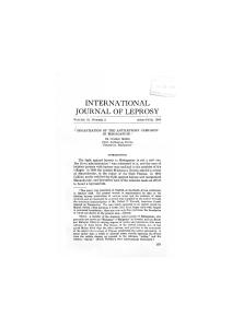
this mycobacterium, which is accompanied by a high (innate) cellular immune reactivity against M. leprae (13). In addition, it is apparent from the immunological and clinical leprosy spectrum in those who progress to the disease despite there being minimal genetic variation between M. leprae isolates reported. This spectrum ranges from tuberculoid (TT) leprosy to lepromatous (LL) leprosy (14) (see Chapter 2.1). TT patients in general show high cellular responses to M. leprae antigens injected in the skin as well as in in vitro T cell assays (15, 16); have low antibody titers to M. leprae antigens; and develop localized granulomatous disease with often few, if any, detectable bacilli in their lesions. At the opposite pole of the spectrum are LL patients unable to generate M. leprae specific Th1 cell responses, instead producing high IgM titers to M. leprae PGL-I with resulting disseminating, progressive infections. The borderline states of leprosy (BL, BB, and BT) are positioned in between and are rather unstable immunologically and prone to the occurrence of leprosy reactions, which are divided into type 1/reversal reactions (RR) and type 2/ erythema nodosum leprosum (ENL) (see Chapter 2.2). The different outcomes of infection with leprosy are most likely caused by host defense mechanisms, which are dominated by the innate and adaptive immune responses (10, 11, 17) (see Chapter 6.2; Chapter 6.3). After recognition of the bacteria by Toll-like receptors (TLRs), NF-kB is activated, resulting in the upregulation of pro-inflammatory cytokines (GM-CSF, IL-1β, TNF-α, IP-10, IL-12) and chemokines inducing the migration and activation of antigen presenting cells (APCs) such as macrophages. Subsequently, APCs migrate to the lymphoid organs in order to present mycobacterial antigens to naïve T cells. Depending on different co-stimulatory or inhibitory molecules and cytokines, different T cells will develop, which may vary from CD4+ Th1 or Th2, CTL, or Th17 cells. Finally, regulatory T cells (Treg), of which the first human variants were described in leprosy (18, 19), are believed to play a role in the M. leprae-specific unresponsiveness encountered in LL patients (20, 21) (see Figure 1). Host resistance to M. leprae is associated with the emergence of a protective Th1-based response dominated by CD4+ Th1 cells and characterized by the secretion of pro-inflammatory cytokines (17). Th1 cells produce IFN-γ and TNF-α, which synergize to activate microbicidal effector mechanisms in human macrophages. In LL and TT leprosy patients, imbalances in cytokine homeostasis in response to M. leprae have been reported (22). LL patients secrete predominantly anti-inflammatory mediators such as IL-10, accompanied by the absence of IFN-γ and other Th1-associated cytokines, in response to M. leprae antigens and mostly lack CD4+ T cells in their lesions. Instead, Th2 T cells producing IL-4, IL-5, and IL-13 dominate in these patients, leading to the production of antibodies as well as IL-4-induced dysregulation of host defenses against M. leprae (23). Conversely, CD4+ Th1 cells predominate in lesions of TT leprosy and these patients produce exacerbated levels of pro-inflammatory cytokines (IFN-γ, IL-15). The cytokines include those produced by Th17 rather than Th1, possibly driven by strong innate immune activation resulting in the release of IL-1β and/or IL-6, TGF-β, and IL-23, all of which are involved in Th17 (24) induction. Since the outcome of the immune response to M. leprae is determined by chemokines and cytokines that act as molecular signals for communication between the cells of the immune system, they are informative tools to predict either protection from or progression to the disease.
Section 7
Diagnostics
3
The International Textbook of Leprosy
FIG 1 Schematic representation of T cells (possibly) involved in the leprosy disease spectrum.
Immunodiagnostics SEROLOGY-BASED The diagnosis and classification of leprosy is largely based on clinical assessment, e.g., hypo-pigmented, anaesthetic skin patches and enlarged affected nerves, and on microscopic detection of acid-fast bacilli in skin slit smears or biopsies of suspected cases (see Chapter 2.1; Chapter 1.1). However, none of these diagnostic approaches are able to detect asymptomatic M. leprae infections. Besides the clinical approach, there are also diagnostic techniques that are based on immunological responses directed against M. leprae. In particular, several lateral flow-, dipstick-, and particle agglutination tests that incorporate the synthetic di- or trisaccharide epitope of phenolic glycolipid-I (PGL-I) have been used in field-based studies (25, 26, 27). The anti-PGL-I antibody (Ab) ELISA test is based on the detection of IgM antibodies against PGL-I, an M. leprae specific, dominant glycolipid component of the cell wall (see Chapter 5.1). Although this test is useful for 4
Part II
Basic Sciences
Immunodiagnostics
the detection of most multibacillary (MB) patients (28, 29), as the antibody levels correlate well with the bacillary load, the detection of anti-PGL-I Ab has limited value in identifying PB leprosy patients (30). Moreover, in areas hyper-endemic for leprosy, more than 50% of young schoolchildren surveyed had positive anti-PGL-I responses (31). Still, the vast majority of individuals with a positive antibody titer will never develop leprosy (25), despite the fact that the presence of anti-PGL-I antibodies was determined to be a risk factor for the development of leprosy (32, 33). In addition to IgM antibodies directed against M. leprae antigens, IgG antibodies against a fusion protein of M. leprae antigens ML0405 and ML2331 designated leprosy IDRI Diagnostic (LID)-1 have been studied (34, 35, 36, 37). Antibody responses towards LID-1 have been shown to be positive in over 95% of those at the lepromatous end of the spectrum (38). In a prospective study involving household contacts of MB patients whose serum antibody responses were analyzed retrospectively to four years prior to the clinical diagnosis of MB leprosy, 7 of 11 patients showed an IgG antibody response to LID-1 up to one year prior to developing clinical symptoms. These responses were strikingly more elevated and occurred much earlier than the increases in the anti-PGL-I IgM response in these same individuals (36). Nevertheless, the responses to LID-1 in PB individuals were rather weak, similar to the percentage of anti-PGL-I positivity, which is usually around 20–40% (36). Serological responses based on IgG Ab against M. leprae major membrane protein (MMP)-II provided slightly less diagnostic potential than IgM Ab against PGL-I for MB patients (88.1% vs. 94.9%), as estimated in a Chinese population (39). However, the number of PB patients testing positive increased significantly (61.1% vs. 38.9%) when using MMP-II. About a third of the leprosy patients’ contacts, 21 individuals, was positive for Ab against MMP-II, seven of whom developed leprosy in the following three years. However, healthy individuals from the same population were not included in this study. The use of the anti-PGL-I assay as a screening procedure to identify those at high risk of developing leprosy in endemic populations is unlikely to be particularly useful in most leprosy control programs, since in previous follow-up studies most of the new cases emerged from the seronegative group in the population (40). The assay could, however, be applied for the epidemiological monitoring of changes in the intensity of infection with M. leprae in a community and for the study of carefully defined groups of contacts during some phases of control programs (41, 42). More for information about the disease than for identification, anti-PGL-I antibody titers can be utilized to monitor efficient treatment by following the antibody clearance in patients. Similarly, following increases in titers may detect treatment failure or relapsing disease (43, 44, 45, 46, 47, 48, 49).
CELLULAR-MEDIATED IMMUNE RESPONSES An alternative test based on immunity to M. leprae antigens, the Mitsuda skin test, measures cellular rather than humoral immunity against lepromin (50, 51). Lepromin consists of a bacillary suspension standardized by the number of M. leprae in the suspension. The reaction to lepromin is measured as induration in millimeters 3–4 weeks after intradermal inoculation and provides
Section 7
Diagnostics
5
The International Textbook of Leprosy
information about the ability of an individual’s T cells to respond to M. leprae and the likelihood of granuloma formation in that individual. In contrast to the tuberculin skin test (TST), which measures cellular immunity to tuberculin (or PPD) and indicates previous infection with M. tuberculosis, the lepromin test does not detect prior M. leprae infection. Rather, its use lies in the determination of the type of immune response (as estimated by tissue injury) that would ensue upon infection with M. leprae. A negative Mitsuda reaction is generally seen in LL/BL patients, indicating the lack of a protective cellular response (52, 53). Leprosin, or Rees’s antigen, consists of proteins extracted from M. leprae and has been used as a 48-hour skin test to determine the cell mediated immune status of individuals (54). In that respect, tests based on the use of leprosin bear a similarity to the TST. Since leprosin and tuberculin are incompletely defined mixtures of mycobacterial antigens, they are of limited diagnostic value due to their inherently high cross-reactivity with other mycobacteria, which results in a test with low specificity. For leprosy, such cross-reactivity is particularly problematic in countries with high incidence rates of TB, routine BCG vaccination, and high levels of exposure to non-pathogenic environmental mycobacteria. One possible approach to avoiding cross-reactivity is the development of tests using M. leprae antigens that can induce cellular immune responses specific to the M. leprae infection.
Immunodiagnostic Tools M. LEPRAE ANTIGENS To develop field-friendly assays based on cellular mediated immune (CMI) responses, an approach similar to that used for the commercially available IFN-γ release assays (IGRAs) for specific diagnosis of M. tuberculosis infection (55, 56) was used for leprosy diagnosis. Since the QuantiferonTB assays exploit M. tuberculosis-specific antigens ESAT-6 (Rv3875), CFP-10 (Rv3874), and TB7.7 (Rv2654), the M. leprae homologs of ESAT-6 and CFP-10 (ML0049 and ML0050) were assayed as leprosy-specific diagnostic tools. Despite limited sequence homology (36% and 40%, respectively) and no cross-reactivity at the serological level (57, 58), the M. leprae homologues of ESAT6 and CFP-10 were recognized by T cells from M. tuberculosis infected individuals. Accordingly, cross-reactions with these assays limit the diagnostic potential of ESAT-6 and CFP-10 in leprosy endemic areas with a high prevalence of tuberculosis (59, 60). Similar data were reported for the M. leprae-specific 45 kDa serine rich antigen (ML0411), which was also recognized by TB patients, probably due to sequence homology with Rv2108 (61). The completion of the M. leprae and M. tuberculosis genome sequences (62, 63) allowed postgenomic approaches in which comparative analyses of annotated mycobacterial genomes were used to select putative open reading frames that were found only in the M. leprae genome and lacked homologues in any of the mycobacterial databases available at that time (61, 64, 65, 66, 6
Part II
Basic Sciences
Immunodiagnostics
67, 68, 69, 70, 71, 72, 73). Bioinformatic analyses of these M. leprae-unique sequences identified several (hypothetical) antigens that were subsequently analyzed for their ability to induce in vitro T cell responses in M. leprae infected individuals, specifically. Since IFN-γ is a stable cytokine, it has been widely used as a surrogate marker for pro-inflammatory immunity against mycobacteria (74). More recently, IFN-γ induced protein 10 (IP-10) has been shown to be a useful biomarker for diagnosis of the M. tuberculosis infection as well (75). Unlike IFN-γ, IP-10 also can be used in HIV infected patients, since it is not affected by low CD4 counts in TB patients with HIV (76). In vitro T cell stimulation assays predominantly assessing IFN-γ production were used to test the potential candidate immunodiagnostic antigens identified by the bioinformatic analysis. The antigens were tested for their potential as diagnostic tools for humans (65, 68, 69, 70, 71, 73, 77) as well as in animal models for leprosy in mice and armadillos (78, 79). Surprisingly, despite being selected for their exclusive presence in M. leprae, so as to avoid T cell cross-reactivity with BCG-vaccinated or M. tuberculosis infected individuals, it was found that most of these M. lepraeunique proteins induced IFN-γ in healthy control individuals (EC) from the same leprosy-endemic areas (70, 73, 80). However, since these EC were living in areas with pockets of high leprosy prevalence, the observed cellular responses towards the M. leprae- unique proteins may have indicated M. leprae-specificity but could be irrelevant to the pathogenic cellular immunity that leads to leprosy. In contrast, the production of IFN-γ as well as IP-10 in response to M. lepraeunique proteins or peptides was able to differentiate between EC groups drawn from areas with different levels of leprosy prevalence. This latter approach allowed the identification of distinct degrees of M. leprae exposure and, thereby, estimations of the risk of infection and, potentially, subsequent transmission (80, 81, 82, 83).
EXPOSURE, INFECTION, AND DISEASE As stated above, discriminatory IFN-γ and IP-10 profiles are observed between different types of leprosy (69, 70, 71) and between EChigh and EClow (80, 81). Regardless, no M. leprae proteins have been identified that can distinguish TT/BT patients from EC or HHC based on IFN-γ or IP-10 production when both groups are drawn from the same leprosy hyper-endemic area and have the identical socioeconomic status. For efficient diagnosis, it is thus imperative to identify new host markers (immunological- or genetic biomarkers) that can be used to discriminate between M. leprae exposure, M. leprae infection, and disease. This goal requires a clear consensus with respect to the different stages that can occur after encountering M. leprae. In general, exposure is defined as the contact of a potential host with a harmful agent, colonization as the invasion of the host by germs without signs or symptoms, and infection as the invasion of a host’s bodily tissues by a disease-causing organism. Infection generally results in the multiplication of the invading germ and the reaction of host tissues to these organisms. Infection may lead to disease, in which case an abnormal, pathological condition that affects part or all of
Section 7
Diagnostics
7
The International Textbook of Leprosy
the host can be observed. Subsequently, the types of immunity associated with these stages (i.e., stage-specific biomarkers) need to be unambiguously identified (see Figure 2).
FIG 2 Schematic overview of the stages of M. leprae infection and their respective biomarker profiles, modified for biomarkers and for leprosy from (127). If the host is capable of eliminating M. leprae without the priming of adaptive immunity (T cells or B-cells), the cytokines and chemokines measured in the blood reflect innate immune responses (stage 1) caused by, for example, natural killer cells, macrophages, or neutrophils. If a combination of specific T cells and innate immune cells cause elimination of M. leprae bacilli, additional blood biomarkers for adaptive immunity may be found in the blood as well as tissues (stage 2). Alternatively, if M. leprae is replicating and invading the host without causing clinical symptoms, biomarkers for early infection need to be identified (stage 3). These biomarkers will be both innate and adaptive, in which the latter differ in quality or quantity from those detected in stage 2, where they lead to clearance of infection. Finally, when M. leprae is disseminating throughout the host’s body (BL/LL) or causing harmful T-cell responses resulting in lesions (TT/BT), pathogenic immune responses predominate and the chronic battlefield between host and pathogen ends in favor of M. leprae. At this stage, the analysis of host blood biomarkers represents the result of pathogenic as well as protective immune responses that are present in leprosy patients (stage 4), a consequence of either vigorous T cell responses in TT/BT or suppressive T cells in LL/ BL patients (19, 22). In stage 4, similar to stages 2 and 3, biomarkers produced by innate immune responses will also be present, as is the case in TB (84, 85). Disease-specific biomarkers likely can be deduced by the comparison of cellular immune markers, such as cytokines and chemokines, but also by genetic markers (86) in leprosy patients, their contacts, and endemic and nonendemic healthy individuals. Biomarker studies including leprosy patients and HHC, but lacking different EC groups (87), are less likely to identify markers for protective innate immunity (stage 1) such 8
Part II
Basic Sciences
Immunodiagnostics
as chemokines secreted by activated macrophages (88, 89). These studies are unlikely to identify markers since the HHC are frequently and intensely exposed to M. leprae and, therefore, produce similar cytokines and chemokines as those found for TT/BT patients (80, 87). Finally, infections with HIV (90, 91, 92, 93, 94), Th2 inducing parasites (95), and also diabetes (96) will produce biomarker profiles of M. leprae-affected individuals, thereby complicating diagnosis. The differential effects of co-infections on host biomarkers for an M. leprae infection and leprosy will require more elaborate studies as well.
HOST PROFILES Although IFN-γ production is used as a surrogate marker of Th1-induced protection, it remains uncertain whether the production of this cytokine in response to M. leprae antigens correlates with either protection against infection or (progression to) disease. Therefore, other analytes, measurable in serum such as cytokines and chemokines, need to be investigated as potential biomarkers. For example, using whole blood assays, IL-1β, CCL4, and MCP-1 could, in contrast to IFN-γ, discriminate between patients (leprosy and TB) and healthy EC living in the same endemic areas in Bangladesh and possibly reflect differences between M. leprae exposure and pathogenic immunity (80). Despite the obvious differences between leprosy patients and HHC on one hand and EC on the other hand, no significant differences were observed between HHC and BT/TT. While some HHC displayed biomarker profiles similar to patients, others seemed to be more like EC. To identify biomarkers in HHC that are specific for (pre-clinical) disease, longitudinal analyses need to be conducted allowing the intra-individual comparison of immune profiles of the cyto-/chemokine responses. Given that host immunity and immuno-pathogenicity in response to M. leprae involves complex interactions between a variety of cells expressing different effector and regulatory molecules, it is rather unlikely that only a single cytokine or chemokine is linearly correlated to protection or to disease. In view of this low likelihood, it is essential to increase the biomarker potential of single cytokines or other markers by using specific combinations of them, also referred to as a biomarker profile. In such a profile, all single markers may not be able to diagnose each patient but, if selected in such a way that each marker is independent of the others in the profile, better performance is expected (97). Since cytokines modulate each other’s effects, their ratios can in fact be more informative than single cytokine values in order to discriminate between different stages of mycobacterial infection. For example, the IFN-γ/ IL-10 ratio has been found to correlate with tuberculosis (TB) severity (98) and combinations of cytokine responses are indicative of active versus latent TB (99). For leprosy, IP-10/IL-10 ratios in unstimulated plasma differed significantly between patients and EC in a small Ethiopian cohort (100), indicating the feasibility of identifying M. leprae infection in endemic areas. Moreover, the onset of type 1 leprosy reactions is associated with an increase in
Section 7
Diagnostics
9
The International Textbook of Leprosy
ratios of pro-inflammatory cytokines versus IL-10 (7), indicating the potential of measuring cytokine ratios for these reactions as well. In summary, since immunity against M. leprae matches the clinical manifestations after infection, it is essential to perform longitudinal studies comparing these immunological relationships. Since the majority of those exposed to M. leprae develop a protective immune response against the bacterium, such follow-up studies need to be large-scale efforts to allow identification of critical host-derived immune biomarker profiles as risk factors in exposed populations in leprosyendemic areas.
TRANSCRIPTOMIC HOST PROFILES Besides host immune profiles, human transcriptome-arrays offer cutting-edge tools for identifying gene expression profiles for leprosy (101). Using a transcriptomic assay dual color Reverse Transcription Multiplex Ligation-dependent Probe Amplification (dcRT-MLPA), a biomarker profile composed of several genes was identified for infection in a follow-up study of TB contacts (102). This dcRT-MLPA platform was also used to identify genetic markers associated with leprosy reactions (12). RNA expression profiles revealed that IFN-induced genes, (V)EGF, and genes associated with cytotoxic T-cell responses (GNLY, GZMA/B, PRF1) were upregulated during T1R, whereas expression of T-cell regulation-associated genes were decreased. Recently in an African TB cohort (South Africa and Malawi) including HIV+ individuals, a 27-gene transcript signature was identified that discriminated between TB patients and latently M. tuberculosis-infected individuals (LTBI). Moreover, the same study showed that a transcript signature including 44 genes could specifically distinguish TB patients from patients with other respiratory diseases (103). These data show the promise of transcriptomic host profiles for diagnosis as well. Combined with functional protein association networks, transcriptomic expression profiles can be used to identify proteins discernible in blood for application as biomarkers in field-friendly assays.
METABOLIC HOST PROFILES Comprehensive approaches to identifying metabolic variations associated with disease are rapidly evolving. The field of metabolomics aims to characterize the concentration (changes) of small molecules occurring in bio-fluids and has contributed to knowledge of various diseases as well as biomarkers of disease (9, 10). As key experimental technologies, mass spectrometry and nuclear magnetic resonance allow the assessment of a cell’s or tissue’s metabolic activity or state, which is subject to environmental stresses. Although this field remains largely unexamined for leprosy, some recent reports showed the utility of metabolomics for the identification of potential contributors to disease pathology in the sera of leprosy patients (104, 105). Thus, the identification of metabolic host profiles may facilitate the development of more targeted treatments for leprosy patients and their contacts. 10
Part II
Basic Sciences
Immunodiagnostics
HOST BIOMARKERS FOR LEPROSY REACTIONS Leprosy often coincides with acute, inflammatory episodes due to augmented anti-mycobacterial host immunity. These reactions represent the major cause of leprosy-related permanent neuropathy and disability. Up to 50% of leprosy patients experience a leprosy reaction at least once, yet no laboratory test is available that allows early diagnosis and treatment that would help prevent nerve damage. Two types of skin- and nerve-damaging reactions are recognized: type 1 reversal reactions (T1Rs) and type 2 reactions (T2Rs), or ENL (see Chapter 2.2). T1Rs are considered a delayed hypersensitivity reaction with the characteristic infiltrations of skin and nerve lesions by CD4+ T-cells producing IFN-γ and TNF-α (106). When patients receive prompt and proper diagnosis and treatment, recovery from inflammatory nerve damage is more likely and the risks of permanent disability are significantly reduced (107). Unfortunately, in endemic contexts, leprosy reactions are frequently misdiagnosed due to a decrease in specialized expertise within integrated health services, and patients can experience significant delays in diagnosis (108). Consequently, if diagnosis and treatment is delayed beyond six months of the first symptoms, the neuropathy is likely to be permanent (109). Therefore, reliable, field-compatible or laboratory tests for the early detection or prediction of leprosy reactions could make significant differences in clinical outcomes. A major obstacle to developing such tests is the lack of dependable, specific biomarkers for leprosy reactions across endemic populations. Substantial evidence points to increased numbers of CD4+ T-cells in skin lesions; high levels of IL-2 receptors, TNF-α, IL-6, IP-10, and IL17F in sera; and increased gene expression of pro-inflammatory cytokines during reactions (110, 111, 112, 113, 114). Consequently, it is conceivable that cytokines, chemokines, antibodies, and also metabolites, which can be measured in body fluids, may prognosticate reactional episodes. Improved knowledge of the relevant biomarker profiles, especially those that are specifically induced by unique M. leprae antigens, will help to accurately identify patients who are developing leprosy reactions.
Immunodiagnostic tests Due to changes in leprosy control programs and the decrease in the specialized expertise required for the early and accurate diagnosis of leprosy, the need for rapid tests that can be applied in nonexpert settings to detect asymptomatic M. leprae infection or predict progression to leprosy may be greater than ever before. A novel rapid diagnostic test, the NDO-LID® test (115), was recently developed for the diagnosis of MB leprosy based on the complementary detection of antibodies against a novel proteinglycolipid conjugate. NDO-LID® is an immunochromatographic test that requires small amounts of serum or whole blood. Like the other serological tests for leprosy, it detects MB patients and, potentially, HHC at a higher risk of developing MB leprosy.
Section 7
Diagnostics
11
The International Textbook of Leprosy
The characteristics of the leprosy disease spectrum, in which the outcome of an M. leprae infection ranges from the strong Th1 immunity in tuberculoid leprosy to the high antibody titers to M. leprae with Th2 cytokine responses in lepromatous leprosy, are pre-eminently suitable for tests that simultaneously detect biomarkers specific for both types of immune responses. Tests of this nature could provide thorough monitoring of the complete immunological leprosy spectrum. Although ELISA techniques, as used in IGRAs, are more widely applied than before, they still require laboratory facilities that are not available at all health centers in leprosy-endemic areas. Lateral flow assays (LFAs) are simple, immunochromatographic assays that detect the presence of target analytes in samples without the need for specialized and costly equipment. Combinations of LFAs with up-converting phosphor (UCP) reporter technology are useful for the detection of a variety of analytes, e.g., drugs of abuse (116); protein and polysaccharide antigens from pathogens like Schistosoma and Brucella (117, 118); bacterial and viral nucleic acids (119, 120); and antibodies against M. tuberculosis, HIV, hepatitis virus, and Yersinia pestis (121, 122, 123). The phosphorescent reporter utilized in UCP-LFAs is excited with infrared light to generate visible light, a process called ‘up-conversion’. UCP-based assays are highly sensitive, since up-conversion does not occur in nature, avoiding interference by auto-fluorescence of other assay components. Importantly, UCP-LF test strips can be stored as permanent records, allowing for re-analysis in a reference laboratory. To accommodate ELISAs to field-applicable tests for leprosy and TB diagnosis or for the monitoring of vaccine and treatment efficacy, LF assays based on up-converting phosphor (UCP-LFAs) were developed for the detection of IFN-γ, IP-10 (Th1), and IL-10 (Treg) as well as antibodies against the M. leprae-specific PGL-I (100, 124, 125, 126). Simultaneous measurement of pro- and anti-inflammatory cellular as well as humoral immunity to M. leprae can be a useful tool in leprosy control programs for the classification of leprosy. It also can allow early diagnosis of leprosy or leprosy reactions, leading to timely treatment and reduced transmission. However, since there is no gold standard for detecting an asymptomatic M. leprae infection, biomarker signatures for the early detection of leprosy need to be identified in the near future.
Acknowledgements AG is supported by the Q.M. Gastmann Wichers Foundation, Netherlands Leprosy Relief Foundation (NLR) together with the Turing Foundation (ILEP#: 701.02.49), the Order of Malta-Grantsfor-Leprosy-Research (MALTALEP), and the EDCTP through a project entitled AE-TBC under Grant Agreement N° IP_09_32040. AG and AvH are supported by the Heiser Program for Research in Leprosy in The New York Community Trust (P13-000392)
12
Part II
Basic Sciences
Immunodiagnostics
References 1.
WHO. 2013. Global leprosy: update on the 2012 situation. Wkly Epidemiol Rec 88:365–379.
2.
Smith WC, Aerts A. 2014. Role of contact tracing and prevention strategies in the interruption of leprosy transmission. Lepr Rev 85:2–17.
3.
WHO. 2013. Global leprosy: update on the 2012 situation. Wkly Epidemiol Rec 88:365–379.
4.
Smith WC, van BW, Gillis T, Saunderson P, Richardus JH. 2015. The missing millions: a threat to the elimination of leprosy. PLoS Negl Trop Dis 9:e0003658.
5.
Siddiqui MR, Velidi NR, Pati S, Rath N, Kanungo AK, Bhanjadeo AK, Rao BB, Ojha BM, Krishna MK, Soutar D, Porter JD, Ranganadha Rao PV. 2009. Integration of leprosy elimination into primary health care in Orissa, India. PLoS One 4:e8351.
6.
Massone C, Clapasson A, Nunzi E. 2013. Borderline lepromatous leprosy in an Italian man. Am J Trop Med Hyg 88:211.
7.
Geluk A, van Meijgaarden KE, Wilson L, Bobosha K, van der Ploeg-van Schip JJ, van den Eeden SJ, Quinten E, Dijkman K, Franken KL, Haisma EM, Haks MC, van Hees CL, Ottenhoff TH. 2014. Longitudinal immune responses and gene expression profiles in type 1 leprosy reactions. J Clin Immunol 34:245–255.
8.
Nolen L, Haberling D, Scollard D, Truman R, Rodriguez-Lainz A, Blum L, Blaney D. 2014. Incidence of Hansen’s Disease – United States, 1994–2011. MMWR Morb Mortal Wkly Rep 63:969–972.
9.
Vermazeren-van Roij J, Brusse E, Koljenovic S, van Hees CL. 2015. Huidafwijkingen en polyneuropathie: denk aan lepra. Ned Tijd Geneeskd 159:A9026.
10. Scollard DM, Adams LB, Gillis TP, Krahenbuhl JL, Truman RW, Williams DL. 2006. The continuing challenges of leprosy. Clin Microbiol Rev 19:338–381. 11. Rodrigues LC, Lockwood DN. 2011. Leprosy now: epidemiology, progress, challenges, and research gaps. Lancet Infect Dis 11:464–470. 12. Geluk, 2013. Biomarkers for leprosy: would you prefer T (cells)? Lepr Rev 84:3–12. 13. Montoya DA, Modlin RL. 2010. Learning from leprosy: insight into the human innate immune response. Adv Immunol 105:1–24.
Section 7
Diagnostics
13
The International Textbook of Leprosy
14. Ridley DS, Jopling WH. 1966. Classification of leprosy according to immunity. A fivegroup system. Int J Lepr Other Mycobact Dis 34:255–273. 15. Kaplan G, Cohn ZA. 1991. Leprosy and cell-mediated immunity. Curr Opin Immunol 3:91–96. 16. Modlin RL, Melancon-Kaplan J, Young SM, Pirmez C, Kino H, Convit J, Rea TH, Bloom BR. 1988. Learning from lesions: patterns of tissue inflammation in leprosy. Proc Natl Acad Sci U S A 85:1213–1217. 17. Ottenhoff, TH. 2012. New pathways of protective and pathological host defense to mycobacteria. Trends Microbiol 20:419–428. 18. Modlin RL, Kato H, Mehra V, Nelson EE, Fan XD, Rea TH, Pattengale PK, Bloom BR. 1986. Genetically restricted suppressor T-cell clones derived from lepromatous leprosy lesions. Nature 322:459–461. 19. Ottenhoff TH, Haanen JB, Geluk A, Mutis T, Ab BK, Thole JE, van Schooten WC, van den Elsen PJ, de Vries RR. 1991. Regulation of mycobacterial heat-shock protein-reactive T cells by HLA class II molecules: lessons from leprosy. Immunol Rev 121:171– 191. 20. Palermo ML, Pagliari C, Trindade MA, Yamashitafuji TM, Duarte AJ, Cacere CR, Benard G. 2012. Increased expression of regulatory T cells and down-regulatory molecules in lepromatous leprosy. Am J Trop Med Hyg 86:878–883. 21. Bobosha K, Wilson L, van Meijgaarden KE, Bekele Y, Zewdie M, van der Ploeg-van Schip JJ, Abebe M, Hussein J, Khadge S, Neupane KD, Hagge DA, Jordanova ES, Aseffa A, Ottenhoff TH, Geluk A. 2014. T-cell regulation in lepromatous leprosy. PLoS Negl Trop Dis 8:e2773. 22. Montoya D, Cruz D, Teles RM, Lee DJ, Ochoa MT, Krutzik SR, Chun R, Schenk M, Zhang X, Ferguson BG, Burdick AE, Sarno EN, Rea TH, Hewison M, Adams JS, Cheng G, Modlin RL. 2009. Divergence of macrophage phagocytic and antimicrobial programs in leprosy. Cell Host Microbe 6:343–353. 23. Yamamura M, Uyemura K, Deans RJ, Weinberg K, Rea TH, Bloom BR, Modlin RL. 1991. Defining protective responses to pathogens: cytokine profiles in leprosy lesions. Science 254:277–279. 24. Chaitanya S, Lavania M, Turankar RP, Karri SR, Sengupta U. 2012. Increased serum circulatory levels of interleukin 17F in type 1 reactions of leprosy. J Clin Immunol 32:1415–1420.
14
Part II
Basic Sciences
Immunodiagnostics
25. Spencer JS, Kim HJ, Wheat WH, Chatterjee D, Balagon MV, Cellona RV, Tan EV, Gelber R, Saunderson P, Duthie MS, Reece ST, Burman W, Belknap R, Mac Kenzie WR, Geluk A, Oskam L, Dockrell HM, Brennan PJ. 2011. Analysis of antibody responses to Mycobacterium leprae phenolic glycolipid I, lipoarabinomannan, and recombinant proteins to define disease subtype-specific antigenic profiles in leprosy. Clin Vaccine Immunol 18:260–267. 26. Oskam L, Slim E, Buhrer-Sekula S. 2003. Serology: recent developments, strengths, limitations and prospects: a state of the art overview. Lepr Rev 74:196–205. 27. Stefani MM, Grassi AB, Sampaio LH, Sousa AL, Costa MB, Scheelbeek P, Neupane KD, Hagge DA, Macdonald M, Cho SN, Oskam L, Buhrer-Sekula S. 2012. Comparison of two rapid tests for anti-phenolic glycolipid-I serology in Brazil and Nepal. Mem Inst Oswaldo Cruz 107(Suppl 1):124–131. 28. Buhrer-Sekula S, Smits HL, Gussenhoven GC, van LJ, Amador S, Fujiwara T, Klatser PR, Oskam L. 2003. Simple and fast lateral flow test for classification of leprosy patients and identification of contacts with high risk of developing leprosy. J Clin Microbiol 41:1991–1995. 29. Goulart IM, Bernardes Souza DO, Marques CR, Pimenta VL, Goncalves MA, Goulart LR. 2008. Risk and protective factors for leprosy development determined by epidemiological surveillance of household contacts. Clin Vaccine Immunol 15:101–105. 30. Spencer JS, Duthie MS, Geluk A, Balagon MF, Kim HJ, Wheat WH, Chatterjee D, Jackson M, Li W, Kurihara JN, Maghanoy A, Mallari I, Saunderson P, Brennan PJ, Dockrell HM. 2012. Identification of serological biomarkers of infection, disease progression and treatment efficacy for leprosy. Mem Inst Oswaldo Cruz 107(Suppl 1):79–89. 31. Barreto JG, Guimaraes LS, Frade MA, Rosa PS, Salgado CG. 2012. High rates of undiagnosed leprosy and subclinical infection amongst school children in the Amazon Region. Mem Inst Oswaldo Cruz 107(Suppl 1):60–67. 32. Beers, vSM., Hatta M, Klatser PR. 1999. Seroprevalence rates of antibodies to phenolic glycolipid-I among school children as an indicator of leprosy endemicity. Int J Lepr Other Mycobact Dis 67:243–249. 33. Douglas JT, Cellona RV, Fajardo TT Jr, Abalos RM, Balagon MV, Klatser PR. 2004. Prospective study of serological conversion as a risk factor for development of leprosy among household contacts. Clin Diagn Lab Immunol 11:897–900. 34. Duthie MS, Hay MN, Morales CZ, Carter L, Mohamath R, Ito L, Oyafuso LK, Manini MI, Balagon MV, Tan EV, Saunderson PR, Reed SG, Carter D. 2010. Rational design and evaluation of a multiepitope chimeric fusion protein with the potential for leprosy diagnosis. Clin Vaccine Immunol 17:298–303.
Section 7
Diagnostics
15
The International Textbook of Leprosy
35. Sampaio LH, Stefani MM, Oliveira RM, Sousa AL, Ireton GC, Reed SG, Duthie MS. 2011. Immunologically reactive M. leprae antigens with relevance to diagnosis and vaccine development. BMC Infect Dis 11:26. 36. Duthie MS, Goto W, Ireton GC, Reece ST, Cardoso LP, Martelli CM, Stefani MM, Nakatani M, de Jesus RC, Netto EM, Balagon MV, Tan E, Gelber RH, Maeda Y, Makino M, Hoft D, Reed SG. 2007. Use of protein antigens for early serological diagnosis of leprosy. Clin Vaccine Immunol 14:1400–1408. 37. Reece ST, Ireton G, Mohamath R, Guderian J, Goto W, Gelber R, Groathouse N, Spencer J, Brennan P, Reed SG. 2006. ML0405 and ML2331 are antigens of Mycobacterium leprae with potential for diagnosis of leprosy. Clin Vaccine Immunol 13:333– 340. 38. Duthie MS, Hay MN, Rada EM, Convit J, Ito L, Oyafuso LK, Manini MI, Goulart IM, Lobato J, Goulart LR, Carter D, Reed SG. 2011. Specific IgG antibody responses may be used to monitor leprosy treatment efficacy and as recurrence prognostic markers. Eur J Clin Microbiol Infect Dis 30(10):1257–1265. 39. Wang H, Liu W, Jin Y, Yu M, Jiang H, Tamura T, Maeda Y, Makino M. 2015. Detection of antibodies to both M. leprae PGL-I and MMP-II to recognize leprosy patients at an early stage of disease progression. Diagn Microbiol Infect Dis 83:274–277. 40. Sinha S, Kannan S, Nagaraju B, Sengupta U, Gupte MD. 2004. Utility of serodiagnostic tests for leprosy: a study in an endemic population in South India. Lepr Rev 75:266–273. 41. Ulrich M, Smith PG, Sampson C, Zuniga M, Centeno M, Garcia V, Manrique X, Salgado A, Convit J. 1991. IgM antibodies to native phenolic glycolipid-I in contacts of leprosy patients in Venezuela: epidemiological observations and a prospective study of the risk of leprosy. Int J Lepr Other Mycobact Dis 59:405–415. 42. Soebono H, Klatser PR. 1991. A seroepidemiological study of leprosy in high- and low-endemic Indonesian villages. Int J Lepr Other Mycobact Dis 59:416–425. 43. Prakash K, Sehgal VN, Aggarwal R. 1993. Evaluation of phenolic glycolipid-I (PGL-I) antibody as a multidrug therapy (MDT) monitor. J Dermatol 20:16–20. 44. Roche PW, Britton WJ, Failbus SS, Neupane KD, Theuvenet WJ. 1993. Serological monitoring of the response to chemotherapy in leprosy patients. Int J Lepr Other Mycobact Dis 61:35–43. 45. Cho SN, Cellona RV, Fajardo TT Jr, Abalos RM, la Cruz EC, Walsh GP, Kim JD, Brennan PJ. 1991. Detection of phenolic glycolipid-I antigen and antibody in sera from new and
16
Part II
Basic Sciences
Immunodiagnostics
relapsed lepromatous patients treated with various drug regimens. Int J Lepr Other Mycobact Dis 59:25–31. 46. Rada E, Ulrich M, Aranzazu N, Rodriguez V, Centeno M, Gonzalez I, Santaella C, Rodriguez M, Convit J. 1997. A follow-up study of multibacillary Hansen’s disease patients treated with multidrug therapy (MDT) or MDT + immunotherapy (IMT). Int J Lepr Other Mycobact Dis 65:320–327. 47. Silva EA, Iyer A, Ura S, Lauris JR, Naafs B, Das PK, Vilani-Moreno F. 2007. Utility of measuring serum levels of anti-PGL-I antibody, neopterin and C-reactive protein in monitoring leprosy patients during multi-drug treatment and reactions. Trop Med Int Health 12:1450–1458. 48. Silva RC, Lyon S, Araos R, Lyon AC, Grossi MA, Lyon SH, Penido RA, Buhrer-Sekula S, Antunes CM. 2008. The result patterns of ML Flow and ELISA (PGL-I) serologic tests in leprosy-endemic and non-endemic areas. Rev Soc Bras Med Trop 41(Suppl 2):19–22. 49. Khadge S, Banu S, Bobosha K, van der Ploeg-van Schip JJ, Goulart IM, Thapa P, Kunwar CB, van Meijgaarden KE, van den Eeden SJ, Wilson L, Kabir S, Dey H, Goulart LR, Lobato J, Carvalho W, Bekele Y, Franken KL, Aseffa A, Spencer JS, Oskam L, Ottenhoff TH, Hagge DA, Geluk A. 2015. Longitudinal immune profiles in type 1 leprosy reactions in Bangladesh, Brazil, Ethiopia and Nepal. BMC Infect Dis 15:477. 50. Roberts PP, Dockrell HM, McAdam KP. 1988. Evidence that the Mitsuda reaction to Mycobacterium leprae can be mediated by lymphocytes responsive to Mycobacterium tuberculosis. Clin Exp Immunol 72:390–393. 51. Maeda SM, Rotta O, Michalany NS, Camargo ZP, Sunderkotter C, Tomimori-Yamashita J. 2003. Comparison between anti-PGL-I serology and Mitsuda reaction: clinical reading, microscopic findings and immunohistochemical analysis. Lepr Rev 74:263– 274. 52. Hanna, 1960. Lepromin. Its use in leprosy and preparation. J Egypt Med Assoc 43:81– 88. 53. Scollard DM LK, Adams LB, Gillis TP, Krahenbuhl JL, Truman RW, Williams DL. 2006. The continuing challenges of leprosy. Clin Microbiol Rev 19:338–381. 54. Smelt AH, Rees RJ, Liew FY. 1981. Induction of delayed-type hypersensitivity to Mycobacterium leprae in healthy individuals. Clin Exp Immunol 44:501–506. 55. Ferrara G, Losi M, D’Amico R, Roversi P, Piro R, Meacci M, Meccugni B, Dori IM, Andreani A, Bergamini BM, Mussini C, Rumpianesi F, Fabbri LM, Richeldi L. 2006. Use in routine clinical practice of two commercial blood tests for diagnosis of infection with Mycobacterium tuberculosis: a prospective study. Lancet 367:1328–1334.
Section 7
Diagnostics
17
The International Textbook of Leprosy
56. Pai M, Kalantri S, Dheda K. 2006. New tools and emerging technologies for the diagnosis of tuberculosis: part I. Latent tuberculosis. Expert Rev Mol Diagn 6:413–422. 57. Spencer JS, Marques MA, Lima MC, Junqueira-Kipnis AP, Gregory BC, Truman RW, Brennan PJ. 2002. Antigenic specificity of the Mycobacterium leprae homologue of ESAT-6. Infect Immun 70:1010–1013. 58. Spencer JS, Kim HJ, Marques AM, Gonzalez-Juarerro M, Lima MC, Vissa VD, Truman RW, Gennaro ML, Cho SN, Cole ST, Brennan PJ. 2004. Comparative analysis of B- and T-cell epitopes of Mycobacterium leprae and Mycobacterium tuberculosis culture filtrate protein 10. Infect Immun 72:3161–3170. 59. Geluk A, van Meijgaarden KE, Franken KL, Subronto YW, Wieles B, Arend SM, Sampaio EP, d Boer T, Faber WR, Naafs B, Ottenhoff TH. 2002. Identification and characterization of the ESAT-6 homologue of Mycobacterium leprae and T-cell cross-reactivity with Mycobacterium tuberculosis. Infect Immun 70:2544–2548. 60. Geluk A, van Meijgaarden KE, Franken KL, Wieles B, Arend SM, Faber WR, Naafs B, Ottenhoff TH. 2004. Immunological crossreactivity of the Mycobacterium leprae CFP10 with its homologue in Mycobacterium tuberculosis. Scand J Immunol 59:66–70. 61. Brahmbhatt S, Hussain R, Zafar S, Dawood G, Ottenhoff TH, Drijfhout JW, Bothamley G, Smith S, Lopez FV, Dockrell HM. 2002. Human T cell responses to peptides of the Mycobacterium leprae 45-kD serine-rich antigen. Clin Exp Immunol 128:140–148. 62. Cole ST, Eiglmeier K, Parkhill J, James KD, Thomson NR, Wheeler PR, Honore N, Garnier T, Churcher C, Harris D, Mungall K, Basham D, Brown D, Chillingworth T, Connor R, Davies RM, Devlin K, Duthoy S, Feltwell T, Fraser A, Hamlin N, Holroyd S, Hornsby T, Jagels K, Lacroix C, Maclean J, Moule S, Murphy L, Oliver K, Quail MA, Rajandream MA, Rutherford KM, Rutter S, Seeger K, Simon S, Simmonds M, Skelton J, Squares R, Squares S, Stevens K, Taylor K, Whitehead S, Woodward JR, Barrell BG. 2001. Massive gene decay in the leprosy bacillus. Nature 409:1007–1011. 63. Cole ST, Brosch R, Parkhill J, Garnier T, Churcher C, Harris D, Gordon SV, Eiglmeier K, Gas S, Barry CE, III, Tekaia F, Badcock K, Basham D, Brown D, Chillingworth T, Connor R, Davies R, Devlin K, Feltwell T, Gentles S, Hamlin N, Holroyd S, Hornsby T, Jagels K, Krogh A, McLean J, Moule S, Murphy L, Oliver K, Osborne J, Quail MA, Rajandream MA, Rogers J, Rutter S, Seeger K, Skelton J, Squares R, Squares S, Sulston JE, Taylor K, Whitehead S, Barrell BG. 1998. Deciphering the biology of Mycobacterium tuberculosis from the complete genome sequence. Nature 393:537–544. 64. Araoz R, Honore N, Cho S, Kim JP, Cho SN, Monot M, Demangel C, Brennan PJ, Cole ST. 2006. Antigen discovery: a postgenomic approach to leprosy diagnosis. Infect Immun 74:175–182.
18
Part II
Basic Sciences
Immunodiagnostics
65. Araoz R, Honore N, Banu S, Demangel C, Cissoko Y, Arama C, Uddin MK, Hadi SK, Monot M, Cho SN, Ji B, Brennan PJ, Sow S, Cole ST. 2006. Towards an immunodiagnostic test for leprosy. Microbes Infect 8:2270–2276. 66. Duthie MS, Ireton GC, Kanaujia GV, Goto W, Liang H, Bhatia A, Busceti JM, Macdonald M, Neupane KD, Ranjit C, Sapkota BR, Balagon M, Esfandiari J, Carter D, Reed SG. 2008. Selection of antigens and prototype test development for a point-of-care leprosy diagnosis. Clin Vaccine Immunol 15(10):1590–1597. 67. Duthie MS, Goto W, Ireton GC, Reece ST, Sampaio LH, Grassi AB, Sousa AL, Martelli CM, Stefani MM, Reed SG. 2008. Antigen-specific T-cell responses of leprosy patients. Clin Vaccine Immunol 15:1659–1665. 68. Dockrell HM, Brahmbhatt S, Robertson BD, Britton S, Fruth U, Gebre N, Hunegnaw M, Hussain R, Manandhar R, Murillo L, Pessolani MC, Roche P, Salgado JL, Sampaio E, Shahid F, Thole JE, Young DB. 2000. A postgenomic approach to identification of Mycobacterium leprae-specific peptides as T-cell reagents. Infect Immun 68:5846– 5855. 69. Spencer JS, Dockrell HM, Kim HJ, Marques MA, Williams DL, Martins MV, Martins ML, Lima MC, Sarno EN, Pereira GM, Matos H, Fonseca LS, Sampaio EP, Ottenhoff TH, Geluk A, Cho SN, Stoker NG, Cole ST, Brennan PJ, Pessolani MC. 2005. Identification of specific proteins and peptides in Mycobacterium leprae suitable for the selective diagnosis of leprosy. J Immunol 175:7930–7938. 70. Sampaio LH, Stefani MM, Oliveira RM, Sousa AL, Ireton GC, Reed SG, Duthie MS. 2011. Immunologically reactive M. leprae antigens with relevance to diagnosis and vaccine development. BMC Infect Dis 11:26. 71. Geluk A, Klein MR, Franken KL, van Meijgaarden KE, Wieles B, Pereira KC, BuhrerSekula S, Klatser PR, Brennan PJ, Spencer JS, Williams DL, Pessolani MC, Sampaio EP, Ottenhoff TH. 2005. Postgenomic approach to identify novel Mycobacterium leprae antigens with potential to improve immunodiagnosis of infection. Infect Immun 73:5636–5644. 72. Geluk A, Ploeg J, Teles RO, Franken KL, Prins C, Drijfhout JW, Sarno EN, Sampaio EP, Ottenhoff TH. 2008. Rational combination of peptides derived from different Mycobacterium leprae proteins improves sensitivity for immunodiagnosis of M. leprae infection. Clin Vaccine Immunol 15:522–533. 73. Geluk A, Spencer JS, Bobosha K, Pessolani MC, Pereira GM, Banu S, Honore N, Reece ST, Macdonald M, Sapkota BR, Ranjit C, Franken KL, Zewdie M, Aseffa A, Hussain R, Stefani MM, Cho SN, Oskam L, Brennan PJ, Dockrell HM. 2009. From genome-based in silico predictions to ex vivo verification of leprosy diagnosis. Clin Vaccine Immunol 16:352–359.
Section 7
Diagnostics
19
The International Textbook of Leprosy
74. Wallis RS, Pai M, Menzies D, Doherty TM, Walzl G, Perkins MD, Zumla A. 2010. Biomarkers and diagnostics for tuberculosis: progress, needs, and translation into practice. Lancet 375:1920–1937. 75. Ruhwald M, Dominguez J, Latorre I, Losi M, Richeldi L, Pasticci MB, Mazzolla R, Goletti D, Butera O, Bruchfeld J, Gaines H, Gerogianni I, Tuuminen T, Ferrara G, EugenOlsen J, Ravn P. 2011. A multicentre evaluation of the accuracy and performance of IP-10 for the diagnosis of infection with M. tuberculosis. Tuberculosis (Edinb) 91:260– 267. 76. Aabye MG, Ruhwald M, Praygod G, Jeremiah K, Faurholt-Jepsen M, Faurholt-Jepsen D, Range N, Friis H, Changalucha J, Andersen AB, Ravn P. 2010. Potential of interferon-gamma-inducible protein 10 in improving tuberculosis diagnosis in HIV-infected patients. Eur Respir J 36:1488–1490. 77. Geluk A, van der Ploeg-van Schip JJ, Teles RO, Franken KL, Prins C, Drijfhout JW, Sarno EN, Sampaio EP, Ottenhoff TH. 2008. Rational combination of peptides derived from different Mycobacterium leprae proteins improves sensitivity for immunodiagnosis of M. leprae infection. Clin Vaccine Immunol 15:522–533. 78. Lahiri R, Randhawa B, Franken KL, Duthie MS, Spencer JS, Geluk A, Krahenbuhl JL. 2011. Development of a mouse food pad model for detection of sub clinical leprosy. Lepr Rev 82:432–444. 79. Pena M, Geluk A, van der Ploeg-van Schip JJ, Franken KL, Sharma R, Truman R. 2011. Cytokine responses to Mycobacterium leprae unique proteins differentiate between Mycobacterium leprae infected and naive armadillos. Lepr Rev 82:422–431. 80. Geluk A, Bobosha K, van der Ploeg-van Schip JJ, Spencer JS, Banu S, Martins SB, Cho SN, Franken KL, Kim HJ, Bekele Y, Uddin MK, Abdul HS, Aseffa A, Pessolani MC, Pereira GM, Dockrell HM, Ottenhoff TH. 2012. New biomarkers with relevance to leprosy diagnosis applicable in areas hyperendemic for leprosy. J Immunol 188:4782– 4791. 81. Martins MV, Guimaraes MM, Spencer JS, Hacker MA, Costa LS, Carvalho FM, Geluk A, van der Ploeg-van Schip JJ, Pontes MA, Goncalves HS, de Morais JP, Bandeira TJ, Pessolani MC, Brennan PJ, Pereira GM. 2012. Pathogen-specific epitopes as epidemiological tools for defining the magnitude of Mycobacterium leprae transmission in areas endemic for leprosy. PLoS Negl Trop Dis 6:e1616. 82. Bobosha K, van der Ploeg-van Schip JJ, Esquenazi DA, Guimaraes MM, Martins MV, Bekele Y, Fantahun Y, Aseffa A, Franken KL, Gismondi RC, Pessolani MC, Ottenhoff TH, Pereira GM, Geluk A. 2012. Peptides derived from Mycobacterium leprae ML1601c discriminate between leprosy patients and healthy endemic controls. J Trop Med 2012:132049. 20
Part II
Basic Sciences
Immunodiagnostics
83. Bobosha K, Tang ST, van der Ploeg-van Schip JJ, Bekele Y, Martins MV, Lund O, Franken KL, Khadge S, Pontes MA, Goncalves HS, Hussien J, Thapa P, Kunwar CB, Hagge DA, Aseffa A, Pessolani MC, Pereira GM, Ottenhoff TH, Geluk A. 2012. Mycobacterium leprae virulence-associated peptides are indicators of exposure to M. leprae in Brazil, Ethiopia and Nepal. Mem Inst Oswaldo Cruz 107(Suppl 1):112–123. 84. Ottenhoff TH, Dass RH, Yang N, Zhang MM, Wong HE, Sahiratmadja E, Khor CC, Alisjahbana B, van CR, Marzuki S, Seielstad M, d van V, Hibberd ML. 2012. Genomewide expression profiling identifies type 1 interferon response pathways in active tuberculosis. PLoS One 7:e45839. 85. Berry MP, Graham CM, McNab FW, Xu Z, Bloch SA, Oni T, Wilkinson KA, Banchereau R, Skinner J, Wilkinson RJ, Quinn C, Blankenship D, Dhawan R, Cush JJ, Mejias A, Ramilo O, Kon OM, Pascual V, Banchereau J, Chaussabel D, O’Garra A. 2010. An interferon-inducible neutrophil-driven blood transcriptional signature in human tuberculosis. Nature 466:973–977. 86. Joosten SA, Goeman JJ, Sutherland JS, Opmeer L, de Boer KG, Jacobsen M, Kaufmann SH, Finos L, Magis-Escurra C, Ota MO, Ottenhoff TH, Haks MC. 2012. Identification of biomarkers for tuberculosis disease using a novel dual-color RT-MLPA assay. Genes Immun 13:71–82. 87. Sampaio LH, Sousa AL, Barcelos MC, Reed SG, Stefani MM, Duthie MS. 2012. Evaluation of various cytokines elicited during antigen-specific recall as potential risk indicators for the differential development of leprosy. Eur J Clin Microbiol Infect Dis 31:1443–1451. 88. Verreck FA, de BT, Langenberg DM, van der ZL, Ottenhoff TH. 2006. Phenotypic and functional profiling of human proinflammatory type-1 and anti-inflammatory type-2 macrophages in response to microbial antigens and IFN-gamma- and CD40L-mediated costimulation. J Leukoc Biol 79:285–293. 89. van der Does AM, Beekhuizen H, Ravensbergen B, Vos T, Ottenhoff TH, van Dissel JT, Drijfhout JW, Hiemstra PS, Nibbering PH. 2010. LL-37 directs macrophage differentiation toward macrophages with a proinflammatory signature. J Immunol 185:1442– 1449. 90. Lockwood DN, Lambert SM. 2011. Human immunodeficiency virus and leprosy: an update. Dermatol Clin 29:125–128. 91. Talhari C, Mira MT, Massone C, Braga A, Chrusciak-Talhari A, Santos M, Orsi AT, Matsuo C, Rabelo R, Nogueira L, de Lima Ferreira LC, Ribeiro-Rodrigues R, Talhari S. 2010. Leprosy and HIV coinfection: a clinical, pathological, immunological, and therapeutic study of a cohort from a Brazilian referral center for infectious diseases. J Infect Dis 202:345–354.
Section 7
Diagnostics
21
The International Textbook of Leprosy
92. Massone C, Talhari C, Ribeiro-Rodrigues R, Sindeaux RH, Mira MT, Talhari S, Naafs B. 2011. Leprosy and HIV coinfection: a critical approach. Expert Rev Anti Infect Ther 9:701–710. 93. Kwobah CM, Wools-Kaloustian KK, Gitau JN, Siika AM. 2012. Human immunodeficiency virus and leprosy coinfection: challenges in resource-limited setups. Case Rep Med 2012:698513. 94. Talhari S, Grossi MA, de Oliveira ML, Gontijo B, Talhari C, Penna GO. 2012. Hansen’s disease: a vanishing disease? Mem Inst Oswaldo Cruz 107(Suppl 1):13–16. 95. Diniz LM, Magalhaes EF, Pereira FE, Dietze R, Ribeiro-Rodrigues R. 2010. Presence of intestinal helminths decreases T helper type 1 responses in tuberculoid leprosy patients and may increase the risk for multi-bacillary leprosy. Clin Exp Immunol 161:142–150. 96. Saraya MA, Al-Fadhli MA, Qasem JA. 2012. Diabetic status of patients with leprosy in Kuwait. J Infect Public Health 5:360–365. 97. Weiner J, III, Kaufmann SH. 2014. Recent advances towards tuberculosis control: vaccines and biomarkers. J Intern Med 275:467–480. 98. Sahiratmadja E, Alisjahbana B, de BT, Adnan I, Maya A, Danusantoso H, Nelwan RH, Marzuki S, van der Meer JW, van CR, d van V, Ottenhoff TH. 2007. Dynamic changes in pro- and anti-inflammatory cytokine profiles and gamma interferon receptor signaling integrity correlate with tuberculosis disease activity and response to curative treatment. Infect Immun 75:820–829. 99. Hur YG, Gorak-Stolinska P, Ben-Smith A, Lalor MK, Chaguluka S, Dacombe R, Doherty TM, Ottenhoff TH, Dockrell HM, Crampin AC. 2013. Combination of cytokine responses indicative of latent TB and active TB in Malawian adults. PLoS One 8:e79742. 100. Bobosha K, Tjon Kon Fat EM, van den Eeden SJ, Bekele Y, van der Ploeg-van Schip JJ, de Dood CJ, Dijkman K, Franken KL, Wilson L, Aseffa A, Spencer JS, Ottenhoff TH, Corstjens PL, Geluk A. 2014. Field-evaluation of a new lateral flow assay for detection of cellular and humoral immunity against Mycobacterium leprae. PLoS Negl Trop Dis 8:e2845. 101. Bleharski JR, Li H, Meinken C, Graeber TG, Ochoa MT, Yamamura M, Burdick A, Sarno EN, Wagner M, Rollinghoff M, Rea TH, Colonna M, Stenger S, Bloom BR, Eisenberg D, Modlin RL. 2003. Use of genetic profiling in leprosy to discriminate clinical forms of the disease. Science 301:1527–1530.
22
Part II
Basic Sciences
Immunodiagnostics
102. Sloot R, Schim van der Loeff MF, van Zwet EW, Haks MC, Keizer ST, Scholing M, Ottenhoff TH, Borgdorff MW, Joosten SA. 2015. Biomarkers can identify pulmonary tuberculosis in HIV-infected drug users months prior to clinical diagnosis. EBioMedicine 2:172–179. 103. Kaforou M, Wright VJ, Oni T, French N, Anderson ST, Bangani N, Banwell CM, Brent AJ, Crampin AC, Dockrell HM, Eley B, Heyderman RS, Hibberd ML, Kern F, Langford PR, Ling L, Mendelson M, Ottenhoff TH, Zgambo F, Wilkinson RJ, Coin LJ, Levin M. 2013. Detection of tuberculosis in HIV-infected and -uninfected African adults using whole blood RNA expression signatures: a case-control study. PLoS Med 10:e1001538. 104. Amaral JJ, Antunes LC, de Macedo CS, Mattos KA, Han J, Pan J, Candea AL, Henriques M, Ribeiro-Alves M, Borchers CH, Sarno EN, Bozza PT, Finlay BB, Pessolani MC. 2013. Metabonomics reveals drastic changes in anti-inflammatory/pro-resolving polyunsaturated fatty acids-derived lipid mediators in leprosy disease. PLoS Negl Trop Dis 7:e2381. 105. Al-Mubarak R, Vander HJ, Broeckling CD, Balagon M, Brennan PJ, Vissa VD. 2011. Serum metabolomics reveals higher levels of polyunsaturated fatty acids in lepromatous leprosy: potential markers for susceptibility and pathogenesis. PLoS Negl Trop Dis 5:e1303. 106. Polycarpou A, Walker SL, Lockwood DN. 2013. New findings in the pathogenesis of leprosy and implications for the management of leprosy. Curr Opin Infect Dis 26:413– 419. 107. Lockwood DN, Saunderson P. 2012. Nerve damage in Leprosy: a continuing challenge for scientists, clinicians and service providers. Int Health 4:77–85. 108. Raffe SF, Thapa M, Khadge S, Tamang K, Hagge D, Lockwood DN. 2013. Diagnosis and treatment of leprosy reactions in integrated services – the patients’ perspective in Nepal. PLoS Negl Trop Dis 7:e2089. 109. Ranque B, Nguyen VT, Vu HT, Nguyen TH, Nguyen NB, Pham XK, Schurr E, Abel L, Alcais A. 2007. Age is an important risk factor for onset and sequelae of reversal reactions in Vietnamese patients with leprosy. Clin Infect Dis 44:33–40. 110. Lockwood DN, Suneetha L, Sagili KD, Chaduvula MV, Mohammed I, van BW, Smith WC, Nicholls P, Suneetha S. 2011. Cytokine and protein markers of leprosy reactions in skin and nerves: baseline results for the North Indian INFIR cohort. PLoS Negl Trop Dis 5:e1327.
Section 7
Diagnostics
23
The International Textbook of Leprosy
111. Sarno EN, Grau GE, Vieira LM, Nery JA. 1991. Serum levels of tumour necrosis factor-alpha and interleukin-1 beta during leprosy reactional states. Clin Exp Immunol 84:103–108. 112. Stefani MM, Guerra JG, Sousa AL, Costa MB, Oliveira ML, Martelli CT, Scollard DM. 2009. Potential plasma markers of type 1 and type 2 leprosy reactions: a preliminary report. BMC Infect Dis 9:75. 113. Scollard DM, Chaduvula MV, Martinez A, Fowlkes N, Nath I, Stryjewska BM, Kearney MT, Williams DL. 2011. Increased CXC ligand 10 levels and gene expression in type 1 leprosy reactions. Clin Vaccine Immunol 18:947–953. 114. Moraes MO, Sampaio EP, Nery JA, Saraiva BC, Alvarenga FB, Sarno EN. 2001. Sequential erythema nodosum leprosum and reversal reaction with similar lesional cytokine mRNA patterns in a borderline leprosy patient. Br J Dermatol 144:175–181. 115. Duthie MS, Raychaudhuri R, Tutterrow YL, Misquith A, Bowman J, Casey A, Balagon MF, Maghanoy A, Beltran-Alzate JC, Romero-Alzate M, Cardona-Castro N, Reed SG. 2014. A rapid ELISA for the diagnosis of MB leprosy based on complementary detection of antibodies against a novel protein-glycolipid conjugate. Diagn Microbiol Infect Dis 79:233–239. 116. Niedbala RS, Feindt H, Kardos K, Vail T, Burton J, Bielska B, Li S, Milunic D, Bourdelle P, Vallejo R. 2001. Detection of analytes by immunoassay using up-converting phosphor technology. Anal Biochem 293:22–30. 117. Qu Q, Zhu Z, Wang Y, Zhong Z, Zhao J, Qiao F, Du X, Wang Z, Yang R, Huang L, Yu Y, Zhou L, Chen Z. 2009. Rapid and quantitative detection of Brucella by up-converting phosphor technology-based lateral-flow assay. J Microbiol Methods 79:121–123. 118. van Dam GJ, de Dood CJ, Lewis M, Deelder AM, van LL, Tanke HJ, van Rooyen LH, Corstjens PL. 2013. A robust dry reagent lateral flow assay for diagnosis of active schistosomiasis by detection of Schistosoma circulating anodic antigen. Exp Parasitol 135:274–282. 119. Zuiderwijk M, Tanke HJ, Sam NR, Corstjens PL. 2003. An amplification-free hybridization-based DNA assay to detect Streptococcus pneumoniae utilizing the up-converting phosphor technology. Clin Biochem 36:401–403. 120. Chen Z, Abrams WR, Geva E, de Dood CJ, Gonzalez JM, Tanke HJ, Niedbala RS, Zhou P, Malamud D, Corstjens PL. 2013. Development of a generic microfluidic device for simultaneous detection of antibodies and nucleic acids in oral fluids. Biomed Res Int 2013:543294.
24
Part II
Basic Sciences
Immunodiagnostics
121. Corstjens PL, Chen Z, Zuiderwijk M, Bau HH, Abrams WR, Malamud D, Sam NR, Tanke HJ. 2007. Rapid assay format for multiplex detection of humoral immune responses to infectious disease pathogens (HIV, HCV, and TB). Ann N Y Acad Sci 1098:437–445. 122. Li L, Zhou L, Yu Y, Zhu Z, Lin C, Lu C, Yang R. 2009. Development of up-converting phosphor technology-based lateral-flow assay for rapidly quantitative detection of hepatitis B surface antibody. Diagn Microbiol Infect Dis 63:165–172. 123. Hong W, Huang L, Wang H, Qu J, Guo Z, Xie C, Zhu Z, Zhang Y, Du Z, Yan Y, Zheng Y, Huang H, Yang R, Zhou L. 2010. Development of an up-converting phosphor technology-based 10-channel lateral flow assay for profiling antibodies against Yersinia pestis. J Microbiol Methods 83:133–140. 124. Corstjens PL, Zuiderwijk M, Tanke HJ, van der Ploeg-van Schip JJ, Ottenhoff TH, Geluk A. 2008. A user-friendly, highly sensitive assay to detect the IFN-gamma secretion by T cells. Clin Biochem 41:440–444. 125. Corstjens PL, de Dood CJ, van der Ploeg-van Schip JJ, Wiesmeijer KC, Riuttamaki T, van Meijgaarden KE, Spencer JS, Tanke HJ, Ottenhoff TH, Geluk A. 2011. Lateral flow assay for simultaneous detection of cellular- and humoral immune responses. Clin Biochem 44:1241–1246. 126. Corstjens PL, Tjon Kon Fat EM, de Dood CJ, van der Ploeg-van Schip JJ, Franken KL, Chegou NN, Sutherland JS, Howe R, Mihret A, Kassa D, d van V, Sheehama J, Simukonda F, Mayanja-Kizza H, Ottenhoff TH, Walzl G, Geluk A. 2015. Multi-center evaluation of a user-friendly lateral flow assay to determine IP-10 and CCL4 levels in blood of TB and non-TB cases in Africa. Clin Biochem 49:22–31. 127. Young DB, Gideon HP, Wilkinson RJ. 2009. Eliminating latent tuberculosis. Trends Microbiol 17:183–188.
Section 7
Diagnostics
25