THE JOURNAL OF BIOLOGICAL CHEMISTRY © 2003 by The American Society for Biochemistry and Molecular Biology, Inc.
Vol. 278, No. 31, Issue of August 1, pp. 28892–28900, 2003 Printed in U.S.A.
Identification and Characterization of DEN1, a Deneddylase of the ULP Family* Received for publication, March 20, 2003, and in revised form, May 14, 2003 Published, JBC Papers in Press, May 19, 2003, DOI 10.1074/jbc.M302890200
Tudeviin Gan-Erdene‡§, Kolli Nagamalleswari‡, Luming Yin‡, Kenneth Wu¶, Zhen-Qiang Pan¶, and Keith D. Wilkinson‡储 From the ‡Department of Biochemistry, Emory University School of Medicine, Atlanta, Georgia 30322 and the ¶Derald H. Ruttenberg Cancer Center, The Mount Sinai School of Medicine, New York, New York 10029-6574
To identify deneddylases, proteases with specificity for hydrolysis of Nedd8 derivatives, a facile method was developed for the synthesis of Nedd8 amidomethylcoumarin (a substrate) and Nedd8 vinyl sulfone (an inhibitor). Deneddylase activity is necessary to reverse the conjugation of Nedd8 to cullin, a modification that regulates at least some ubiquitin ligases. The reaction of Nedd8 vinyl sulfone with L-M(TKⴚ) mouse fibroblast lysates identified two deneddylases. The deubiquitinating enzyme UCH-L3 is labeled by both ubiquitin vinyl sulfone and Nedd8 vinyl sulfone. In contrast, a second and more selective enzyme is labeled only by Nedd8 vinyl sulfone. This protein, DEN1, is a 221-amino acid thiol protease that is encoded by an open reading frame previously annotated as SENP8. Recombinant human DEN1 shows significant specificity for Nedd8 and catalyzes the hydrolysis of Nedd8 amidomethylcoumarin with a Km of 51 nM and a kcat of 7 sⴚ1. The catalytic efficiency of DEN1 acting upon ubiquitin amidomethylcoumarin is 6 ⴛ 10ⴚ4 that of Nedd8 amidomethylcoumarin and its activity on SUMO-1 amidomethylcoumarin is undetectable. This selectivity was unexpected as DEN1 is most closely related to enzymes that catalyze desumoylation. This observation expands to four the number of DUB families with members that can process the C terminus of Nedd8.
Ubiquitin (Ub)1 and the ubiquitin-like proteins (Ubl) constitute a family of proteins that are post-translationally conjugated to other proteins to target them for specific localization (1– 4). Thus, attachment of a Lys48-linked polyubiquitin chain targets proteins to the proteasome, the multicatalytic protein* This work was supported by National Institutes of Health Grants GM30308 and GM066355 (to K. D. W.) and GM061051 (to Z. Q. P.). The costs of publication of this article were defrayed in part by the payment of page charges. This article must therefore be hereby marked “advertisement” in accordance with 18 U.S.C. Section 1734 solely to indicate this fact. The nucleotide sequence(s) reported in this paper has been submitted to the GenBankTM/EBI Data Bank with accession number(s) AAH37443 and AAG21828. § Supported by a grant from the Fogarty Foundation and National Institutes of Health Grant F05 TW05461. 储 To whom correspondence should be addressed: 4017 Rollins Bldg., Dept. of Biochemistry, Emory University School of Medicine, Atlanta, GA 30322. Tel.: 404-727-5980; Fax: 404-727-3452; E-mail: genekdw@ emory.edu. 1 The abbreviations used are: Ub, ubiquitin; Ubl, ubiquitin-like proteins, such as Nedd8, SUMO-1, ISG15, etc.; DUB, deubiquitinating; AMC, 7-amino-4-methylcoumarin; Ub-VS, ubiquitin C-terminal vinyl sulfone; Nedd8-VS, Nedd8 C-terminal vinyl sulfone; MESNA, mercaptoethane sulfonic acid; MALDI-TOF, matrix-assisted laser desorption ionization time-of-flight; E2, ubiquitin carrier protein; E3, ubiquitinprotein isopeptide ligase; UCH, ubiquitin C-terminal hydrolases; ULP, ubiquitin-like proteases.
ase that is responsible for most regulated intracellular proteolysis (5, 6). Ubiquitination is also involved in numerous other processes, including DNA repair, establishment, and maintenance of chromatin structure, receptor internalization, and sorting, and modulation of signal transduction pathways (5, 7). All ubiquitin-like proteins are activated by evolutionarily related E1 enzymes, and passed on (as thiol esters) to a family of E2 conjugating enzymes. In some cases this E2-ubiquitin thiol ester can directly catalyze ubiquitination, whereas in most cases a third enzyme, the ubiquitin ligase, is required to conjugate ubiquitin (and ubiquitin-like proteins) to the target protein (7). Several ubiquitin-like proteins are also conjugated to other proteins and likewise act as targeting signals for the attached proteins (8, 9). Nedd8 is about 50% identical to ubiquitin and its conjugation to the cullin component of several ubiquitin ligases is thought to increase the efficiency of polyubiquitination (4). SUMO-1, -2, and -3 are closely related proteins with about 20% identity to ubiquitin that are attached to numerous proteins in the nucleus and to septins that accumulate at the bud neck of yeast (2, 10 –12). Whereas the precise sites of localization and the mechanisms by which it is accomplished are not known, several important cellular regulators are sumoylated. These include p53, proliferating cell nuclear antigen, topoisomerase II, promyelocytic leukemia protein, and IB␣ (2, 10 –12). Finally, ISG15 is an interferon-stimulated protein with two ubiquitin-like domains that is attached to a poorly characterized set of proteins and results in a cytoskeletal distribution of ISG15 conjugates (3, 13, 14). Modification by ubiquitin-like proteins is reversible. The removal of the ubiquitin domain is catalyzed by processing proteases that have been generically named deubiquitinating (DUB)2 enzymes (15, 16). At least four gene families are well defined. Ubiquitin C-terminal hydrolases (UCH) are thiol proteases with a 230-amino acid core catalytic domain and that show specificity for hydrolysis of leaving groups that are either small or disordered from the C terminus of ubiquitin (17). These enzymes exhibit tight binding of ubiquitin derivatives but will also process small derivatives of Nedd8 (18). There is 2 Nomenclature used is as follows. Deubiquitinating enzyme (DUB) is a general term for any protease with specificity for the hydrolysis of amide or ester bonds at the C terminus of Ub or Ubl. There are four well defined gene families that encode DUBs: UCH, ubiquitin C-terminal hydrolase (Pfam motif PF01088); UBP/USP, ubiquitin-specific proteases (Pfam motif PF00443); ULP, ubiquitin-like protease (Pfam motif PF02902); and JAMM isopeptidases, the Jab1/MPN domain metalloenzymes exemplified by the RPN11 subunit of the proteasome and the CSN5 subunit of the COP9 signalosome. There probably are others. DUBs can be given descriptive names based on the reaction catalyzed by these DUBs. Thus, an enzyme that is specific for ubiquitin is designated as a deubiquitinase, one specific for hydrolysis of Nedd8 derivatives is a deneddylase, etc.
28892
This paper is available on line at http://www.jbc.org
DEN1 Is a ULP Family Deneddylase one UCH in yeast and four in mammals. A family of enzymes more generally able to remove ubiquitin from folded proteins is the ubiquitin-specific protease family (UBP in yeast and USP in man). There are 16 of these enzymes in yeast and about 50 in man (19). The specificity of most of these enzymes is unknown, but it is generally thought that these are specific for ubiquitin. In addition to acting on ubiquitin, one UBP has been reported to hydrolyze ISG15 conjugates (20) and another to hydrolyze Nedd8 derivatives (21). Ubiquitin-like proteases (ULP) remove SUMOs from other proteins, although the exact specificities are ill defined (22, 23). There are two such enzymes in yeast and seven in man. Finally, JAMM isopeptidases (24 – 26) have been shown to deneddylate cullins (the CSN-5 subunit of the COP9/signalosome) and disassemble polyubiquitin chains (the RPN11 subunit of the proteasome). The presence of so many DUBs suggests multiple roles for deubiquitination. These include processing of the Ubl proproteins, reversal of ubiquitination to down-regulate localization processes, and disassembly of polyubiquitin chains to recycle ubiquitin. DUBs have been shown to be oncogenes, to modulate gene silencing and chromatin structure, and to affect developmental processes such as neuronal migration and survival, chemotaxis, differentiation, and eye development (15, 16). One of the outstanding challenges in this field is to define and understand substrate specificity and the physiological roles of these important modulators of the ubiquitin pathway. The only known substrates for Nedd8 conjugation (neddylation) are the cullins (27). Cullins are subunits of many ubiquitin ligases. These multimeric ligases are widespread and their substrate specificity is probably determined by the specific combination of cullins and adapter proteins in the assembled ligase. Optimal activity of these ligases requires neddylation of the cullin component (4, 28, 29). This modification is thought to assist in recruiting the ubiquitin-E2 thiol ester substrate (4, 28, 29). Nedd8-specific DUBs (deneddylases) are required to process the primary gene product by removal of the C-terminal propeptide, as well as to remove Nedd8 from the cullins. Normally, cullin modification is limited to a single Nedd8, although cullins can be hyper-neddylated in vitro. The Deshaies group (26) reported that mutations in the COP9 signalosome resulted in the accumulation of singly neddylated cullins. This suggests that the cullin-containing E3 ligases may be regulated by the metalloprotease activity of the signalosome. Processing of the proprotein of Nedd8 is less well defined, but may be accomplished by UCH enzymes (30). The current work focuses on defining the DUBs that catalyze deneddylation. Using a technique we developed for identification of the corresponding ubiquitin-specific enzymes (31, 32) we have synthesized the C-terminal vinyl sulfone of Nedd8 (Nedd8-VS). This reactive vinyl group is subject to Michael addition and Nedd8-VS is an active site- directed irreversible inhibitor of deneddylases. The addition of an epitope tag to this inhibitor allowed us to purify and identify two deneddylases from L-M(TK⫺) mouse fibroblasts. The same approach can be utilized to synthesize Nedd8-AMC, the C-terminal amidomethylcoumarin of Nedd8, for use as a substrate. The results demonstrate that the DEN1 deneddylase is a member of the ULP family with exquisite specificity for Nedd8. EXPERIMENTAL PROCEDURES
Plasmid Construction and Protein Expression—A cDNA encoding residues 1–75 of human Nedd8 was amplified from pRSNedd8p3 by PCR using the primers 5⬘-CTATAGGGAGACCACAACGG-3⬘ and 5⬘TCTCAGAGCCAACACCAGG-3⬘. The PCR product was digested with NdeI and inserted into pTYB2 (New England Biolabs) digested with
3
B. N. Krantz and K. D. Wilkinson, unpublished data.
28893
NdeI and SmaI prior to ligation. For construction of the FLAG-tagged fusion protein the vector above was digested with NdeI and ligated to a cassette formed by hybridizing two oligonucleotides: 5⬘-pTATGATCGACTACAAAGACGATGACGATAAACA-3⬘ and 5⬘-pTATGTTTATCGTCATCGTCTTTGTAGTCGATCA-3⬘. DNA sequences of inserts were confirmed by automated sequencing. BL21(DE3) transformed with pTYB2-Nedd8 or pTYB2-FLAG-Nedd8 were grown in LB media to an A600 nm of 0.6 – 0.8. Expression of the Nedd81–75-intein-CBD fusion protein was induced with 0.5 mM isopropyl-1-thio--D-galactopyranoside for 3 h at 30 °C. FLAG-Nedd81–75-intein-CBD fusion protein was induced with 50 M isopropyl-1-thio--D-galactopyranoside at 15 °C overnight. Cell pellets from 1 liter of cells were resuspended in 50 ml of 20 mM Tris-HCl, 150 mM NaCl, pH 8.0. After sonication to break the cells, debris was removed by centrifugation at 20,000 ⫻ g for 30 min. Affinity Purification of Nedd8-and FLAG-Nedd8 Thiol Esters—The clarified lysate (50 ml) was incubated with 2.5 ml of chitin beads equilibrated with 20 mM Tris-HCl, 150 mM NaCl, pH 8.0, and incubated with the beads at 37 °C for 2 h. The beads were then poured into a column and the resin was washed with 25 ml of 20 mM HEPES, 50 mM CH3COONa, pH 6.5. The fusion protein was cleaved on the column by applying 2.5 ml of 50 mM MESNA and incubating at 37 °C overnight. The Nedd81–75 thiol ester (or the FLAG-tagged version) were eluted with 4 column volumes of 20 mM HEPES, 50 mM CH3COONa, pH 7.0. The eluted fractions were combined and concentrated 10-fold. Affinity binding, cleavage, and purification were monitored by SDS-PAGE and high performance liquid chromatography. Synthesis of Nedd8 Derivatives and Their Purification—Nedd8amidomethylcoumarin (AMC) was synthesized by reacting 0.5 ml of Nedd81–75 thiol ester (1–3 mg/ml) with 2 ml of 50 mM glycyl-7-amido4-methylcoumarin in 30% hydroxypropyl -cyclodextrin and 0.1 ml of 2 M N-hydroxysuccinimide. The reaction mixture was incubated at 37 °C overnight. FLAG-Nedd8-VS was synthesized by reacting 1 ml of FLAGNedd81–75 thiol ester (1–3 mg/ml) with 0.1 ml of 2 M glycine vinylmethyl sulfone tosylate, a generous gift of Hidde Ploegh (32), pH 7.0, 0.1 ml of 2 M N-hydroxysuccinimide, pH 7.2, and 0.04 ml of 0.5 M Tris base at 37 °C for 3 h. Nedd8 derivatives were purified by ion exchange chromatography as described previously for ubiquitin (33). All steps of synthesis and purification of Nedd8 derivatives were monitored by high performance liquid chromatography as described (34). The identity of the derivatives was confirmed by MALDI mass spectroscopic analysis. Preparation of L-M(TK⫺) Cell Extracts—L-M(TK⫺) cells were cultured in spinner flasks with Dulbecco’s modified Eagle’s medium containing 10% fetal bovine serum and antibiotics, at 37 °C in a 5% CO2 incubator. Cells were harvested by centrifugation at 6,000 ⫻ g for 15 min. The cell pellets were resuspended in HR buffer (50 mM Tris-HCl, pH 7.4, 5 mM MgCl2, 250 mM sucrose, 1 mM dithiothreitol, 2 mM ATP) and lysed by a combination of three cycles of freezing and thawing. Lysates were clarified by centrifugation at 20,000 ⫻ g for 30 min. The final concentration of total protein in the cell lysate was 10 mg/ml. Labeling of Cell Lysates with FLAG-Nedd8-VS and Ub-VS—Deneddylases in L-M(TK⫺) cell lysates were labeled by reacting with the indicated amount of FLAG-Nedd8-VS (0.2 mg/ml) with lysates (10 mg/ml total protein) for 30 min at 37 °C. For prelabeling with Ub-VS, 36 ml of L-M(TK⫺) cell lysate was incubated with 0.54 ml of 0.2 mg/ml Ub-VS for 30 min at 37 °C. After prelabeling, 0.72 ml of 0.2 mg/ml FLAG-Nedd8-VS was added for an additional 30 min at 37 °C. Affinity purification of FLAG-Nedd8-VS-labeled Proteins from LM(TK⫺) Cell Lysates—FLAG-Nedd8-VS-labeled proteins were isolated using anti-FLAG M2 affinity gel (Sigma). The affinity gel (0.6 ml) was equilibrated by washing three times with 0.6 ml of 50 mM Tris-HCl, 150 mM NaCl, pH 7.45, then once with 0.6 ml of 0.1 M glycine HCl, pH 3.0, then five times with 0.6 ml of 50 mM Tris-HCl, 150 mM NaCl, pH 7.45. Labeled proteins were adsorbed onto the gel at 4 °C for 1 h with gentle mixing and the supernatant was removed from the gel. After washing the gel 10 times with 0.6 ml of 50 mM Tris-HCl, 150 mM NaCl, pH 7.45, it was incubated with 0.6 ml of 0.225 mg/ml FLAG peptide in the same buffer at 4 °C for 30 min with gentle mixing. The eluted protein was removed and the affinity gel was eluted two more times with 0.6 ml of 0.15 mg/ml FLAG peptide. After elution, the affinity gel was stripped with 0.6 ml of 0.1 M glycine HCl, pH 3.0. All steps of the purification were monitored by Western blotting. Western Blotting Analysis—Western blotting was performed using the enhanced chemiluminescence (ECL) detection system (Amersham Biosciences). The secondary antibody was horseradish peroxidase-conjugated anti-mouse IgG. Primary and secondary antibodies were prepared in Tris-buffered saline-Tween 20 containing 5% dry milk.
28894
DEN1 Is a ULP Family Deneddylase
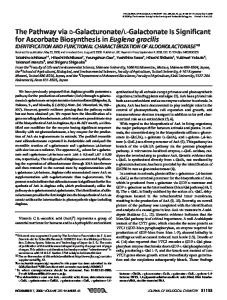
FIG. 2. Hydrolysis of Nedd8-AMC by UCH-L3 and its inhibition by Nedd8-VS. Nedd8-AMC (92 nM) was incubated with buffer or 15 pM UCH-L3 as indicated. At 190 s Nedd8-VS (100 nM) or buffer was added. The reaction was monitored by fluorescence as described previously (36).
FIG. 1. Synthesis and purification of Nedd8 derivatives. The Nedd8 thiol esters were generated and purified by the one-step inteinmediated protein splicing method. The FLAG-Nedd8 thiol ester was reacted with glycyl vinyl sulfone to generate FLAG-Nedd8-VS (30% yield). Nedd8-AMC (5% yield) was synthesized using Nedd8 thiol ester and Gly-AMC. After overnight incubation at 37 °C, both derivatives were purified by ion exchange chromatography as described under “Experimental Procedures.” The molecular weights of the Nedd8 derivatives were determined by MALDI-TOF mass spectroscopic analysis. RESULTS
New Nedd8-specific Inhibitors and Substrates—Identification of deneddylases will be facilitated by the availability of specific inhibitors and substrates. To synthesize inhibitors and substrates of the Ubl protein processing proteases we have adapted the intein fusion protein approach (35) to produce ubiquitin-like proteins activated at their C terminus. In this method, the ubiquitin-like protein, lacking its C-terminal glycine, is produced in bacteria as a fusion protein with a self-splicing intein followed by a chitin binding domain (Fig. 1). Expressed fusion protein was adsorbed to a chitin column and contaminating proteins were removed by washing. Nedd81–75 (or FLAGNedd81–75) was isolated as the C-terminal thiol ester by incubating the column with MESNA. Cleavage from the column is quantitative and the yield of homogeneous Nedd81–75 thiol ester (expected mass 8626.13, observed mass 8626.92) was ⬃3 mg/liter of bacterial culture. Reaction of this thiol ester with nucleophiles (in the presence of N-hydroysuccinimide as a catalyst) leads to a variety of C-terminal derivatives. For instance, reaction of the MESNA ester of FLAG-Nedd81–75 with glycyl vinylmethyl sulfone (32) resulted in the synthesis of FLAG-Nedd8-VS. After purification by ion exchange chromatography, the yield was about 30% (mass expected 9997.50, mass observed 9997.25).
The results above also suggested that we could synthesize substrates of Nedd8-specific processing proteases (deneddylases) using the same chemistry. We chose to synthesize Nedd8-AMC, a substrate whose cleavage should result in the release of the intensely fluorescent AMC group (36). To increase the solubility of the nucleophillic coumarin, we took advantage of the fact that cyclodextrins increase the solubility of many hydrophobic compounds. Reaction of Nedd81–75 thiol ester with glycyl 7-amido-4-methylcoumarin (50 mM) in the presence of 30% hydroxypropyl--cyclodextrin resulted in the synthesis of C-terminal amidomethylcoumarin of Nedd8 (Nedd8-AMC) in about 5% yield. After purification on ion exchange chromatography, Nedd8-AMC was shown to exhibit the expected mass (Fig. 1), fluorescence spectra, and properties as a substrate (Fig. 2). Because the deubiquitinating enzyme UCH-L3 catalyzes the hydrolysis of Nedd8 derivatives we tested if UCH-L3 would hydrolyze Nedd8-AMC. As expected, Nedd8-AMC was completely and efficiently hydrolyzed by UCH-L3 (Fig. 2). When added to this reaction, FLAG-Nedd8-VS was an efficient and irreversible inhibitor of the hydrolysis of Nedd8-AMC by UCH-L3 (Fig. 2). This inhibition was complete, even when the more tightly bound substrate Ub-AMC was used, suggesting that the vinyl sulfone covalently modifies the enzyme. Thus, each of these derivatives was synthesized in good yield and possessed the expected specificity with UCH-L3, a known deneddylase. Detection of Deneddylases in Mammalian Cell Extracts—To detect and identify deneddylases in mammalian cells, we reacted a lysate of L-M(TK⫺) mouse fibroblasts with FLAGNedd8-VS. Based on similar experiments with Ub-VS (31, 32), we expected that only deneddylases would be rapidly and specifically labeled by this reagent. Fig. 3A shows that when this reaction mixture is subjected to SDS-PAGE and Western blotting with anti-FLAG antibodies two proteins are labeled. A major protein reacts to give an adduct with an apparent molecular weight of about 40,000 and a minor protein yields an adduct of apparent molecular weight of about 35,000. Similar results were obtained with lysates of COS and HeLa cells, although the Western blots showed that more cross-reactive
DEN1 Is a ULP Family Deneddylase
28895
FIG. 3. Labeling and purification of Nedd8-specific proteases of L-M (TKⴚ) cell lysates. A, crude L-M(TK⫺) cell lysate was labeled with FLAGNedd8-VS at 37 °C for 30 min. FLAGNedd8-VS-labeled proteins were adsorbed to anti-FLAG M2 affinity gel and eluted with FLAG peptide. Purification of FLAG-Nedd8-VS-labeled proteins was monitored by 4 –20% gradient SDS-PAGE and Western blotting analysis with antiFLAG M5 antibody. The column was regenerated with a low pH wash. -Actin was present in control reactions lacking FLAG-Nedd8-VS. B, cell lysate was preblocked with Ub-VS then labeled with FLAG-Nedd8-VS. The incorporation of FLAG-Nedd8 into Nedd8-specific proteases was monitored by 4 –20% gradient SDS-PAGE Western blotting analysis. Prelabeling blocked the reactivity of UCH-L3 and revealed a new Nedd8specific protease we called DEN1.
background bands were present in the unmodified lysates (data not shown). As these bands would complicate purification of the authentic deneddylases we conducted the rest of our studies with L-M(TK⫺) lysates. To identify the proteins labeled by FLAG-Nedd8-VS we immunopurified the adducts formed by adsorbing the reaction mixture to anti-FLAG M2-Sepharose. Fig. 3A shows that immunoreactive FLAG-Nedd8 is completely adsorbed by the column and can be eluted with FLAG peptide as a competitor. The two most prominent adducts seen in the peptide eluates (lanes 4 – 6) were excised from the gel and subjected to mass spectroscopic analysis. The third band at 35 kDa was not analyzed because of its low abundance. The upper band at about 48 kDa was determined to be -actin and that at about 40 kDa was determined to be UCH-L3 (data not shown). The same results were obtained using MALDI-TOF or LC MS/MS analysis of the digested proteins. -Actin was also identified in control purifications where only lysate was applied to the antibody column and is probably non-specifically bound to the support. Thus, a major deneddylase appears to be UCH-L3 but the identity of the minor 35-kDa band (DEN1, see below) was not obtained in this experiment. To investigate the specificity of these labeling experiments, we first prelabeled the lysates with Ub-VS (to block any Ub-
specific enzymes) and then added FLAG-Nedd8-VS to derivatize any Nedd8-specific enzymes (Fig. 3B). In this experiment, control Western blots showed two weak immunoreactive bands near, but not identical to, the observed adducts (* in Fig. 3B). Labeling with FLAG-Nedd8-VS alone yielded the pattern seen previously. However, reaction of the prelabeled lysates with FLAG-Nedd8-VS revealed an almost complete loss of adduct corresponding to UCH-L3 but persistence of the labeled band at 35 kDa. Increasing the concentration of FLAG-Nedd8-VS 10-fold did not result in the labeling of any additional species. Thus, UCH-L3 reacts with both Ub-VS and FLAG-Nedd8-VS while the adduct at 35 kDa arises from a protein that only reacts with FLAG-Nedd8-VS. This latter reaction can be completely eliminated by incubation with Nedd8 (data not shown), suggesting that the FLAG and vinyl sulfone moieties are not contributing significantly to the specificity of labeling. We propose that the enzyme modified by FLAG-Nedd8-VS to give the 35-kDa band be called deneddylase 1 (DEN1). Note that DEN1 is considerably less abundant than UCH-L3 in these lysates. Identification of DEN1—To identify DEN1, we purified the protein with anti-FLAG M2-Sepharose as described above for UCH-L3. The proteins eluted from the column with the FLAG peptide were separated by SDS-PAGE and the portion of the gel containing the adduct was excised from the gel, digested
28896
DEN1 Is a ULP Family Deneddylase
FIG. 4. LC MS/MS analysis of the DEN1/FLAG-Nedd8-VS adduct. The adduct was purified using the anti-FLAG M2 affinity gel and elution with FLAG peptide. The labeled protein was separated from nonspecific binding proteins by 4 –20% gradient SDS-PAGE and visualized with SYPRO ruby red staining. The immunoreactive band was cut out and analyzed by LC-MS/MS. Among the identified proteins, SENP8 (GenBankTM number AAH37443) was the best candidate for DEN1.
with trypsin, and subjected to LC MS/MS analysis (Fig. 4). Several peptides derived from -actin and -actin-binding proteins (ARP2/3 protein complex 34, tropomodulin, and tropomyosin) were present indicating contamination with cytoskeletal proteins (Table I). As suggested above, these are probably non-specifically bound. Similarly, the ribosomal proteins L5, S3a, and nascent-polypeptide-associated complex ␣ were identified, suggesting that some contamination with abundant ribosomes was also observed. However, none of these proteins are expected to be modified by FLAG-Nedd8-VS and it is assumed that they are trace contaminants. As expected, the band analyzed also contained 4 peptides from Nedd8, as well as five peptides from a previously identified ubiquitin-like protease family member annotated as SENP8 (GenBankTM accession number gi/26006882). Because of its size (25 kDa) and the obvious evolutionary relationship to the ubiquitin system we further investigated whether SENP8 is identical to DEN1. A cDNA encoding the 212-amino acid human SENP8 (GenBankTM accession number AAG21828) was obtained and recombinant protein was expressed in bacteria (see accompanying manuscript, Ref. 47). Purified recombinant SENP8 was used to raise antibodies in rabbits and to purify the antiSENP8 antibody. When lysates of L-M(TK⫺) cells were subjected to Western blotting with affinity purified anti-SENP8 a single band with an apparent molecular mass of ⬃28 kDa was observed (Fig. 5). This protein was unaffected by prelabeling the lysates with Ub-VS, but is quantitatively converted to a 35-kDa adduct upon reaction of the preblocked lysates with FLAG-Nedd8-VS. Likewise, this adduct was adsorbed by the anti-FLAG M2-Sepharose and eluted from the resin by FLAG peptide. Anti-FLAG reactivity and anti-SENP8 reactivity show a similar elution profile. Catalytic Properties of DEN1—The results presented above suggest that DEN1 (SENP8) is a specific deneddylase that has more affinity for, or reactivity with, FLAG-Nedd8-VS than it does with Ub-VS. Accordingly, recombinant human DEN1 was shown to react readily with FLAG-Nedd8-VS (Fig. 6). Quantitative analysis of this titration by SDS-PAGE shows that
nearly 90% of the recombinant DEN1 was modified by equimolar FLAG-Nedd8-VS. Similar titrations with Ub-VS show no modification of DEN1 by this reagent under the same conditions. Thus, DEN1 shows marked specificity and appears to prefer Nedd8 as a substrate over ubiquitin. We quantitated this preference by measuring the steadystate kinetics of DEN1 catalyzed hydrolysis of Nedd8-AMC, Ub-AMC, and SUMO1-AMC.4 At 40 nM each substrate, the observed relative rates of hydrolysis by DEN1 were 60,000, 1, and ⬍0.01, respectively. Thus, at low substrate concentrations the enzyme preferred Nedd8 as a substrate over the others by over 4 orders of magnitude. A full kinetic analysis of this reaction revealed that DEN1-catalyzed hydrolysis of Nedd8AMC proceeded with a Km of 51 nM, a kcat of 7 s⫺1, and a kcat/Km of 1.3 ⫻ 108 M⫺1 s⫺1 (Table II). Because the Km for the DEN1catalyzed hydrolysis of Ub-AMC was over 5 M we could not determine the Km or kcat, but kcat/Km was 2.2 ⫻ 103 M⫺1 s⫺1. Thus, DEN1 is capable of hydrolyzing ubiquitin derivatives, but the selectivity (reflected in the kcat/Km values) is markedly biased toward using Nedd8 as substrate. The true affinity of DEN1 for Ub and Nedd8 was estimated by measuring the Ki for competitive inhibition of hydrolysis of Nedd8-AMC by the free Ubl domains. Ki for Nedd8 was 185 nM while that for ubiquitin was so high that it could not be determined but must exceed 10 M (Table II). Table II also shows that the catalytic properties of UCH-L3 are exactly the converse. UCH-L3 shows very tight binding of Ub, its preferred substrate, with much weaker affinity for pro-Nedd8. In each optimal case (i.e. DEN1 acting upon Nedd8 and UCH-L3 acting on Ub) the reaction is rate limited by diffusion and catalysis itself is very fast. By all criteria DEN1 is a Nedd8 selective processing protease and appears to correspond to the previously identified open reading frame called SENP8. DISCUSSION
Studies on Ub-specific DUBs have benefited greatly from the development of specific substrates and inhibitors. Ubiquitin ethyl ester was the first synthetic substrate described and proved useful in isolating and determining the enzymatic specificity of ubiquitin-C-terminal hydrolases (34, 37). More recently, ubiquitin-AMC has allowed the continuous spectrofluorometric characterization of other DUBs (36). Similarly, the inhibitors ubiquitin aldehyde and ubiquitin vinyl sulfone have been widely used as specific inhibitors and affinity reagents in studies of enzymatic specificity and enzyme structure (31, 32, 38 – 40). All these derivatives can be synthesized using the trypsin-catalyzed transpeptidation of ubiquitin first described over 15 years ago (34). Similar reagents have not been available for the corresponding processing proteases of the ubiquitin-like proteins, in part because of the fact that trypsincatalyzed transpeptidation is not applicable with the other ubiquitin-like proteins. Our intein-based chemistry was developed specifically to make such reagents. The expression of several ubiquitin-like proteins as fusion proteins with the intein-chitin binding domain furnishes a facile route to the synthesis of these derivatives. Trapping of the thiol ester released upon intein-catalyzed cleavage with amines leads to the synthesis of a variety of useful C-terminal derivatives. The current work illustrates the utility of these reagents and has led to the identification of a new deneddylase, one with a surprising specificity and potentially interesting role in the Nedd8 pathway. Nedd8-AMC Is a Good Substrate for Deneddylases—We at4 T. Gan-Erdene, K. Nagamalleswari, L. Yin, K. Wu, Z.-Q. Pan, and K. D. Wilkinson, unpublished data.
DEN1 Is a ULP Family Deneddylase
28897
TABLE I LC MS/MS analysis of the excised 35-kDa adduct shown in Fig. 4 Each protein identified is indicated, along with the GenBank™ protein accession number, the molecular weight, and the number of tryptic peptides identified. Protein
GenBank™ protein accession No.
M
No. peptides identified
␥,-Actin ARP2/3 protein complex subunit 34 Ribosomal protein L5 Tropomodulin 3 Tropomyosin ␣4 SENP8 Nedd8 G-protein  Ribosomal protein S3a Nascent polypeptide-associated complex ␣
gi兩8019561 gi兩5031599 gi兩206734 gi兩8394460 gi储20864610 gi円20890310 gi円4558043 gi兩5174447 gi兩7441114 gi兩20858455
40,992 34,311 35,379 39,478 28,450 26,644 8,555 35,055 29,940 24,582
11 10 7 6 5 5 4 4 4 4
FIG. 6. Active site titration of recombinant human DEN1. The indicated amount of DEN1 was reacted with increasing amounts of FLAG-Nedd8-VS for 30 min at 37 °C and the reaction mixture was separated by 4 –20% gradient SDS-PAGE and visualized with SYPRO ruby red staining. The intensity of staining in each of the bands was quantitated by densitometry. FIG. 5. DEN1 is SENP8. L-M(TK⫺) cell lysate was preblocked with Ub-VS and labeled with FLAG-Nedd8-VS. Labeled proteins were adsorbed to anti-FLAG M2 affinity gel and eluted with FLAG peptide. Purification of FLAG-Nedd8-VS-labeled proteins was monitored by 4 –20% gradient SDS-PAGE and Western blotting analysis with antibodies to the FLAG epitope (panel A) or to human SENP8 (panel B).
tempted to use the intein chemistry described here to synthesize Nedd8-AMC, but the solubility of the nucleophile glycylAMC is so low that little product was observed. -Cyclodextrin has previously been shown to form a 2:2 host-guest complex with coumarins, resulting in increased solubility of the coumarin (41). The orientation of the coumarin in this complex is such that the ␣-amino group of glycyl-AMC protrudes into solvent and is available for reaction. Thus, addition of hydroxypropyl -cyclodextrin to the reaction resulted in a much higher amount of nucleophile and significantly improved yield of product. While the yield of Nedd8-AMC is low, it is severalfold higher than that obtained for Ub-AMC and the reaction can be scaled up to give useful amounts of substrate. UCH and UBP/USP family members are efficient catalysts of C-terminal esters and amides of ubiquitin. We have found that Nedd8-AMC is a sensitive substrate for UCH-L3 (Fig. 2 and Table II), an enzyme previously shown to hydrolyze Nedd8
derivatives (18) and is generally useful as a substrate for deneddylases. This is consistent with the observations that the C-terminal amidomethylcoumarin of ubiquitin (Ub-AMC) is a sensitive and useful substrate for several deubiquitinating enzymes. It is also a specific and sensitive substrate for DEN1 as shown in Table II. Nedd8-VS Is a Specific Inhibitor of Deneddylases—Vinyl sulfones are known to be potent inhibitors of thiol proteases because of the vinyl group undergoing a Michael addition by the active-site thiol. Recent work with Ub-VS and deubiquitinating enzymes confirms this expectation (31, 32). The reaction of ubiquitin vinyl sulfone was shown to be a specific and sensitive way to modify deubiquitinating enzymes. Furthermore, the reaction is exquisitely specific. Borodovsky et al. (31) showed that when cell lysates were reacted with HA-Ub-VS only deubiquitinating enzymes (and two putative DUBs) are labeled. Thus, HA-Ub-VS has wide applicability as a reagent for proteomics studies of DUBs and for discovering new DUBs. The current work shows that Nedd8-VS is an effective and sensitive inhibitor of deneddylases. UCH-L3 is rapidly and selectively modified leading to inhibition of the hydrolysis of Nedd8-AMC (Fig. 2) and Ub-AMC (data not shown). This inhibition is no doubt because of the fact that the substrate-binding
28898
DEN1 Is a ULP Family Deneddylase
TABLE II Steady-state kinetic analysis of DEN1 and UCH-L3 Hydrolysis of Ub1-AMC substrates was determined spectrofluorometrically (36) in a final volume of 120 l. The assays contained the indicated substrates, 50 mM Tris HCl, pH 7.6, 0.1 g/liter ovalbumin, and enzyme at a concentration of 15 pM to 50 nM as needed. The hydrolysis of pro-Nedd8 was monitored by high performance liquid chromatography (34). DEN1
Km Ki (Ubl) kcat kcat/Km
UCH-L3
Nedd8-AMC
Ub-AMC
Ub-AMC
Nedd8-AMC
Pro-Nedd8
51 nM 185 nM 7s⫺1 1.3 ⫻ 108 M⫺1 s⫺1
⬎5,000 nM ⬎5,000 nM ⬎ 0.01 s⫺1 2.2 ⫻ 103 M⫺1 s⫺1
44 nM 350 nM 6.5 s⫺1 1.4 ⫻ 108 M⫺1 s⫺1
⬎ 5,000 nM ND ⬎ 0.5 s⫺1 1 ⫻ 105 M⫺1 s⫺1
⬎ 5,000 nM ND ⬎ 5 s⫺1 1 ⫻ 106 M⫺1 s⫺1
site is occupied with the reagent and the active-site thiol has been inactivated by reaction with the vinyl sulfone. Similarly, DEN1 is rapidly and quantitatively modified by Nedd8-VS (Fig. 6). Detection and Identification of Deneddylases—In analogy to the studies with HA-Ub-VS (31), the introduction of the HA epitope tag to Nedd8-VS would open the way to identification and purification of deneddylases. However, we found that HAtagged Nedd8 is insoluble4 so we turned to the FLAG epitope tag. The specificity of labeling of deneddylases by FLAGNedd8-VS is attested to by the fact that only two proteins are labeled in lysates of mouse L-M(TK⫺) fibroblasts (Fig. 3). By contrast, over 25 deubiquitinating enzymes are labeled in similar extracts (31), suggesting that there are many fewer deneddylases. This is consistent with the fact that the only known substrates for neddylation are the cullins. The 40-kDa band shown in Fig. 3 was identified by LC MS/MS as UCH-L3. This result is consistent with previous observations of UCH-L3 reactivity (18) and our studies with Nedd8-AMC (Table II). We have confirmed that both recombinant UCH-L3 and UCH-L1 are efficiently labeled by FLAGNedd8-VS (data not shown). The ability of UCH-L3 to bind and hydrolyze Nedd8 is because of the fact that the face of ubiquitin that is bound to UCH-L3 is highly conserved in Nedd8 (42). The corresponding surfaces on SUMO-1 and ISG15 are significantly divergent explaining why these Ubl proteins are not substrates for UCH family DUBs. Identification of the 35-kDa DEN1 Adduct—Prelabeling of the extracts with Ub-VS, followed by labeling with FLAGNedd8-VS (Fig. 3) resulted in a markedly diminished labeling of UCH-L3. This is to be expected as UCH-L3 exhibits significant affinity for ubiquitin. In contrast, the 35-kDa adduct was still observed when FLAG-Nedd8-VS was subsequently added. Thus, the protein giving rise to the 35-kDa adduct is selective for Nedd8 and reacts only minimally with Ub-VS. We called this protein DEN1. Mass spectroscopic analysis of the affinity purified 35-kDa DEN1 adduct yielded four Nedd8 peptides and five peptides from a previously uncharacterized 221-amino acid protein annotated as SENP8. This identification was confirmed with affinity purified antibodies to the 212-amino acid human SENP8. Lysates of mouse L-M(TK⫺) fibroblasts contain only one anti-SENP8 immunoreactive band with an apparent molecular mass of 28 kDa (predicted is 25 kDa). This protein does not react with Ub-VS under conditions where UCH-L3 is completely derivatized (Fig. 5). It is, however, quantitatively labeled by FLAG-Nedd8-VS to form the adduct at 35 kDa. These experiments unambiguously show that DEN1 is SENP8. DEN1 is a member of the ULP family of DUBs. ULP family members possess a core catalytic domain of ⬃220 amino acid, usually at the C terminus (Fig. 7). Most members of this family contain N-terminal extensions that are thought to be involved in determining the substrate specificity or localization of these enzymes (22, 43, 44). Yeast contains two ULPs and both hydrolyze SUMO-1 and SUMO-2/3 derivatives, although with different apparent specificity and nuclear localization (23, 45).
There are seven members of this gene family in humans, although little is known about the specific roles of these enzymes (46). Family members contain eight blocks of highly conserved sequence in the catalytic domain (Fig. 7). These motifs (determined independently by sequence analysis) correspond perfectly to the major secondary structural units (44). The crystal structure of the core catalytic domain yeast ULP1 in a complex with SMT3 (the yeast SUMO homolog) reveals that this protein binds primarily to the C-terminal 30 residues of the SMT3 Ubl domain. The sequence of this region is markedly different from that of ubiquitin, Nedd8, and ISG15 and this is thought to be the reason why all ULPs characterized to date show specificity for SUMO and not the other ubiquitin-like proteins. Thus, it is surprising that SENP8 would have significant activity toward Nedd8. For this reason we more carefully characterized the kinetics of catalysis by DEN1. Substrate Specificity of DEN1—DEN1 catalyzes the hydrolysis of Nedd8-AMC and Ub-AMC, but not SUMO1-AMC (Table II). DEN1 exhibits a Km of about 50 nM for Nedd8-AMC suggesting that Nedd8 derivatives could be physiological substrates. The value of kcat/Km suggests that binding is ratelimiting for this substrate with a kcat of 7 s⫺1. The hydrolysis of Ub-AMC by DEN1 exhibits a Km of greater than 5 M. Because we could not saturate the enzyme with Ub-AMC we cannot determine the kcat value, but it can be estimated to be greater than 0.02 s⫺1. The actual binding constants for Nedd8 and ubiquitin were determined by competitive inhibition to be 185 nM and ⬎5 M, respectively (Table II). SUMO-1 is not a substrate and DEN1 does not bind SUMO-1 (47). Conversely, UCH-L3 prefers ubiquitin substrates by a similar magnitude over Nedd8 substrates (Table II). Ub-AMC hydrolysis by UCH-L3 exhibits a Km of 44 nM and a kcat of 6.5 s⫺1, essentially identical to those for DEN1-catalyzed hydrolysis of Nedd8-AMC. The binding constant for ubiquitin binding to UCH-L3 is 350 nM, consistent with the diffusion-limited binding of substrate. UCH-L3 hydrolyzes Nedd8-AMC and proNedd8 with kcat values of greater than 0.5 and 5 s⫺1, respectively, although there is no evidence of saturable binding at concentrations up to 5 M. Thus, UCH-L3 is capable of hydrolyzing Nedd8 derivatives with good catalytic efficiency, although there is little affinity for such substrates. It seems likely that UCH enzymes would have to have the Nedd8 derivatives delivered by a carrier or adapter to efficiently hydrolyze such substrates, How is substrate specificity achieved? DEN1 is a ULP family member and, where tested, the others appear to hydrolyze SUMO derivatives. It is, however, the most divergent ULP (Fig. 7A). In the catalytic core, DEN1 has more differences than similarities. These differences extend into the ULP/Ubl interface and at least seven residues that are predicted to contact the Ubl domain are substantially different from the consensus ULP sequences (Fig. 7). These include Asp-10 (G/K/R in other ULPs), Ser-11 (H/I/L), Gly-33 (N/D), Val-51 (N/S), Glu-57 (K/ Q/R), Pro-77 (K/N/H), and Ala-98 (H/N). A comparison of other DEN1 sequences shows that many of these changes are con-
DEN1 Is a ULP Family Deneddylase
28899
FIG. 7. Comparison of the catalytic core of human ULP enzymes. The indicated core sequences were submitted to the MatchBox server and the displayed blocks of conserved sequence are identified (www.sciences.fundp.ac.be/biologie/ bms/matchbox_submit.shtml). Identities in at least five of the sequences are shaded black and similarities in all seven sequences are shaded gray. Catalytic residues are shown in yellow and the secondary structure of yeast ULP1 (44) is shown above the alignment. Residues that make contact with SMT3 are indicated by the black bars below the alignment. The residues in these contact sites where DEN1/ SENP8 is significantly different from the consensus are shown in red.
served in this subfamily (47). What is striking about these changes is that most result in charge reversal or loss of Hbonding potential. Because these changes would obviate the ionic interactions that have been shown to be important for SMT3 binding to ULP1, it is easy to see why this ULP has little affinity for SUMO derivatives. Conversely, these changes would also generate a much more hydrophobic interface and such changes would be necessary to interact with the more hydrophobic C terminus of Nedd8. A more detailed analysis will require sophisticated molecular modeling and further structural studies. Activities and Substrates of DUBs—Ubiquitin derivatives can be hydrolyzed by at least three DUB families (UCH, UBP/ USP, and JAMM isopeptidase). Previous studies have shown that Nedd8 peptides and fusion proteins can be cleaved by UCH-L3 (18), USP21 (21), and the signalosome (26), members of the same three families. Our finding that DEN1 is a ULP adds a fourth family to those whose members can act as deneddylases. In other words, the primary sequence of the DUB places it in an evolutionarily defined family, but says little about which Ubl is the substrate (Fig. 8). To emphasize this point we have used the term deneddylase in an attempt to accurately describe the activity, as well as to avoid inferring familial relationships from enzymatic activity. Which of the deneddylases is physiologically relevant? Most studies of DUB activities have not quantitated the specificity or selectivity of catalysis. This makes it difficult to know if they are efficient and selective catalysts. The studies reported here are the first quantitative estimates of the catalytic constants for deneddylases. Despite this detailed analysis, we do not
FIG. 8. Activities of DUB family members on different Ubl substrates. An arrow connects the DUB family with the substrates hydrolyzed if at least one member of that family has been reported to hydrolyze a derivative of that Ubl.
know the physiological substrate for DEN1. This enzyme could serve a function in the salvage pathway, reclaiming Nedd8 that had been adventitiously trapped by cellular amines and thiols. Similarly, it could be involved in processing the pro-protein to yield mature Nedd8. Rub1, the yeast homolog of Nedd8, is processed by YUH1. However, deletion of both mouse homologs (UCH-L1 and UCH-L3) leads to only subtle developmental problems suggesting that enzymes other than the UCHs may play a role in this critical processing event. Finally, it is possi-
28900
DEN1 Is a ULP Family Deneddylase
ble that DEN1 is involved in deneddylating cullins, modifiers of ubiquitin-ligase function and the only known substrate for neddylation. We have found that DEN1 is capable of efficiently removing Nedd8 from hyper-neddylated cullin although not from mono-neddylated cullin (47). Thus, DEN1 may play a role in preventing accumulation of hyper-neddylated cullin and maintaining the level of mono-neddylated cullin that is thought to increase the efficiency of ubiquitination by SCF ubiquitin ligases. REFERENCES 1. 2. 3. 4. 5. 6. 7. 8. 9. 10. 11. 12. 13. 14.
15. 16. 17. 18. 19. 20. 21. 22.
Wilkinson, K. D. (1999) J. Nutr. 129, 1933–1936 Wilson, V. G., and Rangasamy, D. (2001) Exp. Cell Res. 271, 57– 65 Loeb, K. R., and Haas, A. L. (1994) Mol. Cell. Biol. 14, 8408 – 8419 Wu, K., Chen, A., Tan, P., and Pan, Z. Q. (2002) J. Biol. Chem. 277, 516 –527 Glickman, M. H., and Ciechanover, A. (2002) Physiol. Rev. 82, 373– 428 Coux, O. (2002) Prog. Mol. Subcell. Biol. 29, 85–107 Hershko, A., and Ciechanover, A. (1998) Annu. Rev. Biochem. 67, 425– 479 Hochstrasser, M. (2000) Nat. Cell Biol. 2, E153–E157 Yeh, E. T., Gong, L., and Kamitani, T. (2000) Gene (Amst.) 248, 1–14 Seeler, J. S., and Dejean, A. (2001) Oncogene 20, 7243–7249 Kim, K. I., Baek, S. H., and Chung, C. H. (2002) J. Cell. Physiol. 191, 257–268 Muller, S., Hoege, C., Pyrowolakis, G., and Jentsch, S. (2001) Nat. Rev. Mol. Cell. Biol. 2, 202–210 Malakhov, M. P., Kim, K. I., Malakhova, O. A., Jacobs, B. S., Borden, E. C., and Zhang, D. E. (2003) J. Biol. Chem. 278, 16608 –16613 Hamerman, J. A., Hayashi, F., Schroeder, L. A., Gygi, S. P., Haas, A. L., Hampson, L., Coughlin, P., Aebersold, R., and Aderem, A. (2002) J. Immunol. 168, 2415–2423 D’Andrea, A., and Pellman, D. (1998) Crit. Rev. Biochem. Mol. Biol. 33, 337–352 Wilkinson, K. D., and Hochstrasser, M. (1998) in Ubiquitin (Peters, J.-M., Harris, J. R., and Finley, D. J., eds) pp. 99 –125, Plenum, Inc., New York Wilkinson, K. D. (1998) in A Handbook of Proteolytic Enzymes (Barrett, A., and Rawlings, N., eds) pp. 660 – 663, Academic Press, London Wada, H., Kito, K., Caskey, L. S., Yeh, E. T., and Kamitani, T. (1998) Biochem. Biophys. Res. Commun. 251, 688 – 692 Amerik, A. Y., Li, S. J., and Hochstrasser, M. (2000) Biol. Chem. 381, 981–992 Malakhov, M. P., Malakhova, O. A., Kim, K. I., Ritchie, K. J., and Zhang, D. E. (2002) J. Biol. Chem. 277, 9976 –9981 Gong, L., Kamitani, T., Millas, S., and Yeh, E. T. (2000) J. Biol. Chem. 275, 14212–14216 Schwienhorst, I., Johnson, E. S., and Dohmen, R. J. (2000) Mol. Gen. Genet. 263, 771–786
23. Li, S. J., and Hochstrasser, M. (1999) Nature 398, 246 –251 24. Yao, T., and Cohen, R. E. (2002) Nature 419, 403– 407 25. Verma, R., Aravind, L., Oania, R., McDonald, W. H., Yates, I. J., Koonin, E. V., and Deshaies, R. J. (2002) Science 298, 611– 615 26. Cope, G. A., Suh, G. S., Aravind, L., Schwarz, S. E., Zipursky, S. L., Koonin, E. V., and Deshaies, R. J. (2002) Science 298, 608 – 611 27. Hori, T., Osaka, F., Chiba, T., Miyamoto, C., Okabayashi, K., Shimbara, N., Kato, S., and Tanaka, K. (1999) Oncogene 18, 6829 – 6834 28. Ohh, M., Kim, W. Y., Moslehi, J. J., Chen, Y., Chau, V., Read, M. A., and Kaelin, W. G., Jr. (2002) EMBO Rep. 3, 177–182 29. Kawakami, T., Chiba, T., Suzuki, T., Iwai, K., Yamanaka, K., Minato, N., Suzuki, H., Shimbara, N., Hidaka, Y., Osaka, F., Omata, M., and Tanaka, K. (2001) EMBO J. 20, 4003– 4012 30. Linghu, B., Callis, J., and Goebl, M. G. (2002) Eukaryot. Cell 1, 491– 494 31. Borodovsky, A., Ovaa, H., Kolli, N., Gan-Erdene, T., Wilkinson, K. D., Ploegh, H. L., and Kessler, B. M. (2002) Chem. Biol. 9, 1149 –1159 32. Borodovsky, A., Kessler, B. M., Casagrande, R., Overkleeft, H. S., Wilkinson, K. D., and Ploegh, H. L. (2001) EMBO J. 20, 5187–5196 33. Wilkinson, K. D. (1988) in Ubiquitin (Rechsteiner, M., ed) pp. 5–38, Plenum Press, New York 34. Wilkinson, K. D., Cox, M. J., Mayer, A. N., and Frey, T. (1986) Biochemistry 25, 6644 – 6649 35. Chong, S., Mersha, F. B., Comb, D. G., Scott, M. E., Landry, D., Vence, L. M., Perler, F. B., Benner, J., Kucera, R. B., Hirvonen, C. A., Pelletier, J. J., Paulus, H., and Xu, M. Q. (1997) Gene (Amst.) 192, 271–281 36. Dang, L. C., Melandri, F. D., and Stein, R. L. (1998) Biochemistry 37, 1868 –1879 37. Mayer, A. N., and Wilkinson, K. D. (1989) Biochemistry 28, 166 –172 38. Hu, M., Li, P., Li, M., Li, W., Yao, T., Wu, J. W., Gu, W., Cohen, R. E., and Shi, Y. (2002) Cell 111, 1041–1054 39. Johnston, S. C., Riddle, S. M., Cohen, R. E., and Hill, C. P. (1999) EMBO J. 18, 3877–3887 40. Melandri, F., Grenier, L., Plamondon, L., Huskey, W. P., and Stein, R. L. (1996) Biochemistry 35, 12893–12900 41. Brett, T. J., Alexander, J. M., and Stezowski, J. J. (2000) Perkin Transactions 2 6, 1105–1111 42. Wilkinson, K. D., Laleli-Sahin, E., Urbauer, J., Larsen, C. N., Shih, G. H., Haas, A. L., Walsh, S. T., and Wand, A. J. (1999) J. Mol. Biol. 291, 1067–1077 43. Barrett, A. J., and Rawlings, N. D. (2001) Biol. Chem. 382, 727–733 44. Mossessova, E., and Lima, C. D. (2000) Mol. Cell 5, 865– 876 45. Li, S. J., and Hochstrasser, M. (2000) Mol. Cell. Biol. 20, 2367–2377 46. Gong, L., Millas, S., Maul, G. G., and Yeh, E. T. (2000) J. Biol. Chem. 275, 3355–3359 47. Wu, K., Yamoah, K., Dolios, G., Gan-Erdene, T., Tan, P., Chen, A., Lee, C-G., Wei, N., Wilkinson, D. D., Wang, R., and Pan, Z.-Q. (2003) J. Biol. Chem. 278, 28882–28891