PRACA ORYGINALNa
Joanna Socha 1, 3, Agnieszka Guzowska 2, Dobromira Tyc-Szczepaniak 1, Aleksandra Szczęsna 2, Lucyna Kępka 1 1 Department of Teleradiotherapy, Maria Skłodowska-Curie Memorial Cancer Centre and Institute of Oncology, Warsaw Head: Prof. L. Kępka, MD, PhD 2 Mazovian Centre for the Treatment of Lung Diseases and Tuberculosis, Otwock, Head: A. Szczęsna, MD, PhD 3 Department of Radiotherapy, Regional Oncology Centre, Częstochowa Head: M. Syguła, MD, PhD
Hypofractionated conformal radiotherapy in combination with chemotherapy in limited disease small cell lung cancer patients Hipofrakcjonowana konformalna radioterapia skojarzona z chemioterapią u chorych na drobnokomórkowego raka płuca w postaci ograniczonej The study has been conducted within statutory activity of M. Sklodowska-Curie Memorial Cancer Center and Institute of Oncology in Warsaw, without additional funding sources
Abstract Aim: To evaluate the results of hypofractionated conformal radiotherapy (RT) in limited disease small cell lung cancer (LD-SCLC) patients, with particular interest in the value of “early” RT, i.e. given before the 3rd chemotherapy (CHT) cycle. Material and methods: Outcome of hypofractionated RT (42 Gy, 2.8 Gy/fraction, given over 19–21 days, using “concomitant boost” technique — elective volume [39 Gy, 2.6 Gy/fraction] and tumour volumes treated during the same fraction) combined with CHT in 100 consecutive LD-SCLC patients, was retrospectively assessed. The outcomes were compared with a previously published series of 117 LD-SCLC patients treated in the same institution with hyperfractionated or conventionally fractionated RT. Results: Forty-two patients (42%) received “early” RT. Grade 3 NCI CTC acute oesophageal toxicity appeared in 5% of patients. There were three treatment-related deaths. Three-year overall survival (OS) rate was 39.4%, median — 24 months in the examined group vs. 26.0%, and 18 months in historical control, P = 0.02. Three-year OS for 78 patients with completed CHT was 42.2%, median — 28 months vs. 30%, and 14 months for 22 patients who received ≤ 3 CHT cycles, (P = 0.03). The actuarial 3-year locoregional failure risk (LRFR) was 34.0% in the examined group vs. 51.0% in the historical control, P = 0.04. Multivariate analysis showed a marginally significant correlation between the “early” use of RT and LRFR: RR = 0.43 (95% CI: 0.17–1.04), P = 0.06, with no significant impact on OS. Conclusions: Shorter duration of RT using hypofractionation results in encouraging outcomes and acceptable toxicity. Completion of all planned CHT cycles is the most important factor for OS. Key words: small cell lung cancer, limited disease, hypofractionated radiotherapy, timing of radiotherapy Pneumonol. Alergol. Pol. 2014; 82: 105–115
Streszczenie Cel: Ocena wyników hipofrakcjonowanej konformalnej radioterapii (RT) u chorych na drobnokomórkowego raka płuca w postaci ograniczonej (DRP-LD), z uwzględnieniem wartości „wczesnej” RT, tzn. rozpoczętej przed podaniem trzeciego kursu chemioterapii (CHT). Materiał i metody: Przeprowadzono retrospektywną ocenę wyników leczenia 100 kolejnych chorych na DRP-LD, leczonych według schematu hipofrakcjonowanej RT skojarzonej z CHT: dawka — 42 Gy po 2,8 Gy, w tym 39 Gy po 2,6 Gy na obszar elektywny (technika concomitant boost — obszar elektywny i zmiany chorobowe leczone w czasie jednej frakcji), 19–21 dni leczenia. Wyniki Address for correspondence: Joanna Socha, MD, PhD, Department of Teleradiotherapy, Maria Skłodowska-Curie Memorial Cancer Centre and Institute of Oncology, ul. Roentgena 5, 02–776 Warsaw, e-mail:
[email protected]; tel. +48 22 5463083, +48 34 367 36 89; fax: +48 22 643 92 87. DOI: 10.5603/PiAP.2014.0016 Praca wpłynęła do Redakcji: 20.02.2013 r. Copyright © 2014 PTChP ISSN 0867–7077
www.pneumonologia.viamedica.pl
105
Pneumonologia i Alergologia Polska 2014, tom 82, nr 2, strony 105–115
leczenia porównano z wynikami grupy historycznej 117 chorych napromienianych w tym samym ośrodku według schematu konwencjonalnej lub hiperfrakcjonowanej RT. Wyniki: „Wczesną” RT otrzymało 42% chorych. Popromienne zapalenie przełyku w stopniu 3 według NCI CTC stwierdzono u 5% chorych. Odnotowano trzy zgony związane z leczeniem. Aktualizowany odsetek 3-letnich przeżyć całkowitych (OS) wyniósł 39,4%, mediana 24 miesiące w grupie badanej v. 26,0% i 18 miesięcy w grupie historycznej, P = 0,02. Trzyletnie OS dla chorych, którzy otrzymali ≤ 3 kursy CHT (22 chorych), wyniosło 30,0%, mediana 14 miesięcy v. 42,2% i 28 miesięcy dla 78 chorych, którzy otrzymali > 3 kursy CHT, P = 0,03. Aktualizowane 3-letnie ryzyko wznowy miejscowo-regionalnej (LRF) wyniosło 34,0% v. 51,0% w grupie historycznej, P = 0,04. Analiza wielowariantowa wykazała trend w kierunku zmniejszenia ryzyka LRF przy zastosowaniu „wczesnej” RT: RR = 0,43 (95%CI: 0,17–1,04), P = 0,06. Czas zastosowania RT nie wpływał znamiennie na OS. Wnioski: Skrócenie leczenia przez zastosowanie hipofrakcjonacji pozwala na uzyskanie dobrych wyników leczenia przy niewielkiej toksyczności. Decydujący wpływ na przeżycie chorych ma realizacja całej zaplanowanej CHT. Słowa kluczowe: drobnokomórkowy rak płuca, postać ograniczona, radioterapia hipofrakcjonowana, czas rozpoczęcia radioterapii Pneumonol. Alergol. Pol. 2014; 82: 105–115
Introduction The value of thoracic radiotherapy (RT) in the treatment of limited disease (LD) small-cell lung cancer (SCLC) was finally established when two meta-analyses showed that it improves overall survival [1, 2]. However, there is still much controversy about timing of RT in relation to chemotherapy (CHT), the way of combining these two methods, the schedule of RT fractionation, the total irradiation dose and the irradiation volume. Treatment results may be improved by using “early” RT with concurrent CHT [3–6], on condition that CHT is delivered as intended [7, 8]. Shortening the duration of RT also leads to an improvement in treatment results; however, accelerated RT is connected with an increased early toxicity, mainly radiation oesophagitis [9, 10]. The best results so far have been obtained with the use of “early” accelerated hyperfractionated RT, i.e. twice-daily treatment with lower dose per fraction [10]. However, due to its significant toxicity, this schedule should only be considered in carefully selected fit patients. The remaining patients receive conventionally fractionated (i.e. with fraction dose of 1.8–2.0 Gy), prolonged (44 days) RT after completion of CHT [11], which makes combined treatment significantly longer. Moreover, twice-daily irradiation is costly, logistically challenging and usually requires hospitalisation, which precludes the adoption of this schedule as a standard regimen in most cancer centres in the world [12–14]. Considerable shortening of the overall treatment time is also achievable by the use of hypofractionated RT, i.e. with higher doses per fraction. The results and tolerance of hypofractionated RT (40 Gy in 15 fractions) did not differ from conventional RT in 215 patients irradiated in the same 106
institution (retrospective data) [15]. However, we lack prospective studies on larger cohorts, which would concern the application of three-dimensional (3D) conformal hypofractionated RT, using contemporary irradiation techniques. So far, such a treatment schedule has not been compared with conventional RT in a randomised study, and the final results of the only randomised prospective study that has compared hypofractionated RT with accelerated hyperfractionated RT will not be available for the next few years [16]. In the present study, we have compared the outcomes of 100 consecutive LD-SCLC patients treated with definitive hypofractionated RT with the outcomes of a historical control group of 117 patients treated at the same centre with conventional or hyperfractionated RT. The value of the shortening of overall treatment time by the use of “early” hypofractionated RT has been additionally assessed.
Material and methods Between 2007 and 2011, in the Department of Radiotherapy, Maria Sklodowska-Curie Memorial Cancer Centre and Institute of Oncology in Warsaw, one hundred consecutive LD-SCLC patients received accelerated hypofractionated RT with curative intent; treatment outcomes and toxicity were retrospectively assessed. A summary of the characteristics of the patients is presented in Table 1. In all cases the diagnosis of SCLC was confirmed histopathologically. The staging procedure included a complete history, physical examination, blood tests, bronchoscopy, chest computed tomography (CT), CT or ultrasound of the abdomen, brain CT or MRI and bone scanning. All patients met the following eligibility criteria for
www.pneumonologia.viamedica.pl
Joanna Socha et al., Hypofractionated radiotherapy in LD-SCLC
Table 1. Clinical characteristics of the study group (100 patients) and the historical group (117 patients) Tabela 1. Charakterystyka grupy badanej (100 chorych) i grupy historycznej (117 chorych) Study group Number of patients (equals percentage)
Historical group Number (percent) of patients
52 48
73 (62) 44 (38)
Median: 59 (range: 41–81)
Median: 57 (range: 43–78)
Karnofsky performance status (KPS)^ [17] 90–100 80 70
85 12 3
116 (99) 1 ( 3 cm in short axis; ^before radiotherapy
radical RT established at our institution: limited stage disease, a Karnofsky performance status (KPS) [17] > 70, no contraindications to chest RT, and no contraindications to PE (cisplatin + etoposide) or KE (carboplatin + etoposide) CHT. Four cycles of PE or KE CHT at 21-day intervals were recommended, but some patients received five or six cycles. The CHT cycle that included RT was elongated to 28 days. RT was to be included after the first or second cycle of CHT, but, due to logistical reasons, some patients received RT after the third cycle or after the end of all CHT cycles. RT was 3D-planned, prescribed to the ICRU reference point, and consisted of 42 Gy, 2.8
Gy/fraction, including total dose to an elective volume of 39 Gy (2.6 Gy/fraction), given over the course of 3 weeks (19–21 days), 5 times a week. “Concomitant boost” technique was used — elective volume and tumour volumes were treated during the same fraction. 6 MV X photons from a linear accelerator were used. Clinical target volume (CTV) and planning target volume (PTV) were determined based on CT, separately for elective volume (CTV1 and PTV1) and for macroscopic disease (CTV2 and PTV2). CTV1 included the following: the ipsilateral hilum, and nodal stations 7, 4R, 4L, 6, 3A, 2R, 2L, and 5 for the left side tumours. In the case of “bulky mediastinal disease” (i.e. an involvement of more than two mediastinal nodal stations or a single LN with a short axis diameter ≥ 3 cm), CTV1 was additionally enlarged to encompass stations 1R and 1L, and supraclavicular areas. CTV2 included the gross tumour with a 5 mm margin and the whole of the nodal stations with pathologically enlarged lymph nodes (i.e. lymph nodes > 1 cm in the short-axis view on CT). In the case of tumour shrinkage within pulmonary parenchyma after CHT, CTV2 encompassed post-CHT tumour volume, but lymph node stations were included in CTV2 in accordance with pre-chemotherapy nodal involvement, even in cases of complete response. PTV1 and PTV2 were created by adding a 1 cm margin to the appropriate CTVs. The mean lung dose could not exceed 20 Gy; less than 35% of the lung volume was to receive more than 20 Gy. Maximum dose to the spinal cord could not exceed 40 Gy. Less than 50% of the heart volume could receive more than 40 Gy. The dose to the oesophagus had to be kept as low as possible. Prophylactic cranial irradiation (PCI) — 25 Gy in 10 fractions — was offered to patients with complete response (CR) or near-complete response (nCR), after completion of treatment. The following conditions were considered relative contraindications to PCI: neurodegenerative changes, dementia syndrome, alcoholism, epilepsy, cerebrovascular diseases and age > 75 years. The lung, oesophageal and haematological toxicity were reported according to the NCI Common Toxicity Criteria, version 3 (NCI CTC) [18]. Moreover, acute lung toxicity was scored using the Southwest Oncology Group (SWOG) scale [19], whereas late pulmonary, and acute and late oesophageal toxicities were reported according to the Radiation Therapy Oncology Group (RTOG) scale [20]. One month after completion of treatment, chest CT was performed to assess response using
www.pneumonologia.viamedica.pl
107
Pneumonologia i Alergologia Polska 2014, tom 82, nr 2, strony 105–115
RECIST (Response Evaluation Criteria in Solid Tumours) criteria [21]. The additional category of nCR was introduced, defined as residual disease visualised on the chest CT, which could not be visible on a chest X-ray. Follow-up examinations were carried out every 3 months. The historical control group included 117 LD-SCLC patients, who received conventionally fractionated or hyperfractionated RT in the Department of Radiotherapy, Maria SklodowskaCurie Memorial Cancer Centre and Institute of Oncology in Warsaw between 1997 and 2006 [22]. Characteristics of the patients and the treatment applied are presented in Tables 1 and 2. To compare the effect of different fractionation schedules, the biologically effective dose (BED) formula, proposed by Fowler [23], was used: BED = nd[1 + d/(a/b)] – ln2(T – Tk) where n – number of fractions, d – dose per fraction; T — total irradiation time; a/b — radiobiological parameter used to describe the response to fractionation (for SCLC = 10 [24]); Tk — time of onset of accelerated tumour repopulation (for SCLC = 28 days [24]). Three-year overall survival (OS) and progression-free survival (PFS) rates were assessed using the Kaplan-Meier method from the start of CHT, as well as the 3-year actuarial locoregional failure risk (LRFR), and the actuarial risk of distant metastases (DM) and brain metastases (BM). Univariate (log-rank) and multivariate (Cox regression model) analyses were used to evaluate the influence of the RT timing (“early” vs. “late”) and other clinical variables, such as: age (< 65 vs. ≥ 65 years), gender, KPS (70-80 vs. 90–100), the presence of “bulky” mediastinal disease, the use of PCI and the number of CHT cycles delivered (≤ 3 vs. >3) on the treatment outcomes. Only predictors with P-value < 0.20 in univariate analysis were included in multivariate analysis. The impact of RT timing on the delivery of RT according to the protocol (i.e. treatment breaks) and CHT compliance was determined using the Mann-Whitney U test. The Wilcoxon test for age and the nonparametric Mann-Whitney U test for the remaining parameters (gender, KPS, the presence of “bulky disease”, the use of PCI, the number of CHT cycles, type of CHT: platinum vs. non-platinum based, RT timing, BED of RT) were used to determine the distribution of patient characteristics within the compared groups. The log-rank test was used to analyse differences in OS, PFS and LRFR between the groups. The Chi-squared test was used to compare RT-induced toxicity between groups.
108
Table 2. Treatment characteristics of the study group (100 patients) and the historical group (117 patients) Tabela 2. Porównanie grupy badanej (100 chorych) i grupy historycznej (117 chorych) w aspekcie zastosowanego leczenia Characteristics
Use of PCI Yes No
Historical Study group group Number Number of (percent) patients (equals of patients percentage) 53 47
46 (39) 71 (61)
78 19 1 0 2
100 (85) 6 (5) 0 1 (1) 10 (9)*
Number of CHT cycles delivered ≤3 >3
22 78
9 (8) 108 (92)
Timing of RT Early^ Late‡
42 58
35 (30)† 82 (70)
0 0 100
0 61 (52) 56 (48)
0 0 100
17 (15) 18 (15) 82 (70)
42 58
0 82 (70)
98
0
1
0
1 0
0 17 (15)
0
18 (15)
0
82 (70)
Type of chemotherapy PE KE PN CAV Other (mostly combinations of above)
RT planning 2D 2D elective volume, 3D boost volume 3D Combination of RT and CHT Concurrent Alternating Sequential RT between 1st and 2nd or 2nd and 3rd cycle of CHT (early sequential RT) RT after CHT RT dose and fractionation 42 Gy in 15 fractions of 2.8 Gy׳׳ (BED = 60 Gy) 39 Gy in 15 fractions of 2.6 Gy (BED = 55,4 Gy) 49.8 Gy in 18 fractions( ׳׳׳BED = 66,4 Gy) 45 Gy b.i.d. of 1.5 Gy (BED = 58 Gy) 56 Gy of 2 Gy (20Gy+20Gy+16Gy₫) (BED = 56 Gy) 44-60 Gy of 2 Gy, median 56 Gy (median BED = 60,2 Gy; range 51,4 Gy – 63,6 Gy)
*two patients did not receive platinum-based chemotherapy; ^ “early” radiotherapy was defined as started before the 3rd cycle of chemotherapy; ‡“late” radiotherapy was defined as started after the 3rd cycle of chemotherapy; †including 17 patients (15%) with hiperfractionated radiotherapy given concurrently with chemotherapy; 18 patients (15%) — alternating radiochemotherapy with conventionally fractionated radiotherapy; ׳׳with 39 Gy, 2.6 Gy/fraction to elective volume; ׳׳׳42 Gy, 2.8 Gy/fraction + 3 additional fractions, each of 2,6 Gy; ₫alternating radiochemotherapy; PCI — prophylactic cranial irradiation; RT — radiotherapy; CHT — chemotherapy; 2D — two-dimensional radiotherapy planning; 3D — three-dimensional radiotherapy planning; b.i.d. — twice daily; PE — Cisplatin + Etoposide; KE — Carboplatin + Etoposide; PN — Cisplatin + Vinorelbine; CAV — Cyclophosphamide +Doksorubicin +Vincristine; BED — biologically effective dose, calculation method given in „Material and methods”
www.pneumonologia.viamedica.pl
Joanna Socha et al., Hypofractionated radiotherapy in LD-SCLC
A P value of less than 0.05 was considered statistically significant. IBM SPSS Statistics v. 20 was used for statistical analysis. “Early” RT was defined as being given before the third cycle of CHT. LRF was defined as tumour progression within the lung and/or regional nodal failure.
Results The median follow up for living patients was 30 months (range 11–17). “Early” RT was administered in 42 patients (42%), and the remaining 58 patients (58%) received “late” RT. Sixteen patients (16%) required RT breaks, in all cases because of haematological toxicity. The median duration of a break was 5 days (range: 2–17). Ten patients with break received “early” RT (24%), and six patients received “late” RT (10%); P = 0.15. Seventy-eight patients (78%) received all planned cycles of CHT: 67% of patients in the “early” RT group and 86% of patients in the “late” RT group, P = 0.03. The reasons for not completing all planned cycles of CHT in the groups with “early” and “late” RT are presented in Table 3. Haematological toxicity was the reason for stop CHT before completion of four planned cycles in 8 (19%) patients with “early” RT, and in 7 (12%) patients with “late” RT. Non-haematological toxicity (i.e. acute oesophageal toxicity) was the reason for discontinuation of systemic treatment in 2 patients (5%) with “early” RT, whereas in the group with “late” RT, non-haematological toxicity did not hinder the continuation of CHT. Fifty-two patients (52%) received PCI.
There was no grade ≥ 3 acute or late pulmonary toxicity observed. Grade 3 (NCI CTC) acute esophageal toxicity was recorded in 5 patients (5%), none of them required nasogastric tube insertion for feeding — according to RTOG scale, this was scored as grade 2. We did not observe grade ≥ 3 late oesophageal toxicity. Grade 3–4 neutropenia and thrombocytopenia were found in 41 patients (41%) and in 11 patients (11%), respectively; grade 3 anaemia was seen in 10 patients (10%). There were three treatment-related deaths (3%). Treatment toxicity according to the timing of RT is presented in Table 4. After completion of treatment, the CR, nCR, partial response and disease stabilization rates were 59%, 24%, 10% and 1%, respectively. In 3 patients (3%) disease progression outside the chest occurred during treatment. Treatment response could not be determined in 3 patients (3%) who had died prior to completion of treatment. Three-year OS rate was 39.4%, and median survival time (MST) was 24 months (Fig. 1). In the group with “early” RT, 3-year OS was 40.0%, MST was 27 months, and in the group with “late” 3-year RT OS was 38.7% and MST 22 months, P = 0.40. Univariate analysis found the number of CHT cycles delivered to be the only significant prognostic factor for OS: 3-year OS rate for patients with completed CHT (78 patients) was 42.2%, vs. 30% for 22 patients who received 3 or less cycles of CHT, with median survival of 28 and 14 months, respectively (P = 0.03) (Fig. 2). Multivariate analysis showed a marginally significant correlation between the fail of delivery of all planned cycles of CHT and OS rates: relative risk (RR) = 1.79 (95% CI: 0.98–3.29), P = 0.059.
Table 3. Reasons for not completing all four courses of chemotherapy according to the timing of radiotherapy Tabela 3. Przyczyny przedwczesnego zakończenia chemioterapii (tzn. podania mniej niż planowane 4 kursy) w grupie chorych z radioterapią „wczesną” i „późną” Reason
”Early” radiotherapy n = 42
„Late” radiotherapy n = 58
Number of patients
Percent of patients
Number of patients
Percent of patients
Performance status, comorbidities
2
5
1
2
Hematologic toxicities
8
19
7
12
1
2
0
0
Non-hematologic toxicities: renal acute esophageal
2
5
0
0
acute pulmonary
1
2
0
0
14
33
8
14
Total
„Early” radiotherapy was defined as started before the 3rd cycle of chemotherapy; „late” radiotherapy was defined as started after the 3rd cycle of chemotherapy.
www.pneumonologia.viamedica.pl
109
Pneumonologia i Alergologia Polska 2014, tom 82, nr 2, strony 105–115
Table 4. Treatment toxicity according to the timing of radiotherapy Tabela 4. Częstość występowania toksyczności leczenia w zależności od czasu rozpoczęcia radioterapii Treatment toxicity and RT timing
Toxicity grade* BD/ND
0
1
2
P-value# 3
4
5
Number (percent) of patients Neutropenia
0.0001
Early RT^
8 (19)
5 (12)
1 (2.5)
2 (4.5)
10 (24)
15 (35.5)
1 (2.5)
Late RT+
11 (19)
14 (24)
11 (19)
6 (10.5)
6 (10.5)
10 (17)
0 (0)
Thrombocytopenia Early RT^ Late RT
+
0.03
8 (19)
11 (26.5)
11 (26.5)
7 (16.5)
4 (9)
1 (2.5)
0
9 (15.5)
30 (51.5)
11 (19)
2 (3.5)
2 (3.5)
4 (7)
0
16 (38)
12 (28.5)
1 (2.5)
0
0
19 (32.5)
17 (29.5)
9 (15.5)
0
0
Anaemia Early RT^ Late RT+
8 (19)
5 (12)
9 (15.5)
4 (7)
Acute esophageal toxicity Early RT^
0
15 (35.5)
Late RT+
0
19 (32.5)
15 (35.5)
8 (19)
4 (9.5)
0
0
27 (47)
11 (19)
1 (1.5)
0
0
Late esophageal toxicity Early RT^
3/5 (7.5/12)
31 (73.5)
Late RT+
3/9 (5/15.5)
44 (76)
2 (4.5)
1 (2.5)
0
0
0
0
2 (3.5)
0
0
0
Acute pulmonary toxicity Early RT^
0
40 (95)
1 (2.5)
1 (2.5)
0
0
1 (2.5)
Late RT+
0
52 (89.5)
5 (9)
1 (1.5)
0
0
1 (1.5)
3/5 (7.5/12)
28 (66.5)
2 (4.5)
4 (9.5)
0
0
0
2/9 (3.5/15.5)
36 (62.5)
10 (17)
1 (1.5)
0
0
0
Late pulmonary toxicity Early RT^ Late RT+
0.15
0.74
0.50
0.72
0.18
*according to NCI Common Toxicity Criteria, version 3, 2006 [18]; #estimated with the use of the chi-square test for all grades of toxicity; ^radiotherapy started before the 3rd cycle of chemotherapy (42 chorych); +radiotherapy started after the 3rd cycle of chemotherapy (58 chorych); p — statistical significance; RT — radiotherapy; BD — no data; ND — not applicable (4 patients had died within 6 months from the start of radiotherapy, before the late toxicity could occurred; in 10 patients late toxicity was not assessed, due to locoregional failure)
No statistically significant correlation between OS and the remaining analysed factors was found. Three-year PFS rate was 31.3%, median PFS was 14 months. Univariate and multivariate analysis did not reveal any statistically significant correlation between PFS and the analysed factors. The actuarial LRFR at three years was 34.0%; 3-year LRFR was 28.4% and 45.1% in the group with “early” and “late” RT, respectively, P = 0.09 (Fig. 3). Multivariate analysis showed a marginally significant correlation between the “early” use of RT and LRFR: RR = 0.43 (95% CI: 0.17–1.04), P = 0.06. The actuarial 3-year DM risk was 54.6%. Performance status was statistically significantly correlated with the risk of DM in univariate analysis. In patients with KPS 90-100, the actuarial 3-year DM risk was 51.3%, median — 31 months; in patients with KPS 70–80 — 74.3% and 12 months, 110
respectively, P = 0.03. In multivariate analysis this factor was not statistically significant. BM was the first site of recurrence in 17 patients (17%); of these, 11 did not receive PCI. The actuarial risk of BM as the first site of recurrence at three years was 29.1% in the whole group of patients. This rate was 22.9% and 38.6% in patients with and without PCI, respectively, P = 0.04. Performance status was significantly better in the historical group, P < 0.001. The number of CHT cycles received (≤ 3 vs. > 3) was significantly lower in the study group (P = 0.001). PCI administration was significantly less frequent in the historical group (P = 0.04). There were no statistically significant differences between the two groups regarding the distribution of the remaining analysed clinical variables potentially influencing the outcomes.
www.pneumonologia.viamedica.pl
Joanna Socha et al., Hypofractionated radiotherapy in LD-SCLC
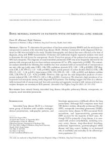
Figure 1. Overall survival in 100 study patients Rycina 1. Odsetek przeżyć całkowitych w grupie badanej (100 chorych) Figure 3. Locoregional failure risk according to the radiotherapy timing: „early” radiotherapy, i.e. started before the 3rd cycle of chemotherapy (42 patients) v. „late” radiotherapy, i.e. started after the 3rd cycle of chemotherapy (58 patients) Rycina 3. Ryzyko wystąpienia wznowy miejscowo-regionalnej w zależności od czasu zastosowania radioterapii — radioterapia „wczesna”, tj. rozpoczęta przed podaniem trzeciego kursu chemioterapii (42 chorych) v. „późna”, tj. rozpoczęta po podaniu trzeciego kursu chemioterapii lub po zakończeniu chemioterapii (58 chorych)
Figure 2. Overall survival according to the number of chemotherapy cycles delivered Rycina 2. Odsetki przeżyć całkowitych w zależności od podania wszystkich zaplanowanych kursów chemioterapii
Treatment toxicity did not differ significantly between the study group and historical controls (Table 5).
Median follow up for the living patients in the historical group was 33 months (range 13–80). In this group, the 3-year PFS rate was 15% and median PFS was 10 months, whereas in the study group it was 31.3% and 14 months, respectively, P < 0.001. In the historical group, the 3-year OS rate was 26% and MST was 18 months, whereas in the study group it was 39.4% and 24 months, respectively, P = 0.02 (Fig. 4). The actuarial LRFR at 3-years was 51% in the historical group and 34.0% in the study group, P = 0.04 (Fig. 5). DM was the most common pattern of failure in both groups. The rate of BM as the first site of failure was 18% in the historical group and 17% in the study group.
Discussion Results of our study show that accelerated hypofractionated RT is a treatment of acceptable toxicity and effectiveness. Changing RT practice
www.pneumonologia.viamedica.pl
111
Pneumonologia i Alergologia Polska 2014, tom 82, nr 2, strony 105–115
Table 5. Treatment toxicity in the study group and in the historical group Tabela 5. Częstość występowania toksyczności leczenia w grupach badanej i historycznej Treatment toxicity
P-value#
Toxicity grade 2
3
4
Number (percent) of patients Acute esophageal toxicity‡
0.64
Study group
24 (24)
0*
0
Historical group
18 (15)
2 (2)
0
Late esophageal toxicity‡ Study group Historical group
BD
3 (3)
0
0
BD
BD
BD
Acute pulmonary toxicity^ Study group
2 (2)
Historical group
7 (6)
0.61
0
0
0
0
Late pulmonary toxicity‡ Study group Historical group
5 (5) 12 (10)
0
0
1 (< 1)
0
Treatment-related deaths Study group
0.14
0.37
3 (3)
Historical group
1 (< 1)
^according to the Southwest Oncology Group (SWOG) scale [19]; ‡according to the Radiation Therapy Oncology Group (RTOG) scale [20]; #estimated with the use of the chi-square test for all grades of toxicity; *according to the NCI Common Toxicity Criteria scale, version 3, 2006 [18], grade 3 esophageal toxicity was recorded in 5 patients, but it was assessed as grade 2 according to the RTOG scale; BD — no data; P — statistical significance
Figure 4: Overall survival in the study group (100 patients) and in the historical group (117 patients)
Figure 5. Locoregional failure risk in the study group (100 patients) and in the historical group (117 patients)
Rycina 4. Odsetki przeżyć całkowitych w grupach: badanej (100 chorych) i historycznej (117 chorych)
Rycina 5. Ryzyko wystąpienia wznowy miejscowo-regionalnej w grupach: badanej (100 chorych) i historycznej (117 chorych)
112
www.pneumonologia.viamedica.pl
Joanna Socha et al., Hypofractionated radiotherapy in LD-SCLC
from previously used RT regimens to trial-defined hypofractionation had a positive impact on the outcomes of LD-SCLC patients treated with RT in the Department of Radiotherapy, Maria Sklodowska-Curie Memorial Cancer Centre and Institute of Oncology in Warsaw. A statistically significant difference was found in OS and PFS (P = 0.02 and P < 0.001, respectively), and LRFR was lower in the study group (P = 0.04). These results have been obtained without enhancement of toxicity, which additionally increases the value of this method of treatment. However, the retrospective nature of the study and lack of uniform RT schedule in the historical group limits our conclusions. Moreover, the comparison with historical control is a limitation itself as it is not possible to exclude the impact on outcome of some changes in the pattern of practice regarding RT and CHT, which could have taken place during 10 years (1997–2007). For instance, some patients in the historical group underwent two-dimensionally (2D) planned RT. However, in the majority of the published studies of thoracic irradiation in SCLC-LD patients, RT was based entirely on conventional 2D treatment planning; and the best treatment results so far were obtained in the era of 2D RT [10]. Furthermore, the range of BEDs in both compared groups was within currently recommended dose limits used in the treatment of LD-SCLC. The application of PCI differed significantly between the two examined groups; however, similar frequencies of BM were found in both groups. The better performance status of patients in the historical cohort may suggest more strict eligibility criteria for definitive RT in this group. The study group consisted of 100 consecutive patients with KPS ≥ 70, so selection criteria was not so rigorous — which is an additional advantage, as the presented RT schedule can be used in a wider population of patients. A negative consequence of less strict qualification criteria in the study group can be the lower proportion of patients who completed the planned CHT, compared to the historical control group; proportions of patients who received > 3 CHT cycles were 78% and 92% in the study group and in the historical cohort, respectively; P = 0.001. Weight loss is an important parameter that could influence the outcomes, but the data on weight loss in the study group were incomplete, and were omitted in the analyses, which also limits the conclusions. The obtained results and data from the literature may suggest that the efficacy of hypofractionated RT (particularly the “early” hypofractiona-
ted RT) is similar to that of hyperfractionated RT, on condition that the planned CHT is completed. In a randomised study conducted in Canada [4], which compared “early” vs. “late” hypofractionated RT (40 Gy in 15 fractions), MST in the “early” arm was 21.2 months and 5-year OS rate was 20%, whereas in the study by Turrisi et al. [10], in the arm with hyperfractionated RT these values were 23 months and 26%, respectively. In the retrospective study by Xia et al. [25] half of the patients received hyperfractionated RT (56 Gy in 40 fractions, 1.4 Gy twice daily), and the other half received hypofractionated RT (55 Gy in 22 fractions, 2.5 Gy/fraction). There was no statistically significant survival difference between both groups, and MST was 27 months. Another retrospective study [26] reported the outcomes of 227 patients who underwent mainly mild hypofractionated irradiation (40 Gy in 15 fractions, 2.67/fraction): MST was 22 months, and 5-year OS rate was 25%. Attempts to modify and intensify the radiochemotherapy regimens used in LD-SCLC have led to improvement in survival in some studies [3, 4, 6, 10, 27]; however, improvement in locoregional control still remains a therapeutic challenge. In the study by Turrisi et al. [10] the LRF rate in the hyperfractionated arm was 36%, and in the study by Bonner et al. [28, 29] an LRF rate of 34% was found in both study arms. This rate was slightly lower in the present study (27%), but the significantly shorter follow-up period should be taken into consideration. The actuarial 3-year LRFR of 34% is comparable to that obtained in other studies [3, 26, 28, 29]. However, the relatively high LRFR in patients with “late” RT (45.1% at 3 years) may suggest that the applied total dose is too low in this group and should be adjusted. The BED of the presented RT schedule is 60 Gy, thus being in the range of the currently recommended doses applied in the treatment of LD-SCLC; however, it would be valuable to modify this schedule in order to increase the biological dose of radiation. In our study, reduced LRFR with “early” RT did not translate into a meaningful survival advantage, which may imply that DM remain the major problem. This may result from the difference in the proportion of patients who received at least four cycles of CHT, which was statistically significant, favouring the “late” over the “early” RT group (86% and 67% of patients, respectively, P = 0.033). A marginally significant correlation between the fail of delivery of all planned cycles of CHT and OS rates was found: RR = 1.79 (95%
www.pneumonologia.viamedica.pl
113
Pneumonologia i Alergologia Polska 2014, tom 82, nr 2, strony 105–115
CI: 0.98–3.29), P = 0.059. The results of other studies also indicate such a relationship. In a British randomised study comparing “early” and “late” hypofractionated RT (40 Gy in 15 fractions), CHT in the “early” arm was completed as intended in 69% of patients, whereas in the “late” arm it was completed in 80%, P = 0.03 [8]. The probability of OS did not differ between the arms, despite the improvement in locoregional control with the use of “early” RT (LRF rate of 26% vs. 37% in “early” and “late” arms, respectively; P = 0.03). The authors conducted a meta-analysis of eight trials and concluded that the optimal delivery of CHT is indispensably necessary to derive any benefit from the “early” use of RT [8]. The newest meta-analysis confirms the key role of completion of all planned cycles of CHT [7]. “Early” RT resulted in an improvement in OS only in trials where the “early” and “late” arms showed similar CHT compliance (HR = 0.79; 95% CI: 0.69–0.91); whereas in the trials where the difference in CHT compliance was ≥ 10% between arms, survival was worse with “early” RT (HR = 1.19; 95% CI: 1.05–1.34) [7]. In the present study we have shown an acceptable toxicity of “early” RT, but the difference between the “early” and “late” groups in the number of CHT cycles delivered warrants caution while interpreting the results. An additional limitation of our study is the lack of precise data concerning the use of chemotherapy, especially the dosages of the cytostatics given to the patients. Similarly as in the study by Spiro et al. [8], in the present study it is also difficult to estimate explicitly, which factors were the main reasons for the disproportion in the CHT compliance. However, it is important to emphasise the limitations of the conclusions drawn from the lack of randomisation and limited number of patients. Moreover, the groups of patients with “early” and “late” RT were distinguished retrospectively, which also hinders interpretation.
Conclusions Accelerated, hypofractionated RT in combination with platinum-based CHT is a treatment of acceptable toxicity and effectiveness in LD-SCLC patients. Overall survival was not significantly affected by the timing of RT, although the use of “early” RT may offer better locoregional disease control. Completion of all planned cycles of CHT is the most important factor for overall survival. The value of hypofractionated radiotherapy should be confirmed in a randomised trial comparing 114
this treatment schedule with hyperfractionated RT — the current “gold standard” in treatment of LD-SCLC.
Conflict of interest The authors declare no conflict of interests.
References 1. Pignon J.P., Arriagada R., Ihde D.C. et al. A meta-analysis of thoracic radiotherapy for small-cell lung cancer. N. Engl. J. Med. 1992; 327: 1618–1624. 2. Warde P., Payne D. Does thoracic irradiation improve survival and local control in limited-stage small-cell carcinoma of the lung? A meta-analysis. J. Clin. Oncol. 1992; 10: 890–895. 3. Jeremic B., Shibamoto Y., Acimovic L., Milisavljevic S. Initial versus delayed accelerated hyperfractionated radiation therapy and concurrent chemotherapy in limited small-cell lung cancer: a randomized study. J. Clin. Oncol. 1997; 15: 893–900. 4. Murray N., Coy P., Pater J.L. et al. Importance of timing for thoracic irradiation in the combined modality treatment of limited-stage small-cell lung cancer. The National Cancer Institute of Canada Clinical Trials Group. J. Clin. Oncol. 1993; 11: 336–344. 5. Sas-Korczyńska B., Sokołowski A., Korzeniowski S. The influence of time of radio-chemotherapy and other therapeutic factors on treatment results in patients with limited disease small cell lung cancer. Lung Cancer 2013; 79: 14–19. 6. Takada M., Fukuoka M., Kawahara M. et al. Phase III study of concurrent versus sequential thoracic radiotherapy in combination with cisplatin and etoposide for limited-stage small cell lung cancer: results of the Japan Clinical Oncology Group Study 9104. J. Clin. Oncol. 2002; 20: 3054–3060. 7. De Ruysscher D., Paris E., Le Pechoux C. et al. A meta-analysis of randomised trials using individual patient data on the timing of chest radiotherapy in patients with limited stage small cell lung cancer. J. Thorac. Oncol. 2011; 6 (Suppl. 2): S641–642, abstr. MO19.03. 8. Spiro S.G., James L.E., Rudd R.M. et al. London Lung Cancer Group. Early compared with late radiotherapy in combined modality treatment for limited disease small-cell lung cancer: a London Lung Cancer Group multicenter randomized clinical trial and meta-analysis. J. Clin. Oncol. 2006; 24: 3823–3830. 9. Pijls-Johannesma M., De Ruysscher D., Vansteenkiste J., Kester A., Rutten I., Lambin P. Timing of chest radiotherapy in patients with limited stage small cell lung cancer: a systematic review and meta-analysis of randomised controlled trials. Cancer Treat. Rev. 2007; 33: 461–473. 10. Turrisi A.T. 3rd, Kim K., Blum R. et al. Twice-daily compared with once-daily thoracic radiotherapy in limited small-cell lung cancer treated concurrently with cisplatin and etoposide. N. Engl. J. Med. 1999; 340: 265–271. 11. Krzakowski M., Orłowski T., Roszkowski K. et al. Drobnokomórkowy rak płuca — zalecenia diagnostyczno-terapeutyczne Polskiej Grupy Raka Płuca. Pneumonol. Alergol. Pol. 2007; 75: 88–94. 12. Kępka L., Danilova V., Saghatelyan T. et al. Resources and management strategies for the use of radiotherapy in the treatment of lung cancer in Central and Eastern European countries: results of an International Atomic Energy Agency (IAEA) survey. Lung Cancer 2007; 56: 235–245. 13. Movsas B., Moughan J., Komaki R. et al. Radiotherapy patterns of care study in lung carcinoma. J. Clin. Oncol. 2003; 21: 4553–4559. 14. Stahel R., Thatcher N., Früh M., Le Péchoux C., Postmus P.E., Sorensen J.B., Felip E.; Panel members. 1st ESMO Consensus Conference in lung cancer; Lugano 2010: small-cell lung cancer. Ann. Oncol. 2011; 22: 1973–1980. 15. Videtic G.M., Truong P.T., Dar A.R., Yu E.W., Stitt L.W. Shifting from hypofractionated to “conventionally” fractionated thoracic radiotherapy: a single institution’s 10-year experience in the management of limited-stage small-cell lung cancer using
www.pneumonologia.viamedica.pl
Joanna Socha et al., Hypofractionated radiotherapy in LD-SCLC
concurrent chemoradiation. Int. J. Radiat. Oncol. Biol. Phys. 2003; 57: 709–716. 16. Gronberg B.H., Halvorsen T.O., Flotten O. et al. Randomized phase II trial comparing two schedules of thoracic radiotherapy (TRT) in limited disease small-cell lung cancer (LD SCLC). J. Clin. Oncol. 2012; 30 (Suppl. 1): abstr. 7027. 17. Karnofsky D.A., Burchenal J.H. The clinical evaluation of chemotherapeutic agents in cancer. W: MacLeod C.M. (ed.). Evaluation of chemotherapeutic agents. Columbia University Press, New York 1949; 191–205. 18. http://ctep.cancer.gov; 17.02.2013. 19. Green S., Weiss G.R. Southwest Oncology Group standard response criteria, endpoint definitions and toxicity criteria. Invest. New Drugs 1992; 10: 239–253. 20. http://rtog.org; 17.02.2013. 21. Therasse P., Arbuck S.G., Eisenhauer E.A. et al. New guidelines to evaluate the response to treatment in solid tumors (RECIST Guidelines). J. Natl. Cancer Inst. 2000; 92: 205–216. 22. Wierzchowski M., Sprawka A., Kępka L. Isolated nodal failure after chemo–radiotherapy in limited disease small cell lung cancer (LD-SCLC). Rep. Pract. Oncol. Radiother. 2009; 14: 58–63. 23. Fowler J.F. Biological factors influencing optimum fractionation in radiation therapy. Acta Oncol. 2001; 40: 712–717.
24. De Ruysscher D., Pijls-Johannesma M., Bentzen S.M. et al. Time between the first day of chemotherapy and the last day of chest radiation is the most important predictor of survival in limited-disaese small-cell lung cancer. J. Clin. Oncol. 2006; 24: 1057–1063. 25. Xia B., Chen G.Y., Cai X.W. et al. Is involved-field radiotherapy based on CT safe for patients with limited-stage small-cell lung cancer? Radiother. Oncol. 2012; 102: 258–262. 26. Giuliani M.E., Lindsay P.E., Sun A. et al. Locoregional failures following thoracic irradiation in patients with limited-stage small cell lung carcinoma. Radiother. Oncol. 2012; 102: 263–267. 27. Perry M.C., Herndon J.E. 3rd, Eaton W.L. Thoracic radiation therapy added to chemotherapy for small-cell lung cancer: an update of Cancer and Leukemia Group B Study 8083. J. Clin. Oncol. 1998; 16: 2466–2467. 28. Bonner J.A., Sloan J.A., Shanahan T.G. et al. Phase III comparison of twice-daily split-course irradiation versus once-daily irradiation for patients with limited stage small-cell lung carcinoma. J. Clin. Oncol. 1999; 17: 2681–2691. 29. Schild S.E., Bonner J.A., Shanahan T.G. et al. Long-term results of a phase III trial comparing once-daily radiotherapy with twice-daily radiotherapy in limited-stage small-cell lung cancer. Int. J. Radiat. Oncol. Biol. Phys. 2004; 59: 943–951.
www.pneumonologia.viamedica.pl
115