Hum. Reprod. Advance Access published June 20, 2012 Human Reproduction, Vol.0, No.0 pp. 1– 10, 2012 doi:10.1093/humrep/des198
ORIGINAL ARTICLE Reproductive biology
Human decidual stromal cells secrete C-X-C motif chemokine 13, express B cell-activating factor and rescue B lymphocytes from apoptosis: distinctive characteristics of follicular dendritic cells R. Mun˜oz-Ferna´ndez 1,†, A. Prados2,†, E. Leno-Dura´n2, A. Bla´zquez 3, J.-R. Garcı´a-Ferna´ndez 4, G. Ortiz-Ferro´n 2, and E.-G. Olivares2,3,5,* 1 Instituto de Parasitologı´a y Biomedicina, Consejo Superior de Investigaciones Cientı´ficas, 18100 Armilla, Granada, Spain 2Instituto de Biopatologı´a y Medicina Regenerativa, Centro de Investigacio´n Biome´dica, Universidad de Granada, 18100 Armilla, Granada, Spain 3Servicio de Ana´lisis Clı´nicos, Hospital Universitario San Cecilio, Granada, Spain 4Consulta de Hematologı´a, Hospital de Baza, 18800 Baza, Granada, Spain 5Departamento de Bioquı´mica y Biologı´a Molecular III e Inmunologı´a, Universidad de Granada, 18012 Granada, Spain
*Correspondence address. Tel: +34-958240731; Fax: +34-958249015; E mail:
[email protected]
Submitted on September 9, 2011; resubmitted on March 22, 2012; accepted on May 8, 2012
background: Decidual stromal cells (DSCs) have classically been considered fibroblastic cells, although their function, cell lineage and origin are not fully understood. We previously demonstrated that human DSCs showed similarities with follicular dendritic cells (FDCs): DSCs expressed FDC-associated antigens, both types of cells are contractile and both are related to mesenchymal stem cells (MSCs). To further characterize DSCs, we investigated whether DSCs and FDCs share any distinctive phenotypical and functional characteristics.
methods: Human FDC lines were obtained from tonsillectomy samples, human DSC lines from elective termination of pregnancy samples and human MSC lines from bone marrow aspirates. We isolated DSC, FDC and MSC lines and compared their characteristics with flow cytometry and enzyme-linked immunosorbent assay. Cell lines were cultured with tumour necrosis factor (TNF) and lymphotoxin (LT)a1b2, cytokines involved in FDC differentiation. Cell lines were also differentiated in culture after exposure to progesterone and cAMP, factors involved in the differentiation (decidualization) of DSC.
results: Like MSCs, DSCs and FDCs expressed MSC-associated antigens (CD10, CD29, CD54, CD73, CD106, a-smooth muscle actin and STRO-1) and lacked CD45 expression, and all three types of cell line showed increased expression of CD54 (ICAM-1) and CD106 (VCAM-1) when cultured TNF and LTa1b2. DSCs and FDCs, however, exhibited characteristics not observed in MSCs: DSCs expressed FDC-associated antigens CD14, CD21 and CD23, B cell-activating factor and secreted C-X-C motif chemokine 13. Moreover, DSC lines but not MSC lines inhibited the spontaneous apoptosis of B lymphocytes, a typical functional attribute of FDC. During culture with progesterone and cAMP, FDCs, like DSCs but in contrast to MSCs, changed their morphology from a fibroblastic to a rounder shape, and cells secreted prolactin.
conclusions: Our results suggest that DSCs and FDCs share a common precursor in MSCs but this precursor acquires new capacities when it homes to peripheral tissues. We discuss these shared properties in the context of immune –endocrine regulation during pregnancy. Key words: decidual stromal cells / follicular dendritic cells / mesenchymal stem cells / C-X-C motif chemokine 13 / B cell-activating factor
†
These authors contributed equally to the work.
& The Author 2012. Published by Oxford University Press on behalf of the European Society of Human Reproduction and Embryology. All rights reserved. For Permissions, please email:
[email protected]
2
Mun˜oz-Ferna´ndez et al.
Introduction Decidual tissue, the maternal component of the maternal–fetal interface, is composed predominantly of typical stromal-type cells as well as glandular cells and leucocytes (Bulmer, 1995). Decidual stromal cells (DSCs) constitute a distinctive stromal cell class that is thought to exert nutritive, endocrine and immune functions that play a key role in embryo implantation and trophoblast development (Zhu et al., 2009). The origin and cell lineage of DSCs are, however, not fully defined. DSCs originate from the proliferation and differentiation (decidualization) of fibroblast-like stromal cell precursors that are detectable in the endometrium (Richards et al., 1995). During the luteal phase of the menstrual cycle, or if pregnancy occurs, DSCs are induced to decidualize by progesterone and other hormones. Decidualized cells become rounder, express desmin in their cytoplasm and secrete prolactin (PRL; Tabanelli et al., 1992). Inflammatory and Th1 cytokines, however, inhibit decidualization (Kanda et al., 1999), which is evidence that this process is regulated by immune and endocrine factors. Furthermore, DSCs support the survival of natural killer cells (NKs) and T lymphocytes (Blanco et al., 2009) and are able to regulate the decidual immune responses that may lead to either a successful pregnancy or abortion (Olivares et al., 1997; Kimatrai et al., 2003, 2005; Blanco et al., 2008). We have been able to isolate and maintain highly purified cultures of DSCs, which has allowed us to demonstrate that human DSCs share some characteristics with follicular dendritic cells (FDCs; Montes et al., 1996; Mun˜oz-Fernandez et al., 2006). FDCs are also stromal cells that exert a supportive activity for B lymphocytes. FDCs are located in the secondary lymphoid organ B-zone, where
they capture and retain native antigens on their cell membrane for prolonged periods to present them to B lymphocytes during the secondary response (El Shikh et al., 2010). Although apparently distinct, DSCs and FDCs are both stromal cells with haematopoietic cell supportive activity, express a-smooth muscle actin (a-SM actin) and exhibit contractile activity (Oliver et al., 1999; Kimatrai et al., 2003, 2005; Mun˜oz-Fernandez et al., 2006). Both types of cell are closely related to mesenchymal stem cells (MSCs; Garcia-Pacheco et al., 2001; Mun˜oz-Fernandez et al., 2006), which suggests that DSCs and FDCs both derive from bone marrow MSCs. Furthermore, DSCs and FDCs are detected in ectopic locations in immunemediated diseases. Ectopic FDC formation occurs in a number of autoimmune diseases and chronic inflammatory situations (Lindhout et al., 1999), and endometrial stromal cells (the endometrial equivalent of DSCs) are detected in ectopic locations in endometriosis, a gynaecological disease with a significant immune component (Hever et al., 2007). Pregnancy modulates the immune system of the mother in order to avoid fetal rejection. This implies changes in decidual immune responses, in which DSCs appear to be involved (Olivares et al., 1997; Kimatrai et al., 2003, 2005; Blanco et al., 2008). In this connection, a recent report showed that FDCs are also involved in the induction of maternal– fetal tolerance in mice (McCloskey et al., 2011). In the present study we show that although DSCs and FDCs expressed MSC-associated antigens, DSCs and FDCs shared some of each other’s distinctive features such as (i) the expression of B cell-activating factor (BAFF) belonging to the tumour necrosis factor (TNF) family, (ii) C-X-C motif chemokine 13 (CXCL13) secretion and the capacity to rescue B cells from apoptosis (properties typical of
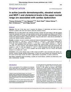
Figure 1 Fresh and cultured human DSCs and FDCs. Fresh DSCs and FDCs (left) were observed 24 h after isolation (bar: 50 mm). FDC exhibited a dendritic morphology with clustered lymphoid cells. After 1 week of culture (right), non-adherent cells died in both preparations, and DSCs and FDCs showed a more polygonal/fibroblastic morphology (bar: 100 mm). After 2 weeks of culture, both types of cell lines exhibited a uniformly fibroblastic morphology (see Fig. 6). This experiment was performed independently four times.
3
Decidual stromal cells and follicular dendritic cells
FDCs; Buettner et al., 2010) and (iii) prolactin (PRL) secretion (a property typical of DSCs). None of these characteristics were found in MSCs. We discuss the implications of these shared properties in the context of immune-endocrine regulation during pregnancy.
Materials and Methods Samples For the FDC lines, tonsil samples were obtained from patients who underwent tonsillectomy for recurrent tonsillitis at the Hospital Universitario Virgen de las Nieves (Granada, Spain). Patients (3–10 years old) were in complete remission before surgery. Informed consent was obtained
from the parents or guardians of each patient. For the DSC lines, samples from elective vaginal terminations of first-trimester pregnancies (6– 11 weeks) were obtained from healthy women aged 20 – 30 years. We excluded women who were using any medication or with infectious, autoimmune or other systemic or local disease. None of the abortions were pharmacologically induced. The specimens were obtained by suction curettage at the Clı´nica El Sur in Ma´laga or the Clı´nica Ginegranada in Granada. Informed consent was obtained from each woman. For the MSC lines, leftover samples of bone marrow aspirates from haematologically normal donors at the Hospital de Baza, in Baza, Granada, were obtained after donors had provided informed consent. This study was approved by the Research and Ethics Committee of the Hospital Universitario Virgen de las Nieves and by the Ethics Committee of the University of Granada.
Figure 2 Antigen expression and C-X-C motif chemokine 13 (CXCL13) secretion by human DSCs and FDCs. (A) Flow cytometry analysis of antigen expression. Almost all DSCs and FDCs were positive for CD10, CD29 and CD73. Both types of cell expressed FDC markers CD14, CD21, CD23, CD54 and BAFF (intracellular). Isotype control (- - -); mAb (—). Percentages show the proportion of cells expressing the antigen. This experiment was performed independently three times. (B) Secretion of CXCL13 by different DSC, FDC and MSC lines.
4
Monoclonal antibodies The monoclonal antibodies (mAbs) used in this study were CD10-phycoerythrin conjugated (PE), CD14-fluorescein isothiocyanate (FITC), CD21-PE, CD23-FITC, CD29-FITC, CD45-FITC, CD73-PE, ICAM-1-FITC (Caltag Laboratories, Burlingame, CA, USA), VCAM-1FITC, BAFF-FITC, CXCL13-Alexa Fluorow 750, STRO-1 (R&D Systems, Minneapolis, MN, USA) and anti-a-SM actin FITC (Sigma-Aldrich, St. Louis, MO, USA). The isotype controls used were immunoglobulin (Ig)M, IgG1-FITC, IgG1-PE or IgG1-APC (Sigma-Aldrich) and IgG2-FITC or IgG2-PE (Caltag Laboratories).
Isolation and culture of DSC, FDC and MSC lines To establish DSC and FDC lines, we used the method described by Montes et al. (1995) and Mun˜oz-Fernandez et al. (2006). Briefly, tissues were thoroughly washed in phosphate-buffered saline (PBS) and finely minced between two scalpels in a small volume of PBS. The suspension was put in a solution of 5 mg/ml Collagenase V (Sigma-Aldrich) for 30 min for DSCs and 2 h for FDCs at 378C. The suspension was diluted in PBS, filtered through gauze and centrifuged at 425g for 10 min. The supernatant was discarded and the cell pellet was suspended in PBS and centrifuged on FicollPaque (Sigma-Aldrich) for 20 min at 600g. To establish MSC lines, bone marrow aspirates were directly diluted in PBS and centrifuged on FicollPaque. Tonsil, decidual or bone marrow cells were collected from the interface, suspended in PBS and washed. The resulting suspension was incubated in culture flasks for 24 h at 378C in Opti-MEM (minimum essential medium; Invitrogen, Grand Island, NY) supplemented with 3% fetal calf serum (FCS), 100 UI/ml penicillin, 100 IU/ml streptomicin and 0.25 mg/ml amphotericin (Sigma-Aldrich). After overnight incubation to allow adherent cells to attach to the flask, non-adherent cells in the supernatant were discarded. The medium was then replaced and changed twice a week. After 1 – 3 weeks, adherent cells were morphologically uniform and covered the whole surface of the 25 cm2 culture flask. Although the different cell lines are referred to generically as DSCs, FDCs or MSCs, in those experiments in which several lines of the same type of cells were included, we used a specific name for each line (DSC MOR, DSC VI, DSC IX, DSC XVI, DSC 1, FDC46, FDC13H, FDC12V, FDC1S, MSC1 and MSC2).
Isolation of fresh DSCs To obtain a fresh cell suspension containing DSCs, we used a protocol similar to that reported by Montes et al. (1996). The decidua was washed in PBS and minced between two scalpels in a small volume of RPMI 1640 with 10% FCS. The suspension was put in a solution of 5 mg/ml Collagenase V (Sigma-Aldrich) for 30 min at 378C. This preparation was filtered through sterile gauze, washed by centrifugation and suspended in the culture medium. The cell suspension was centrifuged at 650g for 30 min over a discontinuous gradient of 20 and 30% Percoll (Sigma-Aldrich). Cells were collected from the 20/30% interphase and washed in PBS. Cell viability was determined by trypan blue exclusion. Only preparations with .95% viable cells were used.
Preparation of purified B cells
Mun˜oz-Ferna´ndez et al. the pellet was suspended in PBS at 106cells/ml. Cell suspension of 100 ml was incubated with 5 ml of the appropriate mAbs for 30 min at 48C in the dark. Cells were washed, suspended in 0.5 ml of PBS and immediately analysed in an FACScan cytometer (BD Biosciences, San Diego, CA, USA). The percentage of Ab-positive cells was calculated by comparison with the appropriate isotype control. For double labelling, we followed the same procedure as for single labelling but a second mAb with a different fluorescent marker was also added. For intracytoplasmic labelling, cells were fixed with 4% paraformaldehyde for 20 min at 48C and permeabilized with cold 0.05% PBS saponine (Merck, Darmstadt, Germany) before the mAbs was added. For indirect labelling, FITC-labelled goat anti-mouse Ig was added after the first mAbs.
Detection of apoptotic tonsil B lymphocytes co-cultured with DSCs, FDCs or MSCs DSC, FDC or MSC lines in the exponential growth phase were plated on 6-well plates on the bottom surface of Transwell plates (Corning Incorporated, New York, NY, USA) at 5 × 10⁴cells/well. The culture medium was removed and 2 ml of a suspension of 5 × 10⁴ B lymphocytes in a complete Opti-MEM culture medium was added directly to the FDCs, DSCs or MSCs, or to the upper compartment of Transwell plates, and incubated at 378C for 72 h. All B lymphocytes were collected from the supernatant, and to identify apoptotic cells we used the method described by Gong et al. (1994). Briefly, cells were washed with PBS, fixed in cold 70% ethanol and then stained with propidium iodide during treatment with RNase (Sigma-Aldrich). Quantitative analysis of sub-G1 cells was carried out in an FACScan cytometer.
Treatment of DSC, FDC and MSC lines DSC, FDC or MSC lines were cultured for 72 h with 10 ng/ml TNF (Sigma-Aldrich) and 10 ng/ml lymphotoxin (LT)a1b2 (Sigma-Aldrich), two cytokines involved in the differentiation of FDCs. To induce decidualization, DSC, FDC or MSC lines were treated with 300 nm progesterone and 500 mm cAMP (Sigma-Aldrich) for 15 days.
Table I Antigen expression by DSC, FDC and MSC lines. Antigens
DSC (n5 10)
FDC (n 5 10)
MSC (n 5 5)
........................................................................................ CD10
+++ a
++ +
+++
CD14
+/2
+
2
CD21
++
++ +
2
CD23
++
++
2
CD29
+++
++ +
+++
CD45
2
2
2
CD54
+++
++ +
+
CD73
+++
++ +
+++
CD106
+/2
+/2
+/2
a-SM actin
+++
++ +
+++
Tonsil B lymphocytes were purified from the supernatant obtained in the preparation of FDCs using the Human B Cell Isolation kit (R&D Systems). The purity of the B lymphocyte preparations was higher than 95%.
BAFF
+++
++ +
2
CXCL13
+++
++ +
2
STRO-1
++
++
++
Flow cytometric analysis
n , number of cell lines analysed. a Flow cytometry reactions: + + +, 67 – 100% positive cells; + +, 34 –66%; +, 5–33%; +/2, some lines positive and some negative.
Cells were detached from the culture flask by treatment with 0.04% EDTA at 378C. The cells were centrifuged, the supernatant was discarded and
5
Decidual stromal cells and follicular dendritic cells
Analysis of culture supernatants The concentration of CXCL13 in supernatants from DSC, FDC or MSC cultures was established by enzyme-linked immunosorbent assay (ELISA; R&D Systems). To determine the concentration of PRL, supernatants from cultures of DSCs, FDCs or MSCs treated with progesterone and 8-bromo-cAMP were collected. The presence of PRL was determined with an ELISA (Roche, Basel, Switzerland).
Statistical analysis Quantitative experiments were performed in triplicate or quadruplicate. The data were analysed with Microsoft Excel 2003 (Microsoft Ibe´rica, Seville, Spain). Student’s t-test was used to compare the results. Values of P , 0.05 were considered significant. The figures show results for a
single experiment, which is representative of three or more separate assays.
Results DSCs exhibit distinctive characteristics of FDCs After 24 h of culture, polygonal and rounded adherent cells appeared in decidua preparations, whereas in tonsil preparations, adherent cells showed a dendritic morphology and were associated with attached lymphoid cells. After 1 week of culture, lymphoid cells bound to FDC detached or died and cultures of DSCs and FDCs formed
Figure 3 BAFF and CXCL13 expression by fresh human DSCs. Fresh DSCs were isolated by Percoll gradients and electronically selected by the expression of CD10. Most fresh CD10+ DSCs were positive for BAFF, and a proportion of them also expressed CXCL13. This experiment was performed independently three times.
6
Mun˜oz-Ferna´ndez et al.
uniform populations of adherent cells with a fibroblastic morphology (Fig. 1). In both types of cell lines, almost all cells were positive for CD10, CD29, CD73 and a-SM actin, and lacked CD45; a proportion of them also expressed CD14, CD21, CD23, CD54 (ICAM-1), CD106, BAFF, CXCL13 and STRO-1, and secreted CXCL-13 (Fig. 2, Table I). Flow cytometric analysis of Percoll-isolated lowdensity decidual cells gated by CD10 expression revealed the presence of cells that, like cultured DSCs and FDCs, expressed BAFF and CXCL13 (Fig. 3). Table I shows that DSCs, FDCs and MSCs expressed the MSC-associated antigens CD10, CD29, CD54, CD73, CD106, a-SM actin and STRO-1, and lacked CD45. However, unlike DSCs and FDCs, MSCs did not express BAFF or CXCL13, and did not secrete CXCL13. We also found that the FDC-associated antigens CD14, CD21 and CD23 were absent from MSC lines (Fig. 2, Table I).
Effect of DSCs, FDCs and MSCs on B lymphocyte apoptosis Like FDCs, DSCs (but not MSCs) inhibited the spontaneous apoptosis of tonsil B lymphocytes (Fig. 5A and B). When FDCs were co-cultured with B lymphocytes in Transwell plates, the protective effect of FDCs on B cells disappeared (Fig. 5C). Likewise, the protective activity of DSCs on B cells was also abrogated in Transwell plate cultures, which demonstrates that this effect also requires DSC-B cell contact (Fig. 5C).
Decidualization of DSCs and FDCs with progesterone and cAMP Like DSCs, progesterone and cAMP changed the morphology of FDCs and induced PRL secretion (Fig. 6). However, the amount of PRL secreted by FDCs was much lower than that secreted by DSCs (P , 0.0001; Fig. 6B). MSC lines cultured with progesterone and cAMP did not secrete PRL (Fig. 6).
Treatment of DSCs, FDCs and MSCs with TNF and LTa1b2
Discussion
Like FDC, DSC and MSC lines treated with TNF and LTa1b2 also increased their expression of CD54 and CD106 (Fig. 4).
In an earlier characterization of DSCs, we reported that these cells expressed FDC antigens (Montes et al., 1996). We demonstrated
Figure 4 Treatment of human DSCs, FDCs and MSCs with TNF and lymphotoxin (LT)a1b2. TNF and LTa1b2 increased the expression of CD54 and CD106 by DSC, FDC and MSC lines. (A) Without cytokines (- - -); with cytokines (—). Percentages show the proportion of cells expressing the antigen. (B) Bars show the percentage of cells expressing the antigen. White bars: untreated cells; black bars: cells treated with cytokines (mean + SD). This experiment was performed independently three times. *P , 0.05; **P , 0.005; ***P , 0.0001.
7
Decidual stromal cells and follicular dendritic cells
Figure 5 Human DSCs and FDCs, but not MSCs, rescue B cells from spontaneous apoptosis. (A) Apoptosis of tonsil B cells cultured without or with DSCs, FDCs or MSCs was studied by quantitative flow cytometry analysis of sub-G1 cells. This experiment was performed independently three times. (B) DSCs and FDCs significantly decreased the spontaneous apoptosis of B cells (mean + SD of three independent experiments). **P , 0.005. (C) Culture in Transwell plates abrogated the protective effect of DSCs or FDCs against B cell apoptosis (mean + SD of three independent experiments).
that DSC and FDC lines exhibited an antigen phenotype and functional properties equivalent to those of their corresponding fresh cells (Montes et al., 1996, Garcia-Pacheco et al., 2001, Mun˜oz-Fernandez et al., 2006). Subsequently, we studied the relationships of both types of cells with myofibroblasts (Oliver et al., 1999; Kimatrai et al., 2003, 2005). We also found that DSCs and FDCs were closely related to MSCs, and that this supported the hypothetical origin of the two former types of cells from MSCs (Garcia-Pacheco et al., 2001, Mun˜oz-Fernandez et al., 2006). The present study confirms the close relationships between DSCs, FDCs and MSCs, because all three cell types expressed the MSC-associated antigens CD10, CD29, CD54, CD73, CD106, a-SM actin and STRO-1, and lacked CD45 expression (Table I; Chamberlain et al. 2007). CD10 is also considered a clinical marker of endometrial stromal cells or DSCs (Sumathi and McCluggage, 2002). Furthermore, TNF and LTa1b2,
increased CD54 and CD106 expression in all three types of cells (Fig. 4). DSCs and FDCs, however, shared some distinctive phenotypical and functional features which were not detected in MSCs. Of these differential properties observed in DSC and FDC lines, PRL secretion during culture with progesterone and cAMP (Fig. 6) is a typical characteristic of DSCs (Tabanelli et al., 1992; Montes et al., 1995), whereas CD14, CD21, CD23, BAFF, CXCL13 expression, the secretion of CXCL13 (Fig. 2, Table I) and the capacity to rescue tonsil B lymphocytes from apoptosis (Fig. 5) are considered distinctive characteristics of FDCa (Lindhout et al., 1997; Park and Choi, 2005; Buettner et al., 2010). Furthermore, we confirmed, as previously reported (Kim et al., 1995), that this latter activity required FDC-B cell contact; likewise, DSCs also required DSC-B contact to inhibit B cell apoptosis (Fig. 5C). We found, however, that the capacity of these DSC lines
8
Mun˜oz-Ferna´ndez et al.
Figure 6 Secretion of prolactin (PRL) by human DSC and FDC lines. (A) DSCs and FDCs were cultured without (left) and with progesterone and cAMP (right) for 2 weeks (Bar: 100 mm). DSCs and FDCs changed from a fibroblastic to a round/dendritic morphology in the presence of progesterone and cAMP. This experiment was performed independently three times. (B) Secretion of PRL by different FDC, DSC and MSC lines (white bars, without progesterone and cAMP; black bars, with progesterone and cAMP). The supernatants of lines MSC 1 and MSC 2 were analysed together with the supernatants of DSC 1, which was studied in the same experiment as a positive control for decidualization. PRL secretion in cultures with progesterone and cAMP was significantly higher in DSCs than in FDCs (the data of the two DSC lines were compared with data of the three FDC lines of the figure, P , 0.0001). MSCs did not secrete PRL.
to secrete CXCL13 in vitro varied (Fig. 2B). Nevertheless, it should be taken into account that DSC lines, like FDC and MSC lines, are normal cells that do not exhibit the in vitro uniformity characteristic of tumour cell lines. The FDC properties detected in the DSC lines are, however, associated with B cell functions (El Shikh et al., 2010), yet B cells are absent from the human decidua (Bulmer, 1995). Interactions between B cells and DSCs may play a role in pathological situations such as the development of uterine B cell lymphomas (Alvarez et al., 1997), infections during pregnancy and inflammation (NhanChang et al., 2008). Nevertheless, the in vitro effects of DSCs on B cells may reflect physiological effects of DSC factors on other types of decidual lymphocytes, as these factors may operate on different types of immune cells (Blanco et al., 2009). High levels of CXCL13 were detected in the decidua and plasma of pregnant women (Nhan-Chang et al., 2008), and according to our results (Fig. 2), DSCs may contribute to the secretion of CXCL13
during pregnancy. Moreover, CXCL13 is a chemokine for regulatory T cells (Lee et al., 2006), and these cells have been detected in human decidua and appear to play a key role in maternal – fetal immune tolerance (Leber et al., 2010). BAFF, a cytokine expressed by FDCs, participates in the rescue of B lymphocytes from apoptosis in the lymphoid follicle, and plays an important role in tolerance modulation and B cell homeostasis (Gorelik et al., 2003). In normal decidua, with no B cells to rescue, the BAFF secreted by DSCs may be involved in the induction of a Th2 response (Phillips et al., 2003; Langat et al., 2008), which is the immune response associated with normal pregnancy (Saito et al., 2010), and in the inhibition of decidual T cell and NK cell apoptosis (Mackay and Leung, 2006; Blanco et al., 2009). Interestingly, mesenchymal cells of the trophoblast are also an important source of BAFF, although, as in the decidua, there are no B cells in this fetal tissue (Langat et al., 2008). The BAFF cytokine is also associated with immune-mediated
9
Decidual stromal cells and follicular dendritic cells
obstetric and gynaecological pathologies such as endometriosis, in which high levels of BAFF have been detected (Hever et al., 2007), and also with recurrent spontaneous abortions, in which the expression of BAFF in the decidua and trophoblasts was found to be reduced (Guo et al., 2008). FDCs and DSCs also shared properties when they were treated with TNF and LTa1b2. These cytokines are differentiation factors for FDCs (Fu and Chaplin, 1999), and they increase the expression of CD54 and CD106, molecules involved in FDC-B cell interactions (Mun˜oz-Fernandez et al., 2006; Fig. 4). Likewise, TNF- and LTa1b2treated DSCs also increased their expression of CD54 and CD106, a property that was also observed in MSCs (Fig. 4; Ren et al., 2010). These molecules, which are highly inducible by cytokines, may be associated with relevant functions of these cells in their respective tissues. Interestingly, CD54 plays a role in DSC physiology, regulating the interaction of DSCs with decidual NK cells (Vigano et al., 1994). Furthermore, the expression of CD54 and CD106 by DSCs may be related to immunosuppressive activity and the maintenance of maternal –fetal tolerance (Blanco et al., 2008) as shown in MSCs (Ren et al., 2010). On the other hand, DSCs differentiate (decidualize) under the effects of progesterone and other hormones. During the luteal phase of the menstrual cycle, or if pregnancy occurs, DSCs or their endometrial counterpart, endometrial stromal cells, are induced to decidualize. The decidualized cells become rounder, express desmin in their cytoplasm and secrete PRL (Tabanelli et al., 1992; Montes et al., 1996; Fig. 6). Locally secreted PRL may modulate the mother’s immune responses (Draca, 1995). Like DSCs, FDCs (but not MSCs) also became rounder and secreted PRL (Fig. 6) under the effects of progesterone and cAMP. Prolactin locally produced by FDCs as a result of the effects of pregnancy hormones may modulate B cell functions (Peeva et al., 2003). In this connection, a recent report showed that FDCs are involved in the induction of tolerance to fetal antigens that occurs during pregnancy (McCloskey et al., 2011). Thus, pregnancy regulates the mother’s immune response, and DSCs and FDCs may be involved in these homeostatic endocrine – immune mechanisms. We cannot assert, however, that FDCs and DSCs are the same type of cells. Although FDCs and DSCs share many characteristics, they also exhibit differences. For example, the amount of PRL secreted by FDCs was significantly lower than that secreted by DSCs (Fig. 6B). What our results suggest is the existence of a common precursor related to MSCs. This hypothesis is supported by the finding of DSCs or FDCs in ectopic locations in immune-mediated diseases (Lindhout et al., 1999; Hever et al., 2007). Under physiological conditions, the common precursor may home either to the secondary lymphoid organs or to the endometrium to develop, respectively, into FDCs or DSCs, acquiring new phenotypical and functional properties such as the expression of BAFF, PRL and CXCL13 secretion, and the ability to rescue B or other lymphocytes when it emigrates to different peripheral tissues. This MSC precursor, however, may also home to ectopic tissues in certain immune diseases.
Acknowledgements We are grateful to the Servicio de Otorrinolaringologı´a, Hospital Universitario Virgen de las Nieves in Granada for providing us with tonsils
specimens. We thank K. Shashok for improving the use of English in the manuscript.
Authors’ roles R.M.-F and A.P. carried out all the experiments. E.L. was responsible for cell culture and A.B. for the analysis of prolactin secretion. R.G.-F was responsible for bone marrow aspirations, G.O.-F. supervised the flow cytometry analysis and E.G.-O. supervised the entire study procedure including conception, design and completion.
Funding This work was supported by the Fondo de Investigaciones Sanitarias, Ministerio de Sanidad (grant PS09/00339) and a Proyecto de Investigacio´n de Excelencia, Consejerı´a de Economı´a, Innovacio´n y Ciencia de la Junta de Andalucı´a (grant CTS-6183).
Conflict of interest None declared.
References Alvarez A, Ortiz J, Sacrista´n F. Large B-cell lymphoma of the uterine corpus: case report with immunohistochemical and molecular study. Gynecol Oncol 1997;65:534 – 538. Blanco O, Tirado I, Mun˜oz-Fernandez R, Abadia-Molina AC, Garcia-Pacheco JM, Pena J, Olivares EG. Human decidual stromal cells express HLA-G: effects of cytokines and decidualization. Hum Reprod 2008;23:144 – 152. Blanco O, Leno-Duran E, Morales JC, Olivares EG, Ruiz-Ruiz C. Human decidual stromal cells protect lymphocytes from apoptosis. Placenta 2009;30:677 – 685. Buettner M, Pabst R, Bode U. Stromal cell heterogeneity in lymphoid organs. Trends Immunol 2010;31:80 –86. Bulmer JN. Immune Cells in Decidua. Oxford: BIOS Scientific Publishers Limited, 1995. Chamberlain G, Fox J, Ashton B, Middleton J. Concise review: mesenchymal stem cells: their phenotype, differentiation capacity, immunological features, and potential for homing. Stem Cells 2007;25:2739–2749. Draca S. Prolactin as an immunoreactive agent. Immunol Cell Biol 1995; 73:481– 483. El Shikh ME, El Sayed RM, Sukumar S, Szakal AK, Tew JG. Activation of B cells by antigens on follicular dendritic cells. Trends Immunol 2010; 31:205– 211. Fu YX, Chaplin DD. Development and maturation of secondary lymphoid tissues. Annu Rev Immunol 1999;17:399 – 433. Garcia-Pacheco JM, Oliver C, Kimatrai M, Blanco FJ, Olivares EG. Human decidual stromal cells express CD34 and STRO-1 and are related to bone marrow stromal precursors. Mol Hum Reprod 2001;7:1151– 1157. Gong J, Traganos F, Darzynkiewicz Z. A selective procedure for DNA extraction from apoptotic cells applicable for gel electrophoresis and flow cytometry. Anal Biochem 1994;218:314– 319. Gorelik L, Gilbride K, Dobles M, Kalled SL, Zandman D, Scott ML. Normal B cell homeostasis requires B cell activation factor production by radiation-resistant cells. J Exp Med 2003;198:937– 945. Guo W, Qu X, Yang M, Zhang W, Liang L, Shao Q, Kong B. Expression of BAFF in the trophoblast and decidua of normal early pregnant women
10 and patients with recurrent spontaneous miscarriage. Chin Med J 2008; 121:309–315. Hever A, Roth R, Hevezi P, Marin M, Acosta J, Acosta H, Rojas J, Herrera R, Grigoriadis D, White E et al. Human endometriosis is associated with plasma cells and overexpression of B lymphocyte stimulator. Proc Natl Acad Sci USA 2007;104:12451–12456. Kanda Y, Jikihara H, Markoff E, Handwerger S. Interleukin-2 inhibits the synthesis and release of prolactin from human decidual cells. J Clin Endocrinol Metab 1999;84:677 –681. Kim HS, Zhang X, Klyushnenkova E, Choi YS. Stimulation of germinal center B lymphocyte proliferation by an FDC-like cell line, HK. J Immunol 1995;155:1101 –1109. Kimatrai M, Oliver C, Abadia-Molina AC, Garcia-Pacheco JM, Olivares EG. Contractile activity of human decidual stromal cells. J Clin Endocrinol Metab 2003;88:844 –849. Kimatrai M, Blanco O, Mun˜oz-Fernandez R, Tirado I, Martin F, Abadia-Molina AC, Olivares EG. Contractile activity of human decidual stromal cells. II. Effect of interleukin-10. J Clin Endocrinol Metab 2005;90:6126 –6130. Langat D, Wheaton D, Platt J, Sifers T, Hunt J. Signaling pathways for B cell-activating factor (BAFF) and a proliferation-inducing ligand (APRIL) in human placenta. Am J Pathol 2008;172:1303–1311. Leber A, Teles A, Zenclussen AC. Regulatory T cells and their role in pregnancy. Am J Reprod Immunol 2010;63:445 –459. Lee B, Chen W, Shi H, Der S, Fo¨rster R, Zhang L. CXCR5/CXCL13 interaction is important for double-negative regulatory T cell homing to cardiac allografts. J Immunol 2006;176:5276 –5283. Lindhout E, Koopman G, Pals S, de Groot C. Triple check for antigen specificity of B cells during germinal centre reactions. Immunol Today 1997;18:573 –577. Lindhout E, van Eijk M, van Pel M, Lindeman J, Dinant HJ, de Groot C. Fibroblast-like synoviocytes from rheumatoid arthritis patients have intrinsic properties of follicular dendritic cells. J Immunol 1999; 162:5949–5956. Mackay F, Leung H. The role of the BAFF/APRIL system on T cell function. Semin Immunol 2006;18:284 –289. McCloskey ML, Curotto de Lafaille MA, Carroll MC, Erlebacher A. Acquisition and presentation of follicular dendritic cell-bound antigen by lymph node-resident dendritic cells. J Exp Med 2011; 208:135–148. Montes MJ, Tortosa CG, Borja C, Abadia AC, Gonzalez-Gomez F, Ruiz C, Olivares EG. Constitutive secretion of interleukin-6 by human decidual stromal cells in culture. Regulatory effect of progesterone. Am J Reprod Immunol 1995;34:188 –194. Montes MJ, Aleman P, Garcia-Tortosa C, Borja C, Ruiz C, Garcia-Olivares E. Cultured human decidual stromal cells express
Mun˜oz-Ferna´ndez et al.
antigens associated with hematopoietic cells. J Reprod Immunol 1996; 30:53 – 66. Mun˜oz-Fernandez R, Blanco FJ, Frecha C, Martin F, Kimatrai M, Abadia-Molina AC, Garcia-Pacheco JM, Olivares EG. Follicular dendritic cells are related to bone marrow stromal cell progenitors and to myofibroblasts. J Immunol 2006;177:280 – 289. Nhan-Chang C, Romero R, Kusanovic J, Gotsch F, Edwin S, Erez O, Mittal P, Kim C, Kim M, Espinoza J et al. A role for CXCL13 (BCA-1) in pregnancy and intra-amniotic infection/inflammation. J Matern Fetal Neonatal Med 2008;21:763– 775. Olivares EG, Montes MJ, Oliver C, Galindo JA, Ruiz C. Cultured human decidual stromal cells express B7– 1 (CD80) and B7– 2 (CD86) and stimulate allogeneic T cells. Biol Reprod 1997;57:609– 615. Oliver C, Cowdrey N, Abadia-Molina AC, Olivares EG. Antigen phenotype of cultured decidual stromal cells of human term decidua. J Reprod Immunol 1999;45:19– 30. Park CS, Choi YS. How do follicular dendritic cells interact intimately with B cells in the germinal centre? Immunology 2005;114:2 – 10. Peeva E, Michael D, Cleary J, Rice J, Chen X, Diamond B. Prolactin modulates the naive B cell repertoire. J Clin Invest 2003;111:275– 283. Phillips T, Ni J, Hunt J. Cell-specific expression of B lymphocyte (APRIL, BLyS)- and Th2 (CD30L/CD153)-promoting tumor necrosis factor superfamily ligands in human placentas. J Leukoc Biol 2003;74:81 – 87. Ren G, Zhao X, Zhang L, Zhang J, L’Huillier A, Ling W, Roberts AI, Le AD, Shi S, Shao C et al. Inflammatory cytokine-induced intercellular adhesion molecule-1 and vascular cell adhesion molecule-1 in mesenchymal stem cells are critical for immunosuppression. J Immunol 2010;184:2321–2328. Richards RG, Brar AK, Frank GR, Hartman SM, Jikihara H. Fibroblast cells from term human decidua closely resemble endometrial stromal cells: induction of prolactin and insulin-like growth factor binding protein-1 expression. Biol Reprod 1995;52:609– 615. Saito S, Nakashima A, Shima T, Ito M. Th1/Th2/Th17 and regulatory T-cell paradigm in pregnancy. Am J Reprod Immunol 2010;63:601 –610. Sumathi VP, McCluggage WG. CD10 is useful in demonstrating endometrial stroma at ectopic sites and in confirming a diagnosis of endometriosis. J Clin Pathol 2002;55:391 – 392. Tabanelli S, Tang B, Gurpide E. In vitro decidualization of human endometrial stromal cells. J Steroid Biochem Mol Biol 1992;42:337– 344. Vigano P, Pardi R, Magri B, Busacca M, Di Blasio AM, Vignali M. Expression of intercellular adhesion molecule-1 (ICAM-1) on cultured human endometrial stromal cells and its role in the interaction with natural killers. Am J Reprod Immunol 1994;32:139– 145. Zhu XM, Han T, Sargent IL, Wang YL, Yao YQ. Conditioned medium from human decidual stromal cells has a concentration-dependent effect on trophoblast cell invasion. Placenta 2009;30:74– 78.