Int. J. Morphol., 29(3):934-938, 2011.
Histochemistry and Morphometric Analysis of Muscle Fibers from Patients with Duchenne Muscular Dystrophy (DMD) Histoquímica y Análisis Morfométrico de las Fibras Musculares de Pacientes con Distrofia Muscular de Duchenne (DMD)
*
**
George Maciel Cavalcanti; *André de Sá Braga Oliveira; *Thiago de Oliveira Assis; Leila Maria Cardão Chimelli; ***Paloma Lys de Medeiros & ****Diógenes Luís da Mota
CALVACANTI, G. M.; OLIVEIRA, A. S. B.; ASSIS, T. O.; CHIMELLI, L. M. C.; MADEIROS, P. L. & MOTA, D. L. Histochemistry and morphometric analysis of muscle fibers from patients with Duchenne muscular dystrophy (DMD). Int. J. Morphol., 29(3):934-938, 2011. SUMMARY: The aim of the study was to analyze the muscle fibers by histochemistry and morphometric methods from patients with Duchenne muscular dystrophy (DMD). Muscle biopsies were taken from the vastus lateralis muscle of five boys between 13 and 15-years of age, with clinical diagnosis of DMD. The histochemistry was performed using myofibrillar ATPases (9.6, 4.6 and 4.3). To morphometrical analysis a computerized semiautomatic system and software Image-Lab was used. ATPase staining showed atrophy of muscle fibers. Fibrosis and adipose deposition occurred in variable degree depending of muscular involvement. The morphometrical analysis showed an increase of size (percentage) to type I fiber than other types in all patients. Furthermore, the type I fiber had a larger cross-sectional area and mean diameter than type IIa and IIb fibers. Both histochemistry and morphometric analysis could be important tools for qualitative and quantitative diagnostics of muscle fibers attacked in this type of disease. KEY WORDS: Histochemistry analysis; Morphometric analysis; Vastus lateralis muscle; Duchenne muscular dystrophy.
INTRODUCTION
The muscular dystrophies (MD) form a wellrecognized heterogeneous group of genetic disorders determined and accompanied by progressive destruction of muscle fibers that expressed with progressive muscle weakness and loss of muscle mass (Zats et al., 1976). Duchenne muscular dystrophy (DMD) has a world-wide incidence of approximately 1:3500 male live births (Emery, 1991) and is an X-linked recessive disorder, considered the most common and most severe muscular dystrophy in childhood, for which there is no effective therapy (Emery, 1993). In most cases, the disease is diagnosed on the basis of gait abnormalities at 4-5 years age. By 8-10 years of age, deterioration of patients condition necessitates wheelchair use. At the same time, by their early teens, neurological and cardiological symptoms are apparent. The disease occurs as a result of mutation within the gene that located at Xp21 and codes for dystrophin protein (Koenig et al., 1989; *
Worton, 1995; El-Harouni et al., 2003). The aim of the study was to analyze muscle fibers of vastus lateralis by histochemistry and morphometric tests in a group of five patients affected by Duchenne muscular dystrophy.
MATERIAL AND METHOD
Subjects. Five patients with Duchenne muscular dystrophy (DMD) were randomly chosen from the Sector of Neuropatologia do Hospital das Clínicas do Rio de Janeiro within the period of August 2004 to June of 2005. These cases were referred by the head of Neuropathology of Clinical Hospital (UFRJ), Profa. Dra. Leila Chimelli, who contributed to the interchange of Pathology Pós-graduation Program (UFPE). The diagnosis of DMD was established
Departamento de Morfologia, Universidade Federal de Pernambuco. Recife-PE, Brasil. Setor de Neuropatologia do Hospital das Clínicas da Universidade Federal do Rio de Janeiro. Rio de Janeiro-RJ, Brasil. *** Departamento de Histologia e Embriologia do Centro de Ciências Biológicas da Universidade Federal de Pernambuco. Recife-PE, Brasil. **** Programa de Pós-graduação em Nutrição da Universidade Federal de Pernambuco. Recife-PE, Brasil. **
934
CALVACANTI, G. M.; OLIVEIRA, A. S. B.; ASSIS, T. O.; CHIMELLI, L. M. C.; MADEIROS, P. L. & MOTA, D. L. Histochemistry and morphometric analysis of muscle fibers from patients with Duchenne muscular dystrophy (DMD). Int. J. Morphol., 29(3):934-938, 2011.
based on neurological findings and myopathic electromyography pattern. The research protocol was approved by the Ethics Committee of Hospital das Clínicas da Universidade Federal de Pernambuco, Recife/PE (CEP/CCS/UFPE-Brazil, no. 179/2004) and informed consent was obtained from every subject. Histochemistry analysis. Skeletal muscle biopsies were taken of vastus lateralis muscles of five patients. Cryosections from the muscle biopsies were processed for histochemistry as described by Brook & Kaiser (1970). The histochemistry was performed using myofibrillar ATPase pre-incubated in medium alkaline and acids (9.6, 4.6 and 4.3). Morphometric analysis. Morphometrical analysis was realized with a computerized semiautomatic system and software Image-Lab. The parameters evaluated were minor diameter (mm) and area (mm2) of skeletal muscle fibers. Statistical analysis. Statistical analysis of data was performed using means ± standard deviations (S.D.). Frequency relative and absolute (%) of muscle fibers was analyzed.
RESULTS
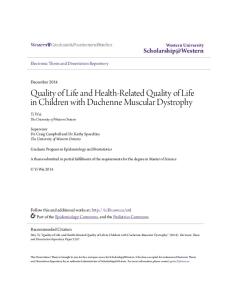
As illustrated in Figure 1 (A, B and C) the characteristic histological features of vastus lateralis muscles in DMD are very similar in the five patients of this study. ATPase staining in DMD has classically shown atrophy in the majority of fibers. There is also evidence of marked variability in fiber size and endomysial and perimysial fibrosis with fatty infiltration. ATPase staining in pH alkaline (Fig. 1) permitted to identify fibers type I or white and fibers type II or dark. ATPase staining in others pH (acids) demonstrated different patterns of fibers (Fig. 1B and C).
Table I. Absolute fiber (%) of types of fiber (I, IIa and IIb) from medial region of vastus lateralis muscles of five patients with DMD. n= number of muscle fibers Muscle fibers Type I
n
%
1762
63.08
Type IIa
605
21.66
Type IIb
426
15.26
2793
100.00
Total
Fig. 1. mATPase histochemical reaction at pH 9.6 (A), 4.3 (B) and 4.6 (C) in cross sections of vastus lateralis muscle from one patient with Duchenne muscular dystrophy (DMD). Type I, I; Type IIa, a; Type IIb, b. Scale bar in (A) represents 100 mm (X200).
935
CALVACANTI, G. M.; OLIVEIRA, A. S. B.; ASSIS, T. O.; CHIMELLI, L. M. C.; MADEIROS, P. L. & MOTA, D. L. Histochemistry and morphometric analysis of muscle fibers from patients with Duchenne muscular dystrophy (DMD). Int. J. Morphol., 29(3):934-938, 2011.
The morphometrical analysis achieved an increased size (percentage) to type I fiber than type IIa and IIb, in all patients (Table I). We observed in Table I, the absolute frequency of main types of fiber like I (63.08%), IIa (21.66%) and type IIb (15.25%). In this disease, affected muscles show a markedly increased variation in fiber diameters (Table II). Furthermore, the type I fiber had a larger cross-sectional area than type IIa and IIb fibers (1107.6 ± 825.3mm2, 1016.9 ± 679.7mm2 and 964.0 ± 626.0mm2) respectively. The mean diameter was observed in the fibers type I when compared with other fibers (IIa and IIb). The results
demonstrated values like 30.7 + 10.7mm, 29.7 + 9.8mm and 28.9 + 10.9mm, respectively (Table III). In 80% of the patients, the fibers type I were observed in bigger number that the fibers type IIa, and this was bigger than the type IIb. The relative frequency of these fibers was respectively: a) patient (1) with 59.31%, 21.31% and 19.37%; b) patient (2) with 51.26%, 31.77% and 16.97%; c) patient (3) with 62.74%, 23.53% and 13.72%; d) patient (4) with 42.91%, 38.33% and 18.75%. However, in 20% of the cases (patient 5) the fibers type IIb (10.59%) showed in bigger numbers than the type IIa (9.05%); but the predominance of type I (80.35%) was maintained (Fig. 2).
Table II. Total area of muscle fiber types from five patients with DMD. Values (Mean± SD) of area (mm2) of fibers type I, IIa and IIb from medial region of vastus lateralis muscles. Fibers Total area in µm2 ( Mean+SD) Medium Minimum Maximum
Type I
1107.6 + 825.3
863.0
165.1
6016.1
Type IIa
964.0 + 626.0
849.8
91.8
3514.9
Type IIb
1016.9 + 679.7
884.9
110.6
4673.3
Table III. Total diameters of the fiber types from five patients with Duchenne muscular dystrophy. Values (Mean±SD) of minor diameter (mm) of fibers type I, IIa and IIb from medial region of vastus lateralis muscles. Fibers Total diameter in µm (Mean+SD) Medium Minimum Maximum
Type I
30.7 + 10.7
28.5
10.5
69.3
Type IIa
28.9 + 10.9
28.5
8.8
62.7
Type IIb
29.7 + 9.8
28.6
8.3
72.0
Fig. 2. Frequency relative (%) of muscle fibers (types I, IIa and IIb) of each patient with Duchenne muscular dystrophy, values percentage over each column.
936
CALVACANTI, G. M.; OLIVEIRA, A. S. B.; ASSIS, T. O.; CHIMELLI, L. M. C.; MADEIROS, P. L. & MOTA, D. L. Histochemistry and morphometric analysis of muscle fibers from patients with Duchenne muscular dystrophy (DMD). Int. J. Morphol., 29(3):934-938, 2011.
DISCUSSION
The muscular dystrophies (MD) constitute a heterogeneous group of genetic disorders determined and accompanied by progressive destruction of muscle fibers with progressive muscle weakness and loss of muscle mass that has been the objective of much research. In this study we observed characteristic changes according to finds in the literature. Toop & Emery (1974) described the predominance of fibers type I in the majority of muscle biopsy of their patients, although fibers type IIb were more affected (15%). Our casuistic showed the same, except in one of the patients that showed increase value for type IIb, suggesting genetic influence or early physiotherapy treatment to avoid the evolution of this disease. Jay & Vajsar (2001) referred to histological changes common in all types of muscular dystrophies such as loss of muscle mass, marked variability in fiber size and endomysial and perimysial fibrosis with fatty infiltration. We reported the same finds and observed the marked variability in fiber size by evaluation of minor diameter of these fibers and the presence of residual fibers. Since that time contributions to the knowledge of muscular dystrophies have been numerous, particularly in relation to the classification on basis of diameter fibers, no one alteration could occur above the 13-years old (Brooke & Engel, 1969). This hypothesis is not valid in our group of patients, one once that we accompanied boys between 13 and 15 years of age. Dubowitz & Brooke (1985) mentioned the predominance of fibers type I associated with genetic disease
and type II with motor disease. Our study showed the great frequency of the fibers type I in all patient corroborating with these authors. Simoneau & Bouchard (1989) demonstrated the percentage of vastus lateralis muscle of Caucasians North Americans 16 to 33years of age. An important find referrer that the vastus lateralis and deltoid muscles maintain the size before weakness appears (Adams et al., 1997; Zats, 2002). Since the beginning of this research we used muscle biopsy of vastus lateralis. Many events that occur in specific tissue were produced by protein diversity and involved regulatory mechanism of genic expression (Muntoni et al., 2003). The same authors mentioned the destruction of muscular fibers and the substitution by fat cells (Kapsa et al., 2003). In general context, these changes reflected in the quality of life of affected patients (Adams et al.; Piccininni et al., 2004). In the cases of Duchenne muscular dystrophy where the biopsy was examined, appearance of the muscles was characteristic and corresponded to that seen by others (Zats; Muntoni et al.; Day & Ranum, 2005). In conclusion both histochemistry and morphometric analysis must be used like important tools for qualitative and quantitative diagnostics of muscle fibers attacked in this type of disease. This observation to reinforce the use these analysis for furthers studies. ACKNOWLEDGEMENTS. The authors wish to thank Alex Benício da Silveira (Department of Pharmacia, UFPE) for excellent technical assistance.
CALVACANTI, G. M.; OLIVEIRA, A. S. B.; ASSIS, T. O.; CHIMELLI, L. M. C.; MADEIROS, P. L. & MOTA, D. L. Histoquímica y análisis morfométrico de las fibras musculares de pacientes con distrofia muscular de Duchenne (DMD). Int. J. Morphol., 29(3):934938, 2011. RESUMEN: El objetivo del estudio fue analizar las fibras musculares mediante histoquímica y métodos morfométricos en pacientes con distrofia muscular de Duchenne (DMD). Se tomaron biopsias musculares del músculo vasto lateral de cinco niños entre 13 y 15 años de edad, con diagnóstico clínico de DMD. La histoquímica se realizó mediante ATPasa miofibrilar (9.6, 4.6 y 4.3). Para el análisis morfométrico se utilizó un sistema semiautomático computarizado y software de imagen de laboratorio. La tinción de ATPasa mostró una atrofia de las fibras musculares. La fibrosis y depósito adiposo se observó en grado variable dependiendo del compromiso muscular. El análisis morfométrico mostró un aumento de tamaño (porcentaje) de fibras tipo I en todos los pacientes. Además, la fibra tipo I tuvo un área de sección transversal y diámetro medio mayor que las fibras tipos IIa y IIb. Tanto la histoquímica y el análisis morfométrico pueden ser herramientas importantes para el diagnóstico cualitativo y cuantitativo de las fibras musculares comprometidas en este tipo de enfermedad. PALABRAS CLAVE: Análisis histoquímico; Análisis morfométrico; Músculo vasto lateral; Distrofia muscular de Duchenne.
937
CALVACANTI, G. M.; OLIVEIRA, A. S. B.; ASSIS, T. O.; CHIMELLI, L. M. C.; MADEIROS, P. L. & MOTA, D. L. Histochemistry and morphometric analysis of muscle fibers from patients with Duchenne muscular dystrophy (DMD). Int. J. Morphol., 29(3):934-938, 2011.
REFERENCES
Adams, R. D.; Victor, M. & Ropper, A. H. Distrofias Musculares. 6a ed. Santiago, McGraw-Hill Interamericana, 1997. Brooke, M. H. & Engel, W. K. The histographic analysis of human muscle biopsies with regard to fiber types. 4. Children's biopsies. Neurology, 19(6):591-605, 1969. Brooke, M. H. & Kaiser, K. K. Muscle fiber types: how many and what kind? Arch. Neurol., 23(4):369-79, 1970. Day, J. W. & Ranum, L. P. RNA pathogenesis of the myotonic dystrophies. Neuromuscul. Disord., 15(1):5-16, 2005. Dubowitz, V. & Brooke, M. H. Muscle Biopsy: A Practical Approach. 2nd ed. London, Philadelphia & Toronto, W. B. Saunders, 1985. El-Harouni, A. A.; Amr, K. S.; Effat, L. K.; Eassawi, M. L.; Ismail, S.; Gad, Y. Z. & El-Awady, M. K. The milder phenotype of the dystrophin gene double deletions. Acta Neurol. Scand., 107(6):400-4, 2003. Emery, A. E. Population frequencies of inherited neuromuscular diseases--a world survey. Neuromuscul. Disord., 1(1):19-29, 1991. Emery, A. E. Duchenne muscular dystrophy. 2nd ed. Oxford and New York, University Press, 1993. Jay, V. & Vajsar, J. The dystrophy of Duchenne. Lancet, 357(9255):550-2, 2001.
Simoneau, J. A. & Bouchard, C. Human variation in skeletal muscle fiber-type proportion and enzyme activities. Am. J. Physiol., 257(4 Pt 1):E567-72, 1989. Toop, J. & Emery, A. E. Muscle histology in fetuses at risk for Duchenne muscular dystrophy. Clin. Genet., 5(3):230-3, 1974. Worton, R. Muscular dystrophies: diseases of the dystrophinglycoprotein complex. Science, 270(5237):755-6, 1995. Zats, M.; Frota-Pessoa, O.; Levy, J. A. & Peres, E. A. Creatine-phosphokinase (CPK) activity in relatives of patients with X-linked muscular dystrophies: a Brazilian study. J. Genet. Hum., 24(2):153-68, 1976. Zats, M. A biologia molecular contribuindo para a compreensão e prevenção das doenças hereditárias. Ciênc. Saúde Coletiva, 7(1):85-99, 2002.
Correspondence to: Prof. Thiago de Oliveira Assis Departamento de Morfologia Universidade Federal da Paraiba (UFPB) Cidade Universitária, S/N. CEP: 58051-900 João Pessoa-Paraíba BRAZIL Phone: +55.83.32167200
Kapsa, R.; Kornberg, A. J. & Byrne, E. Novel therapies for Duchenne muscular dystrophy. Lancet Neurol., 2(5):299310, 2003. Koenig, M.; Beggs, A. H.; Moyer, M.; Scherpf, S.; Heindrich, K.; Bettecken, T.; Meng, G.; Müller, C. R.; Lindlöf, M. & Kaariainen, H. The molecular basis for Duchenne versus Becker muscular dystrophy: Correlation of severity with type of deletion. Am. J. Hum. Genet., 45(4):498-506, 1989. Muntoni, F.; Torelli, S. & Fernili, A. Dystrophin and mutations: one gene, several proteins, multiple phenotypes. Lancet Neurol., 2(12):731-40, 2003. Piccininni, M.; Falsini, C. & Pizzi, A. Quality of life in hereditary neuromuscular diseases. Acta Neurol. Scand., 109(2):113-9, 2004.
938
Email:
[email protected]
Received: 21-01-2011 Accepted: 02-05-2011