Journal of General Virology (2006), 87, 2253–2262
DOI 10.1099/vir.0.81849-0
Hepatitis C virus polyprotein vaccine formulations capable of inducing broad antibody and cellular immune responses Michael Vajdy,1 Mark Selby,13 Angelica Medina-Selby,1 Doris Coit,1 John Hall,1 Laura Tandeske,1 David Chien,1 Celine Hu,1 Domenico Rosa,2 Manmohan Singh,1 Jina Kazzaz,1 Steve Nguyen,1 Steve Coates,1 Philip Ng,1 Sergio Abrignani,2 Yin-Ling Lin,1 Michael Houghton1 and Derek T. O’Hagan1 1
Chiron Vaccines, 4560 Horton Street, Emeryville, CA 94608, USA
Correspondence Michael Houghton
2
Chiron Vaccines, Via Fiorentina 1, 53100 Siena, Italy
[email protected]
Received 21 January 2006 Accepted 30 March 2006
Although approximately 3 % of the world’s population is infected with Hepatitis C virus (HCV), there is no prophylactic vaccine available. This study reports the design, cloning and purification of a single polyprotein comprising the HCV core protein and non-structural proteins NS3, NS4a, NS4b, NS5a and NS5b. The immunogenicity of this polyprotein, which was formulated in alum, oil-in-water emulsion MF59 or poly(DL-lactide co-glycolide) in the presence or absence of CpG adjuvant, was then determined in a murine model for induction of B- and T-cell responses. The addition of adjuvants or a delivery system to the HCV polyprotein enhanced serum antibody and T-cell proliferative responses, as well as IFN-c responses, by CD4+ T cells. The antibody responses were mainly against the NS3 and NS5 components of the polyprotein and relatively poor responses were elicited against NS4 and the core components. IFN-c responses, however, were induced against all of the individual components of the polyprotein. These data suggest that the HCV polyprotein delivered with adjuvants induces broad B- and T-cell responses and could be a vaccine candidate against HCV.
INTRODUCTION Hepatitis C virus (HCV) is the leading cause of parenterally transmitted non-A, non-B viral hepatitis (Armstrong et al., 2000; Choo et al., 1989). Approximately 3 % of the world’s population are infected with HCV (Cohen, 1999) and about 30 000 newly acquired HCV infections occur in the USA annually, mostly as a result of intravenous drug abuse (Alter, 1993). Currently, there is no vaccine available to prevent HCV infection and the only available therapies, IFN-a and ribavirin, are effective in fewer than half of the patients treated (McHutchison et al., 1998; Poynard et al., 1998). Therefore, there is an urgent need for the development of an efficacious vaccine to prevent HCV infection. We previously developed an experimental vaccine comprising a recombinant gpE1 and gpE2 heterodimer that protects chimpanzees against experimental challenge with both homologous (Choo et al., 1994) and heterologous (Coates et al., 2004) genotype 1a strains, which predominate in the USA and also occur worldwide. Natural immunity to 3Present address: Medarex Inc., 521 Cottonwood Drive, Milpitas, CA 95035, USA.
0008-1849 G 2006 SGM
HCV infection has been linked with early and broad Th1type cellular immune responses to non-structural proteins NS3, NS4 and NS5 and the nucleocapsid (C) protein (Diepolder et al., 1995; Ferrari et al., 1997; Gerlach et al., 1999; Tsai et al., 1997). More recent data have shown the importance of Th1-type CD4+ T-cell responses in protection in chimpanzees (Grakoui et al., 2003) and humans (Lechner et al., 2000). To enhance the immunogenicity of recombinant proteinbased vaccines, adjuvants are required. The most widely used adjuvants are insoluble aluminium salts, generically called alum. However, although alum adjuvants have been used for decades and are generally safe, they induce predominantly a Th2-type cytokine response (Lindblad, 2004; Raz & Spiegelberg, 1999; Valensi et al., 1994). Therefore, alternative adjuvants may be required for the successful development of an HCV vaccine. Among many alternative approaches available, polymeric microparticles have been evaluated as vaccine adjuvants (Eyles et al., 2003; O’Hagan & Singh, 2003; Singh et al., 2004a, b). Moreover, in a nonhuman primate model, poly(DL-lactide co-glycolide) (PLG) microparticles have already been shown to induce potent T-cell lymphoproliferative responses against an adsorbed
Downloaded from www.microbiologyresearch.org by IP: 37.44.207.89 On: Sun, 15 Jan 2017 23:09:03
Printed in Great Britain
2253
M. Vajdy and others
recombinant antigen from human immunodeficiency virus (HIV) (Otten et al., 2003). Hence, PLG microparticles appear to be an attractive approach for the development of new-generation adjuvants and in these studies we assessed these microparticles for the delivery of the polyprotein from HCV.
1
2
3
4
Various oil-in-water emulsions have also been developed as alternative adjuvants to alum. The most advanced of these is a squalene oil-in-water emulsion (MF59), which is a potent adjuvant with an acceptable safety profile and has gained approval for licensure in an influenza vaccine product in Europe called Fluad (Ott et al., 1995; Podda & Del Giudice, 2003). To enhance Th1-type responses to vaccines, immunopotentiating adjuvants may be added to ‘delivery’-based adjuvants. CpG is an immune potentiator that has been shown to induce Th1-type responses selectively in smallanimal models (Klinman et al., 2004; Schetter & Vollmer, 2004). In addition, the inclusion of CpG into MF59 or PLG formulations for HIV vaccines has been shown to enhance antibody, as well as Gag-specific, cytotoxic T-cell responses (O’Hagan et al., 2002; Singh et al., 2001). In this study, we designed, cloned, expressed and purified a single polyprotein comprising the HCV core and NS3, NS4a, NS4b, NS5a and NS5b proteins, designated NS345Core. We determined the in vivo immunogenicity of the purified polyprotein formulated with alum, MF59 or PLG in the presence or absence of CpG for induction of B- and T-cell responses in a murine model.
METHODS Construction of HCV-1a DNS3–NS5 (aa 1242–3011) with the core protein (aa 1–121 or 1–173). The plasmid pT7-HCV was
created in a polylinker-modified pUC18 vector (New England Biolabs) to contain full-length HCV-1a cDNA (Choo et al., 1991) preceded by a synthetic T7 promoter. pT7-HCV also contains the complete 59 UTR and the poly(A) version of the 39 UTR followed by an XbaI site. The plasmid pCMV-KMDNS3-5 was generated by ligating a PCR fragment that encodes a Kozak initiator methionine followed by aa 1242 of HCV-1a to a 39 StuI site. This product was ligated to a StuI–XbaI fragment from pT7-HCV along with pCMVKM2 vector (Murphy et al., 1998). The termini of the polyprotein-encoding DNA were modified to be compatible with restriction sites in the pBS24.1 yeast shuttle vector, which contains 2m sequences for autonomous replication in yeast, the a-factor terminator to ensure transcription termination, the yeast genes leu2-d and URA3 for selection, the Col E1 origin of replication and the b-lactamase gene required for plasmid replication in bacteria (Lin et al., 2005). Thereafter, the hybrid ADH2/GAPDH promoter was cloned 59 to the polyprotein-coding sequence to generate pd.DNS3NS5.PJ. This plasmid was transformed into Saccharomyces cerevisiae strain AD3 [MATa, leuD, ura3-52, prb-1122, pep4-3, prc1-407 (CIR0), : : pDM15 (GAP/ADR)]. Clone 5 was analysed for expression and a band with the expected molecular mass of 194 kDa was observed by Coomassie blue staining (Fig. 1). Thereafter, the core was appended on to the C terminus of the DNS3NS5 protein. Two core sequences were used: one terminated at aa 121 and the other at aa 173. 2254
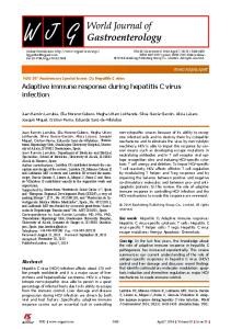
Fig. 1. Expression level of clone pd.DNS3NS5.PJ from the plasmid pd.DNS3NS5 clone #17 shown by Coomassie blue staining. See Methods for details. A protein band of the expected molecular mass of 194 kDa is shown (arrow). Lanes: 1 and 4, size standards; 2, control yeast plasmid; 3, pd.DNS3NS5.PJ colony 1.
The core sequences used the reverse-transcribed version, where aa 9 is Arg rather than Lys and aa 11 is Thr rather than Asn. Strain AD3 was transformed with pd.DNS3NS5.PJ.core121RT clone #6 and pd.DNS3NS5.PJ.core173RT clone #15 and checked for expression. Protein bands of the expected molecular masses of 206 and 210 kDa, respectively, were observed by Coomassie blue staining. Expression levels of the pd.DNS3NS5.PJ.core173RT construct were consistently less (data not shown) than that of the pd.DNS3NS5.PJ.core121RT construct (Fig. 2a). Therefore, in all of the immunogenicity studies, the pd.DNS3NS5.PJ.core121RT construct was used. Purification of the HCV NS345Core121 polyprotein. Yeast cells
expressing insoluble HCV polyprotein intracellularly were lysed by milling with glass beads in a buffer containing 50 mM Tris/HCl, 100 mM sodium chloride, 10 mM dithiothreitol (DTT) and 1 mM PMSF at pH 8?0. The lysate was collected and centrifuged at 39 200 g (Beckman JA-20 rotor at 18 000 r.p.m.) at 22 uC for 30 min. The pellet was washed by resuspending in 50 mM Tris/HCl, 100 mM sodium chloride, 2 M urea, 0?1 % octyl glucoside and 1 mM PMSF at pH 8?0 and mixing well with an OmniMixer. The centrifugation was repeated and the pellet was washed a second time by resuspending in PBS plus 50 mM DTT and 1 % Tween 20 (Sigma). The pH of the suspension was raised to pH 12 with 6 M sodium hydroxide and the suspension was mixed by stirring at room temperature for 30 min. The pH was lowered to pH 8?0 with 6 M hydrochloric acid, more DTT was added to a final concentration of 0?1 M and the suspension was centrifuged as before. HCV polyprotein was extracted from the insoluble pellet by suspending in 50 mM Tris/HCl, 50 mM DTT and 2 % SDS at pH 8?0, raising the pH to 12 for 30 min while stirring, followed by neutralization to pH 8?0. The centrifugation was repeated and the supernatant containing soluble HCV polyprotein was purified further by size-exclusion chromatography using a Superdex 200 column equilibrated with PBS containing 0?1 % SDS and 5 mM DTT. Fractions containing the purest HCV polyprotein as determined by SDS-PAGE were pooled and dialysed using Spectrapore dialysis tubing (molecular mass cut-off of 25 kDa) against water for 6 h at room temperature to
Downloaded from www.microbiologyresearch.org by IP: 37.44.207.89 On: Sun, 15 Jan 2017 23:09:03
Journal of General Virology 87
HCV polyprotein vaccine
(a)
1
2
3
4
5
6
7
temperature for 1 h. The primary antibody was an anti-HCV-C22 monoclonal antibody (specific for the HCV core protein) (developed at Chiron Corporation), which was diluted 1 : 2000 in TBST with 1 % powdered milk and incubated with shaking at room temperature for 1 h. The membrane was washed three times for 5 min each with TBST. The secondary antibody was an alkaline phosphatase-conjugated goat anti-mouse antibody (Boehringer Mannheim) diluted 1 : 2000 in TBST with 1 % powdered milk and incubated with shaking at room temperature for 1 h. The membrane was washed three times for 5 min each with TBST and developed using Western blue stabilized substrate for alkaline phosphatase (Promega). Formulation of the HCV polyprotein with adjuvants. RG503
(b)
1
2
1
175
175
83
83
62
62
47
47
32
32
25
25
16 6
16 6
2
PLG with a 50 : 50 co-polymer composition (intrinsic viscosity of 0?4 from the manufacturer’s specifications) was obtained from Boehringer Ingelheim. Dioctylsulfosuccinate (DSS) and urea were from Sigma. NS345Core121 and MF59C were obtained from Chiron Corporation. Aluminium phosphate was obtained from Superfos. All other reagents were obtained from Sigma. CpG (1826; Oligos Incorporation) was added at a concentration of 25 mg per dose to the delivery system (alum, PLG or MF59) before immunization. PLG/NS345Core121 formulation. PLG microparticles were prepared by a solvent evaporation method as described previously (Kazzaz et al., 2000; Singh et al., 2004a). Briefly, microparticles were prepared by homogenizing 10 ml 6 % (w/v) polymer solution in methylene chloride with 2?5 ml PBS using a 10 mm probe (UltraTurrax T25; IKA-Labortechnik). The water-in-oil emulsion thus formed was then added to 50 ml distilled water containing DSS and the mixture was homogenized at high speed with a 20 mm probe (ES-15 Omni International) for 25 min in an ice bath. This procedure resulted in the formation of water-in-oil-in-water emulsion, which was then stirred at 1000 r.p.m. for 12 h at room temperature and the methylene chloride allowed to evaporate. The size distribution of the resulting microparticles was determined with a particle size analyser (Master Sizer; Malvern Instruments).
Fig. 2. (a) Expression levels of the pd.DNS3NS5.PJ.core173RT and pd.DNS3NS5.PJ.core121RT constructs shown by Coomassie blue staining. See Methods for details. Protein bands of the expected molecular masses of 206 and 210 kDa, respectively, are shown (arrows). Expression of the pd.DNS3NS5.PJ.core173RT construct was considerably lower than that of the pd.DNS3NS5.PJ.core121RT construct. Lanes: 1 and 7, size standards; 2, control yeast plasmid; 3, 4, pd.DNS3NS5.PJ.core121RT colonies 1 and 2; 5 and 6, pd.DNS3NS5.PJ.core173RT colonies 3 and 4. (b) Coomassie blue staining (left) and Western blot (right) of the purified HCV NS345Core121 polyprotein. Lane 1, Pre-stained molecular mass markers (Broad Range; New England Biolabs); lane 2, 0?5 mg Superdex 200 gel filtration pool.
Microparticles with adsorbed protein were prepared by incubating a suspension containing 100 mg blank PLG microparticles with 1 mg protein in PBS with 8 M urea at pH 9. The suspension was allowed to mix on a laboratory rocker (Aliquot Mixer; Miles Laboratories) overnight. The suspension was then dialysed against several changes of water and lyophilized in aliquots containing 250 mg protein each, which comprised ten doses. Alum/NS345Core121 formulation. The alum formulation was
made by incubating 100 mg aluminium phosphate with 1 mg protein in PBS with 8 M urea at pH 5?5 at a final concentration of 0?5 mg ml21. The suspension was aliquotted in vials containing 250 mg protein each, which comprised ten doses. MF59/NS345Core121 formulations. MF59 formulations were prepared by adding 250 ml MF59C (citrate) with 50 ml 5 mg NS345Core121 ml21 and 200 ml PBS and mixing immediately prior
to injection.
remove salts. The purified HCV polyprotein was concentrated by lyophilizing to dryness and redissolving the protein in a minimal volume of buffer (20 mM Tris/HCl, 8 M urea, 5 mM DTT at pH 8?0) to achieve complete solubilization. Buffer exchange into the final buffer was performed by dialysing into a buffer of PBS plus 6 M urea and 5 mM DTT. The expected molecular mass was confirmed by Coomassie blue staining and the antigenicity was confirmed by Western blotting (Fig. 2b). The Western blot was performed by transferring a Tris/glycine gradient gel to 0?2 mm nitrocellulose (BioRad), blocking for 10 min with TBST (Tris-buffered saline with 0?2 % Tween 20) containing 1 % powdered milk and shaking at room http://vir.sgmjournals.org
Mice and immunizations. Female BALB/c or C57BL/6 mice aged 6–8 weeks at the onset of the study were used. Six (proliferation) or ten (ELISPOT and ELISAs) mice per group were used for each immunization. All animal studies were approved by the Chiron Animal Use and Care Committee. Intramuscular (i.m.) immunizations were performed in the thigh with or without anaesthesia once or twice at 3 week intervals. Mice were sacrificed at 8 days after one immunization or at 2 weeks following the second immunization to collect tissues for the proliferation and ELISPOT assays, respectively. Sera and tissue collection. Mice were bled through the retro-
orbital plexus 2 weeks after each immunization to prepare sera
Downloaded from www.microbiologyresearch.org by IP: 37.44.207.89 On: Sun, 15 Jan 2017 23:09:03
2255
M. Vajdy and others for the ELISAs. Popliteal lymph nodes and spleen were meshed through a nylon mesh to prepare single-cell suspensions. Spleens were assayed from pools of all mice in each group unless stated otherwise. Proliferation assay. Popliteal lymph node cells were prepared from six mice per group 8 days after a single immunization to measure proliferative responses against HCV NS345Core121 polyprotein (10 mg ml21). Concanavalin A was used as positive control and cells with no antigen in the medium were used as negative controls. The data are represented as mean c.p.m.±SD of six individual mice per group. Results are representative of one experiment of three. ELISPOT assay. Single-cell suspensions from pooled spleen from
five mice per group were prepared and adjusted to concentrations of 16107 to 36107 cells ml21 in culture medium (50 % RPMI 1640, 50 % alpha-MEM with 10 % heat-inactivated fetal bovine serum, 561025 M 2-mercaptoethanol and 1 % antibiotics). One hundred microlitres from each cell preparation was added in duplicate to the first rows of 96-well PVDF plates (Milipore) pre-coated with rat anti-mouse IFN-c (BD PharMingen) and 2-fold serial dilutions were performed. Pooled synthetic peptides (20mers overlapping by 10 aa, 1–3 mg per well) encompassing the entire core, NS3, NS4, NS5a and NS5b sequences of the HCV-1a genotype or the NS345Core121 protein (10 mg ml21 in 100 ml), synthesized using Fmoc solid-phase methods by Research Genetics or Chiron Mimotopes, were then added to the cells and stimulated overnight for 14–16 h at 37 uC. As a negative control, cells were incubated without the pool of peptides. All peptides had free N and C termini. Following overnight incubation, the plates were washed and biotinylated anti-IFN-c (BD PharMingen) was added. The plates were incubated at room temperature for 2 h and washed. Avidin–peroxidase (BD PharMingen) was added and the plates were incubated for 30 min at 37 uC and then washed. The plates were developed with aminoethyl carbazole solution (Sigma) for 30 min. The results from two experiments are presented as the mean±SD of IFN-c-secreting cells per 16107 mononuclear cells from a minimum of four wells from pools of five mice per group with splenocytes added in duplicate. The background number of spots averaged 20 in the wells with cells without peptide stimulation. Flow cytometric analysis following intracellular and surface staining. Two weeks after the final immunization, spleens were
harvested from two pools of five mice each and single-cell suspensions were prepared. Spleen cells (16106 cells in 200 ml) were cultured at 37 uC in the presence or absence of pools of 20mer overlapping peptides covering the NS3, NS4 and NS5a+b region at a concentration of 10 mg ml21 using 100 ml per well or with monoclonal antibodies directed against CD3 and CD28 (both from BD PharMingen) as a positive control. BD GolgiPlug (BD PharMingen) was added to block cytokine secretion. After 6 h, cells were washed, incubated with BD Fc Block (anti-CD16/32; BD PharMingen) to block Fc-c receptors and stained for cell-surface antigens CD4 and CD8 using the BD Cytofix/Cytoperm kit (BD PharMingen) following the manufacturer’s instructions. Cells were then fixed in 2 % (w/v) paraformaldehyde and stored overnight at 4 uC. The following day, cells were permeabilized and stained for intracellular IFN-c with a phycoerythrin (BD PharMingen)-conjugated mouse IFN-c monoclonal antibody in the presence of 0?1 % (w/v) saponin. Cells were washed and analysed on a FACSCalibur flow cytometer (Becton Dickinson Immunocytometry Systems). A 2-fold increase above the background response of 0?01 % was considered to be positive for CD4+ or CD8+ intracellular IFN-cexpressing cells. For all flow cytometric analyses, the data are presented as means±SD of two pools of five mice each from two experiments. 2256
ELISAs. Serum IgG titres specific for NS345Core121, NS3, NS4, NS5 or core protein were quantified by a standard ELISA. Briefly, ELISA plates (96-well U-bottom plates; Nunc MaxiSorp) were coated with purified NS345Core121 protein at a concentration of 3 mg per well in PBS overnight at 4 uC. After washing with PBS plus 0?03 % Tween 20, the wells were blocked with PBS plus 2 % fetal calf serum for 30 min at 37 uC and serum samples were added in an initial dilution of 1 : 5000. Serial dilutions of 1 : 2 were performed in the blocking reagent. A standard serum was included in each assay as a positive control. The samples and standard sera were incubated at 4 uC overnight and washed with PBS plus 0?03 % Tween 20. The plates were washed and horseradish peroxidase-conjugated goat anti-mouse IgG (Southern Biotechnology Associates) diluted 1 : 10 000 in PBS plus 2 % fetal calf serum was added and incubated for 2 h at room temperature. The plates were washed and developed with tetramethylbenzidine (Kirkegaard & Perry Laboratories) for 15 min and then development was stopped with 2 M HCl. The absorbance of each well was measured using a Titertek absorbance reader at 450 nm. The data are presented as mean titres±SEM of six to ten mice per group. Statistical analysis. Student’s t-test on log-transformed values was
performed for two geometric means assuming equal variances (Ftest, P>0?05), using Microsoft EXCEL.
RESULTS Enhanced serum anti-HCV responses following immunizations with polyprotein in delivery systems To determine whether the designed and yeast-expressed HCV polyprotein could induce B-cell responses in vivo, we immunized mice i.m. with the HCV polyprotein in various delivery systems. We found that the HCV polyprotein alone did not induce antigen-specific serum antibodies (Fig. 3a). In contrast, mixing the HCV polyprotein with the oil-in-water emulsion MF59 or adsorption to PLG microparticles induced serum antibody responses. Immunization i.m. with the HCV polyprotein adsorbed to PLG induced the highest antibody responses, followed by mixing with MF59, whereas adsorption to alum induced very low antibody titres (Fig. 3a). To determine whether differential specific responses were induced to each component of the polyprotein, we next measured the serum antibody responses against each of the vaccine components, i.e. NS3, NS4, NS5, core and the polyprotein, in the group immunized with alum. We found that serum antibody titres were greatest against the NS3 and NS5 proteins, whilst they were considerably lower against the NS4 protein, three orders of magnitude lower than anti-NS3 and -NS5 responses (Fig. 3b). Moreover, the serum antibody responses were also relatively low against the core antigen, two orders of magnitude lower than anti-NS3 and -NS5 titres. These data showed that, although the expressed and purified HCV core polyprotein was immunogenic in mice when given together with various delivery systems, the response against each of the components was different, with the majority of the antibody response being against the NS3 and NS5 components.
Downloaded from www.microbiologyresearch.org by IP: 37.44.207.89 On: Sun, 15 Jan 2017 23:09:03
Journal of General Virology 87
HCV polyprotein vaccine
Fig. 4. T-cell proliferative responses in lymph nodes following immunization with NS345Core121 in the various delivery systems. Six mice per group were immunized once i.m. in the tibialis anterior muscle. Eight days later, single-cell suspensions from popliteal lymph nodes were prepared for the proliferation assay and incubated over 5 days with the NS345Core121 polyprotein. The data are represented as mean c.p.m. ±SD of six individual mice per group and are representative of one experiment of three.
Fig. 3. Serum anti-NS345Core121 (a) or anti-NS3, -NS4, -NS5 and -core IgG (b) responses following immunization with NS345Core121 in the various delivery systems. Ten mice per group were immunized i.m. twice with a 3 week interval. The mice were bled 2 weeks after the second immunization to collect sera for the ELISA. The data are presented as mean serum IgG titres±SD.
T-cell proliferative responses following restimulation of cells with HCV polyprotein As both B- and T-cell responses are considered to be important in immunity against HCV, we next determined the T-cell response. Eight days after a single i.m. immunization with HCV polyprotein in the various delivery systems, single-cell suspensions were prepared from popliteal lymph nodes, which drain the tibialis anterior muscle, and restimulated in vitro in the presence of the HCV polyprotein for 3 days. We found that the polyproteins alone did not induce antigen-specific proliferative responses (data not shown). However, mixing the HCV polyprotein with MF59 or adsorption to alum or PLG microparticles induced T-cell proliferative responses (Fig. 4). Alum induced the lowest proliferative response, whilst the responses were similar following immunization with PLG or MF59. These data showed that immunization with the expressed and purified HCV polyprotein in the various delivery systems was able to induce T-cell proliferative responses. T-cell cytokine responses following restimulation of cells with various peptides derived from the HCV polyprotein As a measure of T-cell responses, we next measured IFN-c responses in the spleen following immunization with HCV http://vir.sgmjournals.org
polyprotein. In order to distinguish which proteins within the polyprotein were the most immunogenic, we stimulated the splenocytes with peptides derived from the core, NS3, NS4, NS5a and NS5b proteins and the whole polyprotein, i.e. NS345Core121. We found that immunization with PLG or alum induced the highest frequency of IFN-c-secreting cells following restimulation with the polyprotein or any of the peptides, whilst immunization with MF59 induced the lowest frequency of IFN-c-secreting cells following restimulation with the polyprotein or any of the peptides (Fig. 5). Restimulation with NS345Core121 generally induced the highest response, whilst restimulation with the core peptide induced the lowest response. NS3, NS4, NS5a and NS5b appeared to be equally immunogenic for the induction of IFN-c responses. Thus, these data showed differential IFN-c responses to various components of the HCV polyprotein. Enhanced serum anti-HCV responses following immunization with polyprotein in various delivery systems plus CpG To enhance the serum anti-HCV polyprotein responses further, we immunized mice i.m. with the HCV polyprotein in the various delivery systems in the presence or absence of the immunopotentiating adjuvant CpG. We found that the HCV polyprotein given as a mixture with MF59 plus CpG induced serum antibody responses that were >20-fold higher compared with protein in MF59 without CpG (Fig. 6a). Immunization i.m. with the HCV polyprotein adsorbed to alum plus CpG also induced >20-fold higher serum antibody responses compared with alum alone. The lowest increase in serum antibody titres (