KUOPION YLIOPISTON JULKAISUJA D. LÄÄKETIEDE 432 KUOPIO UNIVERSITY PUBLICATIONS D. MEDICAL SCIENCES 432
KAI SAVONEN
Heart Rate Response to Exercise in the Prediction of Mortality and Myocardial Infarction A Prospective Population Study in Men
Doctoral dissertation To be presented by permission of the Faculty of Medicine of the University of Kuopio for public examination in Auditorium L21, Snellmania building, University of Kuopio, on Saturday 10 th May 2008, at 12 noon
Kuopio Research Institute of Exercise Medicine Department of Biomedicine, Physiology University of Kuopio Department of Clinical Physiology and Nuclear Medicine Kuopio University Hospital
JOKA KUOPIO 2008
Distributor :
Kuopio University Library P.O. Box 1627 FI-70211 KUOPIO FINLAND Tel. +358 17 163 430 Fax +358 17 163 410 www.uku.fi/kirjasto/julkaisutoiminta/julkmyyn.html
Series Editors:
Professor Esko Alhava, M.D., Ph.D. Institute of Clinical Medicine, Department of Surgery
Author´s address:
Kuopio Research Institute of Exercise Medicine Haapaniementie 16 FI-70100 KUOPIO FINLAND Tel. +358 17 288 4422 Fax +358 17 288 4488 E-mail:
[email protected]
Supervisors:
Professor Rainer Rauramaa, M.D., Ph.D., M.Sc. Kuopio Research Institute of Exercise Medicine
Professor Timo A. Lakka, M.D., Ph.D. Department of Biomedicine, Physiology University of Kuopio
Docent Jari A. Laukkanen, M.D., Ph.D. Lapland Central Hospital Rovaniemi
Reviewers:
Docent Matti Mäntysaari, M.D., Ph.D. Center of Military Medicine / Aeromedical Center Helsinki
Docent Mikko Tulppo, Ph.D. Verve, Research Unit of Exercise Medicine Oulu
Opponent:
Professor Olli Raitakari, M.D., Ph.D. Faculty of Medicine University of Turku
Professor Raimo Sulkava, M.D., Ph.D. School of Public Health and Clinical Nutrition Professor Markku Tammi, M.D., Ph.D. Institute of Biomedicine, Department of Anatomy
ISBN 978-951-27-0952-6 ISBN 978-951-27-1049-2 (PDF) ISSN 1235-0303 Kopijyvä Kuopio 2008 Finland
Savonen, Kai. Heart rate response to exercise in the prediction of mortality and myocardial infarction. A prospective population study in men. Kuopio University Publications D. Medical Sciences. 2008. 165 p. ISBN 978-951-27-0952-6 ISBN 978-951-27-1049-2 (PDF) ISSN 1235-0303 ABSTRACT Heart rate (HR) is one of most easily measured exercise test variables. Although easily measured, the HR reflects a complex, integrated physiologic response: autonomic tone, central and peripheral reflexes, hormonal influences, and factors intrinsic to the heart are all important. During the last two decades exercise test derived HR variables have raised wide interest as prognostic markers of mortality and cardiac events both in asymptomatic persons and in patients with cardiovascular disease (CVD). The results of properly controlled studies suggest a possible bimodal relationship of HR to prognosis, in which both high HR at low workload and inappropriately low HR at maximal or near maximal workload are associated with adverse prognosis. Workload achieved at HR of 100 beats/min (WL100) and HR increase from 40% to 100% of maximal work capacity (HR40-100) were assessed using a maximal, symptom-limited exercise test on an electrically braked cycle ergometer. The complete data on exercise test variables was available for 1679 men in a population based sample of men. During the follow-up of eleven years, deaths were ascertained by linkage to the National Death Registry, and the classification of acute myocardial infarctions was carried out according to the multinational MONICA project protocol. A new variable, HR40-100, quantifying an inappropriately low HR at maximal or near maximal workload was at least as strong a predictor of outcome as previously established related variables. A one standard deviation (SD) decrement in HR40-100 (13 beats/min) was related to an increased risk of all-cause death (relative risk, RR 1.3, 95% confidence interval, CI 1.1 to 1.6) and acute myocardial infarction (RR 1.3, 95% CI 1.1-1.6) in men without coronary heart disease (CHD) at baseline after adjustment for age, cardiovascular risk factors and exercise test variables. After exclusion of men whose test was possibly terminated prematurely because of submaximal effort, symptoms, or findings which could be interpreted to indicate latent CHD, a low HR40-100 still was an independent predictor of outcome. A one SD decrement in WL100 was related to an increased risk of CVD death (RR 1.7, 95% CI 1.3-2.4) and all-cause death (RR 1.7, 95% CI 1.3-2.2) in men without CHD at baseline and in men with known or suspected CHD at baseline after adjusting for risk factors, respectively. The exclusion of men who had an outcome event during the first two years of follow-up did not affect the results. A low WL100 was associated with a high resting HR and a low maximal oxygen uptake, but in survival analyses a low WL100 still provided additional prognostic information beyond these variables. A blunted HR increase during the maximal exercise test is associated with an increased risk of death and adverse cardiac events in men without CHD at baseline. The association is particularly strong when the HR increase during the latter half of the test is considered. On the other hand, a low workload achieved at a submaximal HR of 100 beats/min predicts CVD and CHD mortality in men without CHD and all-cause death in men with known or suspected CHD. Contrary to previous interpretations, an exaggerated HR response at a low workload seems to indicate an increased risk by itself instead of being only a surrogate marker of a low cardiorespiratory fitness. The findings of the current thesis support the hypothesis that a bimodal relationship exists between HR and prognosis in which both an exaggerated HR response at submaximal workload and a blunted HR response at maximal or near maximal workload are associated with an adverse prognosis. In the current study sample several exercise test variables predict outcomes independent of each other and conventional risk factors. This emphasizes the importance of measuring several variables at submaximal and maximal workload and during recovery phase to maximize the prognostic yield obtained from the exercise test. National Library of Medicine Classification: WG 141.5.F9, WG 300 Medical Subject Headings: Cardiovascular Diseases/epidemiology; Cardiovascular Diseases/mortality; Coronary Artery Disease; Exercise Test; Finland; Follow-Up Studies; Heart rate; Men; Myocardial Infarction; Risk Factor
Savonen, Kai. Sydämen sykintätaajuus kuormituskokeessa kuolleisuuden ja akuutin sydäninfarktin ennustajana. Epidemiologinen seurantatutkimus. Kuopion yliopiston julkaisuja D. Lääketiede. 2008. 165 p. ISBN 978-951-27-0952-6 ISBN 978-951-27-1049-2 (PDF) ISSN 1235-0303 TIIVISTELMÄ Sydämen sykintätaajuus, syke, on eräs helpoimmin mitattavista kuormituskoemuuttujista. Näennäisestä yksinkertaisuudestaan huolimatta mitattu syke on monimutkaisen säätelyn lopputulos, jossa autonomisen hermoston reflekseillä, hormonaalisilla tekijöillä ja sydämessä itsessään vaikuttavilla tekijöillä on kullakin oma osuutensa. Edeltäneiden kahden vuosikymmenen aikana kuormituskokeenaikaisen sykkeen on lukuisissa tutkimuksissa havaittu olevan yhteydessä kuolleisuuteen ja sydän- ja verisuonitautitapahtumiin sekä oireettomilla tutkittavilla että sydän- ja verisuonitautipotilailla. Tutkimusten perusteella sykkeen ja ennusteen yhteys on mahdollisesti kaksijakoinen niin, että sekä korostunut sykkeen nousu kevyessä kuormituksessa että toisaalta heikentynyt sykkeen nousu maksimikuormituksessa ovat yhteydessä huonoon ennusteeseen. Tässä itä-suomalaisessa väestöpohjaisessa seurantatutkimuksessa tehtiin maksimaalinen kuormituskoe 1679 miehelle. Kuormituskokeessa määritettiin työmäärä, jonka tutkittava saavutti ennen sykkeen kohoamista yli 100 lyöntiä/min, ja sykkeen nousu välillä 40-100% tutkittavan maksimaalisesta suorituskyvystä. Tutkimuksessa selvitettiin näiden kahden sykemuuttujan ennusteellista merkitystä. Seurattavat päätetapahtumat 11 vuoden seuranta-aikana olivat kokonaiskuolleisuus, kuolleisuus sepelvaltimotautiin ja muihin sydän- ja verisuonisairauksiiin, sekä akuutti sydäninfarkti. Nämä päätetapahtumat luokiteltiin hyödyntäen sairaalapoistotietoja ja valtakunnallisen kuolinsyyrekisterin tietoja. Tutkimuksen keskeisinä löydöksinä todettiin, että heikentynyt sykkeen nousu välillä 40-100% tutkittavan maksimaalisesta suorituskyvystä ennustaa ennenaikaisen kuoleman ja akuutin sydäninfarktin riskiä miehillä, joilla ei ollut sepelvaltimotautia seurannan alkaessa. Tämä uusi heikentynyttä sykkeen nousua maksimikuormituksessa kuvaava muuttuja oli vähintään yhtä voimakas ennustaja kuin aikaisemmissa tutkimuksissa käytetyt vastaavaa ilmiötä kuvaavat muuttujat. Tämä tutkimus osoitti toisaalta sen, että matala saavutettu työmäärä ennen sykkeen kohoamista yli 100 lyöntiä/min ennustaa sydän- ja verisuonitautikuoleman riskiä miehillä, joilla ei ollut sepelvaltimotautia seurannan alkaessa. Sama muuttuja ennusti ennenaikaisen kuoleman riskiä myös miehillä, joilla oli todettu tai epäilty sepelvaltimotauti seurannan alkaessa. Matala työmäärä ennen sykkeen kohoamista yli 100 lyöntiä/min oli yhteydessä korkeaan leposykkeeseen ja huonoon kardiorespiratoriseen kuntoon, mutta elinaikaanalyyseissä sen havaittiin kuitenkin tuovan ylimääräistä ennustearvoa näiden kahden ennestään tunnetun vaaratekijän lisänä. Kokonaisuutena tulokset tukevat hypoteesia, että sekä korostunut sykkeen nousu kevyessä kuormituksessa että heikentynyt sykkeen nousu maksimikuormituksessa ovat yhteydessä huonoon ennusteeseen. Tämä väestötutkimus vahvistaa kliinisen kuormituskokeen merkitystä arvioitaessa myöhempien sydäntapahtumien ja enneaikaisen kuoleman vaaraa, sillä usealla kuormituskoemuuttujalla havaittiin olevan ennusteellista lisäarvoa perinteisiin vaaratekijöihin nähden. Tutkitut sykemuuttujat ennustivat päätetapahtumia sekä sepelvaltimotautia sairastamattomilla miehillä että sitä sairastavaksi todetuilla tai epäillyillä miehillä. Koska syke on eräs helpoimmin mitattavista kuormituskoemuuttujista, sykevasteen arviointi sekä kevyessä että maksimaalisessa kuormituksessa kannattaa ottaa huomioon arvioitaessa tutkittavan ennustetta kuormituskoetulosten perusteella. Yleinen suomalainen asiasanasto: sydän- ja verisuonitaudit, epidemiologia; sydän- ja verisuonitaudit, riskitekijät; syke; sydäninfarkti, miehet, Suomi
“A man’s nature and way of life are his fate, and that which he calls his fate is but his disposition.” Menander (342-291 B.C.)
ACKNOWLEDGEMENTS This study was carried out at the Kuopio Research Institute of Exercise Medicine, Kuopio, Finland. I am deeply indebted to all the people who have contributed to this work. In particular, I wish to thank: Professor Rainer Rauramaa, M.D., Ph.D., M.Sc., the head of the Kuopio Research Institute of Exercise Medicine, my supervisor, for the opportunity to do my thesis in the KIHD study, and to work at the Institute while preparing my thesis. Professor Timo Lakka, M.D., Ph.D., my supervisor, for his invaluable contribution in preparing the manuscripts and for his continuous support and optimism. Docent Jari Laukkanen, M.D., Ph.D., my supervisor, for his invaluable contribution in preparing the manuscripts, as well as for friendship and for being a role model as a physician and researcher. Docent Matti Mäntysaari, M.D., Ph.D., and Docent Mikko Tulppo, Ph.D., the official reviewers of this thesis, for valuable comments and constructive criticism. Co-authors Pirjo Halonen, M.Sc., Professor Jukka T. Salonen, M.D., Ph.D., M.Sc. and Tuomas Rauramaa, M.D., for their important contribution to this work. Professor Emeritus Osmo Hänninen, D.Med.Sc., Ph.D., and Professor Emeritus Esko Länsimies, M.D., Ph.D., for invaluable help in arranging funding for my research work. Docent David Laaksonen, M.D., Ph.D., MPH, for revising the English language of this thesis and for friendship. Professor Esko Vanninen, M.D., Ph.D., the head of the Department of Clinical Physiology of the Kuopio University Hospital, for the opportunity to work in the Department as an EVO-funded researcher. Docent Arto Hautala, Ph.D., Docent Tomi Laitinen, M.D., Ph.D., Docent Hanna-Maaria Lakka, M.D., Ph.D., Docent Heikki Pekkarinen, M.D., Ph.D., Miika Hernelahti, M.D., Ph.D., Riikka Kivelä, Ph.D., Antti Kiviniemi, Ph.D., Jonna Eeva, M.D., M.Sc., Jarno Rutanen, M.D., Pertti Huotari, Ph.Lic., Vesa Kiviniemi, Ph.Lic., and Harri Heikkilä, M.Sc., for friendship and interesting discussions on science and other aspects of life. Kimmo Ronkainen, for his help in providing some important variables from the KIHD database when preparing the third manuscript. Arno Heikelä, M.D., Hannu Litmanen, M.D. and Esko Taskinen M.D., for supervising the exercise tests at the KIHD baseline data collection.
The staff of the former Research Institute of Public Health, for their part in the KIHD baseline data collection. Mrs Kirsti Miettinen, for the loving daycare of our sons for so many times during these years. Kalevi Koivunen, M.Sc., Jari Manninen, Matti Paarma, Juha Pastila, Jussi Ripatti and Timo Tiusanen, for being very important lifelong friends. Ulrich Sturm, M.Sc. and Ari Tuuri, M.Sc., for being so refreshing company during Tuesday evening sauna turns. The whole staff of the Kuopio Research Institute of Exercise Medicine, both present and former, for creating such a warm atmosphere at work and for being like my second family during my years in Kuopio. My parents Tuula and Arto Savonen, for all their support during these years. Finally I want to thank the dearest people in my life, Marja, Olli and Vili for their loving support during all these years. I just cannot find the words to express how much your presence matters to me. This study was supported by the Ministry of Education in Finland, the Finnish Cultural Foundation of Northern Savo, the Foundation of Sports Institutes’, the Maire Taponen Foundation, the Mehiläinen Research Foundation, the Paulo Foundation, the University of Kuopio, the Finnish Medical Foundation, and the Orion-Farmos Research Foundation.
Kuopio, April 2008
Kai Savonen
ABBREVIATIONS CHD CVD ECG HR BP SA AV mV rVLM cVLM VO2max VO2 BMI HDL LDL W WL100 HR40-100 KIHD submaxCRI SBP ICD MONICA AMI SD RR CI ANOVA maxCRI
Coronary heart disease Cardiovascular disease Electrocardiogram Heart rate Blood pressure Sinoatrial Atrioventricular Millivolt Rostral ventrolateral medulla Caudal ventrolateral medulla Maximal oxygen consumption Oxygen consumption Body mass index High-density lipoprotein Low-density lipoprotein Watts Workload achieved at a submaximal heart rate of 100 beats/minute Heart rate increase from 40% to 100% of maximal work capacity in exercise test Kuopio Ischaemic Heart Disease Risk Factor Study Chronotropic response index at submaximal work Systolic blood pressure International Classification of Diseases MONItoring of Trends and Determinants in CArdiovascular Disease Acute myocardial infarction Standard deviation Relative risk Confidence interval Analysis of variance Chronotropic response index at maximal work
LIST OF ORIGINAL PUBLICATIONS This dissertation is based on the following original publications referred to in the text by their Roman numerals I – IV: I
Savonen K, Lakka T, Laukkanen J, Halonen P, Rauramaa T, Salonen JT, Rauramaa R. Heart rate response during exercise test and cardiovascular mortality in middle-aged men. Eur Heart J 2006;27:582-588.
II
Savonen K, Lakka T, Laukkanen J, Rauramaa T, Salonen JT, Rauramaa R. Effectiveness of workload at the heart rate of 100 beats/min in predicting cardiovascular mortality in men aged 42, 48, 54, or 60 years at baseline. Am J Cardiol 2007;100:563-568.
III
Savonen K, Lakka T, Laukkanen J, Rauramaa T, Salonen JT, Rauramaa R. Workload at the heart rate of 100 beats/minute and mortality in middle-aged men with known or suspected coronary heart disease. Heart 2008;94:e14.
IV
Savonen K, Lakka T, Laukkanen J, Rauramaa T, Salonen JT, Rauramaa R. Usefulness of chronotropic incompetence in response to exercise as a predictor of myocardial infarction in middle-aged men without cardiovascular disease. Am J Cardiol 2008;101:992-998.
CONTENTS 1. INTRODUCTION
19
2. REVIEW OF LITERATURE
21
2.1 Sinoatrial node
21
2.2 Cardiac innervation
23
2.3 Regulation of heart rate by central nervous system
27
2.4 Factors modulating heart rate response to neural stimulation
29
2.5 Mechanisms controlling heart rate
31
2.5.1 Arterial baroreceptors and cardiopulmonary lowpressure baroreceptors 2.5.2 Central command and peripheral afferents
32 35
2.5.3 Respiratory sinus arrhythmia, arterial chemoreceptors, and pulmonary and cardiac receptors
37
2.6 Heart rate response to exercise: a synthesis
39
2.7 Central circulatory response to exercise
42
2.8 Factors modulating heart rate response to exercise
43
2.9 Heart rate response to exercise and prognosis
49
2.9.1 Submaximal heart rate and cardiovascular disease events in asymptomatic persons
49
2.9.2 Submaximal heart rate and cardiovascular disease events in patients with known or suspected coronary heart disease
53
2.9.3 Chronotropic incompetence and cardiovascular disease events in asymptomatic persons 2.10 Summary
55 66
3. MAIN HYPOTHESIS AND AIMS OF THE STUDY
69
4. METHODS
70
4.1 Study population
70
4.2 Examination protocol
70
4.3 Exercise testing
71
4.3.1 Assessment of heart rate response to exercise
72
4.3.2 Assessment of cardiorespiratory fitness, exercise electrocardiography and exercise blood pressure
73
4.4 Biochemical analyses
74
4.5 Resting blood pressure, body weight and body mass index
74
4.6 Smoking and alcohol consumption
75
4.7 Baseline diseases and medications
75
4.8 Collection and classification of follow-up events
76
4.8.1 Mortality
76
4.8.2 Acute coronary events
76
4.9 Statistical methods
77
4.9.1 Study I
78
4.9.1 Study II
79
4.9.1 Study III
79
4.9.1 Study IV
80
5. RESULTS
82
5.1 Baseline characteristics
82
5.2 Exercise test findings
82
5.3 Heart rate increase from 40% to 100% of maximal work capacity and mortality in men without coronary heart disease (Study I)
86
5.4 Workload at heart rate of 100 beats/min during exercise test and mortality in men without coronary heart disease (Study II)
90
5.4.1 Workload at heart rate of 100 beats/min, other heart ratederived and exercise test variables, and mortality
92
5.4.2 Workload at heart rate of 100 beats/min and mortality: further adjustments
94
5.5 Workload at heart rate of 100 beats/min during exercise test and mortality in men with coronary heart disease (Study III)
94
5.5.1 Workload at heart rate of 100 beats/min, other heart ratederived and exercise test variables, and mortality
96
5.5.2 Workload at heart rate of 100 beats/min, the use of heart rate-lowering medication, and mortality
97
5.5.3 Workload at heart rate of 100 beats/min and mortality: further adjustments
97
5.6 Heart rate increase from 40% to 100% of maximal work capacity and the risk of acute myocardial infarction in men without cardiovascular disease (Study IV)
98
5.6.1 Heart rate increase from 40% to 100% of maximal work capacity, other chronotropic incompetence variables, and the risk of acute myocardial infarction
99
5.6.2 Heart rate increase from 40% to 100% of maximal work capacity, systolic blood pressure response and the risk of acute myocardial infarction
6. DISCUSSION
100
102
6.1 Methodological aspects
102
6.1.1 Study design
102
6.1.2 Study population
104
6.1.3 Exercise testing
107
6.1.4 Collection and classification of outcome events
109
6.2. Results
110
6.2.1 Heart rate increase from 40% to 100% of maximal work capacity, mortality and the risk of acute myocardial infarction in men without coronary heart disease (Studies I and IV)
111
6.2.2 Workload at heart rate of 100 beats/min during exercise test and mortality (studies II and III)
114
6.2.2.1 Workload at heart rate of 100 beats/min during exercise test and cardiovascular disease mortality in men without coronary heart disease at baseline
118
6.2.2.2 Workload at heart rate of 100 beats/min during exercise test and mortality in men with known or suspected coronary heart disease at baseline 6.3 Clinical implications
120 121
7. SUMMARY AND CONCLUSIONS
125
8. REFERENCES
126
9. ORIGINAL PUBLICATIONS I-IV
19
1. INTRODUCTION Coronary heart disease (CHD) is the leading cause of death in the developed world (1) and may become the leading cause of death in the entire world (2). Although the incidence of CHD has been decreasing over the last two decades, the prevalence is expected to increase given the growing elderly population (3-5). It is important to implement cost-effective strategies that direct the appropriate individuals to the optimal risk reduction procedures through risk prediction (4-6). There is a growing awareness of the need to apply statistical techniques to develop evidence-based scores for better decision making (7). The goal of risk prediction through statistical methods is to provide a logical estimate as to the likelihood of the occurrence of important deleterious clinical events (3-5). The most important outcome is death, but the future risk of nonfatal clinical outcomes is also an important element of risk evaluation (3-5). Based on the worldwide epidemiological experience, the evaluation of cardiovascular risk is based on four time-honoured classical cardiovascular disease (CVD) risk factors: age, serum cholesterol, resting systolic blood pressure and smoking status (8). Exercise testing is not recommended in low-risk asymptomatic subjects due to both lacking evidence of its value and because false positive exercise electrocardiograms (ECGs) are common (9). However, the real issue is not to identify CHD, but to predict outcome (10). Because of this, increasing attention has been focused on the exercise test as a prognostic, as opposed to diagnostic, modality (11,12). It is well known that several exercise test indices in addition to ECG findings, such as exercise capacity (9,13), heart rate (HR) (14,15) and blood pressure (BP) responses (16) to exercise, are strong predictors of CVD events. Exercise is the body’s most common physiologic stress, and it places major demands on the cardiovascular system (17). The exercise test provides a precise and powerful noninvasive tool that permits the study of the regulation of the cardiovascular system under rigorously controlled and highly reproducible conditions, which include the full range of its functional capacity (18). The adaptations that occur during an exercise test allow the body to increase its resting metabolic rate up to 20 times, during which time cardiac output may increase as much as six times (17,19). The obvious advantage to the
20
researcher is that more is learned about how a system operates when it is forced to perform than when it is idle (18). Acute exercise can elicit cardiovascular abnormalities that are not evident at rest, and it can be used to determine the adequacy of cardiovascular function (19). Heart rate is one of the most easily measured exercise test variables. Although easily measured, the HR reflects a complex, integrated physiologic response: autonomic tone, central and peripheral reflexes, hormonal influences, and factors intrinsic to the heart are all important (20-22). Recently, the changes in HR during and after exercise have emerged as powerful measures of risk for future CVD event in their own right (23). An interesting fact from the viewpoint of basic exercise physiology is that the mechanisms mediating the association of exercise HR variables and an increased risk of outcomes are largely unknown (24-26). Office-based assessment of conventional risk factor burden is necessary, but may not accurately estimate risk of future CVD events (27). There is a need for easily available non-invasive methods to detect individuals with an increased risk of CVD events who would probably benefit most from preventive measures (28). The main aim of the current study is identify variables derived from exercise test HR which might serve as useful predictors for future CVD events and possibly provide additional prognostic information to conventional risk factors in a population-based sample of middle-aged men.
21
2. REVIEW OF LITERATURE 2.1 Sinoatrial node The normal heart beat starts in the sinoatrial (SA) node (20,21,29). The normal HR is determined by the firing frequency of SA node (20,21,29). The SA node is a small, flattened, ellipsoid strip of specialized cardiac muscle and associated fibroelastic connective tissue about 3 mm wide, 15 mm long, and 1 mm thick (29-31). It contains clusters of cells, poor in contractile filaments, where the automatic activity resides mostly in the pacemaker or P cells (32). Near the periphery of the SA node lie also another group of cells which are called as transitional cells (33). The SA node is located in the superior lateral wall of the right atrium immediately below and slightly lateral to the opening of the superior vena cava, near the superior end of the sulcus terminalis (20,29,34). Its primary source of blood is from the SA nodal artery, which originates from the right coronary artery in about 60% of humans (29,35,36). The normal cardiac impulses starts at the SA node, passes through the atrial tissue through preferential internodal tracts to the atrioventricular (AV) node where it slows, and then continues down the His-Purkinje system to the ventricular myocardium, where the wave of depolarization terminates when there is no further tissue to depolarize (20,31,37). Further conduction occurs only after a new impulse is formed in the SA node (38). Many cardiac cells, especially the cells of the heart’s specialized conducting system, have the capability of self excitation, a process that can cause automatic rhythmical discharge and conduction (39). The resting potential of a typical cardiac cell is -80 to 90 millivolts (mV) (38,39). When it is depolarized to a certain threshold level (threshold potential), an action potential is produced as a result of a complex series of ionic shifts (37-39). The appearance of the action potential of SA nodal cells is different from that of the typical myocyte (38,39). The normal resting potential of these cells is higher (-55 to -60 mV), and the spontaneous diastolic depolarization is much more pronounced (3840). The slope of the diastolic depolarization determines the rate at which a cell will
22
spontaneously depolarize (automaticy) until it reaches the threshold potential, thus generating an action potential that is then propagated to surrounding cells (38,39,41). The spontaneous cyclic depolarization of primary pacemaker cells in the SA node that establish intrinsic HR arises from the unique time-dependent characteristics of a variety of depolarizing and hyperpolarizing currents (37,39,42). The ionic basis of SA node pacemaker activity is such that action potential configuration is determined mainly by outward hyperpolarizing K+ current, IK, and two depolarizing inward currents, ICa and If, that are carried primarily by Ca2+ and Na+, respectively (37,40,43). At the termination of one SA node action potential, the membrane voltage does not stabilize to a negative level but slowly creeps up with an approximately constant slope, until it reaches the threshold for a new SA node action potential (39,44). The gradual membrane depolarization has been attributed to an overall diminution in the net conductance of hyperpolarizing K+ currents and a constant background inward current caused by the spontaneous inward movement along the concentration gradient for Na+ ions (37,45,46). A major role, however, in the generation and control of the diastolic depolarization is played by a prominent increase in the inward depolarizing current If (37,39,46). An initial membrane depolarization leads to the activation of a transient (Ttype) Ca2+ current (ICa,T) which results in Ca2+ influx into a confined subsarcolemmal space between the sarcolemma and sarcoplasmic reticulum, and [Ca2+] in the subsarcolemmal space begins to increase (37,42). This triggers the focal release of Ca2+ (sparks) from sarcoplasmic reticulum Ca2+ release channels, further increasing [Ca2+] in the space (37). This in turn leads to the activation of forward Na+-Ca2+ exchange (INCX) and further membrane depolarization toward a threshold potential (37). Since the stoichiometry of the exchange is three Na+ for one Ca2+, the current is electrogenic and mediates a net inward current, INaCa (37,42). The interrelated actions of ICa,T, sarcoplasmic reticulum Ca2+ initiates, and INaCa creates a positive feedback loop that culminates in a progressive membrane depolarization to the threshold potential (37). Once threshold is achieved, an L-type inward Ca2+ current (ICa,L) rapidly activates, and an action potential is triggered (42,43,47).
23
The SA node usually has the fastest diastolic depolarization and thus functions as the normal pacemaker of the heart (20,21,29). If the SA node fails, the AV node has the next fastest pacemaker rate (approximately 40-60 beats/min) (21,38,39). 2.2 Cardiac innervation The normal myocardium is richly innervated by the autonomic nervous system (31,48). The heart is supplied by autonomic nerve fibers from superficial and deep cardiac plexuses from which three major cardiac nerves project into the heart (35,49). These nerve networks lie between the bifurcation of the trachea and the ascending aorta, and superior to the bifurcation of the pulmonary artery (35). The parasympathetic supply is from preganglionic cardiac branches of the vagus nerves (21,50,51). The cell bodies of the parasympathetic postganglionic fibers constitute intrinsic ganglia in the vicinity of SA and AV nodes (20,48,50). The postganglionic parasympathetic fibers innervate primarily the atria. There are a few projections to the ventricles, however, and there is increasing evidence to show that the vagal nerves innervate the ventricular myocardium as well (48,52,53). The cell bodies of cardiac afferent vagal neurons are contained within the nodose ganglia inferior to the jugular foramen (35,50,54). The central fibers of these bipolar neurons continue to ascend in the vagus to enter the brain stem (35,50,54). Both pre- and postganglionic cardiac parasympathetic fibers release acetylcholine as neurotransmitter (48,50,51). The effects of acetylcholine on the heart are mediated by muscarinic M2-receptors, but the neural transmission between pre- and postganglionic fibers of both the sympathetic and parasympathetic systems is mediated by nicotinic NN-receptors (35,50,51). Once acetylcholine has been secreted, it persists in the tissue for a few seconds. Thereafter, most is split into acetate and choline by the enzyme acetylcholinesterase (48,55). The sympathetic supply is from postganglionic cardiac sympathetic fibers (20,21,51). The cell bodies of pre- and postganglionic fibers are located in the intermediolateral cell columns of the lateral horns of the superior five or six thoracic segments of the spinal cord, and in the cervical and superior thoracic paravertebral ganglia of the sympathetic
24
trunks, respectively (20,21,51). Sympathetic nerve terminals are located throughout the atria and ventricles (20,21,56). The cardiac afferent sympathetic neurons have their cell bodies in the C6-T6 dorsal root ganglia, and they enter the dorsal horn of the spinal cord (35,50,57). They synapse on cells in the outer part of the dorsal horn, the axons of the second-order neurons immediately decussate and ascend through the spinothalamic tract to reach the thalamus (50,57). Pre- and postganglionic cardiac sympathetic fibers release acetylcholine and noradrenaline as neurotransmitters, respectively (20,50,51). In addition to innervation directly from sympathetic nerve endings, the sympathetic nervous system may have an effect indirectly by stimulating the adrenal medulla to secrete adrenaline and noradrenaline into the circulating blood (50,51,58). These two hormones can further bind to adrenergic receptors in the heart (50,51,58). The effects of noradrenaline and adrenaline on the heart are mediated by α1-, β1- and β2-adrenergic receptors (50,51,58). β1 is the most abundant subtype of adrenergic receptor in the heart, representing approximately 75% of total (20,51,58). Noradrenaline excites mainly α-receptors, but also excites the β-receptors to a lesser extent (39,59). Adrenaline excites both types of receptors approximately eqally (39,59). Noradrenaline is removed from the secretory site in three ways: (i) reuptake into the adrenergic nerve endings (accounting for removal of 50%-80% of the secreted noradrenaline); (ii) diffusion away into the surrounding body fluids; and (iii) destruction by enzymes (32,48,55). The latency of the response of the SA node to vagal stimulation is very short (18,21,48). After a single stimulus, the maximum response has been reported to occur within only 400 milliseconds (18,48,60). Thus, vagal stimulation results in a peak response either in the first or in the second beat after its onset (21,48). On the other hand, following the onset of sympathetic stimulation, there is a latent period of up to 5 seconds. This is followed by a progressive increase in HR, which reaches a steady level in 20 to 30 seconds (18,21,48). Both parasympathetic and sympathetic preganglionic fibers are myelinated, whereas postganglionic fibers do not have a myelin sheath (50,61). The fact that parasympathetic postganglionic fibers are clearly shorter than sympathetic postganglionic fibers partly explains the slower cardiac responses to
25
sympathetic stimuli than to parasympathetic stimuli, because the neural transmission is faster in myelinated fibers (39). The HR is determined from the ECG as the reciprocal of the time interval between two successive R peaks (which reflect depolarization of ventricles) and expressed as beats/minute (17,20). The intrinsic HR, in the absence of any neurohumoral influence, is about 100 to 120 beats/min and declines with age (21,62,63). Without neurohumoral influence maximal HR at peak exercise is 18-24% lower than with intact neurohumoral influence (63,64). In the intact, unblocked individual, the HR at any time represents the net effect of the vagal (parasympathetic) and the sympathetic nerves, which play a key role in the regulation of the HR by modulating the intrinsic pacemaker activity of the heart (19,36,65). In resting conditions, both autonomic divisions are thought to be tonically active with the vagal effects dominant (20,36,51). In normal adults at rest, the HR is about 70 to 85 beats/min and the normal range is 60 to 100 beats/min (29,65,66). SA and AV nodes are the most densely innervated regions of the heart and are most affected by changes in autonomic tone, allowing for neural regulation of the HR (37,48). There is some asymmetry in the distribution of autonomic fibers to the heart, and the SA node is predominantly innervated by fibers from the right side (21,29,67). Weak to moderate vagal stimulation will slow the HR often to as little as one-half normal (39,48). Strong vagal stimulation of the heart can stop the heartbeat for a few seconds, but then the heart usually “escapes” and beats at a rate of 20 to 40 beats/min thereafter, paced by a pacemaker elsewhere than in the SA node (21,36,39). Increased vagal input into the SA node results in the release of acetylcholine from nerve endings in the SA node and the released acetylcholine binds to muscarinic receptors (37,39,48). Acetylcholine can directly activate a specific class of K+ channels (KACh) in SA node cells, which produces a hyperpolarizing current that opposes the effects of depolarizing currents during diastole (37,42,48). Also, the release of acetylcholine slows HR by suppressing membrane-bound adenylate cyclase activity via a G-protein-coupled M2adenylate cyclase mechanism (see next paragraphs) (42,46,47). The former mechanism is more prominent at higher levels of vagal activation and explains acetylcholinemediated action potential hyperpolarization (37,42). The latter mechanism occurs at lower levels of vagal activation and provides a conceptual explanation for a reduction in
26
the rate of diastolic depolarization without prominent membrane hyperpolarization (37,42). Besides these mechanisms vagal activation may affect diastolic depolarization by inducing an upward shift in action potential threshold (37). The net effect of these three mechanisms is to prolong the time required for diastolic depolarization to proceed to an action potential threshold (37). An increase in sympathetic activity forms the principal method of increasing HR above the intrinsic level generated by the SA node to the maximal levels achieved (21,68,69). Strong sympathetic stimulation can increase the HR in adult humans to 180 to 200 beats/min (39,70). Sympathetic stimulation, either directly from sympathetic nerve endings in the heart (noradrenaline) or indirectly by means of circulating adrenaline, accelerates the pacemaker activity of SA node cells (46,47,56). This manifests itself as a marked increase in the rate of diastolic depolarization and an increase in the amplitude of the pacemaker action potential (37,43). The increase in the rate of diastolic depolarization results from cyclic adenosine monophosphate-mediated enhanced intracellular Ca2+ handling and from an increase in the magnitude of If (42,46,47). The binding of sympathetic neurotransmitter to βadrenergic receptor leads to the G-protein-mediated activation of a membrane-bound adenylate cyclase, the formation of cyclic adenosine monophosphate, and subsequent activation of a cyclic adenosine monophosphate-dependent protein kinase A (42,46,47). The active catalytic subunits of protein kinase A phosphorylates sarcoplasmic reticulum Ca2+ pumps and sarcoplasmic reticulum Ca2+ release channels leading to enhanced sarcoplasmic reticulum Ca2+ loading as well as increased Ca2+ sparks frequency (37). These events culminate in a faster increase in [Ca2+] in the subsarcolemmal space and a more robust activation of a depolarizing INCX (37). On the other hand, the channels mediating If are activated by cyclic adenosine monophosphate through the direct binding, and not by protein kinase A-mediated phosphorylation (46,47). The net effect of these cyclic adenosine monophosphate-mediated processes is a shortening of diastolic depolarization and an increase in HR (42,46,47). Still one possible βadrenergic-mediated mechanism involves stimulation of Na+-K+-pump activity (32). The consequent hyperpolarization changes the pacemaker potential in early diastole into
27
the zone required for activity of the If current, so that less time is required to activate this current to initiate the following diastolic depolarization (32). 2.3 Regulation of heart rate by central nervous system All levels of the central nervous system contribute to the regulation of cardiovascular activities, but the main cardiovascular regulating centers are located in the brain stem (20,29,48). Located bilaterally mainly in the reticular substance of the medulla and lower third of the pons is an area called the vasomotor center (35,39). The center transmits parasympathetic impulses through the vagus nerves to the heart and sympathetic impulses through the spinal cord and peripheral sympathetic nerves to the heart and blood vessels of the body (39,48). From the viewpoint of cardiovascular control the most important parts of the vasomotor center are the nucleus tractus solitarius, the ventrolateral medulla, the dorsal motor nucleus and the nucleus ambiguus (71). The nucleus tractus solitarius, which lies in the posterolateral portions of the medulla and lower pons, receives sensory nerve signals from thoracic and abdominal organs mostly via vagus nerves and from the carotid sinuses via the glossopharyngeal nerves (56,72,73). The output signals from the nucleus tractus solitarius controls the activities of those areas in the vasomotor center, which in turn regulate the descending parasympathetic and sympathetic output (72-74). Widespread areas of the higher nervous centers can either excite or inhibit the vasomotor center (56,67,75). The more lateral and superior portions of the reticular substance cause excitation, whereas the more medial and inferior portions cause inhibition (39). The hypothalamus can exert either powerful excitatory or inhibitory effects on the vasomotor center: the posterolateral portions cause mainly excitation, whereas the anterior part can cause mild excitation or inhibition, depending on the precise part of the anterior hypothalamus stimulated (39,75,76). Of various parts of the cerebral cortex, anterior temporal lobe, the orbital areas of the frontal cortex, the anterior part of the cingulate gyrus, the amygdala, the septum and the hippocampus can all either excite or inhibit the vasomotor center, depending on the precise portion of these areas that is stimulated and on the intensity of the stimulus (39,76).
28
The parasympathetic efferent preganglionic neurons are located for the most part in the nucleus ambiguus of the medulla. Lesser numbers are located in the dorsal motor nucleus and the regions in between these two medullary nuclei (20,77,78). Parasympathetic activity to the SA node originates from the central nervous system rather than from peripheral ganglia (76), and severing of the preganglionic fibers, leaving only postganglionic innervation intact, releases the heart from parasympathetic inhibition (79). Preganglionic cardiac vagal fibers are tonically active, with a firing pattern that is pulse synchronous and most active during expiration and reduced during inspiration (respiratory sinus arrhythmia) (71,79). The cardiac vagal neurons in the nucleus ambiguus, however, do not display any pacemaker-like activity such as repetitive or phasic depolarizations or action potentials, but in the absence of synaptic activity those neurons are normally silent (71,80). Synaptic input to cardiac vagal neurons are therefore important in maintaining normal heart rate and cardiac function (79). A major pathway to the nucleus ambiguus originates from the nucleus tractus solitarius, and electrophysiological experiments demonstrate that the pathway is glutamergic (52,81,82). It is still unknown whether the nucleus tractus solitarius neurons relays sensory information project directly to cardiac vagal neurons, or whether there are synapses within the nucleus tractus solitarius before the sensory information is ultimately communicated to cardiac vagal neurons (71,79). Cardiac vagal neurons also have excitatory input from cholinergic nicotinic neurons, which are possibly involved in the respiratory sinus arrhythmia (79), and from dopaminergic neurons, which induce bradycardia via activation of D2-receptors (83). The ventrolateral medulla consists of rostral (rVLM) and caudal (cVLM) parts. A population of rVLM neurons (premotor neurons) project onto the interomediolateral cell column of the spine and constitute the main and final integration center in the brainstem for generating the sympathetic outflow to cardiovascular effector organs (71,72,84). Under normal conditions rVLM transmits signals continuously to the sympathetic preganglionic fibers in interomediolateral cell column via glutamergic transmission (39,71,84). This tonic sympatho-excitatory activity of rVLM is, however, continuously inhibited by gamma-aminobutyric acid-mediated transmission from cVML (71,72,84). cVML in turn receives tonic glutamergic excitatory input from the nucleus tractus
29
solitarius (71,72). Hence the tonic sympatho-excitatory transmission from rVLM to sympathetic preganglionic fibers is modulated by the degree of inhibitory drive from cVLM neurons, which in turn are under the control of the nucleus tractus solitarius (71,72,84). 2.4 Factors modulating heart rate response to neural stimulation A complex interaction between sympathetic and vagal activity may be more important in the modulation of the HR than either branch alone (29). Postganglionic sympathetic and vagal fibers often lie side by side in the walls of the heart (48,85). Therefore, the neurotransmitters and neuromodulators released from nerve fibers of one autonomic division can influence the release of transmitters from the nerve endings of the other division (48,86). When both divisions of the autonomic nervous system are stimulated simultaneously, the resultant cardiac effect is often different from the algebraic sum of the individual responses obtained by stimulating the nerves from the two divisions separately (48,87). A prominent feature of such cardiac autonomic interactions is that the vagal effects tend to predominate over the sympathetic effects with respect to the control of HR (accentuated antagonism) (48,79,87). Two major mechanisms have been suggested to explain the antagonist effects of vagal stimulation on sympathetically induced responses (48,79,87). The first is a presynaptic mechanism, in which acetylcholine reduces the amount of noradrenaline released from sympathetic nerve terminals (48,79,88). The second is a postsynaptic mechanism, in which acetylcholine reduces the magnitude of the response to a given adrenergic stimulus (48,79,87). This second mechanism presumably involves inhibitory Gi-protein-dependent inhibition of cyclic adenosine monophosphate synthesis (79,89). On the other hand, intense sympathetic stimulation attenuates the chronotropic responses to vagal stimulation in the case when sympathetic stimulation antecedes vagal stimulation (90). This profound inhibition of vagal efficacy by antecedent sympathetic activity is believed to be mediated by the release of a specific neuromodulator, neuropeptide Y, from the sympathetic nerve endings (91). Neuropeptide Y inhibits acetylcholine release from vagal nerve endings (90). Additionally, catecholamines can
30
reduce the release acetylcholine by binding to α-adrenergic receptors in the presynaptic region of the parasympathetic nerve fibers (69). Finally, a high concentration of noradrenaline released into the synaptic cleft by a sympathetic postganglionic fiber may limit the subsequent release of further noradrenaline through binding to α2-receptors on the presynaptic nerve terminal (a negative feedback mechanism) (20,69). Opioids modulate parasympathetic control of HR via receptors in the SA node (92), prejunctionally on vagal nerve terminals (93), or within nearby parasympathetic ganglia (93) resulting in attenuation of vagally mediated bradycardia. Angiotensin II exerts inhibitory effect upon the cardiac vagal nerves (94) while having a facilitatory effect on the sympathetic ganglia (18,95,96). Nitric oxide facilitates parasympathetic control of HR by increasing central (97) and peripheral (98-100) vagal neuronal activity. Additionally, nitric oxide might have an inhibitory effect on sympathetic control of HR (101). Vasopressin (or antidiuretic hormone), although having the vasonstrictor action in vascular system, can increase cardiac vagal activity to some extent (102). Besides the pre- and postsynaptic interactions described above, processing probably occurs within the intrinsic cardiac nervous system, which involves afferent neurones, local interconnecting neurones as well as both parasympathetic and sympathetic efferent postganglionic neurones (78). Intrinsic cardiac ganglionic interactions represent the organ component of the hierarchy of intrathoracic nested feedback control loops, which provide rapid and appropriate reflex coordination of efferent autonomic outflow to the heart (78). The chronotropic response of the SA node to a fixed neuronal stimulus can vary as a result of the change in receptors number and activity (32,58). The number of receptors per unit area of the SA node sarcolemma (the receptor density) is not fixed, but can rise or fall in response to certain physiological or pathophysiological circumstances, processes called up- and downregulation (32,58). For example, in congestive heart failure there is a chronic high-level exposure to catecholamines, which causes a reduction in the number of β1-receptors, whereas β2-receptor density remains constant (32,103,104). The second form of the changed response is uncoupling, which refers to a state of the receptor where there is no loss in density, but functional activity is diminished (58,103). The underlying molecular mechanisms of uncoupling are the
31
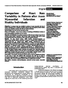
increased levels of inhibitory Gi-proteins, leading to a reduced ratio of Gs/Gi and an impaired Gs-mediated coupling between the β-receptor and adenylate cyclase (103,104). The third form of the changed response is a change in receptor affinity (32). For example, β-agonist catecholamines induce or stabilize a high affinity form of the βadrenergic receptor, which is specific for agonists and binds antagonists rather weakly (32). The chronotropic response of the SA node to neural stimulation also involves genetic variation, which is still poorly understoood (105-107). The Arg389Gly (108,109) and Ser49Gly (58) polymorphisms of the β1-adrenergic receptor are associated with difference in HR at rest, but this has not been a consistent finding, however (110). During exercise there is no difference between Arg389Gly genotypes (111-113). The gene GNAS1 encodes α-subunit of the stimulatory G-protein that couples β1-adrenergic receptor with the adenylyl cyclase (106). The T393C polymorphism of GNAS1 modulates HR response when values at rest, maximal exercise and recovery are considered together (106). The Arg16Gly polymorphism of the β2-adrenergic receptor is associated with difference in HR at rest (107,114), but during low or high intensity exercise the difference in HR does not persist between genotypes (107). The rs324640 polymorphism of the muscarinic M2-receptor gene is associated with a difference in HR recovery after exercise, but maximal HR is not different between the genotypes (105). 2.5 Mechanisms controlling heart rate Although easily measured, the HR reflects a complex, integrated physiologic response: autonomic tone, central and peripheral reflexes, hormonal influences, and factors intrinsic to the heart are all important (Figure 1) (20-22). Some reflexes may increase HR through a decrease in vagal tone, an increase in sympathetic activity, or both, whereas others exert the opposite effects (21,22). In the intact human cardiovascular system several reflexes and control mechanisms operate simultaneously, and the interactions are quite complex (21,22). The relative importance of neural control mechanisms in determining the cardiovascular response to exercise is dependent upon the type of exercise (static or
32
dynamic), the intensity of the exercise, the time after the onset of exercise (immediate, steady state, exhaustion, etc.), and the effectiveness of blood flow to meet the increased metabolic needs of the contracting muscle (22,115). Control mechanisms for the cardiovascular response during exercise are somewhat redundant, rather than additive, and they impinge on the same regulatory neurons in the vasomotor center of medulla, and, possibly, other sites, where integration of afferent information occurs (115,116). Besides neurally mediated reflexes, some humoral factors, such as cortisol, glucagon, growth hormone and thyroxine may play a minor role in modifying the control of HR (20,29,55).
humoral factors
arterial baroreceptors
arterial chemoreceptors
cardiopulmonary low-pressure baroreceptors
pulmonary receptors HEART RATE central command
respiratory influence
cardiac receptors
mechano- and thermosensitive peripheral afferents
chemosensitive peripheral afferents
Figure 1. The overview of mechanisms influencing heart rate. See text for the more detailed discussion about the effects mediated by each individual mechanism.
2.5.1 Arterial baroreceptors and cardiopulmonary low-pressure baroreceptors The function of the arterial baroreceptors is to maintain a normal BP (117-119). The arterial baroreceptors include carotid and aortic baroreceptors, which are spray-type nerve endings located in the walls of the large arteries (29,50,119). Carotid receptors lie in the wall of the internal carotid artery on either side (21,29,120). Each send impulses
33
centrally in the sinus nerve (of Hering), a branch of the glossopharyngeal nerve (50,120,121). Aortic arch receptors on the left lie within the aortic arch, but those on the right lie at the origin of the right subclavian artery and in the adjacent regions of the brachiocephalic artery (29,120,122). From the aortic receptors, activity travels centrally in small vagal branches (50,120,121). The nerve endings in the carotid sinus and aortic arch are activated by expansion of the arterial wall when BP is increased, and this stretching result in increases in discharge frequency in their afferent nerves (arterial baroreflex) (118,119,122). Afferent nerves transmit the baroreceptor activity to the nucleus tractus solitarius, which modulates autonomic outflow to buffer the rise in pressure (21,29,119). Conversely, a fall in BP reduces baroreceptor discharge and trigger adjustments that oppose the hypotension (21,118,119). The baroreceptors respond much more to a rapidly changing pulsatile pressure than to a stationary pressure (39,69). In dogs, arterial rheoreceptors that respond to increased blood flow by sensitizing the function of baroreceptors have been identified in the carotid sinus (123). The existence of rheoreceptors in humans is not known. Because mean arterial BP equals cardiac output times total peripheral resistance, the nucleus tractus solitarius can induce changes in BP by affecting either cardiac output, total peripheral resistance, or both via modulation of neural output (18,118,124). In response to a hypertensive stimulus, there is a rapid decrease in HR due to increased vagal discharge to the heart (18,121,125). In hypotensive stimulus there is an initial rapid increase in HR with withdrawal of vagal tone, followed by a slower rise in HR due to increased sympathetic discharge (18,121,125). The increase in total peripheral resistance (vasoconstriction) plays a major role in response to a hypotensive stimulus. In contrast, a decrease in HR is more important in response to a hypertensive stimulus (18,125). At rest roughly one third of the changes in arterial BP during carotid baroreceptor stimulation are due to changes in HR and two-thirds are dependent on alterations in total peripheral resistance, but the corresponding estimate for the overall baroreflex (carotid and aortic parts combined) is not known (126). Previously it was assumed that aortic baroreceptors operates over a higher range of arterial pressures than carotid receptors (21,39,127), but more recent studies have shown that both
34
baroreceptor populations operate over the same range of pressures (128). The estimated contribution of carotid baroreflex to overall baroreflex control is from 30% to 50 % (128,129). Aortic and carotid baroreceptors summate in their reflex effects (21,125). Because the relationship between total baroreceptor input and response is sigmoid, the type of summation (linear, inhibitory or facilitatory) depends critically on the size of the stimulus and the magnitude of the step in pressure to the baroreceptors (21,125,130). The fact that during exercise BP and HR rise linearly with increases in work rate, whereas at rest the corresponding increases would induce powerful opposing reflexes through the baroreflex has puzzled physiologists for over a century (131,132). The current view is that the arterial baroreflex is reset in direct relation to the intensity of dynamic exercise without a change in sensitivity of the reflex (133-135). The resetting moves the baroreflex set point to the higher BP level so that baroreflex does not oppose the rising arterial BP but even actively tries to elevate BP (via further vagal withdrawal and/or sympathetic excitation to the heart and/or sympathetically mediated vasoconstriction) until the new set point is achieved (136-138). During exercise changes in arterial BP during carotid baroreceptor stimulation are mostly due to changes in total peripheral resistance so that only at light workload HR contributes to changes in BP (138,139). The central command is probably the primary regulator of baroreflex resetting during exercise (138,140,141). The muscle chemoreflex is also able to reset baroreflex, but in normal dynamic exercise it acts more as a modulator of central command-induced resetting (138,141,142). Cardiopulmonary low-pressure baroreceptors monitor blood volume by sensing changes in the filling pressure of the chambers of the heart and pulmonary arteries and veins, as well as changes in cardiac contractility and afterload (18,143,144). Increases in these stimuli activate mechanically sensitive receptors in these structures, stimulating vagal afferent fibers that signal the nucleus tractus solitarius to inhibit sympathetic nervous system activity (18,124,144). The latter results in systemic vasodilatation and a reduction in total peripheral resistance, but physiological changes in cardiopulmonary low-pressure receptor activity cause little if any change in HR (18,124,144). When central venous pressure is normal, cardiopulmonary low-pressure baroreceptors tonically inhibit vasoconstriction induced by arterial baroreflex (18,144,145).
35
The cardiopulmonary low-pressure baroreceptors are not reset by the central nervous system during exercise, but they continuously inhibit arterial baroreceptor-induced vasoconstriction (18,118,144). If this tonic inhibition is eliminated by lower body negative pressure during exercise at a fixed workload, mean arterial BP and HR do not change, but total peripheral resistance increases and stroke volume decreases (146). Likewise, cardiopulmonary low-pressure baroreceptors inhibit the muscle metaboreflexmediated vasoconstriction during dynamic exercise (147). 2.5.2 Central command and peripheral afferents Central command is the term for motor command signals originating from subthalamic neurons involved in locomotion which is believed to be a primary stimulus mediating the autonomic nervous system adjustments to exercise (118,148,149). These signals activate separately both somatomotor and cardiovascular control systems at the onset of exercise (115,149,150). Activation is in direct proportion to the number of motor units required to maintain a given force of contraction (18,143,150). The magnitude of a central command- mediated cardiovascular response during exercise can be independent of force production (e.g. imagined exercise) and dictated more by an individual’s perception of effort (18,116,151). Central command increases HR, cardiac output, and also BP immediately at the onset of exercise by rapid vagal withdrawal, but command signals have a minimal effect on the sympathetic nervous system-mediated vasoconstriction (116,150,152). Because both locomotor and cardiovascular responses, much like the responses to exercise, can be induced by either electrical or chemical stimulation of cells in hypothalamic locomotor region and in mesencephalic locomotor region, the current view is that these two neuroanatomical areas are strongly involved in the central command (115,116,152). Both these areas have connections with the vasomotor center in medulla, which enables them to influence cardiovascular control (18,116,152). Originally it was thought that the central command acts as a pure feed-forward control mechanism (18,143). The close relationship of the central command with the number of motor units recruited and with the perception of effort suggests an important role for feedback from exercising muscles (18,124,153). Brain mapping studies have
36
showed that two regions of the cerebral cortex, the insular cortex and the medial prefrontal cortex, may function to interpret feedback signals from active muscles and elicit appropriate autonomic adjustments via connections to the vasomotor center (153). The muscle chemoreflex is elicited from chemosensitive group III and IV afferent fibers in the muscle whenever muscle blood flow falls below the critical level needed to maintain adequate oxygen transport to the muscle (148,150,154). Release of hydrogen from the working muscles might signal the onset of the reflex, it may actually stimulate not chemically sensitive afferents but rather the conversion of monoprotonated phosphate to its diprotonated form (143,155). Excitatory action potentials from muscle sensory afferents project to the brain via synaptic transmissions in the dorsal root of the spinal cord (115,124,154). This exerts effects on the vasomotor center in medulla (primarily cVLM and rVLM), which in turn inhibit vagal and/or stimulate sympathetic preganglionic neurons, thus producing efferent autonomic nervous system responses to the heart and arterial vasculature (115,124,154). The muscle chemoreflex has a distinct threshold, and in mild exercise this reflex is not tonically active (18,143,150). As the severity of exercise increases to moderate, the margin for any blood flow error decreases and the reflex is tonically active (18,143). The baroreflex normally buffers the muscle chemoreflex by limiting chemoreflex-induced peripheral vasoconstriction so that the rise in arterial BP by the muscle chemoreflex occurs almost solely via an increase in cardiac output (i.e. rise in HR) (156). Besides chemosensitive fibers, group III and IV afferent fibers in muscle also contain mechanosensitive fibers (115,148,154). These fibers are excited by mechanical stimuli (stretch and compression) in the active muscle (115,124,154). Their role in control of HR is not well understood (121,157), but it has been shown that a passive cycling of the legs significantly increased HR above baseline within one second of the onset of limb movement (158). In studies utilizing static muscle contraction this fast tachycardic response is vagally mediated (159), but evidence also exists for a slower sympathetically mediated tachycardiac response induced by mechanosensitive fibers (160). Since mechanoreceptor discharge quickly returns toward control levels during sustained static contractions, it is unlikely that these afferents contribute importantly to the maintained tachycardia in static exercise (157). It is not known, however, whether
37
this waning occurs to the same extent during dynamic contractions as well (18). Nevertheless, the muscle-heart reflex is not necessary for the vagally mediated increase in HR that occurs at the onset of exercise. This increase has been shown to be evoked by attempted exercise in subjects paralyzed with curare-like drugs (i.e. an absent afferent input from muscles) (161). In addition to responding to chemical and mechanical stimuli, many group III and IV sensory afferents are thermosensitive (18,143,155). Muscle temperature, which is normally well below 37°C in the limbs, can increase above 40°C during severe exercise (18,143). Thus, it is possible that these afferents could be stimulated and provide feedback to the central nervous system regarding the thermal status of the active muscle fibers (18,143). While this remains a possibility, experimental findings are insufficient to determine the role of this mechanism in autonomic nervous system-mediated cardiovascular control during exercise (18,124). 2.5.3 Respiratory sinus arrhythmia, arterial chemoreceptors, and pulmonary and cardiac receptors Respiratory sinus arrhythmia refers to the rhythmic variations HR, occurring at the frequency of respiration (21,55,79). Typically HR accelerates during inspiration and decelerates during expiration (21,55,79) as a result of complex central and reflex intearactions involving both vagal and sympathetic branches of autonomic nervous system (162). During exercise respiratory sinus arrhythmia gradually disappears despite gradually increasing respiration (163,164). Peripheral arterial chemoreceptors are situated in the aortic and carotid bodies (21,29,50). They excite nerve fibers that pass through carotid nerves and the vagus nerves into the vasomotor center (39,50,74). Activity in their afferent nerves is increased by arterial hypoxia, hypercapnia, or acidemia (29,50,165). The primary effect of aortic body chemoreceptor stimulation on HR is excitatory (166). Conversely, carotid chemoreceptor stimulation causes a pronounced and consistent bradycardia, but with intact respiratory control it is normally counterbalanced by tachycardia accompanying the respiratory response induced by the same stimulation (29,55,167). Because exercise normally is associated with maintenance of PaO2, normal or reduced arterial carbon
38
dioxide, and maintenance of blood pH within acceptable limits, the arterial chemoreceptors are not normally activated and therefore likely do not play an important role in HR control during conventional dynamic exercise at sea level (20,124). The lungs are richly innervated and lung inflation, with moderate pressures, stimulates airways stretch receptors which results in a reflex increase in HR (168,169). The reflex response during hyperinflation of the lung and also during pulmonary congestion is to cause bradycardia (21). Because breathing frequency, tidal volume and minute ventilation all increase during exercise, it is possible that reflexes activated by lung inflation participate in HR control during exercise. At present, however, there is no compelling evidence for it (124). Atrial receptors are concentrated near the junctions of the superior and inferior venae cavae and the pulmonary veins with the atria (20,21,29). The afferent fibers are contained in the vagus and the efferent pathway within the sympathetic nerves (21,29,170). Atrial receptors are stimulated mainly by stretching due to increases in atrial volume, which results in a reflex increase in HR (20,21,29). Because of the relatively slow time-course of sympathetic responses, the tachycardia following to stimulation of atrial receptors requires up to 30 seconds to reach a stable level (21). Some authors suggest that a larger stimulation of atrial receptors via the increased venous return during exercise may be an important mechanism mediating a normal exercise-induced tachycardia (69,170,171). On the contrary, other authors conclude that the tachycardic reflex mediated by the atrial receptors is weak or nonexistent in humans and thus it does not play any role in exercise-induced tachycardia (18). A direct stretch of SA node can also increase HR to some extent, and this mechanism may be operative in exercise when venous return is increased (39,172). The mechanism might be augmented after heart transplantation (29,172), but in the normal heart this mechanism is largely masked or overridden by other reflex mechanisms (173). A rise in the temperature of the blood reaching SA node as the consequence of muscular work may also increase HR via a direct effect on SA nodal tissue (39,172). Ventricular mechanoreceptors are situated mainly in the left ventricle. Afferent nerves travel either in the vagus or in the sympathetic nerves (170,174). Both populations of receptors can be divided into mechanosensitive and chemosensitive
39
endings (170,174). Normally their activity does not modulate HR either at rest or during exercise, but in the ischemic myocardium their activation results in powerful reflex responses (21,78,174). The vagal afferents mediate reflex cardio-inhibitory, sympathoinhibitory and vasodepressor responses, while activation of sympathetic afferents results in cardio-accelerator, sympatho-excitatory, vasodepressor responses (170,174). Both mechanical and chemical stimuli may be involved in activation of receptors, but chemical stimuli are more important for triggering of reflexes (21,170,174). The anginal pain is mediated by sympathetic afferent fibers and causes tachycardia (21,170,174). The vagal afferents are located nearer to the endocardial than to the epicardial surface, while the reverse is true for the sympathetic afferents (174). Thus subendocardial ischemia stimulates vagal afferents, resulting in bradycardia, whereas transmural ischemia more likely induces tachycardia mediated by sympathetic afferents (174). Left ventricular vagal afferents are preferentially distributed in the inferoposterior wall. In contrast, sympathetic afferents appear to be more uniformly distributed throughout the wall of the left ventricle (174). This probably explains why bradycardia usually occurs when the circumflex branch is occluded or when ischemia involves the inferior and lateral wall of the left ventricle (170,174). Occlusion of the anterior descending branch or ischemia involving the anterior wall is likely to result in an increase in HR (170,174). The chemical stimuli that activate receptors involve substances resulting from myocardial ischemia, including bradykinin, prostaglandins and adenosine (170,174). The bulging or dyskinesis of the ischemic zone may stimulate mechanosensitive vagal afferents, but the increase in discharge lasts only approximately two minutes. In contrast, the increased discharge of chemosensitive vagal afferent persists for the duration of the ischemic event (174). 2.6. Heart rate response to exercise: a synthesis In 1990 Rowell & O’Leary introduced an overall model as to how adjustments of the autonomic nervous system to large-muscle dynamic exercise may be mediated (136). This hypothesis represents the most compelling integrative scheme attempting to explain the primary signals involved and how those signals may interact to produce
40
autonomic nervous system adjustments to exercise (124). The key concept in the model is that BP is the primary variable controlled during exercise and control of HR serves as an adjunct in this scheme (136,150,175). From a resting value up to a rate of 100 beats/min during dynamic upright exercise HR increases rapidly primarily due to the activation of vagal withdrawal (51,65,73). The activation of vagal withdrawal is mainly due to augmented central command (136,150,157). The central command also resets the arterial baroreflex immediately to a higher operating point. Normally the baroreflex does not elicit an increase in sympathetic nervous system activity, however, because the rise in cardiac output (due to fast HR increase) is rapid enough to raise arterial BP to its new operating point (136,157,175). If any difference between the prevailing level of BP and a new, higher baroreflex operating point is detected by baroreceptors they can raise HR via further inhibition of parasympathetic tone (136,150,157). In this setting the muscle chemoreflex is not activated. The exact role of the afferents from muscle mechanreceptors is unknown, but in all probability their effect is negligible (136,150,157). During moderate exercise, when HR exceeds 100 beats/min, the activation of vagal withdrawal by central command still increases cardiac output, but not enough (136,150,175). The fast vagal component of the rise in cardiac output is not sufficient to raise cardiac output to a level that is needed to compensate fully the vasodilation in active muscle (136,150,175). Consequently, arterial BP cannot be increased immediately to its new, reset operating point so there is a pressure error detected by baroreceptors (136,150,175). As a consequence of this error, sympathetic nervous system activity to both the heart and to the resistance vessels increase in correct the pressure error (136,150,175). The sympathetically mediated increase in HR and cardiac output is much slower (by 15- to 20-fold) than the parasympathetically mediated rise (136,150,175). Thus vasoconstriction in resistance vessels all over the body (including active skeletal muscle) becomes a necessary adjunct to increased cardiac output to raise arterial BP as quickly as possible to minimize the pressor error (136,150,175). As workload increases, HR increases due to further sympathetic nervous system activation (20,73,164). The increase in sympathetic nervous system activity can occur due to the arterial barofeflex
41
(via further baroreflex resetting), the muscle chemoreflex (after a threshold has been passed after which this reflex becomes tonically active), or muscle mechanoreceptor activation (20,150,157). Besides direct neural excitation SA node is also stimulated by an increased level of circulating adrenaline, which is secreted from the adrenal medulla (23,73,176). Adrenaline secretion is increased only during moderate to heavy exercise (typically 50% of maximal oxygen consumption or above) and sympathoadrenal activation becomes progressively greater as exercise intensity increases up to maximum (58,176,177). As exercise approaches maximal levels, parasympathetic activity wanes and sympathetic nervous system activity increases such that at maximal oxygen consumption (VO2max) little parasympathetic tone remains and sympathetic activity is greatly elevated (136,157,164). Because during severe exercise HR is at or near maximal level, any further pressor response (i.e. to a fall in a baroreceptor activity or further muscle afferent activation) can only occur via peripheral vasoconstriction in that cardiac output is already at maximal levels (121,136,157). Pharmacological blockade studies have proved the differential contributions of the two autonomic branches during exercise (178). Blockade of vagal control with atropine (muscarinic receptor antagonist) reveals that most of the initial response to exercise, up to a HR of approximately 100 beats/min, is attributable to the withdrawal of tonic vagal activity (164,179,180). Withdrawal of vagal tone has been confirmed using time and frequency domain analyses of HR variability as well (163,164). Conversely, blockade of sympathetic control with β-adrenergic receptor antagonist reveals the importance of augmented sympathetic activity during moderate and heavy exercise (181-183). During light exercise, with workloads of 25% to 40% of VO2max, plasma noradrenaline levels or directly measured muscle sympathetic nervous activity do not significantly increase, confirming the finding that the sympathetic nervous system is more important in the later stages of exercise (136,150,175).
42
2.7 Central circulatory response to exercise During dynamic upright exercise cardiac output (i.e. stroke volume times HR) increases somewhat linearly in proportion to the oxygen consumption (VO2), approximately 6 l/min of cardiac output per 1 l/min of VO2 (19,171,184). An appropriate increase in HR contributes significantly to attaining high levels of cardiac output (23,29,67). Of the two major components of cardiac output, HR and stroke volume, HR is responsible for approximately two thirds of the total increase in cardiac output during dynamic upright exercise (17,185,186). When a normal human exercises maximally in the upright position, the HR increase is 150-300% of resting, while the stroke volume increase is about 10-100% (29,187,188). The stroke volume normally reaches its maximum or almost maximum by the time the cardiac output has increased only halfway to its maximum, after which any further increase in cardiac output must occur by increasing the HR (19,189,190). Thereafter stroke volume levels off (191-193), or there is a small decline (193-195) or increment (196-198) at maximal work intensity. An increase in HR accompanying dynamic exercise also results in an increase in the force of myocardial contraction (i.e. frequency-force relationship, or the staircase phenomenon) (20,37,143). The increase in force is secondary to a transient imbalance in cellular Ca2+ influx and efflux (favoring influx), an increase in sarcoplasmic reticulum Ca2+ content, and a larger sarcoplasmic reticulum Ca2+ release during each excitationcontraction coupling cycle (18,37,143). Although a sufficient increase of HR is essential to raise cardiac output at heavier workloads, the absolute cardiac output which a person can attain is determined by the magnitude of maximal stroke volume (68,184,199). The relationship between HR and VO2 or work intensity is approximately linear (19,200,201). It has been suggested that the HR might increase relatively less than VO2 as the work rate becomes very heavy (171,202,203), but two studies have shown the opposite (204,205). In both studies HR rose slightly more steeply above anaerobic threshold (approximately 50-60% of VO2max) than below anaerobic threshold, but the HR/work intensity -relationship was linear (205).
43
The linear relationship between HR and VO2 or work intensity is widely employed in a number of submaximal exercise tests (200,206-216), in which VO2max is estimated based on HR measured in a single or several submaximal workloads (69,217,218). By utilizing a linear HR-workload relationship a straight line is fitted to measured HR values (69,217,218). This line is extrapolated to the predicted maximal HR; the corresponding estimated maximal workload can then be approximated (69,217,218). VO2max can then be estimated by using the relationship between work rate and VO2 (69,217,218). 2.8. Factors modulating heart rate response to exercise In the same person under standardized conditions, the variation from day to day in HR at a given VO2 is 3-5 beats/min depending on the relative workload (%VO2max), provided the state of training is the same (219-221). The HR at a given VO2 is related to the maximal stroke volume (222,223), but it is not a measure of maximal cardiorespiratory fitness, unless the maximal HR is considered (69,224). Despite limitation some researchers have, however, considered the workload which a person can attain at some predetermined HR as a good estimate of cardiorespiratory fitness (225227). There is a tendency for persons with a low resting HR to have a low HR at a fixed submaximal workload (14,228,229), but this has not been observed consistently (171,230). Concerning maximal HR, persons with a high maximal HR have a higher HR at the same relative workload compared with the persons with a low maximal HR (230,231). Furthermore, an inverse (229) or non-existent (230) relationship has been observed between maximal HR and HR at a fixed submaximal workload, which emphasizes a fundamental difference when expressing a submaximal workload in terms of either relative or absolute work. The physiological limit on maximal HR in normal subject is determined by the steepness of the diastolic depolarization slope of SA nodal cells before they reach threshold potential, thus generating an action potential that is then propagated to surrounding cells (38,39). When HR reaches 195 beats/min in humans during severe exercise, ventricular diastolic filling time is only 0.12 seconds compared to 0.55
44
seconds at rest (HR 70 beats/min) (41,143,232). It seems logical that a limit would be approached where an increase in HR would not effectively increase cardiac output due to decreased diastolic filling; not only would the heart receive less blood to pump, but the degree of coronary artery perfusion would decrease (17,20). Although this theoretical limitation is reasonable, there is little experimental work to support it (17,20,196). Age. HR at a given submaximal VO2 (and workload) has been reported to be the same for individuals of the same gender and state of training regardless of age (14,229,233). On the other hand, HR at a fixed submaximal workload has also been suggested to be higher (15,226,234) or lower (228,235,236) in older subjects, but in these observations the state of training may differ between individuals. The decline of maximal HR with age is a well-known phenomenon (217,237,238). A comprehensive review of the literature compiling over 23 000 subjects aged 5 to 81 years revealed that age alone accounted for 75% of the variability in maximal HR; other factors added only about 5% (239). Of mechanisms underlying age-related decrease in maximal HR, cardiac chronotropic responsiveness to β-adrenoceptor stimulation has been shown to be preserved (240) or reduced (241-243) in the elderly. Subclinical atherosclerosis accompanying aging has also been proposed as a mechanism (69). The slope of the decay of exercise maximal HR with age is very similar to the slope of the intrinsic HR with age, suggesting that the decline of maximal HR is independent of autonomic influence, but has more to do with the SA node and the myocardium (20,244,245). The age-related decline in maximal HR is steeper in men who have a low cardiorespiratory fitness (246) and are physically inactive (17). Gender. HR at a given submaximal workload is higher in women (229,247,248). Maximal HR does not differ between genders (17,239,249) or is slightly lower (229,250,251) in women. Menstrual cycle. In women HR at a fixed submaximal workload has been reported to be higher during the mid-luteal phase (252), but most studies have reported no change in submaximal (253-255) or maximal HR (255-257) during the menstrual cycle. Body height and weight. The persons with the heavier body weight have been reported to have a lower HR at a fixed submaximal workload (222,236), and the
45
association is more pronounced when lean body weight is considered instead of total body weight (258,259). Height and body weight (lean or total) do not affect on maximal HR (20,51,222). Obesity. The overweight persons have a higher HR at a fixed submaximal workload than normal weight persons. The relationship is valid especially when exercising on a treadmill, but also when on a cycle ergometer (251,260,261). In two studies, however, there was no relationship between body mass index (BMI) and HR at a given submaximal workload on a treadmill (14,262), and in one study (228) an inverse relationship was observed between BMI and HR at a given submaximal workload on a cycle ergometer. Overweight has not been reported to have an effect on maximal HR (20,51,261), except one study in which overweight was associated with lower maximal HR (263). Mode of exercise. HR is slightly higher at a given submaximal VO2 on a cycle ergometer than on a treadmill (264,265). Maximal HR probably does not differ between a cycle ergometer and treadmill (266-268), although it has been suggested to be slightly lower with cycle ergometer testing (29,239,269). Both HR at a given submaximal workload and maximal HR are lower on a cycle ergometer in the supine position (29,184,270). Total mass of working muscles. The HR at a given submaximal workload is higher when the dynamic exercise is performed with the arms than with the legs (171,271,272). The maximal HR with arm exercise is 88-100% of the maximal HR in leg exercise (171,273,274). Exercise protocol. Large increments in workload combined with a short duration of the step may result in HR not rising to a steady state level at that workload (275). Consequently, HR at that submaximal workload is lower than the actual HR at the identical work provided that steady state would have been achieved (275). The maximal HR is not markedly different between protocols as long as the same exercise mode is used (268,276). Pedal frequency. When exercising with cycle ergometer, HR at a fixed submaximal workload may be slightly lower with a pedal frequency of 40 to 50 revolutions/minute than at clearly higher pedal frequencies (277). Habituation. Habituation to repeated exercise tests has been found to lead to a reduction in HR response to a fixed submaximal workload, but the habituation effect is difficult to separate from a training response (278,279). Other studies have, however,
46
shown no appreciable habituation effect (220). True maximal effort. Maximal effort should always be confirmed objectively before an attempt is made to measure a maximal HR (29). Objective measures of appropriate maximal effort include respiratory exchange ratio >1.10 and blood lactate level >7-8 mmol/l (17,20,29). Older individuals might be more afraid to achieve true maximal exertion but this effect may disappear on repeated testing (20). Even if the true VO2max is achieved in exercise it is still possible that maximal HR is even slightly higher than HR measured at the workload corresponding to VO2max (280). Sampling interval of HR measurement. The difference between measured HR and true HR (determined by the last 30 seconds of each minute during exercise) is inversely related to sampling interval (281). The 6second rhythm strip at the end of each minute represents a reasonable balance between convenience and precision for measuring HR during exercise (281). Environment. A hot environment causes a higher HR at a fixed submaximal workload than exercise at a low ambient temperature (171,282,283). Also a high relative humidity elicits a higher HR at a fixed submaximal workload (70). Maximal HR can even reach slightly higher values under hyperthermia than in normothermia (18). Again, HR at a given submaximal workload is lower in a cold environment (284) and also maximal HR is lower compared with neutral temperature (285). Emotional factors, nervousness, excitement and apprehension may raise the HR during exercise of light and moderate intensity (69,171,217). The heavier the workload, however, the less pronounced is this nervous effect on the HR such that it does not affect maximal HR (69). Dehydration. In dehydrated state HR at a given submaximal workload is higher than in euhydrated state (171,190,238), whereas acute expansion of blood volume decreases HR (286-288). Acute plasma volume expansion does not affect maximal HR (289), but in a dehydrated state maximal HR might be slightly higher than in the euhydrated state (18). Level of fitness. HR at a fixed workload seems to be inversely related to a maximal cardiorespiratory performance of a subject (238,290,291), although maximal HR modulates this relationship (69). A high cardiorespiratory fitness accelerates the rate of attainment of the steady-state HR at submaximal work (275,292). Maximal HR has
47
been reported to be the same (293-295), higher (229,230,296) or lower (239,250,297) in subjects with a high cardiorespiratory fitness. Training. Within the same subject endurance-typed exercise training reduces HR at a given submaximal workload, and reduced physical activity has the opposite effect (29,298,299). The rate of attainment of the steady-state HR at submaximal work occurs more rapidly after training as well (300). Endurance training does not change the maximal HR or slightly reduces it (301-303). Cessation of endurance-type exercise training may increase maximal HR (304). HR at a given submaximal workload has been reported to be lower (14,251,271) and maximal HR (230) higher, respectively, in subjects with a higher level of self-reported physical activity. Bed rest. After prolonged bed rest HR at a given submaximal workload is higher than before bed rest (190,280,305). The maximal HR is either the same (280) or increased after the bed rest (305,306). Medications and alcohol. The effects of various medications on HR during exercise are summarized in Table 1. After acute ingestion of alcohol, HR at a fixed submaximal workload is increased (307) or unaltered (308) but maximal HR is not affected (307). Arterial oxygen content. A reduced arterial oxygen content in anemia or after hemoglobin blocked by carbon monoxide (as acutely after smoking) results in a higher HR at a fixed submaximal workload (171,238,311). Parenthetically, also smokeless tobacco increases HR at submaximal work (70,312). The increased blood hemoglobin concentration lowers HR at a given submaximal workload (218,313), but does not affect on maximal HR (313,314). Long-term smokers seem to have a lower HR at a given submaximal workload (262,315,316), but unaltered (14,15,317) and higher (318) HR values have also been reported. Long-term smokers have been reported to have a lower maximal HR (230,263,315). Thyroid gland function. Hyperthyroid patients may have a high HR at a fixed submaximal workload (238,319), whereas hypothyroid patients may have the reverse (320).
48
Table 1. Medications affecting heart rate during exercise (20,308,309). Medications atropine agents blocking β-adrenergic receptors nitrates calcium channel blockers dihydropyridine agents diltiazem, verapamil digitalis hydralazine, minoxidil centrally acting antihypertensives (clonidine, methyldopa, moxonidine) antiarrhytmic agents quinidine, disopyramide propafenone amiodarone bronchodilatators sympathomometic anticholinergic methylxanthines psychotropic medications antidepressants major tranquilizers cold medications with sympathomimetic agents thyroid medications anorexiants / diet pills * † ‡
Effect on heart rate during exercise (↑ = increase, ↓ = decrease, ↔ = no effect) ↑ ↓*† ↑ or ↔ ↑ ↓ or ↔ ↓‡ ↑ or ↔ ↔ or ↓ ↑ or ↔ ↔ or ↓ ↓ ↑ or ↔ ↑ or ↔ ↑ or ↔ ↑ or ↔ ↑ or ↔ ↑ or ↔ ↑ ↑ or ↔
β-blockers with intrinsic sympathomimetic activity have a reduced effect. The effect increases with a relative intensity of exercise (64,183,310). Heart rate decreases in patients with controlled atrial fibrillation and possibly congestive heart failure, but heart rate is not significantly altered in patients with sinus rhythm.
Circadian rhythm and seasons. HR display a circadian rhythm so that HR at a fixed submaximal workload is slightly higher at early afternoon (a peak around 13.30) compared with morning or late afternoon (321,322). On maximal HR the effect of circadian rhythm is negligible (322). HR at a fixed submaximal workload has been shown to be lower in the summer than in other seasons (251). Eating and sleeping before the test. HR at a fixed submaximal workload is increased for an hour or more after a heavy meal (70,217). An abnormally short sleep the night before exercise may raise HR at a fixed submaximal workload (70). Genetics. The heritability estimates for HR at workload of 50 Watts (W) in cycle ergometer, and walking at submaximal speed on treadmill have been reported as 57% (323) and 32% (324), respectively. The workload that a person can attain at a
49
submaximal HR of 150 beats/min is characterized by a significant familial resemblance, but the heritability as a percent of the age and gender-adjusted phenotypic variance is only 135 bpm vs. HR 0.1 for removal) from conventional risk factors, including alcohol consumption, BMI, cigarette smoking, cardiac insufficiency, history of myocardial infarction, diabetes, myocardial ischemia during the exercise test, serum LDL and HDL cholesterol, and systolic and diastolic BP at rest. The additional predictive value brought by WL100 beyond other HR-derived and exercise test variables was explored by entering WL100 into a Cox model that included age, examination year, testing protocol, use of HR-lowering medication, conventional risk factors chosen by stepwise selection, and the HR and exercise test variables in turns. Because the use of HR-lowering medication affects WL100, the association of WL100 with mortality was examined separately in men who did not use HR-lowering medication and in men who used such medication. Difference in WL100 between two different testing protocols was tested using linear regression analysis after adjustment for age and use of HR-lowering medication. To address specifically the effect of two different exercise test protocols the stepwise selection was performed separately in corresponding subgroups. In supplementary analyses, the sample was restricted to subjects who had none of the following: history of cancer, history of cardiomyopathy, stroke or claudication, history of chronic obstructive pulmonary disease, bronchial asthma or pulmonary tuberculosis, or dizziness, dyspnea, chest pain, symptoms of cardiac insufficiency, arrhythmia, ischemic ECG changes or change in BP as a cause of discontinuation of a test. 4.9.4 Study IV The difference in HR40-100 between men whose test was terminated because of reason potentially caused by latent CVD (dizziness, dyspnea, chest pain, arrhythmia, ischemic ECG changes or change in BP) or because of submaximal effort and other men was tested with the independent samples t-test. The Pearson correlation coefficients between
81
chronotropic incompetence variables were calculated. Cox proportional hazards’ regression models were fitted to compute the relative risk of AMI associated with a low HR40-100, expressed as a continuous or dichotomous variable. Two different sets of covariates were used: 1) age and examination year; and 2) age, examination year, alcohol consumption, BMI, cigarette smoking, diabetes, VO2max, myocardial ischemia during exercise test, serum LDL and HDL cholesterol, systolic and diastolic BP at rest, SBP response, and SBP at 2 minutes after peak exercise. Age and examination year were forced into the model, and backward stepwise selection, with a p-value of >0.10 for removal, was used for the rest of the variables. In separate supplementary analyses, we excluded the subjects who died because of CHD within the next year after experiencing AMI, and men whose test was terminated because of reason potentially caused by a latent CVD or because of submaximal effort. Comparisons among chronotropic incompetence variables was addressed in a separate Cox model in which first stepwise selection was performed among the rest of the variables, and then forward stepwise selection was performed among chronotropic incompetence variables, with a p-value of