Journal of Neurology, Neurosurgery, and Psychiatry, 1980, 43, 76-81
Haemorrhagic complications of multiple sclerosis JOSEPH JANKOVIC, HOWARD DERMAN, AND DAWNA ARMSTRONG From Baylor College of Medicine, Departments of Neurology and Pathology, Texas Medical Center, Houston, Texas, USA SUMMARY Neurological exacerbations in multiple sclerosis patients are usually attributed to relapses of the disease. This report emphasises that other conditions, such as spontaneous CNS haemorrhage, may be responsible for the clinical deterioration. We describe two patients appropriately diagnosed as having multiple scleiosis who developed spontaneous CNS haemorrhage.
When a clinical diagnosis of multiple sclerosis is made, subsequent neurological events are usually attributed to relapses of the disease. However, other conditions may occur in patients with multiple sclerosis or may simulate that disease.' There have been reports of primary brain tumours2 and other neurological conditions occurring in patients with multiple sclerosis. Occasionally there is a problem of differentiating it from the collagen-vascular disorders.`' Two patients appropriately diagnosed as having multiple sclerosis who subsequently developed spontaneous CNS haemorrhage are described. The objectives of this report is to emphasise the importance of careful evaluation of each exacerbation of the clinical state of a multiple sclerosis,patient, and to illustrate that aetiological possibilities, other than relapse, must be included in the differential diagnosis of clinical exacerbation. The possible mechanisms of haemorrhages in patients with multiple sclerosis will be discussed. Case reports CASE 1
This 31-year-old woman, previously diagnosed as having multiple sclerosis, was recently admitted to the Neurosensory Center of Houston with a four-day history of headache, confusional state, and right-sided weakness involving face, arm, and Address for reprint requests: Dr J Jankovic, Baylor College of Medicine, Department of Neurology, Texas Medical Center, Houston, Texas 77030, USA.
Accepted 19 June 1979
76
leg. Ten years previously she had an acute loss of central vision in the right eye. Six months later, she suddenly lost vision in the left eye and developed weakness and numlbness in the right arm and the right leg. The neurodiagnostic investigation, which included cerebral arteriogram and pneumoencephalogram, was normal. The CSF examination was normal except for 8 mononuclear and 10 polymorphonuclear cell per mm.3 Her neurological deficit gradually diminished except for residual mild right hemiparaesthesias and impaired visual acuity of 20/200 in the right eye and 20/30 in the left eye. Four years later she developed right retro-orbital pain, right facial numbness, and vertigo. These symptoms resolved spontaneously within two to three days. Since that time she had had approximately five episodes per year suggesting multifocal CNS involvement. Five months before admission her neurological examination revealed right optic nerve atrophy, with a visual acuity of 20/200. There was moderate right leg weakness and bilateral hyperreflexia. Pain perception was impaired below T3 dermatome on the left, and bilateral cerebellar ataxia was present. Twelve days before admission she developed severe bifrontal and retro-orbital pains accompanied by nausea and vomiting. These symptoms resolved within two days, but six days later she was found in her room lethargic, confused, and vomiting. She was admitted to a hospital where she was treated for aspiration pneumonitis. After four days, she was transferred to the Neurosensory Center of Houston where the neurolo,gical examination revealed a drowsy woman with marked neck rigidity able to follow
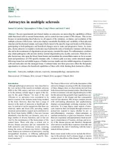
Haemorrhagic complications of Multiple Sclerosis only simple verbal commands. Comprehensive language testing was consistent with the syndrome of transcortical motor aphasia. The visual acuity in the right eye was 20/800 and in the left eye 20/50_ Bilateral optic nerve atrophy, right homonymous hemianopsia and left gaze preference were present. The right corneal reflex was depressed and there was mild right facial weakness involving the upper and lower part of the face. The gag reflex was absent bilaterally and bilateral tongue weakness was noted. There was right hemiplegia, bilaterally increased muscle tone, and right hemisensory deficit. The deep tendon reflexes were symmetrical and brisk, and the plantar responses were extensor. All routine laboratory tests including coagulation studies were within normal range. Lumbar puncture revealed grossly bloody cerebrospinal fluid with opening pressure of 130 mm H20. Computerised axial tomography (CAT) scan showed evidence of left frontal lobe haematoma with surrounding oedema and intraventricular extension (fig 1). Cerebral angiography demonstrated 10 mm left-to-right shift of the pericallosal artery, suggestive of a left frontal mass. In addition, diffuse luminal irregularities of the major intracranial vessels were present. The segmental narrowing was most pronounced on the left in the area of the haematoma and was suggestive of vascular spasm. Repeat arteriogram 12 days later revealed some improvement in the segmental narrowing. Right hemiparesis and transcortical motor aphasia persisted and therefore a left frontal craniotomy was performed and an intracerebral haematoma was evacuated. No apparent specific cause for the haematoma was apparent at surgery. Multiple biopsy samples of the wall of the haematoma were taken.
77
Pathology Eight specimens of blood clot, brain, and necrotic tissue were examined separately. They revealed necrotic brain containing recent haemorrhage, swollen axons, and polymorphonuclear cells. There were several vessels occluded by thrombus and some surrounded by haemosiderin pigment suggesting a previous haemorrhage. A biopsy specimen showed cerebral white matter exhibiting demyelination and astrogliosis with some preservation of axons (fig 2). Adjacent to this was necrotic or infarcted brain with macrophages and numerous proliferating small blood vessels. No vascular malformation was identified. Histochemical stains on a portion of the tissue revealed Oil Red 0 material in the cortex, and normal blood vessels. PAS and Hirsch Pfieffer stains for metachromasia were negative. Immunofluorescence for complement components C3, C4, IgG, IgA, and fibrinogen were all negative. Postoperatively the language difficulty and the right hemiparesis gradually improved. At the time of discharge, six weeks after admission, the patient had a mild comprehension deficit with persistent echolalia, slight right hemiparesis, and residual left optic nerve atrophy. CASE 2 A 46-year-old man was evaluated at the Houston Veterans Hospital for progressive quadriparesis. His neurological problems began 11 years previously with diplopia. Neurological examination then revealed nystagmus on both vertical and horizontal gaze, increased on gaze to the right. There was limitation of conjugate gaze to the right and right abducens nerve palsy. All
Fig 1 Case 1. A scan showing left frontal lobe haematoma with surrounding oedema.
78
Joseph Jankovic, Howard Derman, and Dawna Armstrong
diagnostic studies were normal including right and left carotid arteriograms and a rectilinear brain scan. The patient's ocular abnormalities improved and he had no neurological exacerbations until a second admission eight years later. His complaint then was a progressive right anterior abdominal numbness which demarcated in the midline at T1O segmental level. The numbness descended to involve the right thigh and medial aspect of the knee over a period of a week. There was no bowel or bladder dysfunction, weakness, or pain. Neurological examination revealed *a marked upward nystagmus and convergent nystagmus on downward gaze. There was a right internuclear ophthalmoplegia and a slight gaze paresis to the right. An area of sensory loss on the right extended from the umbilicus to the right knee. Joint position sense was reduced in both toes. Deep tendon reflexes were brisk in both legs, more on the right. There was transient right knee and ankle clonus and an extensor plantar reponse on the right. The patient circumducted his right leg when he walked. All diagnostic studies including myelogra.phy were normal. Cerebrospinal fluid examination revealed 13 white cells (6 polymorphonuclear cells and 7 lymphocytes) per mm3, protein of 0-75 g/l and a glucose level of 3-4 mmol/l. Electrophoresis of cerebrospinal fluid revealed increase in levels of gamma globulins (IgG level 0-102 g/l; normal 0-005-/0 016 g/l). The diagnosis of multiple sclerosis was made and the patient was treated with a 10-day course of ACTH which was coincidental with partial improvement. Over the ensuing year the patient continued to work regularly but had continuous numbness along the right mid-abdomen and increasing right leg limp. In July 1978 the patient experienced an acute onset of severe neck pain with weakness in both arms and left leg. There was right internuclear ophthalmoplegia, horizontal and vertical nystagmus on both right and left lateral gaze. Funduscopic examination was normal. Strength in upper and lower extremities was graded as three out of five with proximal strength slightly greater than distal strength. The Babinski sign was present on the right. The sensory level remained at TIO dermatome on the right side. Sphincter tone was normal. A 10-day course of ACTH therapy was given for presumed acute exacerbation of multiple sclerosis. He was discharged and returned to the hospital five days later in acute urinary retention. Neurological examination this time was unchanged except for
complete paralysis of both legs, 1/5 strength in the left arm, and 3/5 strength in the right arm. Cerebrospinal fluid examination revealed xanthochromic fluid with a protein of 0-87 g/l and a sugar level of 3-5 mmol/l, 30 white cells 12 polymorphonuclear cells and 18 lymphocytes per mm3.3 In the ensuing 24 hours the patient developed a progression of the quadriparesis. A myelogram revealed widening of the spinal cord at the level of CS and C6 segments with a complete block of the contrast medium at that level. A bilateral laminectomy at C4-C6 vertebrae was performed and a large intramedullary haematoma was evacuated. After surgery the patient regained some strength in his right arm. Additional studies revealed abnormal visual evoked responses from both eyes. His sedimentation rate of 41 mm/hr was attributed to urinary tract
Y.
. fr
Fig 2 Case 1. Multiple biopsy samples of wall of haematoma. Left upper: thrombosed vessel and surrounding neurones and inflammatory reaction. Right upper: white matter showing gliosis in areac demyelination. Left lower: wall of haematoma, macrophages, and neovascularity. Right lower: haemorrhage into brain.
Haemorrhagic complications of Multiple Sclerossi infection; antinuclear antibody test, LE cell preparation, and latex fixation were normal.
Pathology The sections revealed abnormal spinal cord tissue containing a large haematoma which showed recent accumulation of red cells and haemosiderin and hyalinised tissue from a previous haemorrhage. The surrounding neuropil was abnormal showing areas of acute inflammation with prominent dilated and hyalinised blood vessels and necrosis. Thromibi were present in some of the small vessels. There was some neural tissue showing gliosis (fig 3).
.t ,,
0
,
S f~~~~~~~~~A
F,~~~~~~3
-i., -r'->
Fig 3 Case 2. Tissue adjacent to haematonia. Upper: cord showing oe4ema and dilated small vessels. Lower: cord showing disrupted thrombosed vessel.
Discussion There is sufficient clinical, laboratory, and histological evidence to support the diagnosis of multiple sclerosis in the two patients. In both patients the recent clinical exacerbations were initially attributed to a relapse of their multiple sclerosis. However, in case 1, the repeated similar episodes of focal right hemiparesis,
79
aphasia, and headache were a clue to the correct diagnosis of left frontal haematoma. In Case 2 the progressive quadriparesis led to the appropriate studies and surgical procedure. In previous reports, vascular episodes in patients with multi;ple sclerosis have usually been attributed to an underlyinig vasculitis or so-called "cryptic" arteriovenous malformation. However, the possibility of haemorrhage into a demyelinating plaque has not, to our knowledge, been considered previously. Such haemorrhage may simulate an acute relapse of multiple sclerosis and may be attributed clinically to the natural course of the disease. In diseases with multiple neurological manifestations, the possibility of CNS vasculitis must be excluded. Polyarteritis nodosa8 may present clinically like multiple sclerosis. The literature contains reports of several patients suspected of having multiple sclerosis who were later proved to have a collagen-vascular disorder. Fulford et al described four such patients and reviewed the literature. They, with others, have pointed out that systemic lupus erythematosus may simulate multiple sclerosis or that the two diseases may be present in the same patient or in members of the same family. The term "lupoid sclerosis" has been proposed for those patients showing features of both diseases. There is, however, no convincing evidence that the two diseases represent a spectrum of a single disorder with a similar pathogenetic mechanism. CNS haemorrhage may occur in patients with systemic lupus erythematosus,9 in our patients, the diagnosis of systemic lupus erythematosus is unlikely in view of the negative collagen-vascular disease investigations and histology. The possibility of a cryptic arteriovenous malformation producing haemorrhage has been mentioned. Abroms et a!0 described two adolescent girls who were suspected of having multiple sclerosis and who developed spontaneous intracerebral haemorrhage. No specific aetiology for the haemorrhage was found and therefore it was attributed to the presence of a "cryptic" arteriovenous malformation. In our case the angiogram in Case 1, and the myelogram in Case 2 as well as inspection at the time of surgical evacuation of the haematomac and histological examination failed to reveal evidence of vascular malformations. Furthermore, both of our patients had classical clinical features of an exacerbating and remit.ting course with multiple CNS lesions, and in Case 1 areas of demyelinisation were observed in biopsy tissue.
80
Joseph Jankovic, Howard Derman, and Dawna Armstrong With no obvious source for the haemorrhage 2 Reagan TJ, Freiman IS. Multiple cerebral gliomas in multiple sclerosis. J Neurol Neurosurg in either patient, we suggest the possibility of a Psychiatry 1973; 36:523-8. demyelinating lesion contributing or setting the 3 Aita JF, Bennett DR, Anderson RE, Ziter F. stage for the focal haemorrhage. In the past, Cranial CT appearance of acute multiple several observers have proposed a vascular sclerosis. Neurology (Minneap) 1978; 28:251-5. aetiology to explain the rapid development of clinical symptoms and signs in multiple sclerosis 4 Cendrowski W, Stepien M. Clinical variant of lupus erythematosus resembling multiple patients. The characteristic relation of a sclerosis. Europ Neurol 1974; 11:373-6. demyelinating lesion to small blood vessels gives 5 Fulford KWM, Catterall RD, Delhanty JJ et al. some support to the vascular hypothesis." A collagen disorder of the nervous system prePutnam"2 advocated venous thrombosis as a senting as multiple sclerosis. Brain 1972; 95: possible pathogenetic process. Others reported 373-86 . increased capillary fragility,'3 subcutaneous 6 PE. Comparison of neurologic manifesKaplan and various coagulation abhaemorrhages,4" tations of systemic lupus erythematosus, perinormalities'6" in multiple sclerosis patients arteritis nodosa, and multiple sclerosis. Illinois Macchi'9 observed that the most common Med J 1976; 149:144-9. vascular change was enlargement and dilatation 7 Shepherd DI, Downie AW, Best PV. Systemic of vessels in plaques. Computerised tomography lupus erythematosus and multiple sclerosis. wi.th contrast enhancement has revealed evidence Trans Am Neurol Assoc 1974; 99:172-6. of breakdown in the integrity of the blood-brain 8 Waisburb H. Polyarteritis nodosa complicated by barrier at the edges of acute demyelinating a multiple sclerosis-like syndrome. Can J Neurol plaques3 2021 in multi sclerosis patients. This Sci 1974; 1:250-2. suggests alteration in the vascular structures 9 Johnson RT, Richardson EP. The neurological leading to increased permeability and extramanifestations of systemic lupus erythematosus. vasation of blood constituents. A clinical-pathological study of 24 cases and review of literature. Medicine (Baltimore) 1968; Although focal haemorrhages are not usually 47:337-69. regarded as pathological features of multiple sclerosis it is possible that under certain con- 10 Abroms IF, Yessayan L, Shillito J, Barlow CF. Spontaneous intracerebral haemorrhage in ditions, particularly in acute lesions, such changes patients suspected of multiple sclerosis. J Neurol may occur. The combination of enhanced platelet Neurosurg Psychiatry 1971; 34:157adhesiveness,"' venous thrombosis,12 increased vascular fragility,"-" transient thrombocyt- 11 Fog T. On the vessel-plaque relationships in the brain in multiple sclerosis. Acta Neurolog Scand openia,22 increased fibrinolytic activity,23 24 and the 1964; 40 (Supplement 10):9-15. proximity of the demyelinating lesion to blood 12 Putnam TJ. Evidence of vascular occlusion in vessels may set the stage for focal haermorrhagic multiple sclerosis and encephalomyelitis. Arch diathesis leading to the development of a Neurol Psychiatry 1937; 37:1298-1321. haematoma. The proposed mechanism for HH, Alexander L, Ehrentheil OF, haemorrhage is speculative, but it suggests further 13 Shulman Gross R. Capillary resistance studies in multiple studies which may explain the haemorrhagic sclerosis. J Neuropathol Exp Neurol 1950; 9: complications in patients afflicted with multiple 420-9. sclerosis. 14 Sibley WA, Kiernat J, Laguna JF. The modificaWe thank Dr B Cooper and Dr A Evans who participated in the care of Case 1. Dr C Mattioli and Dr EH Dew helped with the histological examination. The Neuroradiology Department of Methodist Hospital performed and helped with the interpretation of the arteriogram and the CAT scan on Case 1. References I
Lehman RAW, Fieger HG. Arachnoid cyst producing recurrent neurological disturbances. Surg Neurol 1978; 10:134-6.
15 16
17
18
tion of experimental allergic encephalomyelitis with epsilon aminocaproic acid. Neurology (Minneap) 1978; 28:102-5. Swank RL. Subcutaneous haemorrhages in multiple sclerosis. Neurology (Minneap) 1958; 8: 497-9. Feldman S, Izak G, Nelken D. Blood coagulation studies and serotonin determinations in serum and cerebrospinal fluid in multiple sclerosis. Acta Psychiatr Scand 1957; 32:37-43. Millac P. Platelet stickiness in multiple sclerosis. Dtsch Nervenheil 1967; 191:74-9. Millar JHD, Merrett JD, Dalby AM. Platelet stickiness in multiple sclerosis. J Neurol Neurosurg Psychiatry 1966; 29:187-9.
Haemorrhagic complications of Multiple Sclerosis 19 Macchi G. The pathology of the blood vessels in multiple sclerosis. J Neuropathol Exp Neurol 1954; 13:378-84. 20 Lebow S, Anderson DC, Mastri A, Larson D. Acute multiple sclerosis with contrast-enhancing plaques. Arch Neurol 1978; 35:435-9. 21 Sears ES, Tindall RSA, Zarnow H. Active multiple sclerosis. Enhanced computerised tomographic imaging of lesions and the effect of corticosteroids. Arch Neurol 1978; 35:426-34.
81 22 Fog T, Kristensen I, Helweg-Larsen HF. Blood platelets in disseminated sclerosis. Arch Neurol Psychiatry 1955; 73:267-85. 23 Menon IS, Dewar HA, Newel DJ. Fibrinolytic activity of venous blood in patients with multiple sclerosis. Neurology (Minneap) 1969; 19:101-4. 24 Persson I. Variations in the plasma fibrinogen during the course of multiple sclerosis. Arch Neurol 1955; 74:17-30.