Gluten-Sensitive Enteropathy (Celiac Disease) Controversies in Diagnosis and Classification Arzu Ensari, MD, PhD
disease, or gluten-sensitive enteropaNthy,Context.—Celiac is a chronic inflammatory disorder of the small intestine characterized by malabsorption after ingestion of gluten in individuals with a certain genetic background. Clinical presentation can vary from full-blown malabsorption to subtle and atypical symptoms. Diagnosis currently relies on clinicopathologic studies including mucosal biopsy, serologic tests, and the effects of a diet free of gluten on the symptoms. Mucosal pathologic features are also variable, ranging from mild abnormalities, including intraepithelial lymphocytosis, to completely flat mucosa. Since patients with minimal histologic lesion of intraepithelial lymphocytosis often present with normal serologic findings, biopsy diagnosis becomes more important for identifying such individuals. Classification of mucosal pathology in gluten-sensitive enteropathy has been a subject of controversy among pathologists and needs to be revised according to the current understanding of the disease. eliac disease, also known as celiac sprue, nontropical C sprue, gluten-induced enteropathy, or gluten-sensitive enteropathy (GSE), is a chronic inflammatory disorder of the small intestine characterized by malabsorption after ingestion of wheat gluten or related proteins in rye (secalins) and barley (hordeins) in individuals with a certain genetic background.1–3 The pathogenesis involves a T-cell–mediated immune response and autoreactive B lymphocytes that produce autoantibodies directed against gliadin, endomysium, or tissue transglutaminase in individuals with a genetic susceptibility related to human leukocyte antigens HLA-DQ2 and HLA-DQ8.4–6 The classical textbook description of celiac disease with malabsorption syndrome and associated flat mucosa comes from previous studies when the initial peroral jejunal biopsies revealed the typical advanced mucosal lesion. It is probably useful within the context of this review that the term celiac disease be restricted to that definition and that the remaining body of the iceberg— comprising the latent celiac disease, the monosymptomAccepted for publication September 3, 2009. From the Department of Pathology, Ankara University Medical School, Ankara, Turkey. The author has no relevant financial interest in the products or companies described in this article. Reprints: Arzu Ensari, MD, PhD, Department of Pathology, Ankara University Medical School, Sihhiye 06100, Ankara, Turkey (e-mail:
[email protected]). 826
Arch Pathol Lab Med—Vol 134, June 2010
Objectives.—To highlight the variations in clinical and pathologic presentation of gluten-sensitive enteropathy, to emphasize the importance of small-intestinal biopsy evaluation in the diagnosis, and to propose a new classification of mucosal pathology in gluten-sensitive enteropathy, in an effort to overcome the problems related to the classification systems currently available. Data Sources.—A review of the literature on clinicopathologic features and the morphologic spectrum of gluten-sensitive enteropathy is presented. Conclusions.—Considering that there are many entities in the differential diagnosis of gluten-sensitive enteropathy, because of the varied clinicopathologic spectrum of the disease, diagnosis depends on good clinicopathologic communication. The classification that is presented in this review is a simple and practical approach to improve clinicopathologic correlation in gluten-sensitive enteropathy. (Arch Pathol Lab Med. 2010;134:826–836) atic patients, and relatives of patients with celiac disease showing milder mucosal pathology—be discussed under the wider term of gluten-sensitive enteropathy or simply gluten sensitivity. DIAGNOSIS With the description of the spectrum of mucosal pathology and the availability of genetic and highly reliable serologic markers, a substantial change has occurred in the diagnosis of GSE. Most importantly, the presence of total flat mucosa, with no detectable villi on its surface, is no longer necessary provided other histologic features of GSE and specific antibodies are present in a patient with genetic susceptibility.7 Although diagnosis currently relies on clinicopathologic studies including mucosal biopsy, serologic tests, and the effects of a diet free of gluten on the symptoms, there is still a need for consensus guidelines set by the major scientific organizations associated with GSE. According to the revised criteria of the European Society for Paediatric Gastroenterology and Nutrition,8 an initial characteristic smallintestinal abnormality should be regarded as essential and there should be a clear-cut response to strict gluten-free diet (GFD) with clinical recovery within weeks. In asymptomatic patients, a second follow-up biopsy under a GFD is advised to demonstrate the histologic recovery of the mucosa, which usually does not develop before 6 months.9 The American Gastroenterological Association Controversies in Diagnosis and Classification—Ensari
mandates a biopsy to confirm the diagnosis in people suspected of having GSE,10 whereas the US National Institutes of Health, in its recent consensus statement, recommends biopsies only after a positive serologic finding or when serologic results are nondiagnostic.11 In summary, the diagnostic scheme of GSE consists of the following:
. Clinical history and symptomatology . Serologic testing (tissue transglutaminase, endomysial antibodies, and antigliadin antibodies)
. Histologic findings in proximal small-intestinal biopsy . Clinical and serologic (optionally histologic) response to a GFD7,12,13 CLINICAL SPECTRUM OF GSE Symptomatology of GSE is not associated with the severity of the mucosal lesion but is mainly related to the length of the affected bowel.14–16 Clinical presentation varies from full-blown malabsorption with weight loss, diarrhea, and steatorrhea to more subtle symptoms such as folate- or iron-deficiency anemia, flatulence, episodic diarrhea, loose stools, neurologic problems, osteoporosis, and vitamin K and D deficiencies in as many as 50% of patients.2,3,17 It also varies with the age of the patient, the duration of the disease, and the presence of extraintestinal findings.12 In children, usually within few months of introducing the child to wheat-based foods, the classic syndrome of chronic diarrhea, steatorrhea, abdominal distension, and failure to thrive appears between 6 months and 2 years of age.14,18 Both weight (40% below 10th centile) and growth (25% below 10th centile) are affected in these children, although weight is affected more often.19 Other children with fewer symptoms may escape detection and will perhaps not be diagnosed until adulthood.9 If undiagnosed, however, GSE can lead to permanent short stature and pubertal delay as well as the sequelae of nutrient deficiency such as iron deficiency or megaloblastic anemia and rickets.9,19,20 Patients presenting with symptoms as adults may also have short stature and/or historical symptoms indicative of ongoing disease since childhood. Current estimates show that the incidence of GSE is 1:200 or higher in wheat-eating populations such as Western Europe and North America, while the incidence continues to rise in Eastern societies, possibly as a result of ‘‘western-style’’ eating habits.14 Prompt diagnosis and treatment of GSE not only eases symptoms and improves quality of life but also has the potential to decrease long-term risks for lymphoma, gastrointestinal cancers, dermatitis herpetiformis, osteopathy, endocrine abnormalities, infertility, cardiomyopathy, and other autoimmune disorders.14,17,21 Latent Gluten Sensitivity The term latent gluten sensitivity is used to descibe symptoms in patients with evidence of gluten sensitivity but without full-blown symptoms of sprue.22–25 Terms that have been applied to such cases include subclinical, silent, or occult celiac disease; gluten-sensitive disease with mild enteropathy; low-grade enteropathy; minimally symptomatic enteropathy; and potential celiac disease.25–29 This form of GSE is characterized by the presence of no or only mild changes in the proximal small-intestinal mucosa, subtle symptoms when investigated carefully, and isolated positivity of serologic tests. These cases prove to be difficult to diagnose because of the atypical symptoms Arch Pathol Lab Med—Vol 134, June 2010
that do not immediately suggest a gastrointestinal cause. Indeed, almost half of individuals with latent gluten sensitivity may be apparently well, while others may have a sense of malaise that is difficult to define. Such cases require vigorous histopathologic evaluation, as the findings may occur along a spectrum of normal mucosa with increased numbers of intraepithelial lymphocytes (IELs) to flat mucosa, and they should not be missed since patients still remain predisposed to cancer or lymphoma by virtue of chronic gluten ingestion.22,23,27–29 Serologic Tests Widespread availablity of serologic tests has permitted physicians to test their patients for GSE without further aid from pathology. Serologic tests are based on the use of immunoglobulin (Ig) A isotypes and include antigliadin antibodies as well as connective tissue antibodies such as reticulin antibodies, endomysial antibodies (EMAs), and tissue transglutaminase (tTG) antibodies.7,30–33 In most patients, serologic positivity supports the diagnosis and these tests are very useful for screening and follow-up.7,32–34 The enzyme-linked immunosorbent assay–based clinical test for IgA anti-tTG antibodies has a high sensitivity and specificity and has recently become the serologic test of choice for GSE, largely replacing other antibody tests.30,31 While EMA is as sensitive and specific as tTG, the immunofluorescent test used for detection of EMA is time-consuming and more subjectively interpreted than the tTG test.32,33 Serologic positivity usually correlates with the degree of mucosal damage, while minimal histologic lesion of intraepithelial lymphocytosis (IELosis) often presents with normal serologic findings.7,35–37 In parallel with this view, a previous study34 demonstrated that increasing titers of tTG predicted higher levels of villous flattening. Similar findings were observed in pediatric patients who had tTG levels above 100 units and showed advanced (Marsh type 3) lesions in their biopsy specimens.34,38 Taken together, these findings indicate that a negative serologic result is not sufficient to rule out GSE with a Marsh type 1 lesion. Human leukocyte antigen (HLA) testing to detect susceptible HLA subtypes is also used in the routine diagnostic workup of GSE.7,39 While HLA-DQ2 is found in 90% to 95% of patients with GSE, most of the remaining cases are associated with HLA-DQ8. Since these HLA alleles are found in up to 40% of the general population, their presence does not aid the diagnosis directly, but the absence of DQ2 or DQ8 virtually excludes GSE.7 It is therefore justified to state that, despite the development of new serologic tests and genetic analysis, small-intestinal biopsy continues to be the gold standard for the diagnosis of GSE, particularly in less severely affected patients with mild mucosal abnormality. Small-Intestinal Biopsy: Site and Number? Similar to its wide variation in clinical manifestations, GSE has a wide spectrum of histologic abnormalities, which makes interpretation of small-intestinal biopsy specimens problematic for the pathologist. The damage to the small-intestinal mucosa classically involves the proximal small intestine including duodenum and upper jejunum and extends distally for a variable length into the ileum.14,40,41 Healing of the small-bowel mucosa, on the other hand, takes place in a distal to proximal direction.15 This may take at least 6 months and may even be prolonged Controversies in Diagnosis and Classification—Ensari
827
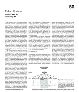
Figure 1. Normal duodenal mucosa with 3 to 4 long villi with a villous to crypt ratio of .3:1 and normal number of intraepithelial lymphocytes (hematoxylin-eosin, original magnification 3200). Figure 2. Vertical orientation of the villi to the muscularis mucosae (hematoxylin-eosin, original magnification 3100).
up to 24 months after treatment with GFD.42 Because of the slow tempo of IEL (gd subtype in particular) loss from the epithelium and entry after gluten ingestion, IEL count may be the last feature to return to normal after a GFD.41,43 The number of small-intestinal biopsies has substantially increased over the years, partly because of the increased awareness by clinicians of the atypical forms of GSE, as well as the increased use of upper gastrointestinal endoscopy. Previously, small-intestinal biopsies were always taken by suction capsule, conventionally positioned at the distal duodenum, duodeno-jejunal junction (ligament of Treitz), or proximal jejunum under fluoroscopic control.12,14,16 Although, capsule biopsies are usually bigger and easier to orientate, swallowing the capsule is discomforting for the patient and is more labor-intensive and time-consuming for the physician. In the past decade, duodenal biopsies have almost entirely replaced capsule biopsies of jejunal mucosa for the diagnosis of GSE in most gastroenterology units, essentially because endoscopy has the advantages of saving time and reducing the risk of failure and false-negative findings.12,44–48 Also, multiple targeted biopsies can be taken during endoscopy because, in most patients with flat mucosa, the duodenum shows typical endoscopic features described as mosaic appearance, scalloping, or reduction of duodenal folds.12,14,45,46 Newer endoscopic methods, such as push enteroscopy and double-balloon enteroscopy, allow access to the entire length of small bowel,15 but are more timely and costly compared with upper gastrointestinal endoscopy. Obtaining biopsy samples of adequate size from across a circular mucosal fold is as important as the biopsy site itself.12,14,15 Pathologists should be aware that biopsy forceps can crush and destroy tissue, causing hemorrhage in the sample and thus making the evaluation of specimen difficult. Superficial biopsy samples lacking muscularis mucosa can cause separation of the villous bases, resulting in shorter and thicker villi that can easily be misinterpreted in favor of a diagnosis of GSE. Conflicting reports exist in the literature regarding the distribution pattern of mucosal pathologic features along the small bowel.16,49–51 Although it was widely accepted in the past that villous flattening rarely coexisted with 828
Arch Pathol Lab Med—Vol 134, June 2010
histologically normal mucosa, currently, many investigators believe that GSE can exhibit a patchy distribution, that is, areas with villous flattening may occur in proximity to areas with mild villous shortening and also in areas with normal histologic features, particularly in pediatric patients.50,51 This notion may lead to false-negative diagnosis, particularly when there is inadequate sampling. However, the optimal number of biopsy specimens necessary to confirm the diagnosis of GSE is still not known. There are no recommendations in the guidelines of the North American Society for Pediatric Gastroenterology, Hepatology and Nutrition,52 while the American Gastroenterology Association has recommended 6 biopsy specimens as necessary for the diagnosis of GSE.53 In practice, it seems reasonable to suggest that at least 4 endoscopic biopsy samples must be taken from distal duodenum with 2 samples from the bulbus to detect patchy and subtle mucosal lesions in GSE. Biopsy specimens from the bulbus, however, should be interpreted with caution because this area is susceptible to peptic injury and contains prominent Brunner glands, which can lead to obliteration of the villi, with an artifactually flat mucosal appearance.12,47,54 What Is ‘‘Normal’’? Pathologists should be capable of recognizing normal features of small-intestinal mucosa so as to interpret the abnormalities associated with GSE correctly. It is generally accepted that the presence of at least 3 or 4 consecutive villi with a normal villous to crypt ratio in a biopsy sample is sufficient to consider as normal.12,15 Normal smallintestinal mucosa has long, slender villi, with this ratio ranging between 3:1 to 5:1 depending on the site of the biopsy; shorter villi are found in the duodenum, whereas the height of the villi increases distally from jejunum to ileum (Figure 1). Normal distribution of IELs along the villi shows a characteristic decrease from the base of the villus toward the villous tip and resembles the musical ‘‘decrescendo’’ sign.55 However, the pathologist should be aware of the possible patchiness of mucosal lesions when making a decision of normal mucosa and ruling out GSE. For accurate histopathologic assessment of villus to crypt ratio, it is essential that the biopsy specimen be Controversies in Diagnosis and Classification—Ensari
properly oriented with the luminal surface upwards such that the villi should be vertical to the muscularis mucosae (Figure 2).12,13,15 Specimens can be oriented on a supporting medium (eg, a strip of filter paper, a piece of tissue, or dental wax) with the naked eye or with the assistance of a dissecting microscope; embedded in wax; and cut through vertical plan.13 In tangentially cut sections, artifactual shortening of the villi and falsely increased lymphocytes in the surface epithelium can occur.12,14 The approach to biopsy specimen by the pathologist is as important as the correct orientation and involves low-power microscopic examination for architectural abnormalities, as well as cellular distribution, and is followed by a high-power view to assess cellular content and epithelia. Although the exact numbers are not known, few inflammatory cells comprising plasma cells, lymphocytes, eosinophils, and macrophages are found in the lower one-third of the lamina propria, while villous lamina propria is considered as ‘‘empty’’ in normal small-intestinal mucosa.12,15 In the presence of inflammation, however, inflammatory cells, including polymorphonuclear leukocytes, infiltrate upper parts of the lamina propria and cause obliteration of the villi. THE MORPHOLOGIC SPECTRUM OF GSE Histologic evidence of GSE depends on abnormalities in either architecture (villous shortening and crypt hyperplasia) or the number of IELs or both. In its classical form, GSE results in shortened, widened villi or even totally flat mucosa with hyperplastic crypts. Overall thickness of the mucosa remains relatively unchanged, but villous to cyrpt ratio (normally 3:1 in distal duodenum and 2:1 in bulbus) decreases as the villi become shortened. These architectural changes are preceded by an increase in the number of IELs as compared to normal numbers, corresponding to the cellmediated immune nature of the disorder. It is this group of cases that should be actively sought in biopsy specimens in which GSE is part of the clinical differential diagnosis.14,56 Marsh56 was the first to describe the morphologic continuum of GSE, which ranges from normal villous architecture with IEL increase as the only abnormality to flat mucosa with crypt hyperplasia, increase in the number of IELs, epithelial destruction, and increased lamina propria inflammation. Marsh classification will be discussed in detail in association with other classification schemes in the following sections. Intraepithelial Lymphocytes Intraepithelial lymphocytes have been considered to be responsible for the epithelial damage observed in GSE, although the exact mechanism is still not known. Most IELs are T lymphocytes, which are mostly cytotoxic T cells expressing ab T-cell receptor (TCR) on their surface. The population specifically expanded in GSE is the CD3+/ CD42/CD82, gd TCR–bearing IELs, which is only 5% of the total in normal mucosa.57,58 Over the years, the cutoff value, and thus the normal number of IELs, has been subject to major variation and a significant reduction has occurred in the highest value for normal. The trend toward a lower normal number of IELs is reflected by the decreasing numbers of IELs, including 25, 22, or even 20 IELs per 100 epithelial cells, incorporated into revised classification schemes.59–62 The reasons for this can partly be attributed to the change in biopsy site (see previous sections)—normal jejunal mucosa has a higher number of IELs than duodenal mucosa—and also to the Arch Pathol Lab Med—Vol 134, June 2010
decrease in section thickness over the years and to the application of immunohistochemistry to define IELs. Currently, the normal upper limit of IELs is accepted as 20 lymphocytes per 100 enterocytes (a ratio of 1 IEL per 5 enterocytes) in hematoxylin-eosin sections, whereas 25 IELs per 100 enterocytes (or a ratio of 1:4) is considered the upper limit of normal in CD3-immunostained slides.62 It remains, however, to be determined whether lowering the upper limit for IELs will adversely affect the specificity of small-bowel biopsy in the diagnosis of GSE, since it may cause overlaps with other causes of IELosis. At this point, it would be wise to state that the greater the number of duodenal biopsy samples seen by the pathologist, the more accurately will be defined the normal range of IELs. To Count or Not To Count? Although there are various ways of enumerating IELs, almost all are impractical for incorporation into the routine pathology practice. Counting IELs per 100 enterocytes (usually in a total of 300–500 epithelial cells) has been the most widely used method, either with the aid of CD3 immunostaining or without.56,57,60–65 Recently, an alternative method of screening for GSE has been proposed in which IEL counts in villous tips (5 well-oriented villi, 20 enterocytes at the tip of each) can be used for a rapid assesment. The normal average number of IELs according to ‘‘villous-tip’’ method is less than 5 per 20 enterocytes, while counts between 6 to 12 per 20 epithelial cells are considered suggestive of GSE.66,67 Routine application of CD3 immunohistochemistry has been suggested as a better means of evaluating the number and distribution of IELs when there is normal villous architecture in a biopsy specimen.62,68 In my experience, immunohistochemistry may, indeed, be very useful when there is a suspected, rather than a definite increase in IELs. In practice, however, I strongly believe that the distibution pattern of IELs within the epithelium is more valuable than the actual counts, since accurate quantification can be difficult because of nuclear overlap and resemblance of IELs to enterocyte nuclei and granulocytes. Patients with GSE, including those with normal villi in their biopsy specimen, lack the normal decrescendo pattern as a result of increased density of IELs in the distal tips of the villi, thus causing a diffuse infiltration of the villous epithelium.55,67–69 Therefore, a practical approach would be to scan the villi and look for the loss of normal decrescendo pattern or the presence of a diffuse and uniform infiltration. This approach would also serve to correctly interpret cases with mild increases in IEL numbers that may overlap with those of healthy individuals. It should be stressed, however, that the loss of normal distribution pattern is not a diagnostic, but a suggestive feature of GSE. In my experience, as well as that of others,66,68 immunohistochemical staining with CD3 helps to highlight the distribution pattern of IEL within the villous epithelium, though there are some contradictory reports55 in the literature (Figure 3, A and B). Causes of IELosis Intraepithelal lymphocytes are active components of the mucosal immune system, which undergoes threat from luminal antigens such as gluten, microorganisms, drugs, and other toxic molecules, all of which can cause an increase in IEL numbers. Although IELosis in a normal mucosa is an increasingly reported pathologic feature in GSE, it is by no means diagnostic, since overlap in the IEL Controversies in Diagnosis and Classification—Ensari
829
Figure 3. A, Normal decrescendo pattern of villous intraepithelial lymphocytes (IELs). B, Villous showing diffuse infiltration of IELs (anti-CD3 immunostain, streptavidin-biotinperoxidase, original magnifications 3400).
counts occurs between patients with and without GSE. Various pathologic processes, including food allergies other than GSE, Helicobacter pylori–assocated duodenitis, giardiasis, graft-versus-host disease, tropical sprue, viral enteritis, injury caused by nonsteroidal anti-inflammatory drugs, chemoradiotherapy-induced enteritis, autoimmune enteropathy, immunodeficiencies, and Crohn disease can induce IELosis with or without associated architectural changes.12,14,15 Some of these entities are discussed in the differential diagnosis section and are listed in Table 1. Lamina Propria Although considerable amount of research has focused mainly on IELs, lamina propria seems to have an even more important part in the pathogenesis of GSE. The presentation of gliadin peptides by antigen-presenting cells bearing DQ2 or DQ8 to lamina propria helper T cells leads to T cell activation and secretion of various cytokines. These events take place in the lamina propria and are considered to precede IEL infiltration.56 Because of the immunopathologic basis of the disease, the pathologists encounter a complex and heterogenous population of inflammatory cells in the lamina propria comprising Table 1. Conditions Included in the Differential Diagnosis of Gluten-Sensitive Enteropathy IELosis
Villous Shortening/Flattening
HP-associated gastroduodenitis Nongluten food hypersensitivity Infections (Giardia, Cryptosporidium, etc) Bacterial overgrowth Drugs (primarily NSAIDs) IgA deficiency Common variable immunodeficiency Crohn disease Small-bowel allograft rejection
Microvillus inclusion disease Autoimmune enteropathy Tropical sprue Refractory sprue Collagenous sprue Radiochemotherapy Graft-versus-host disease Nutritional deficiencies EITCL
Abbreviations: EITCL, enteropathy-induced T-cell lymphoma; GSE, gluten-sensitive enteropathy; HP, Helicobacter pylori; IELosis, intraepithelial lymphocytosis; IgA, immunoglobulin A; NSAIDs, nonsteroidal anti-inflammatory drugs. 830
Arch Pathol Lab Med—Vol 134, June 2010
plasma cells that locally produce antigliadin and EMAs in addition to T cells that include predominantly helper T cells as well as a few cytotoxic cells. Also, neutrophils, eosinophils, and mast cells may be found in varying numbers, although grading this inflammatory reaction is difficult and impractical.15 Since none of these changes is specific to GSE, IELosis and architectural changes affecting villous to crypt ratio remain as the main diagnostic parameters of pathologic evaluation. CLASSIFICATION OF GSE On the basis of substantial amount of clinical research, including a sequence of dynamic studies, Marsh56 first introduced the morphologic continuum of gluten sensitivity. Marsh classification is composed of 4 consecutive states of mucosal damage (types 1–4) as follows. Infiltrative lesion: Increased numbers of IELs in the villus epithelium in an otherwise normal mucosa with normal villous to crypt ratio (Marsh type 1). A type 1 lesion may be observed in patients with GSE who follow a GFD and indicate that minimal amounts of gliadin are still ingested or that the patient is not yet in full remission. More importantly, it is found in latent GSE, in family members of patients with GSE (potential candidates for celiac disease), and in some individuals with dermatitis herpetiformis.22,35,55,56,61 An increase in the number of IELs is the first and most sensitive index of gluten effect on the mucosa and, although not specific, is the most characteristic histologic feature of GSE.70 Gluten sensitivity has also been known to affect other parts of the gastrointestinal tract, including esophagus, stomach, and large intestine and, interestingly enough, IELs are also increased in these areas.71,72 In accordance with this information, rectal biopsy, performed before and after a rectal gluten challenge,73–75 was proposed as a simpler means of diagnosing GSE. However, it has not gained international acceptance as a useful clinical tool and thus, pathologists are currently unlikely to receive rectal biopsy specimens taken for the diagnosis of GSE other than for research purposes. Hyperplastic lesion: Crypt hyperplasia with normal villi showing increased numbers of IELs (Marsh type 2). The importance of this lesion is that it was first described by Controversies in Diagnosis and Classification—Ensari
Mowat and Ferguson76 in a neonatal mouse model of graft-versus-host disease. It therefore clearly represents another distinctive, T-cell–mediated immune response of intestinal mucosa. Type 2 lesion is only very rarely, if ever, encountered in the biopsy samples of patients with GSE and has mainly been observed under experimental conditions or time-dose–related gluten challenge studies.77–81 The results of these studies demonstrated that crypt hyperplasia was the first architectural change in the evolution of the mucosal lesion, initiated by the increase in IELs. Crypt hyperplasia or elongation of the length of the crypt is a process that precedes villous shortening despite the general misconception that it is a compensatory feature occurring in response to villous shortening.77 Elongation may be caused by the interaction of stromal cells with the epithelium through the secretion of various cytokines and influx of inflammatory cells releasing growth factors. In the advanced stages of the disease, matrix metalloproteinases and their tissue inhibitors may also play a role in the development of mucosal architectural changes through their degradative action on the stromal cells of the lamina propria.12,15,56,70 Although I believe that the original type 2 (hyperplastic) lesion clearly exists within the spectrum of gluten sensitivity, in routine practice we do not see these lesions in the form that they were originally described by Marsh. In his schematic drawings, the type 2 lesion has always been illustrated as normal villi with elongated crypts.21,78,82 In practice, however, when there is crypt hyperplasia, it is always accompanied by shortened villi. Similarly, for treated patients with celiac disease, one observes restoration of villi (still short, though) together with crypt hyperplasia, which again is different from Marsh’s original type 2 lesion.41,77 This led to a confusion among pathologists who have (naturally) interpreted Marsh type 2 lesion as mucosa containing shortened villi and hyperplastic crypts, simply because this pattern is what they see in biopsies. Indeed, in many articles published by other authors,7,14,49 the photographs illustrating Marsh type 2 lesions are those of mucosae with shortened villi and crypt hyperplasia, in contrast to Marsh’s original definition.56 This type of lesion occurs only in strictly time-dose–dependent gluten challenge studies, where it is possible to witness mucosal abnormalities in a more dynamic manner. In one such study,77 in which jejunal biopsy was performed before and at 12, 36, 60, and 84 hours after challenge during a 5-day time course, the entire spectrum of mucosal changes was reproduced initiating with the infiltrative lesion in which dose-dependent lymphoid infiltration into normal villous epithelium had occurred. This was followed by enlargement of crypts to produce the infiltrative-hyperplastic lesion without any villous shortening. Larger challenges produced villous shortening followed by flattening with further increases in crypt size.77 Destructive lesion: Flat mucosa with crypt hyperplasia and increased IEL numbers (Marsh type 3). Atrophic lesion: Flat mucosa with crypt hypoplasia and mild inflammation (Marsh type 4). This is the very rare hypoplastic lesion that is characterized by a flat mucosa with only a few crypts and normal IEL counts. It is usually observed in patients with refractory sprue or patients who develop enteropathy-induced T-cell lymphoma (EITCL).56,70 Despite the fact that Marsh is a gastroenterologist, his efforts to understand the mucosal pathology of GSE have Arch Pathol Lab Med—Vol 134, June 2010
made the most powerful impact on the interpretation of small-intestinal biopsies by pathologists. His classification has not only enhanced our understanding of the biology of the disease but has also facilitated the recognition of cases with increased IEL counts in an otherwise normal mucosa. One problem arising from Marsh classification is that the cutoff for the number of IELs has not been defined. This is mainly because of the strict morphometric techniques that Marsh used to enumerate IELs in his studies, as well as other cells of interest in small-intestinal mucosa. Cells were enumerated with respect to a standard test area of 104 mm2 of muscularis mucosae in order to define the absolute numbers in a 3-dimensional fashion,65,77,79-81,83 which, in day-to-day practice, is far too time-consuming and unpractical to be applied to routine pathology. The updated classification,59 proposed after 4 years, has largely overcome this problem by indicating that 40 IELs per 100 enterocytes should be the cutoff point for normal, a figure possibly derived from jejunal mucosal biopsies, which used to be taken previously for the diagnosis of GSE. Section thickness may have also contributed to the high IEL counts in the past, since sections 3- to 4-m thick are taken in most pathology laboratories now. Upper limit of IELs, on the other hand, is 25 per 100 enterocytes in the duodenal mucosa, which is currently the preferred biopsy site for the diagnosis of GSE. Emphasis made on milder mucosal lesions by defining upper limits of normal IEL counts is a valuable impact of the Oberhuber classification, although, in routine practice, it is not always necessary nor possible to enumerate the IELs; rather, an estimate is made by looking at their distribution within the epithelium. The second revision in this classification was the subgrouping of type 3 lesion (flat mucosa) into 3 grades with regard to the severity of villous shortening (‘‘atrophy’’ in the original report). The authors59 have classified Marsh type 3 lesions as follows: Type 3A: Mild villous atrophy. Mild villous flattening indicates minor or moderate degree of shortening and blunting of villi. Type 3B: Marked villous atrophy. Marked villous flattening indicates that only short tentlike remainders of the villi are present. Type 3C: Completely flat mucosa. Total villous flattening or flat mucosa indicates that no more villi can be recognized and that the surface is flat. Although the Marsh classification, as modified by Oberhuber et al,59 is used by many pathologists, both for diagnosis and also to assess the response to therapy, this classification has the potential to cause significant reproducibility problems leading to increased intraobserver and interobserver variations because of the greater number of diagnostic categories. Moreover, definitions of type 3A lesions (as mild or moderate villous shortening) and type 3B lesions (as marked villous shortening) lack objective criteria with respect to villous to crypt ratio, thereby increasing the subjectivity of interpretation. Recently, a new and relatively simpler classification was proposed by Corazza and Villanaci.84 Their 3-grade classification maintains the original type 1 (infiltrative) lesion as grade A, while type 2 (hyperplastic) lesion is totally left out by the authors, stating that it has no additional use for diagnosing cases that would already be identified by the increased IEL counts. Oberhuber type 3A and 3B lesions are grouped into a single grade as grade B1, Controversies in Diagnosis and Classification—Ensari
831
whereas type 3C, the flat mucosa, is maintained as grade B2. Marsh type 4 (atrophic) lesion has been made obsolete by the recent finding that refractory sprue, ulcerative jejunitis, and EITCL are all characterized by aberrant clonal T-cell expansions, as demonstrated by immunohistochemical and molecular techniques.12,85–88 In summary, Corazza and Villanaci84 have proposed that lesions characterizing GSE should be classified as nonatrophic (grade A) and atrophic (grade B) and that grade B lesions should be split into (1) grade B1, characterized by a villous to crypt ratio of less than 3 to 1, and (2) grade B2, characterized by completely flat mucosa with no detectable villi. Although there are no obvious problems with this proposal, I would object to the use of the term grade to classify the mucosal lesions, since the severity of mucosal lesion does not necessarily indicate clinical severity. Moreover, the term grade is used for tumor differentiation, thereby, making it inappropriate for classifying the mucosal pathology of GSE. I would personally suggest that we retain the term type, not only because it is accepted both by the gastroenterologists and pathologists, but also as a tribute to Marsh. I would also object to the use of the term atrophy to define villous architectural changes, as dynamic studies in the past have shown that the mucosa does not at all undergo a process of atrophy, but rather it demonstrates a hyperplastic state characterized by elongation of the crypts and widening of the lamina propria by inflammation. This observation is also supported by the finding that the height of the mucosa is not changed when an infiltrative lesion progresses to a flat lesion.77–81 Highlighting the term atrophy as the major feature of immunologically driven pathologic processes that are induced by gluten fails to include the condition of most patients, who often present with milder lesions devoid of ‘‘atrophy,’’ or more appropriately, villous flattening.56,70 The ‘‘New’’ Version of an ‘‘Old’’ Classification The above discussion highlights the problems regarding the classification systems currently used for GSE. The growing number of duodenal biopsies taken for suspected GSE, however, potentiates the pathologist’s need for a more reproducible and standardized classification, which is equally appealing to clinicans. At this point, I would like to propose an updated version of the original Marsh classification, which, from a pathologist’s point of view, is much simpler and user-friendly. In the proposed classification, mucosal pathologic features will be defined in 3 groups, mainly depending on the degree of cellular and architectural abnormalities. Type 1: Normal villi with IELosis (Figure 4, A). This type corresponds to Marsh type 1, also present in the Oberhuber classification, and to grade A in Corazza and Villanaci’s proposal. Type 2: Shortened villi (,3:1 or ,2:1 in bulbus) with IELosis and crypt hyperplasia (Figure 4, B). This type corresponds to types 3A and 3B in the Oberhuber classification and to grade B1 in Corazza and Villanaci’s proposal. Type 3: Completely flat mucosa with IELosis and crypt hyperplasia (Figure 4, C). This type corresponds to Marsh type 3, and also to type 3C in the Oberhuber classification, and to grade B2 in Corazza and Villanaci’s proposal. While types 1 and 3 are exactly identical to their counterparts in the original Marsh classification, type 2, in the current proposal, comprises cases with crypt hyper832
Arch Pathol Lab Med—Vol 134, June 2010
plasia and any degree of villous shortening but not flattening (ie, villi can still be identified). As already stressed, villous shortening must be evaluated according to the biopsy site, since the normal villous to crypt ratio varies throughout the small intestine. Thus, types 2 and 4 of the original Marsh classification have been made obsolete in this proposal because of the reasons already discussed in the previous paragraphs. The similarity of this new version to the original Marsh classification may help to gain a wider acceptance from both pathologists and clinicians; however, further validation through clinicopathologic studies is necessary. Comparison of all 4 classifications is presented in Table 2. VARIANTS OF GSE Patients with incomplete or no response to GFD are considered within the context of refractory sprue. Diagnosis of refractory sprue is rarely made and should be strictly limited to patients with clinical features of GSE not responding to GFD for at least 6 months. It can be either primary, as lack of initial response to diet, or secondary, as unresponsiveness to diet in the form of a relapse. Primary refractory sprue can include many different pathologic conditions mimicking GSE, comprising collagenous sprue, ulcerative jejunitis, and EITCL.85,86 Histopathologically, most cases with refractory sprue show flat or near flat mucosa with a dense mononuclear infiltrate of mainly plasma cells and lymphocytes in the lamina propria and a massive increase in IELs (Figure 5, A). The lymphocytes in the villous, crypt epithelium, and lamina propria are normal to medium in size, with a normal cytologic appearance (Figure 5, B). However, most cases of refractory sprue have abnormal T-cell phenotypes (CD103+ but CD42/CD82/TCR2) showing clonal expansions as proved by monoclonal reaarangements of the TCR gd gene. These features have led to the idea that such cases represent a form of ‘‘in situ’’ T-cell lymphoma.12,15,85–90 Collagenous sprue refers to a variant entity having a thick collagen table beneath the surface epithelium of a mucosa, which also has other typical features of GSE, including IELosis.12,14,15,91 Patients with long-standing malabsorption can develop EITCL as the most fearsome complication of GSE. They usually suffer from severe malabsorption refractory to GFD, causing weight loss and complications such as perforations and bleeding due to ulcerative lesions. Histopathologically, the mucosa is flat with ulcerations. An atypical population of neoplastic lymphocytes infiltrate the mucosa. Lymphoma cells are pleomorphic large cells that are double-negative (CD8– and CD42) for cell surface markers, but almost always positive for CD30, which is also associated with poor prognosis.85290 DIFFERENTIAL DIAGNOSIS Differential diagnosis of GSE involves a great variety of disorders with architectural abnormalities and/or IELosis (see Table 1). Among these, only conditions that are likely to be encountered by the pathologist in routine duodenal biopsies will be briefly mentioned here. Chronic H pylori–associated duodenitis: Helicobacter pylori gastritis can cause an increase in IELs in duodenal bulb, which causes a major diagnostic confusion with GSE. This is particularly a problem when distal duodenum is not biopsied, since it is usually normal in H pylori infection.54 Helicobacter pylori colonizes the duodenal Controversies in Diagnosis and Classification—Ensari
Figure 4. New proposal for classification of mucosal pathology in gluten-sensitive enteropathy. A, Type 1 lesion showing normal mucosa with diffuse increase in intraepithelial lymphocytes (IELs). B, Type 2 lesion with villous shortening and diffuse increase in IELs. C, Type 3 lesion with completely flat mucosa and diffuse increase in IELs (anti-CD3 immunostain, streptavidin-biotin-px, original magnification 3200 [A]; hematoxylineosin, original magnifications 3200 [B and C]).
mucosa only in areas of gastric foveolar metaplasia, which can lead to active duodenitis.92–94 Features useful in distinguishing this condition from GSE are the heavy neutrophilic infiltration of the lamina propria, as well as the surface epithelium; relatively less architectural damage in terms of villous shortening; and the presence of foveolar metaplasia, though the latter may also be seen in GSE if the biopsy is taken from the bulbus affected by H pylori (personal observation). Food allergy: Hypersensitivity to food antigens other than gluten, including cow’s milk, soy protein, fish, rice, and chicken may also be associated with increased numbers of IELs, as well as architectural changes in the form of patchy or diffuse disease in any part of the gastrointestinal tract in affected individuals. Lymphoid hyperplasia in the bulbus is a common feature of food allergy together with eosinophilic infiltration of the
lamina propria and IELosis, especially in children, while villous shortening is only rarely encountered.14,95,96 Infections: A wide variety of infectious agents including viruses, parasites, and bacteria can affect small-intestinal mucosa. Since it is beyond the scope of this review to discuss these in detail, a general view will be given. Most infections cause only mild and nonspecific alterations in the intestinal mucosa including IELosis, lamina propria inflammation, and rarely, architectural changes, whereas some, such as giardiasis; Whipple disease; opportunistic infections including microsporidiosis, cryptosporidiosis, and CMV can be diagnosed by identifying the microorganism in biopsy specimens. Mucosal histologic features in bacterial overgrowth, either due to gastric hypochlorhydria or dysmotility, may be normal but often show architectural changes as well as increased IEL counts in a patchy fashion.12,14,97,98
Figure 5. A, Flat mucosa with crypt hypoplasia and diffuse lymphoid infiltration in the lamina propria. B, Medium-sized lymphocytes infiltrating lamina propria and surface epithelium (hematoxylin-eosin, original magnifications 3200 [A] and 3400 [B]). Arch Pathol Lab Med—Vol 134, June 2010
Controversies in Diagnosis and Classification—Ensari
833
Table 2. Classification Schemes For Pathologic Evaluation of Gluten-Sensitive Enteropathy Marsh,56 1992
Type 1 Type 2 Type 3 Type 4
Oberhuber et al,59 1999
Type Type Type Type Type Type
1 2 3A 3B 3C 4
Corazza & Villanaci,84 2005
Grade A Grade A Grade B1 Grade B1 Grade B2 obsolete
‘‘New’’ Proposal
Type 1 Type 1 Type 2 Type 2 Type 3 obsolete
Autoimmune enteropathy: This condition, caused by autoantibodies to enterocytes, primarily affects children. Histologic findings of villous flattening and crypt hyperplasia are similar to GSE, but the resultant secretory diarrhea is unresponsive to GFD or total parenteral nutrition. There may be some degree of IEL increase, but neutrophils are more prominent in the surface epithelium.12,14,15,99 Crohn disease: Although the exact frequency of upper gastrointestinal involvement in Crohn disease is not known, it can represent up to 30% to 50% of cases in some centers. Granulomas are uncommon, but when found, they are usually more often in the stomach than the duodenum and can be helpful in the differential diagnosis.12,14,100,101 REPORTING Histopathologically, the manifestations of GSE display a range in severity. Also, a long list of entities cause a pathologic picture similar to that of GSE, thereby complicating the histopathologic diagnosis and the pathologic reporting. I believe that the small-intestinal biopsy is essential in the diagnosis of GSE and that a pathology report should always be descriptive unless the serologic and clinical findings are indicative of GSE. Considering that there are many entities in the differential diagnosis, the pathologist should always communicate with the clinician and obtain serologic and clinical data before making a definite diagnosis.12,14,15 It is important that pathologists recognize the biopsy site, which may alter their interpretation of what is ‘‘normal,’’ and that they report the most severely affected part of the biopsy specimen, as the lesion may show some patchiness. Therefore, the pathology report should include the biopsy site and number of biopsy specimens, a description of the architecture in terms of villous to crypt ratio, the presence of damage to surface epithelium, evidence of IELosis, and the type and degree of lamina propria inflammation. Description of mucosal pathologic features should be followed by a descriptive diagnosis and classification of the lesion. At the end, a comment on possible diagnosis or differential diagnosis may also be included. When indicated, the pathologist may recommend serologic testing, follow-up biopsy, or a second opinion from an experienced gastrointestinal pathologist. CONCLUSIONS Widely varying clinical presentations and mucosal pathologic features of GSE make the diagnosis difficult for the pathologist. Although considered as characteristic, IELosis can be observed in a number of other diseases affecting the small intestine, thereby complicating the diagnostic picture as there is no cutoff for IEL counts for distinguishing GSE from other causes of IELosis. Therefore, in routine workup, the pathologist should not feel 834
Arch Pathol Lab Med—Vol 134, June 2010
obliged to enumerate IELs, but rather should scan through the biopsy pieces to see whether there is a definite increase in IELs and whether it is diffuse or absent from villous tips. CD3 immunostaining is recommended to highlight the distribution pattern of IELs. Counting is useful when there is no serologic correlation. It may also help when there is no obvious increase in IEL counts or when the biopsy specimen is not properly oriented. An IEL count of more than 20 per 100 enterocytes in hematoxylin-eosin sections (.25 in CD3 immunostaining) should be considered as a definite increase and reported as ‘‘compatible with a diagnosis of GSE’’ if it is diffusely distributed over the villi. As our understanding of the disease has changed over the years, a need for modification of the original classification of mucosal pathology has emerged. A simpler and clarified version, comprising identical counterparts of original Marsh types 1 and 3 lesions, and the ‘‘new’’ type 2 lesion, which is redefined as crypt hyperplasia with shortened but not flattened villi, is proposed in this review. This proposal is based not only on the vast amount of information available in the literature but also on the research data from Marsh’s laboratory and from almost 20 years of personal experience in small-intestinal pathology. References 1. Shewry PR, Tatham AS, Kasarda DD. Cereal proteins and coeliac disease. In: Marsh MN, ed. Coeliac Disease. London, United Kingdom: Blackwell Scientific Publications; 1992:305–342. 2. Farrell RJ, Kelly CP. Coeliac sprue. N Eng J Med. 2002;346(3):180–188. 3. Chand N, Mihas AA. Coeliac disease: current concepts in diagnosis and treatment. J Clin Gastroenterol. 2006;40(1):3–14. 4. Sollid LM, Markussen G, Ek V, et al. Evidence for a primary association of coeliac disease to a particular HLA-DQ a/b heterodimer. J Exp Med. 1989;169(1): 345–350. 5. Fasano A, Catassi C. Current approaches to diagnosis and treatment of coeliac disease: an evolving spectrum. Gastroenterology. 2001;120(3):636–651. 6. Schuppan D. Current concepts of coeliac disease pathogenesis. Gastroenterology. 2000;119(1):234–242. 7. Green HR, Rostami K, Marsh MN. Diagnosis of coeliac disease. Best Pract Res Clin Gastroenterol. 2005;19(3):389–400. 8. Walker-Smith JAGS, Schmerling DM, Visakorpi JK. Revised criteria for diagnosis of coeliac disease: reports of Working Group of European Society of Paediatric Gastroenterology and Nutrition. Arch Dis Child. 1990;65(8): 909–911. 9. Shidrawi RG, Przemioslo R, Davies DR, et al. Pitfalls in diagnosing celiac disease. J Clin Pathol. 1994;47(8): 693–694. 10. Ciclitira PJ, King AL, Fraser JS. AGA technical review on coeliac sprue: American Gastroenterological Association. Gastroenterology. 2001;120(6): 1526–1540. 11. NIH Consensus Statement on Coeliac Disease. NIH Consens State Sci Statements. 2004;21(1):1–22. 12. Serra S, Jani PA. An approach to duodenal biopsies. J Clin Pathol. 2006; 59(11):1133–1150. 13. Babbin BA, Crawford K, Sitaraman SV. Malabsorption work-up: utility of small bowel biopsy. Clin Gastroenterol Hepatol. 2006;4(10):1193–1198. 14. Owens SR, Greenson JK. The pathology of malabsorption: current concepts. Histopathology. 2007;50(1):64–82. 15. Dickson BC, Streutker CJ, Chetty R. Coeliac disease: an update for pathologists. J Clin Pathol. 2006;59(10):1008–1016. 16. Ravelli A, Bolognini S, Gambarotti M, Villanaci V. Variability of histologic lesions in relation to biopsy site in gluten sensitive enteropathy. Am J Gastroenterol. 2005;100(1):177–185. 17. Antonioli DA. Celiac disease: a progress report. Mod Pathol. 2003;16(4): 342–346. 18. Fasano A. Clinical presentation of coeliac disease in the paediatric population. Gastroenterology. 2005;128(4 suppl 1):S68–S73. 19. Altuntas B, Kansu A, Ensari A, Girgin N. Coeliac disease in Turkish shortstatured children and the value of antigliadin antibody in diagnosis. Acta Paediatr Jpn. 1998;40(5):457–460. 20. Altuntas B, Filik B, Ensari A, Zorlu P, Tezic¸ T. Can zinc deficiency be used as a marker for the diagnosis of coeliac disease in Turkish children with short stature? Paediatr Int. 2000;42(6):682–684. 21. Marsh MN. Celiac and allied sprue syndromes. Semin Gastrointest Dis. 1992;3(4):214–233. 22. Marsh MN. Screening for latent gluten sensitivity: questions many but answers few. Eur J Gastroenterol Hepatol. 1996;8(1): 3–6.
Controversies in Diagnosis and Classification—Ensari
23. Holmes GKT. Potential and latent coeliac disease. Eur J Gastroenterol Hepatol. 2001;13(9):1057–1060. 24. Karnam US, Felder LR, Raskin JB. Prevalence of occult coeliac disease in patients with iron-deficiency anaemia: a prospective study. South Med J. 2004; 97(1):30–34. 25. Ferguson A, Arranz E, O’Mahony S. Clinical and pathological spectrum of coeliac disease-active, silent, latent, potential. Gut. 1993;34(2):150–151. 26. Bardella MT, Minoli G, Ravizza D, et al. Increased prevalence of coeliac disease in patients with dyspepsia. Arch Intern Med. 2000;160(10):1489–1491. 27. Green PH, Shane E, Rotterdam H, Forde KA, Grossbard L. Significance of unexpected coeliac disease detected at endoscopy. Gastrointest Endosc. 2000; 51(1):60–65. 28. Ferguson A, Aranz E, O’Mahony S. Definitions and diagnostic criteria of latent and potential coeliac disease. Dynamic Nutr Res. 1992;12:119–122. 29. Troncone R, Greco I, Mayer M, et al. Latent and potential coeliac disease. Acta Paediatr Suppl. 1996;412:10–14. 30. Dieterich W, Ehnis T, Bauer M, et al. Identification of tissue transglutaminase as the autoantigen of celiac disease. Nature Med. 1997;3(7):797–801. 31. Dieterich W, Laag E, Schopper H, et al. Autoantibodies to tissue transglutaminase as predictors of celiac disease. Gastroenterology. 1998; 115(6):1317–1321. 32. Rostom A, Dube C, Cranney A, et al. The diagnostic accuracy of serologic tests for coeliac disease: a systematic review. Gastroenterology. 2005;128(4 suppl 1):S38–S46. 33. Hill ID. What are the sensitivity and specificity of serologic tests for celiac disease: do sensitivity and specificity vary in different populations? Gastroenterology. 2005;128(4 suppl 1):S25–S32. 34. Donaldson MR, Book LS, Leifermann KM, Zone JJ, Nehausen SL. Strongly positive tissue transglutaminase antibodies are associated with Marsh 3 histopathology in adult and paediatric coeliac disease. J Clin Gastroenterol. 2008;42(3):256–260. 35. Brown I, Mino-Kenudson M, Deshpande V, Lauwers GY. Intraepithelial lymphocytosis in architecturally preserved proximal small intestinal mucosa. Arch Pathol Lab Med. 2006;130(7):1020–1025. 36. Tursi A, Brandimarti G, Giorgetti G, Gigliobianco A, Lombardi D, Gasparrini G. Low prevalence of antigliadin and antiendomysial antibodies in subclinical/silent coeliac disease. Am J Gastroenterol. 2001;96(5):1507–1510. 37. Rostami K, Kerikheart J. Sensitivity of antiendomysial and antigliadin antibodies in untreated coeliac disease: disappointing in clinical practice. Am J Gastroenterol. 1999;94(4):888–894. 38. Barker CC, Mitton C, Jevon G, et al. Can tissue transglutaminase antibody titers replace small-bowel biopsy to diagnose coeliac disease in select paediatric populations? Paediatrics. 2005;115(5):1341–1346. 39. Liu E, Rewers M, Eisenbarth GS. Genetic testing: who should do the testing and what is the role of genetic testing in the setting of coeliac disease? Gastroenterology. 2005; 128(4 suppl 1):S33–S37. 40. Marsh MN. Mucosal pathology in gluten sensitivity. In: Marsh MN, ed. Coeliac Disease. London, United Kingdom: Blackwell Scientific Publications; 1992:136–191. 41. Marsh MN, Ensari A, Morgan SR. Evidence that gluten sensitivity is an immunologic disease. Curr Opin Gastroenterol. 1993;9:994–1000. 42. Wahab PJ, Meijer JW, Mulder CJ. Histologic follow-up of people with coeliac disease on a gluten-free diet: slow and incomplete recovery. Am J Clin Pathol. 2002;118(3):459–463. 43. Kutlu T, Brousse N, Rambaud C, Deist LeF, Schmitz J, Cerf-Bensussan N. Numbers of T cell receptor ab but not of TCR gd intraepithelial lymphocytes correlate with the grade of villous atrophy in coeliac patients on a long term normal diet. Gut. 1993;34(2):208–214. 44. Meijer JW, Wahab PJ, Mulder CJ. Small intestinal biopsies in celiac disease: duodenal or jejunal? Virchows Arch. 2003;442(2):124–128. 45. Thompson M, Kitching P, Jones A, et al. Are endoscopic biopsies of small bowel as good as suction biopsies for diagnosis of enteropathy? J Paediatr Gastroenterol. 1999;29(4):438–441. 46. Achkar E, Carey WD, Petras R, et al. Comparison of suction capsule and endoscopic biopsies of small bowel mucosa. Gastrointest Endosc. 1986;32(4): 278–281. 47. Riestra S, Dominguez F, Fernandez-Ruiz E, et al. Usefulness of duodenal biopsy during routine upper gastrointestinal endoscopy for diagnosis of celiac disease. World J Gastroenterol. 2006;12(31):5028–5032. 48. Kori M, Gladish V, Ziv-Sokolovskaya N, Huszar M, Beer-Gabel M, Reifen R. The significance of routine duodenal biopsies in paediatric patients undergoing upper intestinal endoscopy. J Clin Gastroenterol. 2003;37(1):39–41. 49. Pais WP, Duerksen DR, Pettigrew NM, Bernstein CN. How many duodenal biopsy specimens are required to make a diagnosis of celiac disease? Gastrointest Endosc. 2008;67(7):1082–1087. 50. Bonamico M, Mariani P, Thanasi E, et al. Patchy villous atrophy of the duodenum in childhood coeliac disease. J Paeditr Nutr. 2004;38(2):204–207. 51. Manuel PD, Walker-Smith JA, France NE. Patchy enteropathy in childhood. Gut. 1979;20(3):211–215. 52. Hill ID, Dirks MH, Liptak GS, et al. Guideline for the diagnosis and treatment of celiac disease in children: recommendations of the North American Society for Pediatric Gastroenterology, Hepatology and Nutrition. J Paeditr Gastroenterol Nutr. 2005;40(1):1–19.
Arch Pathol Lab Med—Vol 134, June 2010
53. Rostom A, Murray JA, Kagnoff MF. American Gastroenterological Association (AGA) Institute technical review on the diagnosis and management of coeliac disease. Gastroenterology. 2006;131(6):1981–2002. 54. Vogelsang H, Hanel S, Steiner B, Oberhuber G. Diagnostic duodenal bulb biopsy in coeliac disease. Endoscopy. 2001;33(4):336–340. 55. Goldstein NS. Proximal small bowel mucosal villous intraepithelial lymphocytes. Histopathology. 2004;44(3):199–205. 56. Marsh MN. Gluten, major histocompatibility complex and the small intestine: a molecular and immunobiologic approach to the spectrum of gluten sensitivity (‘‘coeliac sprue’’). Gastroenterology. 1992;102(1):330–354. 57. Arato A, Hacsek G, Savilahti E. Immunohistochemical findings in the jejunal mucosa of patients with coeliac disease. Scand J Gastroenterol Suppl. 1998;228:3–10. 58. Maki M, Holm K, Collin P, Savilahti E. Increase in gamma/delta T cell receptor bearing lymphocytes in normal small bowel mucosa in latent coeliac disease. Gut. 1991;32(11):1412–1414. 59. Oberhuber G, Granditsch G, Vogelsang H. The histopathology of coeliac disease: time for a standardized report scheme for pathologists. Eur J Gastroenterol Hepatol. 1999;11(10):1185–1194. 60. Hayat M, Cairns A, Dixon MF, O’Mahony S. Quantitation of intraepithelial lymphocytes in human duodenum: what is normal? J Clin Pathol. 2002;55(5): 393–394. 61. Mahadeva S, Wyatt JI, Howdle PD. Is a raised intraepithelial lymphocyte count with normal duodenal villous architecture clinically relevant? J Clin Pathol. 2002;55(6):424–428. 62. Veress B, Franzen L, Bodin L, Borch K. Duodenal intraepithelial lymphocyte-count revisited. Scand J Gastroenterol. 2004;39(2):138–144. 63. Ferguson A, Murray D. Quantitation of intraepithelial lymphocytes in human jejunum. Gut. 1971;12(12):988–994. 64. Ferguson A. Intraepithelial lymphocytes of the small intestine. Gut. 1977; 18(11):921–937. 65. Crowe PT, Marsh MN. Morphometric analysis of intestinal mucosa, VI: principles of enumerating intra-epithelial lymphocytes. Virchows Arch. 1994; 424(3):301–306. 66. Biagi F, Luinetti O, Campanella J, et al. Intraepithelial lymphocytes in the villous tip: do they indicate potential coeliac disease? J Clin Pathol. 2004;57(8): 835–839. 67. Jarvinen TT, Collin P, Rasmussen M, et al. Villous tip intraepithelial lymphocytes as a marker of early-stage celiac disease. Scand J Gastroenterol. 2004;39(5):428–433. 68. Mino M, Lauwers GY. Role of lymphocytic immunophenotyping in the diagnosis of gluten-sensitive enteropathy with preserved villous architecture. Am J Surg Pathol. 2003;27(9):1237–1242. 69. Goldstein NS, Underhill J. Morphologic features suggestive of gluten sensitivity in architecturally normal duodenal biopsy specimens. Am J Clin Pathol. 2001;116(1):63–71. 70. Marsh MN, Crowe PT. Morphology of the mucosal lesion in gluten sensitivity. Baillieres Clin Gastroenterol. 1995; 9(2):273–293. 71. Alsaigh N, Odze R, Goldman H, Antonioli D, Ott MJ, Leichtner A. Gastric and oesophageal intraepithelial lymphocytes in paediatric coeliac disease. Am J Surg Pathol. 1996;20(7):865–870. 72. Ensari A, Marsh MN, Loft DE, Morgan S, Moriarty K. Morphometric analysis of intestinal mucosa, V: quantitative histological and immunohistochemical studies of rectal mucosae in gluten sensitivity. Gut. 1993;34(9):1225–1229. 73. Loft DE, Marsh MN, Sandle GI, et al. Studies of intestinal lymphoid tissue, XII: epithelial lymphocyte and mucosal responses to rectal gluten challenge in coeliac sprue. Gastroenterol. 1989;97(1):29–37. 74. Ensari A. Gluten Sensitivity: The Cellular and Molecular Biology of Mucosal Inflammation. [dissertation]. Manchester, United Kingdom: University of Manchester; 1995. 75. Ensari A, Marsh MN, Morgan SR, et al. Diagnosing coeliac disease by rectal gluten challenge: prospective study based on immunopathology, computerized image analysis and logistic regression analysis. Clin Sci (Lond). 2001; 101(2):199–207. 76. Mowat AM, Ferguson A. Intraepithelial lymphocyte count and crypt hyperplasia measure the mucosal component of the graft versus host reaction in mouse small intestine. Gastroenterology. 1982;83(2):417–423. 77. Marsh MN, Loft DE, Garner V, Gordon D. Time/dose responses of celiac mucosae to graded oral challenges with Frazer’s fraction III of gliadin. Eur J Gastroenterol Hepatol. 1992;4:667–673. 78. Marsh MN. Grains of truth: evolutionary changes in small intestinal mucosa in response to environmental antigen challenge. Gut. 1990;31(1):111–114. 79. Marsh MN. Studies of intestinal lymphoid tissue, XI: the immunopathology of cell-mediated reactions in gluten sensitivity and other enteropathies. Scan Microsc. 1988;2(3):1663-1684. 80. Marsh MN. Studies of intestinal lymphoid tissue, XIII: immunopathology of the evolving coeliac sprue lesion. Pathol Res Pract. 1989;185(5):774–777. 81. Marsh MN. The immunopathology of the small intestinal reaction in gluten-sensitivity. Immunol Invest. 1989;18(1–4):509–531. 82. Marsh MN. Mechanisms of diarrhoea and malabsorption in glutensensitive enteropathy. Eur J Gastroenterol Hepatol. 1993;5(10):784–795. 83. Marsh MN, Hinde J. Morphometric analysis of small intestinal mucosa, III: the quantitation of crypt epithelial volumes and lymphoid cell infiltrates, with reference to coeliac sprue mucosae. Virchows Arch A Pathol Anat Histopathol. 1986;409(1):11–22.
Controversies in Diagnosis and Classification—Ensari
835
84. Corazza GR, Villanaci V. Coeliac disease. J Clin Pathol. 2005;58(6):573–574. 85. Bagdi E, Diss TC, Munson P, Isaacson PG. Mucosal intraepithelial lymphocytes in enteropathy-associated T-cell lymphoma, ulcerative jejunitis, and refractory coeliac disease constitute a neoplastic population. Blood. 1999;94(1):260–264. 86. Cellier C, Delabesse E, Helmer C, Patey N, et al. Refractory sprue, celiac disease, and enteropathy-associated T cell lymphoma. Lancet. 2000;356(9225): 203–208. 87. Farstad IN, Lundin KEA. Gastrointestinal intraepithelial lymphocytes and T cell lymphomas. Gut. 2003;52(2):163–164. 88. Robert ME, Ament ME, Weinstein WM. the histologic spectrum and clinical outcome of refractory and unclassified sprue. Am J Surg Pathol. 2000; 24(5):676–687. 89. Daum S, Cellier C, Mulder C. Refractory coeliac disease. Best Pract Res Clin Gastroenterol. 2005;19(3):413–424. 90. Farstad IN, Johansen FE, Vlatkovic I, et al. Heterogeneity of intraepithelial lymphocytes in refractory sprue: potential implications of CD30 expression. Gut. 2002;51(3):372–378. 91. Weinstein WM, Saunders DR, Tytgat GN, et al. Collagenous sprue—an unrecognized type of malabsorption. N Eng J Med. 1970;283(24):1297–1301. 92. Voutilainen M, Juhola M, Farkkila M, Sipponen P. Gastric metaplasia and chronic inflammation at the duodenal bulb mucosa. Dig Liver Dis. 2003;35(2): 94–98.
836
Arch Pathol Lab Med—Vol 134, June 2010
93. Wyatt JI, Rathbone BJ, Sobala GM, et al. Gastric epithelium in the duodenum: its association with H. pylori and inflammation. J Clin Pathol. 1990; 43(12):981–986. 94. Walker MM, Dixon MF. Gastric metaplasia: its role in duodenal ulceration. Aliment Pharmacol Ther. 1996;10(suppl 1):119–128. 95. Justinich CJ. Update in gastrointestinal allergic diseases. Curr Opin Pediatr. 2000;12(5):456–459. 96. Rothenberg ME. Eosinophilic gastrointestinal disorders (EGID). J Allergy Clin Immunol. 2004;113(1):11–28. 97. Oberhuber G, Kastner N, Stolte M. Giardiasis: a histologic analysis of 567 cases. Scand J Gastroenterol. 1997;32(1):48–51. 98. Durand DV, Lecomte C, Cathebras P, Rousset H, Godeau P. Whipple disease: clinical review of 52 cases. Medicine (Baltimore). 1997;76(3):170– 184. 99. Ruemmele FM, Brousse N, Goulet O. Autoimmune enteropathy: molecular concepts. Curr Opin Gastroenterol. 2004;20(6):587–591. 100. Wright CL, Riddell RH. Histology of stomach and duodenum in Crohn’s disease. Am J Surg Pathol. 1998;22(4):383–390. 101. Yao K, Yao T, Iwashita A, et al. Microaggregate of immunostained macrophages in noninflamed gastroduodenal mucosa: a new useful histological marker for differentiating Crohn’s colitis from ulcerative colitis. Am J Gastroenterol. 2000;95(8):1967–1975.
Controversies in Diagnosis and Classification—Ensari