CASE REPORT
Giant Inflammatory Fibroid Polyp IRINA TUDOSE1, F. ANDREI1, V. CALU2, FLORICA STĂNICEANU3, A. MIRON2 1
Pathology Department, “Elias” Emergency Hospital, Bucharest, Romania 2 Surgery Department, “Elias” Emergency Hospital 3 Pathology Department, “Colentina” Hospital
Inflammatory fibroid polyps are uncommon but well documented polypoid lesions occurring in gastrointestinal tract, most commonly in the stomach followed by ileum and rarely in the colon, duodenum or esophagus, especially in the sixth decade of life. The lesions are characterized by variable proliferation of fibroblasts and small vessels which may involve the whole thickness of the digestive wall, in an edematous connective tissue accompanying a marked inflammatory cell infiltrate which contains numerous eosinophils. We present the case of a 33 years old male patient with a solid, cylindrical-shaped 11/6.5/5 cm tumor arising from the inferior 1/3rd of the esophagus and eso-gastric region, projecting into the lumen of the stomach. The morphological features and the immnunohistochemical profile were consistent with the diagnosis of IFP. This case has peculiar clinical and pathological particularities (sex and age of the patient, location and large dimensions of the lesion) that should be kept in mind when evaluating a mesenchymal tumor of the eso-gastric junction. Key words: Inflammatory, giant fibroid polyp.
Inflammatory fibroid polyp (IFP) represents a benign lesion of the digestive tract, most commonly affecting the gastric antrum (70%) and ileum (20%) [1], especially in the sixth decade of life. Cases have been also reported in the colon, duodenum and rarely in the esophagus [2, 3]. It was first described as a submucosal ‘granuloma with eosinophilic infiltration’ in 1949 by Vanek [4], but the term “IFP” is now generally accepted after Ranier and Helwig proposed it in 1953 [5]. IFP is a benign lesion of uncertain origin, although some reports suggest the myofibroblastic or vascular origin [6, 7]. Recently it has been generally accepted that this is not a neoplasm, but is a reactive process to physical, chemical or microbiological stimuli [8]. However, the mutations in PDGFRA (platelet-derived growth factor receptor alpha, chromosome 4q12) were identified in up to 70% of the cases and are identical to those seen in GIST, suggesting a neoplastic feature of IFP [9]. The utility of PDGFRA mutation testing to confirm the diagnosis of IFP is minimal, but it is important to realize that not all PDGFRA-mutated mesenchymal neoplasms in the gastrointestinal tract are gastrointestinal stromal tumors (GISTs) [10]. ROM. J. INTERN. MED., 2012, 50, 2, 179–185
CASE REPORT
We present the case of a 33 years old male patient admitted in our hospital with complaints of nausea, epigastric pain, recent progressive disphagia (initially for solids and later on for liquids), anemia and high temperature of 38°C for at least two months. The past medical history included a duodenal ulcer 14 years ago, documented with barium swallow. There was no history of another previous abdominal surgery, besides appendectomy. The patient denies alcohol consumption and quitted smoking three years ago. Other medical complaints were about some allergic-type skin manifestation including urticaria, drug eruption and possible scalp eczema. The last laboratory analysis revealed progressive severe anemia with 8g/dl of haemoglobin, 23000 leukocytes with a prevalence of neutrophils and erythrocyte sedimentation rate of 120 mm/h. Evaluations also included CT scan and gastroscopy. The most recent gastroscopic exam was performed in our clinic and revealed an intraluminal solitary tumor, projecting into the lumen, covered by focally ulcerated mucosa, sharply demarcated from the normal digestive wall, at the distal 1/3rd of the esophagus and eso-gastric
180
Irina Tudose et al.
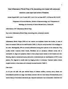
region with a significantly reduced lumen of cardia. We received a superior gastric resection specimen including the eso-gastric junction (Fig. 1). While opening, we identified a solid, cylindrical-shaped
2
11/6.5/5 cm mass, projecting into the lumen, covered by focally ulcerated mucosa. On the section, the specimen had a yellowish-grey appearance with congestive areas (Fig. 3).
Figs. 1–2. Intraluminal tumor, projecting into the lumen, covered by focally ulcerated mucosa, sharply demarcated from the normal digestive wall, at the distal 1/3rd of the esophagus and eso-gastric region.
3
Giant Inflammatory Fibroid Polyp
The presumptive clinical diagnosis was that of GIST or leiomyoma. Histopathological exam revealed a tumor arising from submucosal compartment, with variable cellularity, composed of spindle shaped and stellate cells having bland nuclei and clear/Alcian Blue positive cytoplasm. The cells were admixed with many congested blood vessels and fine collagen fibers in an edematous stroma. There was an abundant inflammatory infiltrate comprising numerous eosinophils, lymphocytes and plasma cells (Fig. 3). The lesion involved the entire thickness of the digestive
181
wall with focal ulceration of the overlying mucosa, very well demarcated from the surrounding normal tissue, but not encapsulated (Figs. 4–5). All the nodules from the adipose tissue surrounding the specimen were analyzed and the slides revealed reactive changes consisting in follicular hyperplasia and sinusal histiocytosis. The immunohistochemistry analysis revealed that the spindle lesional cells were negative for CD117, S100, CD99, bcl-2, Desmin and ALK and positive for CD34 (Figs. 7–10). The Ki67 index was less than 1%.
Fig. 3. The lesion had grey-whitish appearance with congestive areas on the section.
Fig. 4. Hematoxylin-eosin stain, OB4. Well demarcated tumor with large area of ulceration at the eso-gastric junction; haemoragic areas in the tumor periphery.
182
Irina Tudose et al.
Fig. 5. Hematoxylin-eosin stain, OB10. The tumor arising in the submucosal compartment of the esophagus; mucosa is ulcerated.
Fig. 6. Hematoxylin-eosin stain, OB20. Spindle shaped and stellate lesional cells having bland nuclei; very edematous stroma with many congested blood vessels.
Fig. 7. CD117 negativity of the lesional cells.
4
5
Giant Inflammatory Fibroid Polyp
Fig. 8. S100 negativity of the lesional cells.
Fig. 9. CD34 positivity of the lesional cells and in vessels.
Fig. 10. Desmin negativity of the lesional cells.
183
184
Irina Tudose et al.
The differential diagnosis included GIST, neurinoma, inflammatory myofibroblastic tumor, gastric plexiform fibromyxoma, leiomyoma and solitary fibrous tumor. GIST is an intramural tumor with CD117 positive cells while IFP is a submucosal lesion with CD117 negative cells. Both entities share similarities regarding CD34 positivity and stromal aspects, except the presence of numerous eosinophils which is a feature of IFP. Neurinoma is another entity which had to be excluded in our case. Usually neurinoma arises from lamina propria, it has no eosinophils or other inflammation in the stroma and have CD34 positivity in a small percent of cases (15–30%). In our case, the negativity of S100 supported the diagnosis of IFP. Inflammatory myofibroblastic tumor (IMT) appears often in young people and represented another lesion for differential diagnosis. IMT is frequently associated with systemic signs and symptoms, histopathologically showing more numerous plasma cells than eosinophils in the stroma and nuclear pleomorphism of the lesional cells. Immunohistochemical markers settled the differential diagnosis because cells proliferating in IMT are Desmin and ALK positive and CD34 negative. Gastric plexiform fibromyxoma is a multinodular intramural tumor, CD34 negative, centered on muscularis propria, with no intrinsic inflammatory infiltrate. Our lesion was differentiated from leiomyoma both by the histopathological aspect and Desmin negativity. Solitary fibrous tumor arises in the serosa and has no inflammation, so it was easily excluded in our case.
6
The morphological features and the immunohistochemical profile were consistent with the diagnosis of IFP. DISCUSSION
IFPs are benign lesions with no metastatic potential [11]. They occur mostly in the sixth decade of life and recent studies have reported a predominance among females. Although it generally presents as a polypoid mass in the gastric antrum, it can be seen along all the gastrointestinal tract. It is believed to represent a reactive, nonneoplastic condition, but its histogenesis remains controversial. Some have suggested an infectious etiology for IFPs; however, no causative agent has ever been identified [12]. We present this case because it has particularities regarding clinical and pathological findings. First, it is unusual for this lesion to appear in an young male, with no important past medical history. The lesion has arisen in the submucosal compartment of the esophagus and eso-gastric junction, which represents a very rare location for IFPs. The macroscopic aspect and the large dimension of the lesion makes our case more interesting because IFPs rarely exceed 5–6 cm in diameter [13, 14]. Recent papers have described IFPs of large size, but no lesion interested the esophagus or the eso-gastric region [14]. Since IFPs have no distinctive radiologic and clinical findings, histologic confirmation is necessary in all the cases to exclude malignancy.
Polipii fibroizi inflamatori (PFI) reprezintă leziuni polipoide rare, dar bine documentate, care interesează tractul gastrointestinal, apărând mai ales la nivelul stomacului, ileonului şi mai rar la nivelul colonului, duodenului sau esofagului, cu precădere la pacienţi în decada 6 de viaţă. Leziunile sunt caracterizate de o proliferare variabilă a fibroblaştilor şi a vaselor sangvine de calibru mic, de multe ori cu interesarea întregii grosimi a peretelui tubului digestiv, într-un ţesut conjunctiv edematos cu marcat infiltrat inflamator incluzând frecvente eozinofile. Prezentăm cazul unui pacient în vârstă de 33 de ani care prezintă clinic o formaţiune tumorală polipoida, cvasi-cilindrică, cu dimensiuni de 11/6/5 cm, localizată la nivelul treimii inferioare esofagiene, protruzivă în stomac. Aspectul histopatologic şi profilul imunohistochimic au susţinut diagnosticul de PFI. Acest caz prezintă particularităţi clinice şi patologice (sexul şi vârsta pacientului, localizarea şi dimensiunea mare a formaţiunii), particularităţi care ar trebui luate în consideraţie atunci când evaluăm o tumoră mezenchimală de joncţiune eso-gastrică. Corresponding author: Irina Tudose “Elias” Emergency Hospital, Pathology Department, Bucharest, Romania E-mail:
[email protected]
7
Giant Inflammatory Fibroid Polyp
185
REFERENCES 1. 2. 3. 4. 5. 6. 7. 8. 9. 10. 11. 12. 13. 14.
WYSOCKI A.P., TAYLOR G., WINDSOR J.A., Inflammatory fibroid polyps of the duodenum: a review of the literature. Dig Surg 2007; 24 (3): 162–168. POLLICE L., BUFO P., Inflammatory fibroid polyp of the rectum. Path Res Pract 1984, 178: 508–512. OTT D.J., WU W.C., SHIFLETT D.W., PENNELL T.C., Inflammatory fibroid polyp of the duodenum. Am J Gastroenterology 1980, 73: 62–64. VANEK J., Gastric submucosal granuloma with eosinophilic infiltration. Am J Pathol 1949, 25: 397–411. HELWIG E.B., RANIER A., Inflammatory fibroid polyps of the stomach. Surg Gynecol Obstet 1953; 96: 355–367. NAVAS-PALACIOS J.J., COLINA-RUIZDELGADO F., SANCHEZ-LARREA M.D., CORTES-CANSINO J., Inflammatory fibroid polyps of the gastrointestinal tract. An immunohistochemical and electron microscopic study. Cancer 1983, 51: 1682–1690. WILLE P., BORCHARD F., Fibroid polyps of intestinal tract are inflammatory-reactive proliferations of CD34 positive perivascular cells. Histopathology 1998, 32: 498–502. LIVOLSI V.A., PERZIN K.H., Inflammatory pseudotumors (Inflammatory fibrous polyps) of the esophagus: a clinicopathologic study. Am J Digestive Dis 1975, 20: 475–481. SCHILDHAUS H.U., CAVLAR T., BINOT E., BUTTNER R., WARDELMANN E., MERKELBACH-BRUSE S., Inflammatory fibroid polyps harbour mutations in the platelet-derived growth factor receptor alpha (PDGFRA) gene. J Pathol 2008, 216 (2): 176–82. PLESEC TH.P., Gastrointestinal Mesenchymal Neoplasms other than Gastrointestinal Stromal Tumors: focusing on their molecular aspects. Pathological Research International 2011, Article ID 952569. GONUL I.I., ERDEM O., ATAOGLU O., Inflammatory fibroid polyp of the ileum causing intussusception – a case report. Turk J Gastroenterol, 2004, 15 (1): 49–62. ODZE R.D., JOHN GOLDBLUM J., Surgical Pathology of the GI Tract, Liver, Biliary Tract and Pancreas, 2nd edition, Philadelphia, 2009. COULIER B., MALDAGUE P.H., BROZE B., GIELEN I., Ileal inflammatory fibroid polyp causing ileocolic intussusception. JBR-BTR 2008, 91: 149–152. REHMAN S., GAMIE Z., WILSON T.R., COUP A., KAUR G., Inflammatory fibroid polyp (Vanek’s tumor), an unusual large polyp of the jejunum – case report. Cases Journal 2009, 2: 7152.
Received March 31, 2012