ED ITOR IA LS
14. Engel J Jr. The timing of surgical intervention for mesial temporal lobe epilepsy: a plan for a randomized clinical trial. Arch Neurol 1999;56:133841. 15. Kwan P, Brodie MJ. Early identification of refractory epilepsy. N Engl J Med 2000;342:314-9. Copyright © 2001 Massachusetts Medical Society.
G ENETIC C LUES TO THE C AUSE OF P RIMARY P ULMONARY H YPERTENSION
P
RIMARY pulmonary hypertension is a devastating disease that is invariably fatal without definitive therapy. The disorder is more appropriately called a syndrome, since it is characterized by a set of clinical and pathophysiological features common to a variety of underlying causes. The proposed causes of primary pulmonary hypertension range widely, from environmental (e.g., hypoxia) to autoimmune (e.g., systemic lupus erythematosus) to drug related (e.g., dexfenfluramine use). Although genetic determinants no doubt indirectly influence the propensity for primary pulmonary hypertension to develop in response to these factors, a heritable form of primary pulmonary hypertension, familial primary pulmonary hypertension, has also been recognized since 1948.1 The incidence of familial primary pulmonary hypertension in the general population ranges from 1 to 2 cases per million, accounting for 6 percent of the 187 cases of primary pulmonary hypertension in the registry of the National Institutes of Health 2; its pattern of inheritance appears to be autosomal dominant with incomplete penetrance, since the disease develops in no more than 20 percent of persons at risk. Familial primary pulmonary hypertension is an uncommon form of an uncommon disease, yet its heritability provides the opportunity for the use of contemporary genetic approaches to elucidate the molecular basis of the disease. Two related articles in this issue of the Journal 3,4 further our understanding of the genetic and molecular determinants of familial forms of primary pulmonary hypertension. Newman and colleagues3 report the results of a detailed genetic study of a large kindred with familial primary pulmonary hypertension. In the light of the findings in earlier studies of genetic linkage that the locus for familial primary pulmonary hypertension is found on chromosome 2q31–32, the investigators surveyed the known genes in this large chromosomal region for biologically plausible candidates. They identified a unique member of the transforming growth factor b (TGF-b) receptor family, known as bone morphogenetic protein receptor II, as a possible candidate because this receptor can modulate vascular-cell growth. Their logical hunch proved correct when they detected a missense mutation — a substitution of guanine for thymine at position 354 — in exon 3 of the gene for bone morphogenetic pro-
tein receptor II (BMPR2) in all affected family members. In earlier studies, over 25 other mutations in BMPR2 had been identified, with each mutation transmitted within a given family.5 To understand how a mutation in BMPR2 may lead to primary pulmonary hypertension, we must first review the vascular pathological features of the disease. Pulmonary arterial lesions in patients with primary pulmonary hypertension are characterized by medial hypertrophy, concentric laminar intimal fibrosis, and plexiform lesions with obstruction of the arterial lumen, aneurysmal dilatation, and proliferation of interconnected vascular channels (occlusive arteriopathy). The laminar changes and plexiform lesions are associated with proliferation of both vascular smooth-muscle cells and endothelial cells. In some cases of primary pulmonary hypertension, these proliferative changes may represent adaptive (or maladaptive) responses to an exogenous stimulus, such as hypoxia. In familial primary pulmonary hypertension, however, the genetic findings suggest that vascular-cell proliferation is the primary event in the development of lesions. As a member of the TGF-b receptor family, bone morphogenetic protein receptor II is poised to regulate cell proliferation in response to ligand binding. The ligands for the TGF-b receptor family include TGF-b and its superfamily members, bone morphogenetic protein and activin. These growth factors have pleiotropic effects on endothelial and vascular smoothmuscle cells that depend on the environmental and developmental context of the signal, as well as the specific TGF-b receptor family members to which they bind. For example, endothelial-cell proliferation is potently inhibited by TGF-b; however, proliferation can be stimulated in the presence of vascular endothelialcell growth factor and fibroblast growth factor.6 Similarly, TGF-b and bone morphogenetic protein can inhibit migration and proliferation of vascular smoothmuscle cells; however, both can also promote growth of vascular smooth-muscle cells under certain conditions: TGF-b can induce mitogen synthesis and its release from quiescent vascular smooth-muscle cells that, in turn, support cell proliferation,7 and low concentrations of bone morphogenetic protein directly stimulate proliferation of vascular smooth-muscle cells. As a complement to its actions as an inhibitor of cell growth, TGF-b can also induce apoptosis of endothelial and vascular smooth-muscle cells. Even in this context pleiotropy is apparent, however, since TGF-b has been shown to protect vascular cells from apoptosis induced by other agents.8 These actions of the TGF-b superfamily on vascular cells are mediated by a family of cognate receptors. Broadly speaking, the TGF-b receptor family comprises three classes: types I, II, and III.8 The TGF-b superfamily exerts its principal effects by binding to heteromeric complexes of type I and type II receptors, which are transmembrane signaling molecules with
N Engl J Med, Vol. 345, No. 5 · August 2, 2001 · www.nejm.org · 367
The Ne w E n g l a nd Jo u r n a l o f Me d ic i ne
serine–threonine kinase activity. The type I receptors include the TGF-b receptor I, bone morphogenetic protein receptor I, and activin receptor I; the type II receptors include the TGF-b receptor II, bone morphogenetic protein receptor II, and activin receptor II. Specific members of the TGF-b superfamily can associate with various type I and type II receptors to evoke pleiotropic cellular responses through various signaling and transcriptional pathways. The current paradigm for signaling through this system is that after ligand binding, type II receptors recruit and phosphorylate type I receptors, which in turn activate downstream signaling molecules of the Smad class by phosphorylation. The Smad proteins are a family of at least nine gene products, each of which is involved in some way in mediating cellular responses to specific members of the TGF-b superfamily. For example, the binding of TGF-b to heteromeric complexes of the TGF-b receptor I and the TGF-b receptor II leads to phosphorylation of Smad2 and Smad3, whereas the binding of bone morphogenetic protein to heteromeric complexes of bone morphogenetic protein receptor I and bone morphogenetic protein receptor II leads to phosphorylation of Smad1, Smad5, and (probably) Smad8. Phosphorylated Smads then bind to the one Smad family member that is not phosphorylated, Smad4, forming a complex that translocates to the nucleus, where it interacts with specific proteins to modulate gene expression. Depending on the specific ligand, heteromeric receptors, cell type, downstream signals, and transcriptional program, the ultimate cellular response can either promote or inhibit proliferation. Much of the published experimental work on these TGF-b–mediated responses has been performed with the use of neoplastic cells. Studies with vascular cells have been less numerous but are nevertheless intriguing and relevant to this discussion. A recent study showed that expression of type II receptors is significantly decreased in vascular cells within atheromas,9 and this suppression appears to convert the cells from an antiproliferative to a proliferative phenotype10; transfection of these cells with the complementary DNA for a type II receptor partially reverses this change in phenotype.8 On the basis of these and similar data, one can postulate that the mutation in BMPR2 found in the family of patients with primary pulmonary hypertension that is described by Newman and colleagues3 leads to a loss of the inhibitory action of bone morphogenetic protein on the growth of vascular smooth-muscle cells in the pulmonary vasculature, thereby supporting the proliferative responses underlying the development of pulmonary hypertension. Recent in vitro data provide evidence of this action of bone morphogenetic protein on vascular smooth-muscle cells,11 and functional in vitro studies of mutations in type II receptors confirm the adverse consequences of certain mutations in BMPR2 on ligand specificity and signaling events.12
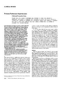
The arteriopathy of primary pulmonary hypertension involves not only proliferation of vascular smoothmuscle cells but also, importantly, proliferation of endothelial cells. Evidence for the expression of bone morphogenetic protein receptor II by endothelial cells13 or for an effect of bone morphogenetic protein on endothelial-cell growth is limited. Thus, we are left with the conclusion that the endothelial proliferative response observed in familial primary pulmonary hypertension may be a secondary consequence of the increase in peripheral vascular resistance, the increase in pulmonary arterial pressure, and the subsequent endothelial activation (or injury) produced by the partial loss of inhibition of vascular smooth-muscle-cell growth by the mutant bone morphogenetic protein receptor II (Fig. 1). That the observed mutation is partially functional is indicated by the finding that a homozygous mutation in the kinase domain of bone morphogenetic protein receptor II is lethal in mouse embryos.14 A second study in this issue, by Trembath and colleagues,4 offers additional insights into the role of the endothelial cell in the pathobiology of another inherited form of primary pulmonary hypertension. These investigators studied a rare subgroup of patients with another uncommon disease, hereditary hemorrhagic telangiectasia (Rendu–Osler–Weber syndrome), in whom pulmonary hypertension developed. The pathological features identified in these patients’ blood vessels consist of the vascular dilatations and arteriovenous fistulaes characteristic of hereditary hemorrhagic telangiectasia but also the occlusive arteriopathy of primary pulmonary hypertension. Mutations in two related gene products, endoglin and activin-receptor– like kinase I, have been found in patients with hereditary hemorrhagic telangiectasia. Endoglin is an accessory protein member of the type III receptor class that facilitates assembly of type I and type II receptor complexes,15 and activin-receptor–like kinase 1, an activin type I receptor, appears to be particularly abundant in the pulmonary vasculature, primarily in endothelial cells.16 Trembath and colleagues identified mutations in the gene for activin-receptor–like kinase 1 (but not the gene for endoglin) in members of families with hereditary hemorrhagic telangiectasia who also had primary pulmonary hypertension. What are the implications of this observation for our understanding of the molecular mechanisms underlying the development of primary pulmonary hypertension? A recent report by Oh and colleagues17 helps address this question. These investigators showed that the binding of TGF-b to heteromeric complexes of activin-receptor–like kinase 1 and TGF-b receptor II leads to inhibition of TGF-b–dependent transcriptional activation mediated by another type I receptor, activin-receptor–like kinase 5, which forms a complex with TGF-b receptor II in endothelial cells. The binding of TGF-b to activin-receptor–like kinase 5 nor-
368 · N Engl J Med, Vol. 345, No. 5 · August 2, 2001 · www.nejm.org
ED ITORIA LS
Familial primary pulmonary hypertension
Primary pulmonary hypertension associated, with hereditary hemorrhagic telangiectasia, , TGF-b receptor II
Endoglin
TGF-b
Mutant ALK1 Mutant BMPR-II BMPR-I
Pi
BMP
Pi
–, Pi Pi
+
Smad1, Smad5, Smad8
Smad1, Smad5, Smad8 Smad4 Smad2, Smad3
Smad4
Pi
Pi
Cell-growthB stimuli TGF-b ALK5
TGF-b receptor II Endoglin
Proliferation of endothelial cellsB AngiogenesisB B
Epigenetic factors
Epigenetic factors
Proliferation of vascularB smooth-muscle cell
PulmonaryB vascularB resistance
Activation and injuryB of endothelial cells
Additional geneticB factors
PulmonaryB vascularB resistance
Additional geneticB factors
Pulmonary hypertension
Figure 1. Hypothetical Model of the Role of Mutations in BMPR2 and ALK1 in the Development of Primary Pulmonary Hypertension. In familial primary pulmonary hypertension, the mutation in BMPR2 leads principally to stimulation of the growth of pulmonary arterial vascular smooth-muscle cells (left-hand side) through the action of growth-promoting factors unopposed by the growthinhibitory pathway of mutant bone morphogenetic protein receptor II (BMPR-II). The binding of ligand (BMP) to the complex of BMPR-I and BMPR-II causes receptor-dependent phosphorylation (Pi denotes inorganic phosphate) of Smad1, Smad5, and (probably) Smad 8, which associate with Smad4 to modulate transcription of factors that regulate cell growth. Mutant BMPR-II is less effective at transducing Smad phosphorylation than is wild-type BMPR-II, leading to proliferation of pulmonary arterial vascular smooth-muscle cells, an increase in pulmonary vascular resistance, and secondary activation and injury of pulmonary arterial endothelial cells, ultimately causing migration and proliferation of pulmonary arterial endothelial cells and adaptive (or maladaptive) angiogenesis (plexiform lesions). These latter changes further increase pulmonary vascular resistance and cause pulmonary hypertension. In primary pulmonary hypertension associated with hereditary hemorrhagic telangiectasia, activin-receptor–like kinase 1 (ALK1), which is found principally in pulmonary arterial endothelial cells (right-hand side), binds to TGF-b receptor II and endoglin in the presence of ligand (TGF-b) to form a heterotrimeric receptor complex that phosphorylates Smad1 and Smad5. The phosphorylated Smads bind to Smad4 and, in turn, modulate gene expression to suppress proliferation of pulmonary arterial endothelial cells (minus sign denotes suppression of proliferation). The ALK5-dependent pathway, which opposes the ALK1-dependent pathway, also binds the complex of TGF-b receptor II and endoglin to form the heterotrimeric receptor complex in the presence of TGF-b. This complex phosphorylates Smad2 and Smad3, which also bind to Smad4 to form a complex that modulates gene expression to promote proliferation of pulmonary arterial endothelial cells (plus sign denotes promotion of proliferation). Mutant ALK1 is less able to signal through Smad1 and Smad5 to inhibit the growth-promoting actions of TGF-b signaling through ALK5, thereby leading to migration and proliferation of pulmonary arterial endothelial cells and adaptive (or maladaptive) angiogenesis that causes an increase in pulmonary vascular resistance and pulmonary hypertension. Epigenetic and additional genetic factors modify these responses in a person harboring either of these mutations.
N Engl J Med, Vol. 345, No. 5 · August 2, 2001 · www.nejm.org · 369
The Ne w E n g l a nd Jo u r n a l o f Me d ic i ne
mally promotes proliferation of endothelial cells, and competitive binding to activin-receptor–like kinase 1 attenuates this proliferative response. Unopposed TGF-b signaling through the activin-receptor–like kinase 5 pathway would therefore be predicted to increase migration and proliferation of capillary endothelial cells, resulting in arteriovenous connections without an intervening capillary bed.17 There is evidence of this proposed mechanism in the finding that mice with a complete deficiency of activin-receptor–like kinase 1 die in mid-gestation with severe vascular abnormalities, including fusion of capillary plexuses into cavernous vessels and hyperdilatation of large vessels.17 Taken together, these data suggest that in patients with primary pulmonary hypertension associated with hereditary hemorrhagic telangiectasia, pulmonary vascular hemodynamics are altered by vascular malformations induced by abnormal endothelial proliferative responses through unopposed TGF-b signaling mediated by activin-receptor–like kinase 5. This occurs through increased flow through arteriovenous malformations, increased resistance through blind vascular channels, or both (Fig. 1). These complementary studies are intriguing, because genetic targets have finally been identified in certain forms of primary pulmonary hypertension. As with any good study, these reports raise as many questions as they answer. First, they are limited by the lack of functional studies of the specific mutations in cellular systems or animal models that can be used to identify the specific molecular mechanisms by which the mutations lead to changes in the pulmonary vasculature. In the case of the mutation in BMPR2, for example, animal models would be particularly helpful in understanding why the vascular changes occur in the pulmonary, but not in the systemic, vasculature. Second, the observation that mutations in two different, but mechanistically related, TGF-b receptors can produce the same clinical phenotype suggests that these two receptors may interact directly to modulate vascular-cell growth. However, there is as yet no evidence of a direct interaction between activin-receptor–like kinase 1 and bone morphogenetic protein receptor II. Smad1 is phosphorylated by activin-receptor–like kinase 1 as well as by bone morphogenetic protein receptor I, but each type I receptor phosphorylates Smad1 through a different recognition domain.18 These results can then be interpreted to mean that each mutation produces primary pulmonary hypertension by a different mechanism, perhaps in a celltype–specific manner — that is, the mutation in BMPR2 leads to primary pulmonary hypertension by inducing proliferation of primarily pulmonary vascular smooth-muscle cells and the mutation in activinreceptor–like kinase 1 leads to primary pulmonary hypertension by inducing proliferation of primarily endothelial cells (Fig. 1). Third, the incomplete penetrance of the primary
pulmonary hypertension in both of these reports and the finding that approximately one quarter of patients with “sporadic” primary pulmonary hypertension have a mutation in BMPR2 suggest that there are either epigenetic determinants of disease expression (i.e., determinants that are not heritable) or additional genetic determinants. Put another way, these observations suggest that the individual genomic and environmental context influences the phenotypic expression of this complex disease trait. For example, do these mutations lead to primary pulmonary hypertension only when a person is exposed to an environmental stimulus, such as hypoxia? Or are there other, as yet unidentified, disease-modifying genes that modulate disease expression? These questions can be answered only through the study of relevant animal models and of kindreds in which penetrance of the disease is incomplete. In an animal model of hereditary hemorrhagic telangiectasia induced by targeted disruption of the gene for endoglin, phenotypic heterogeneity of the disorder was manifest and depended on the genetic background of the mouse strain from which the knockout was created.19 These data suggest that disease-modifying genes influence the ultimate phenotype of this disorder and support the concept of such a mechanism in humans with the mutation as well. Similarly, the observation that familial primary pulmonary hypertension is characterized by genetic anticipation (i.e., that the disease becomes increasingly severe or occurs at an earlier age with successive generations)20 also suggests that disease-modifying genes influence phenotype, especially since the mutations in BMPR2 in patients with familial primary pulmonary hypertension are in the coding region of the gene and are invariant among generations within an affected family. At the very least, these reports highlight new genetic markers for use in identifying family members who may be at risk for the disease. Natural-history studies and close follow-up of persons harboring the mutant alleles before the development of elevated pulmonary pressures will give us further insights into the relation between these genotypes and this devastating and perplexing phenotype. JOSEPH LOSCALZO, M.D., PH.D. Boston University Medical Center Boston, MA 02118
REFERENCES 1. Lange F. Die essentielle Hypertonie de Lungenstrombahn und ihr familiäres Vorkommen. Dtsch Med Wochenschr 1948;73:322-6. 2. Rich S, Dantzker DR, Ayres SM, et al. Primary pulmonary hypertension: a national prospective study. Ann Intern Med 1987;107:216-23. 3. Newman JH, Wheeler L, Lane KB, et al. Mutation in the gene for bone morphogenetic protein receptor II as a cause of primary pulmonary hypertension in a large kindred. N Engl J Med 2001;345:319-24. 4. Trembath RC, Thomson JR, Machado RD, et al. Clinical and molecular genetic features of pulmonary hypertension in patients with hereditary hemorrhagic telangiectasia. N Engl J Med 2001;345:325-34. 5. Deng Z, Morse JH, Slager SL, et al. Familial primary pulmonary hy-
370 · N Engl J Med, Vol. 345, No. 5 · August 2, 2001 · www.nejm.org
ED ITORIA LS
pertension (gene PPH1) is caused by mutations in the bone morphogenetic protein receptor-II gene. Am J Hum Genet 2000;67:737-44. 6. Pepper MS, Vassalli J-D, Orci L, Montesano R. Biphasic effect of transforming growth factor-b1 on in vitro angiogenesis. Exp Cell Res 1993;204: 356-63. 7. Battegay EJ, Raines EW, Seifert RA, Bowen-Pope DF, Ross R. TGF-b induces bimodal proliferation of connective tissue cells via complex control of an autocrine PDGF loop. Cell 1990;63:515-24. 8. McCaffrey TA. TGF-bs and TGF-b receptors in atherosclerosis. Cytokine Growth Factor Rev 2000;11:103-14. 9. McCaffrey TA, Du B, Fu C, et al. The expression of TGF-b receptors in human atherosclerosis: evidence for acquired resistance to apoptosis due to receptor imbalance. J Mol Cell Cardiol 1999;31:1627-42. 10. McCaffrey TA, Consigli S, Du B, et al. Decreased type II/type I TGFb receptor ratio in cells derived from human atherosclerotic lesions: conversion from an antiproliferative to profibrotic response to TGF-b1. J Clin Invest 1995;96:2667-75. 11. Dorai H, Vukicevic S, Sampath TK. Bone morphogenetic protein-7 (osteogenic protein-1) inhibits smooth muscle cell proliferation and stimulates the expression of markers that are characteristic of SMC phenotype in vitro. J Cell Physiol 2000;184:37-45. 12. Carcamo J, Zentella A, Massague J. Disruption of transforming growth factor b signalling by a mutation that prevents transphosphorylation within the receptor complex. Mol Cell Biol 1995;15:1573-81. 13. Atkinson C, Stewart S, Imamura T, Trembath RC, Morrell NW. Immunolocalisation of BMPR-II and TGF-b type I and II receptors in primary plexogenic pulmonary hypertension. J Heart Lung Transplant 2001; 20:149. 14. Beppu H, Miyazono K. The roles of TGF-beta superfamily in mouse early development. Tanpakushitsu Kakusan Koso 2000;45:Suppl 13:210915. (In Japanese.) 15. Barbara NP, Wrana JL, Letarte M. Endoglin is an accessory protein that interacts with the signaling receptor complex of multiple members of the transforming growth factor-b superfamily. J Biol Chem 1999;274:58494. 16. Panchenko MP, Williams MC, Brody JS, Yu Q. Type I receptor serinethreonine kinase preferentially expressed in pulmonary blood vessels. Am J Physiol 1996;270:L547-L558. 17. Oh SP, Seki T, Goss KA, et al. Activin receptor-like kinase 1 modulates transforming growth factor-b1 signaling in the regulation of angiogenesis. Proc Natl Acad Sci U S A 2000;97:2626-31. 18. Chen Y-G, Massague J. Smad1 recognition and activation by the ALK1 group of transforming growth factor-b family receptors. J Biol Chem 1999;274:3672-7. 19. Bourdeau A, Faughnan ME, Letarte M. Endoglin-deficient mice, a unique model to study hereditary hemorrhagic telangiectasia. Trends Cardiovasc Med 2000;10:279-85. 20. Loyd JE, Butler MG, Foroud TM, Conneally PM, Phillips JA III, Newman JH. Genetic anticipation and abnormal gender ratio at birth in familial primary pulmonary hypertension. Am J Respir Crit Care Med 1995;152:93-7. Copyright © 2001 Massachusetts Medical Society.
S ACRED S ECRETS — T HE P RIVACY OF M EDICAL R ECORDS Whatsoever I shall see or hear concerning the life of men, in my attendance on the sick, or even apart therefrom, which ought not to be noised abroad, I will keep silence thereon, counting such things to be as sacred secrets. The Hippocratic Oath
P
ERSONAL medical information is far from private in the United States. Insurers use identifiable medical records for risk rating, employers use them for hiring and firing, health systems for quality assurance, pharmaceutical firms for marketing, banks for assessing loan risk, and the government for the detection of fraud. It is difficult to obtain health insurance without
consenting to the release of present and future medical data into an information market that is largely unregulated. Very few people are happy about these uses of their medical records. In a recent Gallup poll, 78 percent of respondents said it is very important that their medical records be kept confidential, 82 percent opposed letting insurance companies see their medical records without permission, and 95 percent opposed access by banks.1 Over the past five years, polling data have consistently shown that Americans want their medical records to be confidential, fear the use of their medical records to discriminate against them, and want stronger protections for information from identifiable medical records. The struggle between patients and commercial interests is reflected in the medical-privacy bills recently introduced in Congress. Some of these bills would codify unfettered access to medical records by a wide range of “authorized users,” with loose standards for patient consent.2 Others would erect very comprehensive legal barriers to unauthorized access or misuse and strict standards for consent.3 Neither approach has attracted overwhelming political support, because of the divisiveness of the issue itself. Federal privacy regulations issued by the Clinton administration will create major new privacy protections, but they will also leave large loopholes for certain commercial organizations and government agencies to gain access to records without patients’ consent or even awareness.4 Although reservations about the use of medical information have been in existence for some time, the dramatic increase in genetic testing has prompted fears that patients will suffer discrimination on the basis of genotype, even when it is weakly predictive of phenotype. These fears will intensify as DNA-based testing for hereditary conditions proliferates. In addition to the voluntary testing of adults, there will be an expansion of mandatory screening of newborns. Mandates were originally enacted by states for non–DNA-based screening for inborn errors of metabolism, but these statutes are now being used to expand mandatory screening into DNA-based testing. The number of DNA-based tests that could potentially become mandatory for newborns is extensive. As a consequence of these trends, medical records are likely to contain more and more genetic information with the potential for use in discriminating against individual persons or groups. Thirty-seven states have enacted legislation prohibiting discrimination in employment or insurance on the basis of genetic information. Although these statutes are a step in the right direction, they actually provide little practical protection, since the burden of proof is on the patient and discrimination is difficult to prove. In addition, many states require physicians to handle genetic information with a higher level of confidentiality than the rest of the medical record. This
N Engl J Med, Vol. 345, No. 5 · August 2, 2001 · www.nejm.org · 371