Gastric Intestinal Metaplasia and Early Gastric Cancer in the West: A Changing Paradigm Justin M. Gomez, MD, and Andrew Y. Wang, MD, FACG, FASGE
Dr Gomez is a clinical instructor in the Department of Medicine and Dr Wang is an associate professor of medicine in the Division of Gastroenterology and Hepatology at the University of Virginia in Charlottesville, Virginia.
Abstract: Gastric cancer remains the fifth leading cancer diagno-
Address correspondence to: Dr Andrew Y. Wang Division of Gastroenterology and Hepatology Box 800708 University of Virginia Health System Charlottesville, VA 22908 Tel: 434-924-1653 Fax: 434-244-7590 E-mail:
[email protected]
nal metaplasia, which can increase the risk of gastric cancer by
sis worldwide, and it is the third leading cause of cancer-related mortality. The incidence of gastric cancer within the United States, however, has remained substantially lower than elsewhere, which has led to a lack of screening and surveillance in clinical practice. Patients with known premalignant lesions, such as gastric intestias much as 6-fold, might benefit from surveillance guidelines to detect gastric cancer at an earlier, potentially curative stage. Chromoendoscopy with optical magnification, narrow-band imaging, and other image-enhanced endoscopic techniques are commercially available to assist in the diagnosis of premalignant gastric lesions and early gastric cancer. Furthermore, endoscopic mucosal resection and endoscopic submucosal dissection have become more widely available and offer potentially curative endoscopic resection for dysplastic lesions of the stomach and early gastric cancers, which is an alternative to traditional surgical resection.
G
Keywords Early gastric cancer, endoscopic submucosal dissection, esophagogastroduodenoscopy, gastric intestinal metaplasia, staging
astric cancer is the fifth leading cancer diagnosis worldwide, with approximately 952,000 cases diagnosed in 2012, and it is the third leading cause of cancer-related death, with approximately 723,000 deaths annually.1 More than 70% of these cases occur in the developing world, and roughly 50% occur in Southeast Asia.2 Despite this high global incidence of disease, incidence within North America has remained significantly lower at 21,000 new cases and 12,000 gastric cancer–related deaths in 2012.3 Early gastric cancer is defined as adenocarcinoma confined to the mucosa or submucosa.4 A multicenter retrospective study of 2191 patients with gastric cancer undergoing surgical resection found that early gastric cancer represented approximately 20% of all surgically resected cancers in North America, but early gastric cancer accounted for 50% of resected cancers in Japanese centers.5 There are multiple proposed explanations for this geographic variability in cancer stage at the time of diagnosis. As Japan has a higher overall incidence of gastric cancer than the West, the Japanese have instituted screening protocols that augment early diagnosis.
Gastroenterology & Hepatology Volume 10, Issue 6 June 2014 369
GOMEZ AND WANG
There are also differences between Japanese and Western pathologists in the classification of early gastric cancers, which can affect international cancer statistics and clinical studies of this disease. Western pathologists require the presence of invasion into the lamina propria for the diagnosis of gastric cancer, whereas Japanese pathologists can make this diagnosis based on the presence of cytologic and architectural atypia without requiring the presence of mucosal invasion. This difference is illustrated in a study in which Japanese and Western pathologists reviewed 35 gastric biopsies and found histologic agreement in only 31% of the cases.6 The Vienna classification of gastrointestinal (GI) neoplasia was, in part, developed with the hope of arriving at a common nomenclature for the diagnosis of early GI malignancies.7 Gastritis and Premalignant Lesions of the Stomach Gastric adenocarcinoma is thought to arise through a cascade that was first described by Correa in 19888 and involves nonatrophic gastritis, atrophic gastritis, gastric intestinal metaplasia (IM), gastric dysplasia, and, ultimately, gastric cancer. The underlying mechanism for this process remains unclear, but it is thought to be linked to Helicobacter pylori infection.8,9 Atrophic gastritis and gastric IM confer an increased risk of progression to gastric cancer, as these conditions are the background from which dysplasia and, ultimately, adenocarcinoma develop. Gastritis is an inflammatory condition of the gastric mucosa that is histopathologically diagnosed by an inflammatory-cell infiltrate, consisting primarily of mononuclear cells including lymphocytes and plasma cells. The phenotypic distinction between nonatrophic gastric and atrophic gastritis is well recognized. Atrophic gastritis is defined as loss of the normal gastric glandular epithelium and replacement by either metaplastic glandular structures or fibrosis of the lamina propria.10 Several classification schemes have been developed to further define chronic gastritis. The updated Sydney system is the most widely accepted for classification of gastritis. The Sydney system was developed to standardize histopathologic grading and topography of chronic gastritis, as well as to provide information regarding the underlying etiology of gastritis.11 More recently, a collaborative group of gastroenterologists and pathologists created the Operative Link on Gastritis Assessment (OLGA) staging system. The OLGA staging system integrates the atrophy score obtained on biopsy and the atrophy topography obtained through biopsy mapping to stratify patients’ cancer risk and guide further prognostic decision-making. Rugge and colleagues followed a cohort of 93 dyspeptic patients for more than 12 years to assess the prognostic
value of the OLGA staging system and its ability to predict progression to gastric cancer.12 The study found that the only 2 patients in whom invasive neoplasia developed were classified as OLGA grade III/IV, which significantly predicted neoplasia at the end of the follow-up period. Pathologically, gastric IM is defined by the loss of normal gastric epithelium and replacement with an intestinal phenotype containing goblet cells, Paneth cells, and absorptive cells.13-15 Gastric IM may be further classified, based on histologic appearance with hematoxylin and eosin staining, into complete and incomplete gastric IM. Complete (type I) gastric IM is characterized by the presence of a small intestinal–type mucosal phenotype with goblet cells containing sialomucins interspersed among absorptive cells and with a well-defined brush border. In contrast, incomplete (type III) gastric IM is characterized by a colonic-type mucosal phenotype with tortuous crypts lined by tall columnar cells containing abundant sulfomucins. A hybrid form of gastric IM (type II, also considered incomplete) exists that expresses a mixture of gastric and intestinal mucins.11 Significance of Gastric Intestinal Metaplasia The prevalence of gastric IM in the general population remains difficult to ascertain given the asymptomatic nature of the lesion. Sonnenberg and colleagues conducted a large retrospective study of 78,985 patients undergoing esophagogastroduodenoscopy (EGD) with biopsy across the United States and found that the prevalence of gastric IM was 7%.16 The frequency of gastric IM appears to be equal between men and women and demonstrates a linear age-dependent rise.16 Patients at increased risk for development of gastric IM and cancer include those with a first-degree family history of gastric cancer and patients from ethnic backgrounds in which gastric cancer is prevalent.17 A meta-analysis, published in 2010, showed that a first-degree family history of gastric cancer increased the incidence of gastric cancer, with an odds ratio of 1.98 (95% CI, 1.36-2.88).18 Since Correa first described the cascade by which gastric IM progresses to gastric cancer,8 gastric IM has become well-accepted as a premalignant lesion. A large Japanese study of 1246 patients with gastric IM followed for a mean of 7.8 years found that the relative risk of progression to gastric cancer was 6.4 (95% CI, 2.6-16.1).19 The largest Western study of gastric IM performed by a Dutch team of investigators in 61,707 patients found that gastric cancer developed in 874 cases, with a 10-year incidence of 1.8%.20 The prevalence of disease and the incidence of progression to gastric cancer take on greater significance in comparison with another premalignant lesion—Barrett esophagus—which has well-defined screening and sur-
370 Gastroenterology & Hepatology Volume 10, Issue 6 June 2014
G A S T R I C I N T E S T I N A L M E TA P L A S I A A N D E A R LY G A S T R I C C A N C E R I N T H E W E S T
veillance recommendations. Estimates of the prevalence of Barrett esophagus in the Western population are variable, but a US study of 961 patients found a prevalence of 6.8%.21 Likewise, the annual incidence of progression from Barrett esophagus to adenocarcinoma ranges between 0.12% and 0.5%.22,23 The progression from gastric IM to gastric adenocarcinoma is highly associated with the histologic subtype of IM. The incomplete patterns (types II and III) of gastric IM are associated with the greatest risk of progression to gastric cancer.13,24-29 A study completed in Spain found that the incidence of gastric cancer in patients with incomplete IM was 18.2% (16 of 88 patients) compared with 0.96% (1 of 104 patients) in those with complete IM over a mean follow-up period of 12.8 years.30 However, in clinical practice, pathologists do not typically make the distinction between different types of gastric IM. Pathologically, this distinction may be difficult to make, as incomplete and complete gastric IM can coexist, and the finding of gastric IM can be very focal even on a small biopsy specimen. Gastric IM is thought to be a breakpoint in the process towards carcinogenesis. Several attempts have been made at inducing regression of gastric IM by treating H pylori infection. A meta-analysis of 2658 patients with atrophic gastritis and gastric IM found that atrophic gastritis in the antrum may be reduced through treatment of H pylori infection; however, atrophic gastritis in the corpus or the presence of gastric IM anywhere in the stomach was not impacted.31 As a result of the increased risk of gastric cancer in patients with atrophic gastritis and gastric IM, the European Society of Gastrointestinal Endoscopy published surveillance guidelines in 2012 pertaining to these patient populations. These guidelines recommend that endoscopic surveillance be offered to patients with extensive atrophic gastritis or gastric IM every 3 years.32 In North America, there are currently no consensus guidelines regarding the management of gastric IM. American Society for Gastrointestinal Endoscopy guidelines, published in 2006, state that there are insufficient data on gastric IM within the US population to recommend endoscopic surveillance; however, it was stated that patients at increased risk due to ethnic background or family history may benefit from surveillance endoscopy.26 Although not a definitive guideline, an expert review article published by Correa and colleagues in the American Journal of Gastroenterology in 2010 proposed a surveillance algorithm for gastric IM.25 The authors recommended that patients with extensive gastric IM (defined as IM present in at least 2 gastric locations or moderate or marked IM in at least 2 biopsy specimens) or incomplete/type III gastric IM found on index EGD undergo surveillance EGD with mapping or serum pepsinogen (PG) levels at 1 year. Repeat surveillance EGD every 3 years was suggested if extensive
IM/atrophy or incomplete-type IM persists. Extensive atrophy was defined as a serum PGI level of less than 70 μg/L and a PGI/PGII ratio of less than 3. Diagnostic Endoscopy for Premalignant Gastric Lesions and Early Gastric Cancer Patients with gastric cancer are typically asymptomatic until advanced disease is present. EGD is the diagnostic modality of choice for diagnosing premalignant gastric lesions and gastric cancers. Once a dysplastic gastric lesion is identified endoscopically, endoscopic ultrasonography (EUS) can be helpful in providing T- and N-staging, particularly if an advanced gastric cancer is found. The Paris system for classification was created in 2002 to better classify superficial lesions found in the luminal GI tract that might be amenable to endoscopic resection.33 Possibly due to the lower prevalence of early gastric cancers in the West and to a lack of clinical practice guidelines in this field, endoscopic training during GI fellowship in the United States has not traditionally focused on the screening or surveillance of premalignant gastric lesions to identify early gastric cancers. Each of the following sections on the diagnosis and endoscopic management of premalignant gastric lesions and early gastric cancers is predicated on the systematic practice of carefully inspecting the cleaned mucosa of the entire stomach.34 Without this level of endoscopic attention, dysplastic lesions and early gastric cancers can easily be missed. Despite the ability of conventional white-light endoscopy to detect advanced gastric cancer, it is not as reliable for the diagnosis of premalignant gastric lesions.35 Highdefinition magnifying chromoendoscopy has provided a more reliable method of detecting gastric IM (sensitivity of 76% and specificity of 84%) and gastric dysplasia (sensitivity of 97% and specificity of 81%).36,37 However, chromoendoscopy requires more time, specialized training, and endoscopes equipped with optical magnification so as to properly visualize pits and other surface characteristics. In response to the challenges of dye-based chromoendoscopy, narrow-band imaging (NBI) and other commercially available image-enhanced endoscopic techniques have been developed to provide optical enhancement at the push of a button. Magnification endoscopy using NBI has been shown to distinguish between malignant and nonmalignant gastric lesions, with a sensitivity of 97% and specificity of 84%.38 Gastric biopsies are helpful in confirming the presence of preneoplastic or dysplastic lesions (Figure 1). The Sydney system for the classification and grading of gastritis was originally designed to provide more standardization to biopsy reporting. The system recommends 5 biopsies, including 2 from the antrum, 1 from the incisura, and 2 from the corpus. Although this approach provides adequate
Gastroenterology & Hepatology Volume 10, Issue 6 June 2014 371
GOMEZ AND WANG
A
B
C
D
E
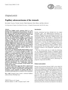
Figure 1. An esophagogastroduodenoscopy was undertaken to evaluate for gastric intestinal metaplasia and dysplasia in a patient with suspected autoimmune metaplastic atrophic gastritis (antiparietal cell antibody was positive). Note the overall paucity of rugal folds seen on the endoscopic images. Multiple cold biopsies were obtained from the antrum (A), incisura (B), and lesser curve (C) of the stomach, which are the typical “hot spots” for early gastric cancer. Other biopsies were obtained from other areas of the body and fundus to evaluate for autoimmune gastritis. Narrow-band imaging (D) and chromoendoscopy (E) using 0.8% indigo carmine were used to survey for dysplasia, which was not found. Histopathology demonstrated focal intestinal metaplasia in the background of likely autoimmune metaplastic atrophic gastritis.
372 Gastroenterology & Hepatology Volume 10, Issue 6 June 2014
G A S T R I C I N T E S T I N A L M E TA P L A S I A A N D E A R LY G A S T R I C C A N C E R I N T H E W E S T
assessment for the presence of H pylori gastritis, there is significant controversy over its ability to detect gastric IM and dysplasia. Unlike atrophic gastritis, which has a more diffuse phenotype, gastric IM and dysplasia tend to be multifocal and might be missed without additional sampling.11 As a result, several studies have examined the number of biopsies needed to accurately diagnose gastric IM. A multicenter Dutch study of a population with a low prevalence of gastric cancer evaluated the yield of an endoscopic strategy that included 12 nontargeted biopsies and additional targeted biopsies for detecting gastric IM, dysplasia, and cancer.39 The researchers found that a biopsy protocol that consisted of 7 nontargeted biopsies, including 3 from the antrum, 1 from the incisura, and 3 from the corpus, was able to accurately diagnose 97% of gastric IM and 100% of dysplasia and cancer cases. Nontargeted biopsies from the angularis (40%), antrum (35%), and lesser curvature of the corpus (30%) resulted in the highest yields of premalignant conditions. Staging of Early Gastric Cancers Gastric cancer is commonly staged using the tumor-nodemetastasis system, as proposed by the American Joint Committee on Cancer.40 Early gastric cancer is defined as a T1 lesion that invades the mucosa (lamina propria or muscularis mucosa, T1a) and/or the upper portion of the submucosa (early T1b lesion, referred to as superficial submucosal invasion, or as a Sm1 lesion in Japan) but does not invade the muscularis propria.41 However, the T-stage of early gastric cancer does not preclude the possibility of lymph node metastases. The incidence of lymph node metastasis in tumors that invade the submucosa can be as high as 10% to 15%.42-45 An Italian study of 652 cases of surgically resected early gastric cancer found that patients with lymph node metastasis were significantly more likely to have larger primary tumors and submucosal invasion than those patients without lymph node involvement (24% vs 5%, respectively).42 Larger tumor size (>4 cm) was associated with nodal metastasis in 30% of cases vs only 9% for tumors smaller than 2 cm. The prognostic importance of nodal metastases has been demonstrated in multiple studies, including the aforementioned Italian study, which found a 10-year survival rate of 92% in patients with no lymph node metastasis.42 However, 10-year survival rapidly dropped to 82%, 73%, and 27% in the presence of 1 to 3, 4 to 6, or greater than 6 involved lymph nodes, respectively.42,46 As a result, the ability to accurately predict and/or diagnose nodal metastases is crucial in correctly identifying those patients with early gastric cancers who might benefit from endoscopic resection as opposed to those who should undergo surgical resection and lymph node dissection.
In a landmark study, Gotoda and colleagues reviewed over 5000 patients who underwent gastrectomy with meticulous R2-level lymph node dissection and developed criteria for curative endoscopic therapy of early gastric cancer using endoscopic mucosal resection (EMR).44 Using these data, expanded criteria also were developed to identify lesions that might be appropriate for resection by endoscopic submucosal dissection (ESD).47 The expanded criteria for ESD of early gastric cancers includes differentiated mucosal cancer, without ulceration, of any size; differentiated mucosal cancer, with ulceration, 3 cm or less in size; and differentiated submucosal cancer (Sm1 ≤500 µm), 3 cm or less in size. In addition to these criteria, lesions must be without lymphovascular invasion on final pathology after ESD; otherwise, gastrectomy with lymph node dissection is indicated. When these criteria are met, the risk of lymph node metastasis has been estimated to be as low as 0%.47 Generally speaking, EUS provides accurate locoregional staging of gastric cancer. A meta-analysis of 5601 patients with gastric cancer who underwent EUS to stage the depth of invasion of their primary tumor found that the sensitivity and specificity of T1-2 vs T3-4 tumors was 86% and 91%, respectively.48 However, EUS was less useful for the accurate determination of lymph node status, with a sensitivity and specificity of only 69% and 84%.48-50 Okada and colleagues found that EUS was associated with high accuracy for diagnosing the depth of invasion for patients with well-differentiated early gastric cancer, 30 mm in diameter or smaller, and for those with undifferentiated early cancer, 20 mm in diameter or smaller.51 The researchers recommended that differentiated early gastric cancers that were greater than 30 mm in size should be considered carefully in terms of EUS-based treatment decisions. Early gastric cancers with ulceration and larger lesions were associated with more incorrect diagnoses of depth of tumor invasion by EUS. Given the difficulty that EUS has at diagnosing submucosal invasion (T1b)52 and differentiating superficial from deeper submucosal invasion (Sm1 vs Sm2 or deeper), EUS is not considered mandatory prior to endoscopic resection of early gastric cancers.41 In fact, studies have demonstrated that careful evaluation of the surface characteristics of early gastric cancers may be all that is required to accurately predict which lesions might possess deep submucosal invasion, which would make them unresectable.53 Endoscopic Therapy for Dysplastic Gastric Lesions and Early Gastric Cancer Although gastrectomy remains the mainstay of treatment for advanced gastric cancer, endoscopic therapies have provided a curative treatment alternative for patients with dysplastic
Gastroenterology & Hepatology Volume 10, Issue 6 June 2014 373
GOMEZ AND WANG
A
B
C
D
E
F
Figure 2. An 8-mm nodule was found in a gastric body. Narrow-band imaging (A) demonstrated an irregular capillary pattern with thickened vessels suggestive of dysplasia. Cap- and band-assisted endoscopic mucosal resection (B) was performed with complete resection (C) of the lesion. The mucosal defect was closed with endoclips (D). The specimen was resected as a single piece (E), and it was affixed to a foam board with pins (not shown) and delivered for tissue fixation and staining. Low-power histopathology (20× magnification using hematoxylin and eosin staining) showed resection of the mucosa and submucosa with deep and lateral margins negative for dysplasia (F). (continued on page 375)
374 Gastroenterology & Hepatology Volume 10, Issue 6 June 2014
G A S T R I C I N T E S T I N A L M E TA P L A S I A A N D E A R LY G A S T R I C C A N C E R I N T H E W E S T
H
G
Figure 2. (continued from page 374) High-power histopathology (200× magnification using hematoxylin and eosin staining) of the resected gastric lesion showed low-grade dysplasia and intestinal metaplasia on a background of chronic inflammation (G). Helicobacter pylori infection was not identified. An Alcian blue stain highlighted goblet cells (H) indicative of intestinal metaplasia.
gastric lesions and certain patients with early gastric cancer. The Japanese have a set of well-accepted criteria, as mentioned above, that specify which lesions are amenable to endoscopic resection either by using EMR or ESD.44,46 EMR is a well-established therapeutic modality for the treatment of mucosally based GI neoplasia, including early gastric cancers that are differentiated and no more than 2 cm in size.47 EMR (also called mucosectomy) typically involves various methods to lift and isolate a mucosally based lesion, such as submucosal saline injection, an endoscopic cap and suction technique, or a cap- and band-assisted approach, all of which are followed by hot snare excision (Figure 2). One of the largest series of patients undergoing EMR for early gastric cancer in the West was conducted in Germany. This study found that, of 39 patients with early gastric cancer treated by EMR who were followed, 38—or 97%—achieved clinical remission after initial resection.54 However, during a mean follow-up of 57 months, 29% of patients were found to have metachronous lesions that were ultimately treated successfully with repeated endoscopic resections. Piecemeal EMR of larger lesions (typically >2 cm in size) is associated with a higher rate of local recurrences than en bloc resection by ESD. A Japanese study of 149 patients followed over 10 years found that dysplastic lesions removed by piecemeal EMR were significantly more likely to have unclear horizontal margins. As a result, the local recurrence rate following piecemeal EMR was found to be 30%, whereas no recurrence was observed in the en bloc resection group. However, no patients died of gastric cancer in either group over the 10-year follow-up period.55 As mentioned above, ESD was developed in Japan in response to the inadequacy of EMR for the oncologic
resection of large early gastric cancers that were confined to the surface epithelium (Tis), mucosa (T1a lesions), or the upper portion of the submucosa (early T1b lesions).41 ESD provides a way to achieve en bloc endoscopic resection of large dysplastic lesions and early gastric cancers, including some early gastric cancers with superficial submucosal involvement. ESD involves submucosal lifting using saline and other injectates followed by the use of various electrosurgical knives to perform circumferential incision followed by submucosal dissection (Figure 3). Skill with endoscopic hemostasis techniques is critical to performing successful ESD, as bleeding control from exposed submucosal vessels is often required.56 Complete resection rates are significantly higher in patients undergoing ESD than EMR due to the ability of ESD to enable the resection of large lesions en bloc. A study by Oka and colleagues that included 711 patients who underwent EMR and 185 patients who underwent ESD for early gastric cancers found that 58% of the lesions removed by EMR were done so in a piecemeal fashion, whereas 83% of lesions removed by ESD were removed en bloc.57 The rate of complete resection was lower among patients undergoing EMR (24%) compared with those who had ESD (83%). Furthermore, local recurrence of disease developed in 5% of patients who had incomplete resection by EMR compared with no patients who had ESD.54,57 Although the overall success rate for attaining disease-free recurrence is higher with ESD than EMR, ESD is more time-intensive, requires specialized training due to a steep learning curve, and is associated with more complications.41 In the above-mentioned study by Oka and colleagues, intraprocedural bleeding occurred in 7.6% of patients who had EMR compared with 22.6% of patients who underwent ESD, but there was no significant difference in postprocedural bleeding between the
Gastroenterology & Hepatology Volume 10, Issue 6 June 2014 375
GOMEZ AND WANG
A
B
C
D
E
F
Figure 3. An area of severe dysplasia, measuring approximately 2.5 cm, was identified along the posterior wall of the antrum and involved approximately one-third of the prepyloric channel (A). This lesion was too large to remove by endoscopic mucosal resection in a single piece; as such, endoscopic submucosal dissection was performed. The circumference of the lesion was marked (B), and circumferential incision followed by submucosal dissection (C) was performed. Exposed submucosal vessels were treated with atraumatic coagulating forceps (D) and endoclips (E) to reduce the risk of delayed bleeding. The resected specimen was affixed to a foam board and sent for histopathologic tissue fixation (F). Complete endoscopic resection was achieved with negative circumferential and deep margins on histopathology.
376 Gastroenterology & Hepatology Volume 10, Issue 6 June 2014
G A S T R I C I N T E S T I N A L M E TA P L A S I A A N D E A R LY G A S T R I C C A N C E R I N T H E W E S T
EMR and ESD groups.57 The risk of perforation in those who had ESD was 9.7% compared with only 0.5% for those who underwent EMR.57,58 EMR and the international emergence of ESD have greatly changed the therapeutic landscape and approach to managing patients with dysplastic lesions of the stomach and early gastric cancers. However, in routine practice, many patients present with gastric lesions that do not meet the expanded Japanese criteria for endoscopic resection. These patients, and those who might have suspected nodal involvement as identified by other endoscopic features or by radiographic studies, should be managed with partial or subtotal gastrectomy.59 The Changing Paradigm in the West Although gastric cancer remains a leading cause of cancerrelated death worldwide, the overall survival of patients with early gastric cancer has gradually improved over the past several decades. The most dramatic improvement has been seen in geographic regions where gastric cancer is more prevalent. Improved outcomes are likely due to multifactorial reasons related to screening protocols for gastric cancer in East Asia and other high-prevalence areas, as well as the evolution of improved methods for endoscopic diagnosis, staging, and treatment of this disease. Significant evidence now supports that gastric IM is associated with an increased risk of gastric cancer. However, despite this evidence regarding the malignant potential of gastric IM, significant confusion exists among practicing gastroenterologists regarding how these lesions should be managed, particularly in Western nations.60,61 Up until now, the lower prevalence of disease within Western nations compared with East Asian nations made endoscopic screening and surveillance impractical. Interestingly, screening and surveillance recommendations are well established in the West for IM of the esophagus (Barrett esophagus), which likely has a lower incidence for progression to esophageal cancer than does gastric IM for progression to gastric cancer. With advances in endoscopes and processors now making high-definition imaging and optical enhancements such as NBI widely available, premalignant gastric lesions and early gastric cancers are being diagnosed more accurately and reliably. Furthermore, with EMR being practiced worldwide and with ESD becoming increasingly available in the West, many early gastric cancers might now be endoscopically cured without the need for surgical resection. Taken together, endoscopists now have the ability to identify premalignant gastric lesions and offer definitive endoscopic therapy when dysplastic lesions and early gastric cancers are diagnosed. The hope is that these technologic and procedure-related advances will prompt a
paradigm shift away from cursory evaluation of the gastric mucosa on EGD and identification of gastric cancers too late for endoscopic—and sometimes even surgical—cure and towards surveillance of premalignant gastric lesions so that gastric cancers will be identified early and be amenable to EMR or ESD. Although North American consensus guidelines are not yet available for the diagnosis and management of premalignant lesions or early malignancies of the stomach, the renewed interest and research into this field is evident. In the absence of consensus guidelines, it appears reasonable for endoscopists to screen patients at increased risk for gastric cancer. If premalignant lesions, such as multifocal gastric IM or autoimmune metaplastic atrophic gastritis, are found and confirmed, then it would be reasonable to offer surveillance endoscopy, probably at 3-year intervals. If dysplastic gastric lesions or early cancers are found, then these patients should be referred for EMR or ESD, depending on lesion size and surface characteristics. The authors have no relevant conflicts of interest to disclose. References 1. Pisani P, Parkin DM, Ferlay J. Estimates of the worldwide mortality from eighteen major cancers in 1985. Implications for prevention and projections of future burden. Int J Cancer. 1993;55(6):891-903. 2. Ferlay J, Shin HR, Bray F, Forman D, Mathers C, Parkin DM. Estimates of worldwide burden of cancer in 2008: GLOBOCAN 2008. Int J Cancer. 2010;127(12):2893-2917. 3. International Agency for Research on Cancer. GLOBOCAN 2012: estimated cancer incidence, mortality, and prevalence worldwide in 2012. http://globocan. iarc.fr. Accessed April 17, 2014. 4. Gotoda T. Endoscopic resection of early gastric cancer: the Japanese perspective. Curr Opin Gastroenterol. 2006;22(5):561-569. 5. Noguchi Y, Yoshikawa T, Tsuburaya A, Motohashi H, Karpeh MS, Brennan MF. Is gastric carcinoma different between Japan and the United States? Cancer. 2000;89(11):2237-2246. 6. Schlemper RJ, Itabashi M, Kato Y, et al. Differences in diagnostic criteria for gastric carcinoma between Japanese and western pathologists. Lancet. 1997;349(9067):1725-1729. 7. Schlemper RJ, Riddell RH, Kato Y, et al. The Vienna classification of gastrointestinal epithelial neoplasia. Gut. 2000;47(2):251-255. 8. Correa P. A human model of gastric carcinogenesis. Cancer Res. 1988;48(13): 3554-3560. 9. De Luca A, Iaquinto G. Helicobacter pylori and gastric diseases: a dangerous association. Cancer Lett. 2004;213(1):1-10. 10. Genta RM. Recognizing atrophy: another step toward a classification of gastritis. Am J Surg Pathol. 1996;20(suppl 1):S23-S30. 11. Dixon MF, Genta RM, Yardley JH, Correa P. Classification and grading of gastritis. The updated Sydney System. International Workshop on the Histopathology of Gastritis, Houston 1994. Am J Surg Pathol. 1996;20(10):1161-1181. 12. Rugge M, de Boni M, Pennelli G, et al. Gastritis OLGA-staging and gastric cancer risk: a twelve-year clinico-pathological follow-up study. Aliment Pharmacol Ther. 2010;31(10):1104-1111. 13. Filipe MI, Muñoz N, Matko I, et al. Intestinal metaplasia types and the risk of gastric cancer: a cohort study in Slovenia. Int J Cancer. 1994;57(3):324-329. 14. Watanabe H. Intestinal metaplasia—the effect of acid on the gastric mucosa and gastric carcinogenesis. J Toxicol Pathol. 2010;23(3):115-123. 15. Leung WK, Sung JJ. Review article: intestinal metaplasia and gastric carcinogenesis. Aliment Pharmacol Ther. 2002;16(7):1209-1216. 16. Sonnenberg A, Lash RH, Genta RM. A national study of Helicobactor pylori infection in gastric biopsy specimens. Gastroenterology. 2010;139(6):1894-1901.e2; quiz e12.
Gastroenterology & Hepatology Volume 10, Issue 6 June 2014 377
GOMEZ AND WANG
17. Gomez JM, Frye JW, Patrie JT, et al. The presence of gastric intestinal metaplasia in patients undergoing EGD with biopsy is associated with a family history of gastric cancer in the United States. J Gastroenterol Hepatol Res. 2013;2:479-482. 18. Rokkas T, Sechopoulos P, Pistiolas D, Margantinis G, Koukoulis G. Helicobacter pylori infection and gastric histology in first-degree relatives of gastric cancer patients: a meta-analysis. Eur J Gastroenterol Hepatol. 2010;22(9):1128-1133. 19. Uemura N, Okamoto S, Yamamoto S, et al. Helicobacter pylori infection and the development of gastric cancer. N Engl J Med. 2001;345(11):784-789. 20. de Vries AC, Kuipers EJ. Epidemiology of premalignant gastric lesions: implications for the development of screening and surveillance strategies. Helicobacter. 2007;12(suppl 2):22-31. 21. Rex DK, Cummings OW, Shaw M, et al. Screening for Barrett’s esophagus in colonoscopy patients with and without heartburn. Gastroenterology. 2003;125(6):1670-1677. 22. Bhat S, Coleman HG, Yousef F, et al. Risk of malignant progression in Barrett’s esophagus patients: results from a large population-based study. J Natl Cancer Inst. 2011;103(13):1049-1057. 23. Sharma P, Falk GW, Weston AP, Reker D, Johnston M, Sampliner RE. Dysplasia and cancer in a large multicenter cohort of patients with Barrett’s esophagus. Clin Gastroenterol Hepatol. 2006;4(5):566-572. 24. Tava F, Luinetti O, Ghigna MR, et al. Type or extension of intestinal metaplasia and immature/atypical “indefinite-for-dysplasia” lesions as predictors of gastric neoplasia. Hum Pathol. 2006;37(11):1489-1497. 25. Correa P, Piazuelo MB, Wilson KT. Pathology of gastric intestinal metaplasia: clinical implications. Am J Gastroenterol. 2010;105(3):493-498. 26. Hirota WK, Zuckerman MJ, Adler DG, et al; Standards of Practice Committee, American Society for Gastrointestinal Endoscopy. ASGE guideline: the role of endoscopy in the surveillance of premalignant conditions of the upper GI tract. Gastrointest Endosc. 2006;63(4):570-580. 27. Yagi K, Nakamura A, Sekine A. Intestinal metaplasia of gastric cardia and carditis in Japanese patients with Helicobacter pylori infection. Digestion. 2004;70(2):103-108. 28. Correa P. Chemoprevention of gastric cancer: has the time come? J Clin Oncol. 2003;21(23 suppl):270s-271s. 29. Matsukura N, Suzuki K, Kawachi T, et al. Distribution of marker enzymes and mucin in intestinal metaplasia in human stomach and relation to complete and incomplete types of intestinal metaplasia to minute gastric carcinomas. J Natl Cancer Inst. 1980;65(2):231-240. 30. González CA, Pardo ML, Liso JM, et al. Gastric cancer occurrence in preneoplastic lesions: a long-term follow-up in a high-risk area in Spain. Int J Cancer. 2010;127(11):2654-2660. 31. Wang J, Xu L, Shi R, et al. Gastric atrophy and intestinal metaplasia before and after Helicobacter pylori eradication: a meta-analysis. Digestion. 2011;83(4):253-260. 32. Dinis-Ribeiro M, Areia M, de Vries AC, et al; Sociedade Portuguesa de Endoscopia Digestiva. Management of precancerous conditions and lesions in the stomach (MAPS): guideline from the European Society of Gastrointestinal Endoscopy (ESGE), European Helicobacter Study Group (EHSG), European Society of Pathology (ESP), and the Sociedade Portuguesa de Endoscopia Digestiva (SPED). Virchows Arch. 2012;460(1):19-46. 33. The Paris endoscopic classification of superficial neoplastic lesions: esophagus, stomach, and colon: November 30 to December 1, 2002. Gastrointest Endosc. 2003;58(6 suppl):S3-S43. 34. Emura F, Gralnek I, Baron TH. Improving early detection of gastric cancer: a novel systematic alphanumeric-coded endoscopic approach. Rev Gastroenterol Peru. 2013;33(1):52-58. 35. Atkins L, Benedict EB. Correlation of gross gastroscopic findings with gastroscopic biopsy in gastritis. N Engl J Med. 1956;254(14):641-644. 36. Areia M, Amaro P, Dinis-Ribeiro M, et al. External validation of a classification for methylene blue magnification chromoendoscopy in premalignant gastric lesions. Gastrointest Endosc. 2008;67(7):1011-1018. 37. Dinis-Ribeiro M, da Costa-Pereira A, Lopes C, et al. Magnification chromoendoscopy for the diagnosis of gastric intestinal metaplasia and dysplasia. Gastrointest Endosc. 2003;57(4):498-504.
38. Li HY, Dai J, Xue HB, et al. Application of magnifying endoscopy with narrow-band imaging in diagnosing gastric lesions: a prospective study. Gastrointest Endosc. 2012;76(6):1124-1132. 39. de Vries AC, Haringsma J, de Vries RA, et al. Biopsy strategies for endoscopic surveillance of pre-malignant gastric lesions. Helicobacter. 2010;15(4):259-264. 40. Washington K. 7th edition of the AJCC cancer staging manual: stomach. Ann Surg Oncol. 2010;17(12):3077-3079. 41. Wang AY. The international emergence of endoscopic submucosal dissection for early gastric cancer. Gastrointest Endosc. 2011;73(5):928-931. 42. Roviello F, Rossi S, Marrelli D, et al. Number of lymph node metastases and its prognostic significance in early gastric cancer: a multicenter Italian study. J Surg Oncol. 2006;94(4):275-280, discussion 274. 43. Yasuda K, Shiraishi N, Suematsu T, Yamaguchi K, Adachi Y, Kitano S. Rate of detection of lymph node metastasis is correlated with the depth of submucosal invasion in early stage gastric carcinoma. Cancer. 1999;85(10):2119-2123. 44. Gotoda T, Yanagisawa A, Sasako M, et al. Incidence of lymph node metastasis from early gastric cancer: estimation with a large number of cases at two large centers. Gastric Cancer. 2000;3(4):219-225. 45. Seto Y, Nagawa H, Muto T. Impact of lymph node metastasis on survival with early gastric cancer. World J Surg. 1997;21(2):186-189, discussion 190. 46. Tsujitani S, Oka S, Saito H, et al. Less invasive surgery for early gastric cancer based on the low probability of lymph node metastasis. Surgery. 1999;125(2):148-154. 47. Gotoda T. Endoscopic resection of early gastric cancer. Gastric Cancer. 2007;10(1):1-11. 48. Mocellin S, Marchet A, Nitti D. EUS for the staging of gastric cancer: a metaanalysis. Gastrointest Endosc. 2011;73(6):1122-1134. 49. Yoshida S, Tanaka S, Kunihiro K, et al. Diagnostic ability of high-frequency ultrasound probe sonography in staging early gastric cancer, especially for submucosal invasion. Abdom Imaging. 2005;30(5):518-523. 50. El Abiad R, Gerke H. Gastric cancer: endoscopic diagnosis and staging. Surg Oncol Clin N Am. 2012;21(1):1-19. 51. Okada K, Fujisaki J, Kasuga A, et al. Endoscopic ultrasonography is valuable for identifying early gastric cancers meeting expanded-indication criteria for endoscopic submucosal dissection. Surg Endosc. 2011;25(3):841-848. 52. Kim JH, Song KS, Youn YH, et al. Clinicopathologic factors influence accurate endosonographic assessment for early gastric cancer. Gastrointest Endosc. 2007;66(5):901-908. 53. Choi J, Kim SG, Im JP, Kim JS, Jung HC, Song IS. Endoscopic prediction of tumor invasion depth in early gastric cancer. Gastrointest Endosc. 2011;73(5):917-927. 54. Manner H, Rabenstein T, May A, et al. Long-term results of endoscopic resection in early gastric cancer: the Western experience. Am J Gastroenterol. 2009;104(3):566-573. 55. Horiki N, Omata F, Uemura M, et al. Risk for local recurrence of early gastric cancer treated with piecemeal endoscopic mucosal resection during a 10-year follow-up period. Surg Endosc. 2012;26(1):72-78. 56. Wang AY, Emura F, Oda I, Cox DG, Kim HS, Yeaton P. Endoscopic submucosal dissection with electrosurgical knives in a patient on aspirin therapy (with video). Gastrointest Endosc. 2010;72(5):1066-1071. 57. Oka S, Tanaka S, Kaneko I, et al. Advantage of endoscopic submucosal dissection compared with EMR for early gastric cancer. Gastrointest Endosc. 2006;64(6):877-883. 58. Oda I, Suzuki H, Nonaka S, Yoshinaga S. Complications of gastric endoscopic submucosal dissection. Dig Endosc. 2013;25(suppl 1):71-78. 59. Lee JH, Yom CK, Han HS. Comparison of long-term outcomes of laparoscopy-assisted and open distal gastrectomy for early gastric cancer. Surg Endosc. 2009;23(8):1759-1763. 60. Frye JW, Gomez JM, Bleibel W, Shami VM, Sauer BG, Wang AY. Lack of consensus regarding management of gastric intestinal metaplasia among practicing gastroenterologists indicates a need for societal guidelines. Gastrointest Endosc. 2013;77(5):AB261-AB262. 61. Frye JW, Sauer BG, Gomez JM, Shami VM, Northup PG, Wang AY. Gastroenterologists lack consensus regarding if, how, and when to screen or survey for premalignant gastric lesions: results of a pilot survey. Am J Gastroenterol. 2012;107(suppl 1s):S43-S44.
378 Gastroenterology & Hepatology Volume 10, Issue 6 June 2014