Documento descargado de http://www.elsevier.es el 27/01/2017. Copia para uso personal, se prohíbe la transmisión de este documento por cualquier medio o formato.
Rev Esp Med Nucl Imagen Mol. 2014;33(4):215–226
Special collaboration
Functional neuroimaging in the diagnosis of patients with parkinsonism: Update and recommendations for clinical use夽,夽夽 J. Arbizu a,∗,1 , M.R. Luquin b,1 , J. Abella c , R. de la Fuente-Fernández c , R. Fernandez-Torrón d , ˜ h, D. García-Solís e , P. Garrastachu f , J.M. Jiménez-Hoyuela g , M. Llaneza c , F. Lomena C. Lorenzo-Bosquet i , M.J. Martí j , J.C. Martinez-Castrillo k , P. Mir l , M. Mitjavila m , J. Ruiz-Martínez n , L. Vela o a
Grupo de Trabajo de Neuroimagen de la SEMNIM, Servicio de Medicina Nuclear, Clínica Universidad de Navarra, Pamplona, Spain Grupo de Trastornos del Movimiento de la SEN, Departamento de Neurología, Clínica Universidad de Navarra, Pamplona, Spain Servicio de Neurología, Hospital A, Marcide, Ferrol, Spain d Servicio de Neurología, Hospital Universitario Donostia, Área de Neurociencias, Instituto de Investigación Biodonostia, CIBERNED, San Sebastián, Spain e Servicio de Medicina Nuclear, UDIM, Hospital Universitario Virgen del Rocío, Centro de Investigación Biomédica en Red sobre Enfermedades Neurodegenerativas (CIBERNED), Sevilla, Spain f Servicio de Medicina Nuclear, Hospital San Pedro, CIBIR, Logro˜ no, Spain g Servicio de Medicina Nuclear, Hospital Universitario Virgen de la Victoria, Málaga, Spain h Servicio de Medicina Nuclear, Hospital Clinic, Barcelona, Spain i Servicio de Medicina Nuclear, Hospital Vall d’Hebron, CETIR-ERESA, Unitat Teknon, Barcelona, Spain j Unidad de Parkinson y Trastornos del Movimiento, Servicio de Neurología, ICN, CIBERNET, Hospital Clinic, Barcelona, Spain k Servicio de Neurología, Hospital Ramón y Cajal, Madrid, Spain l Unidad de Trastornos del Movimiento, Servicio de Neurología y Neurofisiología Clínica, Instituto de Biomedicina de Sevilla (IBiS), Hospital Universitario Virgen del Rocío, Centro de Investigación Biomédica en Red sobre Enfermedades Neurodegenerativas (CIBERNED), Sevilla, Spain m Servicio de Medicina Nuclear, Hospital Universitario Puerta de Hierro, Madrid, Spain n Unidad de Parkinson y otros Trastornos del Movimiento, Servicio de Neurología, Hospital Universitario Donostia, CIBERNED, Instituto de Salud Carlos III, San Sebastián (Guipúzcoa), Spain o Servicio de Neurología, Hospital Universitario Fundación Alcorcón, Alcorcón, Spain b c
a r t i c l e
i n f o
Keywords: Parkinson’s disease Parkinsonism Scintigraphy SPECT PET Differential diagnosis
a b s t r a c t Functional neuroimaging has been traditionally used in research for patients with different parkinsonian syndromes. However, the emergence of commercial radiotracers together with the availability of single photon emission computed tomography (SPECT) and, more recently, positron emission tomography (PET) have made them available for clinical practice. Particularly, the development of clinical evidence achieved by the functional neuroimaging techniques over the past two decades have motivated a progressive inclusion of several biomarkers in the clinical diagnostic criteria for neurodegenerative diseases that occur with parkinsonism. However, the wide range of radiotracers designed to assess the involvement of different pathways in the neurodegenerative process underlying parkinsonian syndromes (dopaminergic nigrostriatal pathway integrity, basal ganglia and cortical neuronal activity, myocardial sympathetic innervation), and the different neuroimaging techniques available (scintigraphy, SPECT and PET), have generated some controversy concerning the best neuroimaging test indicated for the differential diagnosis of parkinsonism. In this article, a panel of nuclear medicine and neurology experts has evaluated the functional neuroimaging techniques emphazising practical considerations related to the diagnosis of patients with uncertain origin parkinsonism and the assessment of Parkinson’s disease progression. © 2014 Elsevier España, S.L. and SEMNIM. All rights reserved.
Neuroimagen funcional en el diagnóstico de pacientes con síndrome parkinsoniano: actualización y recomendaciones para el uso clínico r e s u m e n Palabras clave: Enfermedad de Parkinson Parkinsonismo
Las técnicas de neuroimagen funcional se han utilizado tradicionalmente en la investigación de los pacientes que presentan un síndrome parkinsoniano. Sin embargo, la aparición de radiofármacos
夽 Please cite this article as: Arbizu J, Luquin MR, Abella J, de la Fuente-Fernández R, Fernandez-Torrón R, García-Solís D, et al. Neuroimagen funcional en el diagnóstico de pacientes con síndrome parkinsoniano: actualización y recomendaciones para el uso clínico. Rev Esp Med Nucl Imagen Mol. 2014;33:215–226. 夽夽 This consensus document has been endorsed by the Sociedad Espanola ˜ ˜ de Neurología (SEN) and the Sociedad Espanola de Medicina Nuclear e Imagen Molecular (SEMNIM). ∗ Corresponding author. E-mail address:
[email protected] (J. Arbizu). 1 These authors contributed equally to this work and should be considered as first authors. 2253-8089/$ – see front matter © 2014 Elsevier España, S.L. and SEMNIM. All rights reserved.
Documento descargado de http://www.elsevier.es el 27/01/2017. Copia para uso personal, se prohíbe la transmisión de este documento por cualquier medio o formato.
216 Gammagrafía SPECT PET Diagnóstico diferencial
J. Arbizu et al. / Rev Esp Med Nucl Imagen Mol. 2014;33(4):215–226
comerciales junto a la disponibilidad de equipos de tomografía por emisión de fotón único (SPECT) y más recientemente de la tomografía por emisión de positrones (PET), han permitido su empleo rutinario en la práctica clínica. Precisamente el desarrollo y grado de evidencia clínica alcanzado por los biomarcadores de neuroimagen durante las 2 últimas décadas ha conllevado que progresivamente se estén incluyendo en los criterios clínicos de diagnóstico de enfermedades neurodegenerativas que cursan con un síndrome parkinsoniano. No obstante, la diversidad de radiofármacos que permiten evaluar la funcionalidad de las vías anatómicas involucradas en la neurodegeneración presente en los diferentes síndromes parkinsonianos (vía nigroestriatal dopaminérgica, actividad neuronal de los ganglios basales y la corteza, inervación simpática miocárdica), junto a las técnicas de neuroimagen (gammagrafía, SPECT y PET) han originado cierta controversia con respecto a la indicación de las pruebas de neuroimagen como exploración complementaria. En esta revisión realizada por un panel de expertos en medicina nuclear y neurología se analizan las técnicas de neuroimagen funcional disponibles haciendo especial énfasis en las consideraciones prácticas del diagnóstico de pacientes con un síndrome parkinsoniano de origen incierto y la valoración de la progresión de la enfermedad de Parkinson. © 2014 Elsevier España, S.L. y SEMNIM. Todos los derechos reservados.
Introduction to the clinical problem In recent years there has been a steady inclusion of different neuroimaging biomarkers in the criteria for clinical diagnosis of neurodegenerative diseases. Undoubtedly, this change is due to the notable progress and advances undergone by imaging techniques in the last decade. Specifically, functional techniques in nuclear medicine, such as positron emission tomography (PET) or single photon emission computed tomography (SPECT), have greatly contributed to our knowledge regarding the physiopathology of different neurodegenerative diseases and also to diagnosis in the earliest phases of disease, when structural changes are not yet evident. The diagnosis of neurodegenerative diseases presenting as a parkinsonian syndrome may be complex in the early phases due to the initial overlapping of symptoms between different diseases. Diagnostic accuracy improves with disease progression, when some atypical signs, incompatible with the diagnosis of idiopathic Parkinson’s disease (PD), become evident. In this scenario, the possibility of in vivo noninvasive evaluation of the integrity of the dopaminergic nigrostriatal pathway, neuronal activity of the basal ganglia and cortex, as well as myocardial sympathetic innervation may be useful to complement the clinical diagnosis, thereby improving the specificity and facilitating decision making. There is currently a great diversity of functional neuroimaging techniques with common objectives, but there is also a certain degree of controversy regarding the diagnostic capability of each technique. Therefore, it is necessary to determine the utility of each and every technique in order to establish recommendations for their use in the clinical diagnosis of patients with parkinsonism of uncertain origin. This document is the result of the consensus reached by a panel of neuroimaging experts from the Spanish Society of Nuclear Medicine and Molecular Imaging (SEMNIM) and experts in movement disorders from the Spanish Society of Neurology (SEN), following an exhaustive review of the literature.
Clinical characteristics of parkinsonian syndromes Parkinsonism is defined as a clinical syndrome characterized by a combination of the following cardinal symptoms: resting tremor, rigidity, bradykinesia, loss of postural reflexes, gait impairment, motor blockade or freezing phenomenon. Parkinsonism can be a clinical manifestation of hereditary and nonhereditary neurodegenerative diseases but it can also be secondary to multiple causes including structural, infectious, pharmacological, toxic or traumatic events. The most frequent form of degenerative parkinsonism is PD. In contrast to PD, other types of parkinsonism called Parkinson’s plus syndromes or atypical parkinsonism do not respond to conventional levodopa treatment or show mild and transitory
response. Atypical parkinsonisms include multiple system atrophy (MSA) with its parkinsonian and cerebellar atrophy variants, progressive supranuclear palsy (PSP) and corticobasal degeneration (CBD) (Table 1).1 Although PD patients may exhibit different clinical manifestations and disease progression, they present some common characteristics that can help in the differential diagnosis of the disease with other degenerative parkinsonisms. PD has an asymmetrical presentation since the beginning of the disease and remains asymmetrical throughout the evolution of the disease. The presence of resting tremor is characteristic. Despite certain differences, depending on the age of onset, progression of PD is slower than atypical parkinsonisms, and gait and balance impairment are usually present in advanced stages of PD. Motor manifestations of PD significantly improve with dopaminergic agents but most develop motor complications (motor fluctuations and dyskinesias) after 5–8 years of levodopa therapy. Non-motor symptoms are very common in PD patients, especially in advanced stages of the diseases being cognitive decline, depression, anxiety, dysautonomia, fatigue and pain the most frequent. The underlying pathology of these symptoms is unknown and, in some cases, these symptoms precede the appearance of the classical motor profile by many years. MSA usually appears as a parkinsonian syndrome in combination with cerebellar, pyramidal and/or dysautomonic symptoms/signs. The scarce response to levodopa, the prominence and precocity of the dysautonomy, and more rapid and torpid progression differentiates MSA from PD. PSP classically appears as a rigid-akinetic symmetric syndrome with predominant axial rigidity and early alteration in balance, frequent falls, vertical supranuclear gaze palsy, bulbar syndrome and frontal cognitive dysfunction. In recent years, other phenotypes have been described including parkinsonian-type PSP in which patients initially present a parkinsonism with resting tremor and
Table 1 Classical clinical characteristics of degenerative parkinsonisms.
Progression l-DOPA response Parkinsonism Asymmetry Dysautonomy Pyramidalism Cerebellar syn. Oculomotor deficits Apraxia in extremities
EP
AMS
PSP
CBD
Slow Excellent Mixed + Late − − − −
Rapid Mild and initial R-A − Early intense + +/− + −
Rapid Absent R-A − – + − ++ −
Rapid Absent R-A ++ – +/− − + +
MSA: multiple system atrophy; CBD: corticobasal degeneration; PD: Parkinson’s disease; Mixed: with resting tremor, bradykinesia with/without rigidity; PSP: progressive supranuclear palsy; R-A: parkinsonism with rigidity and bradykinesia.
Documento descargado de http://www.elsevier.es el 27/01/2017. Copia para uso personal, se prohíbe la transmisión de este documento por cualquier medio o formato.
J. Arbizu et al. / Rev Esp Med Nucl Imagen Mol. 2014;33(4):215–226
217
Table 2 Summary of the characteristics and differences of the SPECT and PET tomographic techniques. Characteristics
SPECT
PET
Equipment Radioisotopes Distribution Static acquisition Dynamic acquisition Reconstruction Spatial resolution Sensitivity Specificity Relative quantification Parametric quantification Voxel-based analysis Multimodality Availability
Gamma camera Gamma emitters Commercial 30–40 min of duration Neurodedicated equipment Automated 12–16 mm Lower than PET Fewer radioligands. High for DT Striatal uptake ratio (SUR) Very limited use Possible, limited by spatial resolution Fusion SPECT-CT and SPECT-MR All nuclear medicine departments
Devoted tomograph Positron emitters Commercial and radiotracer synthesis with cyclotron 10–20 min of duration Dynamic and list mode Automated 4–6 mm High Wide list of radioligands of greater affinity and specificity SUV. Striatal uptake ratio (SUR) Adequate hardware, software and radioligands Wide use, expanding Fusion PET-CT and PET-MR Wide availability, but still inferior to SPECT
SUV: standard uptake value in PET studies; DT: dopamine transporter.
a mild response to levodopa; clinical types with gait blockade or pure akinesia; and a corticobasal syndrome or nonfluent progressive aphasia.2 These different phenotypes make clinical differential diagnosis difficult when dealing with PD or frontotemporal degeneration. CBD appears with the so-called corticobasal syndrome, meaning a very symmetric akinetic-rigid syndrome, generally accompanied by dystonia of the affected extremities and cortical signs with apraxia, cortical sensitive alteration, and alien hand and myoclonus in response to stimuli.
Evaluation of dopaminergic system by PET and SPECT Striatal dopaminergic neurotransmission system The nigrostriatal pathway originates in the dopaminergic neurons of the pars compacta of the substantia nigra and in the retrorubral field and innervates the dorsal striatum (caudate and putamen nuclei). Degeneration of this pathway is responsible for the main signs and symptoms of PD and other neurodegenerative parkinsonisms.3 Dopamine is synthetized from tyrosine in the cytosol of the dopaminergic neurons. Tyrosine is metabolized to levodopa by tyrosine hydroxylase and subsequently, it is decarboxylated to dopamine by DOPA-decarboxylase. Dopamine is stored in vesicles located in the terminal part of the neuronal axon by a vesicular monoamine transporter type-2 (VMAT-2) and is released to the synapsis by exocytosis. The dopamine released into the synaptic space binds to the postsynaptic dopaminergic receptors; the dopamine not bound to receptors is recaptured by the presynaptic transporters and stored in the presynaptic vesicles for subsequent reuse. Dopamine is degraded by catechol-O-methytransferase (COMT). There are five types of postsynaptic dopaminergic receptors which constitute two families, type D1 (D1 and D5 receptors) and type D2 (D2, D3 and D4 receptors), both commonly related to motor activity. Dopamine synthesis is autoregulated by the inhibition of tyrosine hydroxylase, the release of dopamine into the synaptic cleft and the density of postsynaptic dopaminergic receptors. This autoregulation system is responsible for compensating the loss of dopaminergic neurons which occurs in PD and, consequently, the delay in the appearance of PD symptoms. An increase in the number of postsynaptic D2 receptors and an increase in dopamine turnover are the two most important mechanisms induced so as to compensate for the reduction of striatal dopamine levels. The proper functioning of the compensation systems delays the appearance
of PD symptoms until neural dopaminergic degeneration in the substantia nigra reaches 50–60%. SPECT and PET SPECT and PET are two nuclear medicine techniques that yield 3D images of the striatal dopaminergic system (Table 2). Both techniques require previous injection of radiotracers or radioligands (Table 3) and the significance of their striatal uptake differs depending on the radioligand used. Radioligands with affinity for molecules which participate in the synthesis of dopamine and with affinity for dopamine transporters evaluate the presynaptic or nigrostriatal neurons. The radioligands with affinity for the dopaminergic receptors assess the integrity of the postsynaptic or striato-telencephalic neurons. The information contained in the PET and SPECT images is functional (integrity of the nigrostriatal pathway) and molecular (distribution and concentration of DOPAdecarboxylase, transporters, receptors). Anatomical information of the striate body can be added using PET/MR or SPECT/MR fusion images4 . Striatal PET imaging A large number of PET tracers allow pre- and postsynaptic dopaminergic evaluation. With this technique we can study different aspects of the presynaptic neuron: (1) the concentration or activity of the amino acid membrane transporters, the activity of DOPA-decarboxylase and the vesicular dopaminergic Table 3 Principal PET and SPECT radiotracers which allow evaluation of pre-and postsynaptic dopaminergic activity. Tracer
Mechanism
Synapsis
Dopamine synthesis Vesicular transporter VMAT-2 Dopamine transporter Striatal D2/D3 antagonist Striatal D2/D3 antagonist Extraestriatal D2/D3 antagonist Striatal D1 antagonist Striatal D2 agonist
Pre Pre Pre Post Post Post Post Post
Striatal D2/D3 antagonist Striatal D2/D3 antagonist Dopamine transporter Dopamine transporter Dopamine transporter
Post Post Pre Pre Pre
PET 18
F-DOPA C-DTBZ F-FP-CIT 11 C-raclopride 18 F-falipride 11 C-FLB-457 11 C-SCH 23390 11 C-MNPA 11 18
SPECT 123 I-IBZM 123 I-epidepride 123 I--CIT 123 I-FP-CIT 99m Tc-TRODAT
Post: postsynaptic; Pre: presynaptic.
Documento descargado de http://www.elsevier.es el 27/01/2017. Copia para uso personal, se prohíbe la transmisión de este documento por cualquier medio o formato.
J. Arbizu et al. / Rev Esp Med Nucl Imagen Mol. 2014;33(4):215–226
218
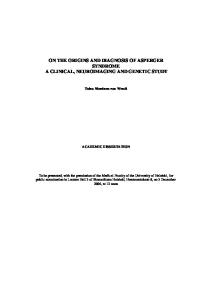
reserve are studied with 18 F-DOPA and 18 F-FMT (6-18 F-fluoro-mtirosine); (2) the density of the VMAT-2 transporter using 11 C-DTBZ (11 C-dihydrotetrabenazine), and (3) the concentration or density of the presynaptic dopamine transporter protein using 11 C-dMP (d-threo-methylphenidate), 11 C or 18 F-CFT (2-carbomethoxy-3[4-fluoro] tropane), 11 C or 18 F-PE2I, 18 F-FP-CIT. Postsynaptic neurons integrity can be evaluated using striatal dopamine D2/D3 receptor antagonist radiotracers such as 11 Craclopride or 11 C-falipride; extrastriatal dopamine D2/D3 receptor antagonists such as 11 C-FLB-457; striatal dopamine D1 receptor antagonists such as 11 C-SCH 23390 and 11 C-NNC 112; and dopamine D2 receptor agonists such as 11 C-MNPA.5 All these tracers are used almost exclusively in research. 18 FDOPA is the only radioligand used clinically in the differential diagnosis of parkinsonian syndrome of degenerative origin. Striatal SPECT imaging The number of radioligands used with SPECT is smaller than that used with PET. The most widely used presynaptic radioligand for SPECT is 123 I-N-3-fluoropropyl-2b-carbomethoxy3b-(4-iodophenyl) nortropane (123 I-FP-CIT). Dopamine postsynaptic receptors are studied with 123 I-IBZM, a benzamide analog of raclopride with affinity for striatal D2 receptors.5 This analog is not registered as a radiotracer in Spain, and it is used for the differential diagnosis between PD and atypical parkinsonisms by means of a foreign medication policy. PET with 18 F-DOPA 18 F-DOPA is the most commonly used PET tracer in the study of PD and atypical parkinsonisms, presenting very good sensitivity and specificity values as well as good correlation with disease severity and progression, similar to cerebral SPECT with 123 I-FP-CIT, but with greater spatial resolution of the striatal images because it allows separate analysis of the caudate and the putamen nuclei and identification of the accumbens nucleus (Fig. 1). Discontinued use of anti-parkinsonian drugs is recommended (l-Dopa for 6–12 h, and dopaminergic antagonists for 24 h) prior to 18 F-DOPA PET, and carbidopa should be administered (150 mg) 60 min before injection of 18 F-DOPA. This allows inhibition of the peripheral dopadecarboxylase and thereby, improves DOPA availability in the presynaptic dopaminergic terminals. In the clinical routine, PET images are acquired 70 min after the injection of 111–185 MBq of 18 F-DOPA, and static 3D can be used with an approximate duration of 10–20 min. Ninety-minute dynamic acquisition is used in research, a requirement for absolute quantification of the decarboxylation constant (K3 or Ki ). As in SPECT studies using 123 I-FP-CIT (see below), 18 F-DOPA PET can be evaluated visually by analyzing its distribution in the striatum. Quantitative analysis is performed by analyzing the uptake
Caudate
of regions of interest. In the case of static images, the average activity in the regions with specific uptake (striatum) and a region with non-specific uptake (occipital) is obtained, and the ratio between the two is calculated for each hemisphere (SUR). In dynamic studies, the parameter that is quantified is the decarboxylation constant (Ki ), obtained by modeling a simplified compartmental system (Patlak plot with region of reference) from which a parametric image of the Ki parameter can be created. 18 F-DOPA is currently registered and commercialized in Spain by Cis Bio International (DOPACIS® ) and by Barnatrón SA (NeuroPET® ). SPECT with 123 I-FP-CIT 123 I-FP-CIT
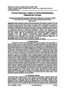
or ioflupane (registered as DaTSCAN by GE Healthcare) is a cocaine analog that presents high affinity for the dopamine transporter (Ki 3.5 nM), with good specificity (selectivity TDA/SERT of 2.8/1), and therefore, it is an excellent marker for nigrostriatal neuron viability and functionality. Uptake is concentrated in the axonal terminal of the nigrostriatal neuron where there is a high concentration of DAT, achieving maximum uptake in equilibrium at 3–6 h of endovenous administration of 150–250 MBq (normally 185 MBq). To perform this study it is recommended that cocaine intake be discontinued 2 days prior; cessation of other drug intake is as follows: amphetamine, 7 days; methylamphetamine, 3 days; mazindol, 3 days; phentermine, 14 days; modafinil, 3 days; bupropion, 8 days and benzatropine, 5 days. In addition, lugol should be administered orally to block thyroid uptake of 123 I. Visual analysis of SPECT images is sufficient for diagnosis (Fig. 2). Complementary quantification of striatal compared to occipital uptake (SUR) can be useful for detecting significant differences with respect to healthy subjects.6 False positives (pathologic striatal uptake with an intact nigrostriatal pathway) are infrequent and related to pharmacological interferences (drugs which block dopamine transporters) or methodological artifacts.7 False negatives (normal striatal uptake with degeneration of the nigrostriatal pathway) are even less frequent, with the positive predictive value of SPECT being greater than 90% in almost all the series. SPECT with 123 I-FP-CIT detects changes in striatal uptake in patients with premotor symptoms of PD (olfactory disorders, REM sleep phase behavior disorders) and in asymptomatic subjects who are carriers of genetic mutations in cases of familial PD. Evaluation of postsynaptic neuronal activity by 18 FDG PET An increase in glutamatergic neuronal activity (excitatory synaptic activity) determines an increase of cerebral glucose metabolism through the neuron–glia metabolic unit. The 2-[18 F]
Accumbens
Putamen
A
B
C
D
Fig. 1. Patterns of normality and pathology of cerebral 18 F-DOPA PET. (A) Normal pattern, axial slice of 18 F-DOPA PET: preserved striatal uptake in both caudate and putamen nuclei. (B) Normal pattern, coronal slice of 18 F-DOPA PET: preserved striatal uptake in both caudate, putamen and accumbens. (C) Asymmetric pathologic pattern of 18 F-DOPA PET: right putaminal decreased uptake and normal uptake in the left putamen nucleus and both caudate nuclei. (D) Symmetric pathologic pattern of 18 F-DOPA PET: absence of uptake in both putamen nuclei and preserved uptake in the caudate nuclei.
Documento descargado de http://www.elsevier.es el 27/01/2017. Copia para uso personal, se prohíbe la transmisión de este documento por cualquier medio o formato.
J. Arbizu et al. / Rev Esp Med Nucl Imagen Mol. 2014;33(4):215–226
219
Fig. 2. Normal and pathological patterns of 123 I-FP-CIT SPECT. (A) Axial anatomic slice of the encephalon showing the caudate and putamen nuclei. (B) Normal pattern of 123 I-FP-CIT SPECT: symmetric uptake of caudate and putamen nuclei. (C) Asymmetric pathologic pattern of 123 I-FP-CIT SPECT: decreased uptake of the left putamen nucleus and normal uptake of both caudate nuclei and right putamen nucleus corresponding to a patient with right unilateral PD. (D) Asymmetric pathologic pattern of 123 I-FP-CIT SPECT: lack of uptake in left putamen nucleus. Decreased uptake in right putamen nucleus. Normal uptake in caudate nuclei corresponding to a patient with right unilateral PD. SPECT detected right nigroputaminal alteration before the appearance of motor symptoms in the left hemibody. (E) Bilateral pathological pattern of 123 I-FP-CIT SPECT: bilateral putaminal decreased uptake, with more preserved uptake in caudate nuclei corresponding to a patient with bilateral right unilateral PD. (F) Bilateral pathologic pattern of 123 I-FP-CIT SPECT: bilateral global striatal decreased uptake, accompanied by a relative increase in extrastriatal uptake. This pattern is unusual in PD, being more typical of Lewy body dementia and atypical parkinsonisms. When this pattern appears pharmacological interferences should be ruled out.
fluoro-2-deoxy-d-glucose (18 FDG) glucose analog is incorporated into the cells by glucose transporters. Once inside the cells, 18 FDG is phosphorylated to 18 FDG-6-phosphate by the hexokinase enzyme. The presence of 18 F blocks its isomerization to fructose-6-phosphate, and therefore, 18 FDG does not follow the glucolysis metabolic pathways or the glycogen synthesis pathways. The retention of 18 FDG-6-phosphate is proportional to the glucose brain tissue consumption. Therefore, 18 FDG is used for evaluating regional cerebral glycidic metabolism as a biomarker for neuronal and synaptic activity.8 In the case of neurodegenerative diseases accompanied by a parkinsonian syndrome, 18 FDG allows evaluation of the neuronal activity in the basal ganglia. Thus, it has been suggested that 18 FDG PET is a good marker for postsynaptic activity of the nigrostriatal pathway. Patients should fast for at least 4 h before PET scanning so as to reduce the levels of glycemia (from 110 to 160 mg/dl) because elevated glycemia levels reduce the signal ratio of gray/white matter and increase the stochastic noise in the image, thereby making it more difficult to interpret the study. The use of insulin for correcting the elevated glycemia levels has not been shown to be effective and therefore, the studies should be carried out in previously controlled normoglycemic states.9 In addition, it is important to know the medication that the patient is taking because certain central nervous system drugs may influence cerebral glucose metabolism.10 Consequently, this type of medication should be withdrawn prior to the study whenever the patient situation allows for this. In patients presenting parkinsonism, it is important to know if they are taking levodopa or dopaminergic agonists drugs because of their possible modulation of 18 FDG uptake.11
Following intravenous administration of 125–250 MBq of 18 FDG (generally 150 MBq), patients are required to rest for 40 min in supine position in a quiet, dimly lit room, without talking, reading or doing any physical activity. When patients are not collaborative, and sedation is required, this should be initiated at least 30 min after tracer injection. The acquisition protocol consists in a brain static image (10–15 min) based on the characteristics of the PET-CT scanner. The resulting images are reoriented according to the orbito-meatal angle; qualitative (visual) or quantitative analysis is performed. In visual analysis, possible areas of hypometabolism must be recognized and when a structural lesion is suspected, coregistration with the morphologic CT or MR studies is recommended. Thereafter, we should identify whether or not the findings correspond to any of the patterns described in neurodegenerative diseases (Fig. 3). Several voxel-based programs of quantitative analysis are available for complementing the visual analysis. Evaluation of sympathetic innervation by myocardial scintigraphy with 123 I-metaiodobenzylguanidine 123 IMyocardial innervation scintigraphy with 123 metaiodobenzylguanidine ( I-MIBG) shows specific neuronal noradrenergic uptake and is a functional marker of the integrity and distribution of the postganglionic presynaptic terminals. 123 I-MIBG competes with NA for the uptake in the postganglionic sympathetic terminals, allowing in vivo visualization of the sympathetic innervation. 123 I-MIBG does not bind to the postsynaptic receptors nor is it degraded by the COMT and MAO enzymes that
Documento descargado de http://www.elsevier.es el 27/01/2017. Copia para uso personal, se prohíbe la transmisión de este documento por cualquier medio o formato.
J. Arbizu et al. / Rev Esp Med Nucl Imagen Mol. 2014;33(4):215–226
220
18F-FDG
A
B
C
Fig. 3. 18 FDG PET image of a normal subject (A) with predominant, homogeneous and symmetric activity in the basal ganglia; with PD (B) similar to the normal subject with a greater activity in the posterior region of the putamen nucleus; and with MSA-P type (C) in which a marked reduction is observed in the posterior region of the putamen in both hemispheres.
metabolize the endogenous catecholamines. After administration of 111–370 MBq of 123 I-MIBG (generally 370 MBq), there is rapid myocardial uptake with the greatest flow to the extraneuronal spaces. In the following hours, 123 I-MIBG actively enters the sympathetic terminal, especially that of the left ventricle, washing out from non-neuronal tissue. Neuronal accumulation of 123 I-MIBG reaches its maximum at 3–4 h, showing active neuronal uptake without passive components (Fig. 4).12 Administration of 123 I-MIBG is performed while resting, avoiding substances or drugs that might alter its uptake and after having performed thyroid blockade by the oral administration of a solution of lugol or 500 mg of potassium perchlorate. Thorax planar images, anterior projection, are acquired early at 15–20 min and delayed acquisition is at 3–4 h, with 10-min image duration, an energy window of 159 keV ± 20% and a matrix of 256 × 256. The use of medium energy collimators, either all-purpose (LEAP) or high resolution (LEHR), is recommended due to the presence of photons with higher energy photopeaks in 123 I than in those of 99m Tc. However, for practical reasons some centers perform the study with low energy collimators.13,14 Regional myocardial uptake of 123 I-MIBG tends to be heterogeneous, being somewhat less in the apex and inferior wall. This heterogeneity limits qualitative evaluation of the images and has led to the development of semiquantitative parameters. The heartto-mediastinum ratio is used as an index of global 123 I-MIBG uptake (heart/mediastinum (H/M) ratio). These H/M ratios are obtained by drawing regions of interest in the anterior image of the thorax and, particularly, the entire cardiac area. To obtain the H/M ratio,
123I-MIBG
A
B
the mean of the counts per pixel of the myocardium is divided by the mean of the counts per pixel of the mediastinum. The washout rate is obtained as follows: (counts per pixel in the myocardium at 15 min − counts per pixel in the myocardium at 4 h)/counts per pixel in the myocardium at 15 min. The washout reflects the tone of the sympathetic nervous system and therefore, an increase in washout could be an early marker for sympathetic dysfunction, showing not only a lower number of sympathetic cardiac terminals but also an increase in spillover or a reduced capability for maintaining the NA in the sympathetic terminals. Both a reduction of the delayed H/M ratio and an increase in the rate of washout are parameters which indicate alteration of cardiac sympathetic innervation. Although some studies have reported reference values of these ratios (H/M ratio range of 1.9–2.8, with a mean of 2.2 ± 0.3), each center should adjust their ratios depending on the circumstances of their equipment and the protocols used.13–15 Differential diagnosis of tremor and parkinsonian syndrome Although PD tremor and essential tremor (ET) are not difficult to differentiate (Table 4), in some cases neither anamnesis nor clinical examination allows definite clinical diagnosis. However, in some cases patients with ET frequently develop PD. When a patient with
Table 4 Differences between Parkinsonian tremor and essential tremor. Finding
Parkinson’s disease
Essential tremor
Age at onset (years) Family history Frequency of tremor (Hz) Characteristics of the tremor
55–75 +/− 4–6 Pronation–supination
10–80 ++ 5–10 Flexion–extension
Increases Diminishes Diminishes Diminishes (micrography), increases (trembling) Increases − Re-emerging (latency) +/− Asymmetric Face, jaw, lips, chin
Diminishes Increases Increases
Influencing factors Resting Action Concentration Writing
Fig. 4. Myocardial innvervation study by planar scintigraphy of the anterior thorax obtained at 4 h after the administration of 123 I-MIBG, (A) with normal cardiac uptake and (B) with diminished cardiac uptake in a patient with PD.
Walking Alcohol Postural tremor Kinetic tremor Tremor of extremities Other localizations Modified from Jankovic.21
Diminishes Diminishes Without latency Yes Quite symmetric Head, voice
Documento descargado de http://www.elsevier.es el 27/01/2017. Copia para uso personal, se prohíbe la transmisión de este documento por cualquier medio o formato.
J. Arbizu et al. / Rev Esp Med Nucl Imagen Mol. 2014;33(4):215–226
ET presents resting tremor, the added presence of PD must be ruled out. Sometimes this is a tremor of wide frequency range which is transmitted on resting when the patient is not completely relaxed. Diagnosis is further complicated if the tremor is asymmetric, if the muscle tone is increased, and if the movements of the patient are relatively slow. In this scenario, functional neuroimaging studies may be of great value in the differential diagnosis. PET or SPECT studies of the presynaptic dopaminergic pathway have a sensitivity of 90% when differentiating between PD and patients with ET or healthy controls.16 However, the reference pattern for comparison is based on clinical criteria17,18 and not on pathological criteria. With this pattern, the pretest probability (the grade of clinical uncertainty) will be, at the most, equal to the post-test probability, thereby demonstrating the weakness of the diagnostic studies in this field. As pointed out by De la Fuente-Fernández,19 new neuropathological studies are needed for evaluating the diagnostic accuracy of the neuroimaging tests. Another interesting aspect of the neuroimaging tests is that 10–15% of patients with clinical criteria of PD, assessed by expert neurologists, show normal 18 F-DOPA or 123 I-FP-CIT uptake in the striatum. These individuals have been denominated “SWEDD” (subjects without evidence of dopaminergic degeneration). These patients usually present a resting tremor phenotype which some define as dystonic tremor, and slow movement with doubtful bradykinesia. The evolution and follow-up imaging studies indicate that these patients most likely do not present PD, thereby supporting the normal results obtained from the functional neuroimaging tests.20,21 Patients with isolated resting tremor consistently show abnormal presynaptic dopamine uptake in the striatum (Figs. 1 and 2) whereas patients with isolated postural tremor have normal uptake. Some patients diagnosed with ET show a reduction in dopaminergic uptake in the putamen nucleus, within the same range as patients with PD. This suggests early nigrostriatal dysfunction. However, there is no sufficient clinical follow-up of these patients which could confirm the development of a typical PD. A reduction in the uptake of 18 F-DOPA in the putamen nucleus has also been described in asymptomatic relatives of patients with PD and in patients with isolated postural tremor who are phenomenologically identical to ET. An overlapping between PD and ET seems evident since PD is more prevalent in families with ET and vice versa. Neuroimaging of the dopaminergic pathway is usually useful for differentiating the two entities. In fact, the technical specifications of 123 I-FP-CIT (DaTSCAN® ) include the indication of differential diagnosis between ET and diseases related to PD. It is difficult to sustain a diagnosis of PD with preservation of the dopaminergic pathway. Similarly, if there were an alteration of the dopaminergic pathway, diagnosis of ET would be possible but with a high suspicion that it could evolve to PD.
Differential diagnosis of atypical degenerative parkinsonisms The prognosis of atypical parkinsonisms is quite different from that of PD and therefore, complementary functional neuroimaging tests may be necessary in the initial phases when there is greater diagnostic uncertainty. Cerebral metabolism with 18 FDG PET in atypical parkinsonisms In PD, cerebral 18 FDG PET is normal or shows an increase in uptake in the putamen nucleus while in atypical parkinsonisms, hypometabolic patterns have been described in the basal ganglia, the thalamus or cortex, depending on the causal entity (Table 5).
221
Multiple system atrophy The most characteristic finding observed in the parkinsonian variant of MSA (MSA-P type) is a reduction in the uptake of 18 FDG in both putamen nuclei with a rostro-caudal gradient (Fig. 3). This finding has a sensitivity of almost 95% and a specificity of 100%.22 In these patients, decreased uptake can also be detected in the thalamus, brainstem and in cortical areas. Thus, the second consensus for the diagnostic criteria of MSA has established that hypometabolism in the putamen nucleus, mesencephalic region and cerebellum is an additional characteristic for diagnosing a possible MSA-P type.23 In the study by Tang this pattern has a positive predictive value of 88% in the first 2 years of the disease and of 100% at 5 years.24 In patients with cerebellar variant of MSA (MSA-C type), hypometabolism of the anterior cerebellar hemispheres and the vermis may be detected one year after symptom onset, although putaminal hypometabolism can also be observed and is common among the parkinsonian variants.25 The presence of this metabolic pattern is of great value for differentiating these patients from others with predominant cerebellar symptomatology such as spinocerebellar ataxias or other diseases which occur with ataxia and rigid-akinetic symptoms, and consequently, the second consensus for the diagnostic criteria of MSA established that hypometabolism in the putamen nucleus is an additional characteristic for the diagnosis of MSA-C type.23 Unlike patients with a parkinsonian phenotype, a cortical metabolic deficit is not detected in patients with a hereditary form of the cerebellar variant.
Progressive supranuclear palsy Patients with PSP present reduced 18 FDG uptake in the caudate, putamen (caudo-rostral pattern) and prefrontal cortex,25 although the earliest sign in these patients is a reduced metabolism in the brainstem. It is important to note that frontal hypometabolism is not specific of this condition because it may be observed in other neurodegenerative diseases such as the variant of frontotemporal dementia with a predominant behavioral alteration, Huntington’s disease, PD, and patients with depression. Statistical parametric analyses of 18 FDG PET images have shown a sensitivity of 88%, a specificity of 94% and a positive predictive value of 91% for the diagnosis of MSA, which may rise to 100% in the early phases of the disease.24 However, the data obtained in the last longitudinal follow-up study analyzing patients suspected of having atypical parkinsonisms had a slightly lower sensitivity of 73% and a specificity of 95.2%.26
Corticobasal degeneration In these patients, the greatest reduction in 18 FDG uptake is produced asymmetrically in the posterior frontal, inferior parietal and superior temporal regions, together with a more marked hypometabolism in the thalamus and striatum on the opposite side of the most affected extremities.25 Using voxel by voxel analysis, it is possible to differentiate the metabolic pattern of patients with CBD (greater parietal hypometabolism) from that of patients with PSP (greater mesencephalic and thalamic hypometabolism).27
Lewy body dementia In these patients, a symmetric hypometabolism of 18 FDG in the association cortex, including the occipital visual cortex28 is the most common finding with a sensitivity of 83–90% and a specificity of 86–88%, thereby differentiating Lewy body from Alzheimer’s disease dementia. Nonetheless, this technique has not been shown to be more accurate than 123 I-FP-CIT SPECT.
Documento descargado de http://www.elsevier.es el 27/01/2017. Copia para uso personal, se prohíbe la transmisión de este documento por cualquier medio o formato.
J. Arbizu et al. / Rev Esp Med Nucl Imagen Mol. 2014;33(4):215–226
222
Table 5 Summary of the most characteristic findings of the functional neuroimaging tests in patients with atypical parkinsonisms. MSA-P
MSA-C
CBD
PSP
Lewy BD
PET 18 FDG
↓ putamen, pallidum, caudate, cerebellum
↓ anterior cerebellum, putamen, pallidum, caudate, thalamus
↓ asymmetric frontal, parietal, temporal, thalamus and striatum
↓ occipito-lateral, prefrontal, parieto-temporal
PET 18 F-DOPA
↓ putamen, caudate, external globus pallidum, red nucleus, locus coeruleus ↓ posterior putamen > caudate ↓ striatum
↓ putamen, caudate, external globus pallidum, red nucleus, locus coeruleus ↓ posterior putamen > caudate ↓ striatum
↓ asymmetric caudate and putamen
↓ mesencephalus, prefrontal medial cortex, thalamus, caudate, anterior cingulate ↓ caudate, anterior and posterior putamen
↓ asymmetric striatum
↓ striatum
↓ striatum
Normal
↓ striatum
↓ striatum
SPECT 123 I-FP-CIT SPECT 123 I-IBZM; PET 11 C-Raclopride
↓ putamen, caudate, prefrontal limbic and associative areas
MSA-P: parkinsonian multiple system atrophy variant; MSA-C: cerebellar multiple system atrophy variant; CBD: corticobasal degeneration; LBD: Lewy body dementia; PSP: progressive supranuclear palsy.
Dopaminergic presynaptic functionality PET or SPECT studies of the presynaptic dopaminergic pathway in patients with atypical parkinsonisms show decreased tracer uptake in the striatum which is similar to that of patients with PD with the same time of evolution (Table 5). However, these studies are of great value in differentiating Lewy body dementia from other dementias and therefore, they have been included among the diagnostic criteria.29 Dopaminergic postsynaptic studies 123 I-IBZM SPECT in patients with MSA shows reduced striatal uptake of the radioligand in the early phases of the disease; this fact contrasts with PD in which the radiotracer uptake is increased. However, this pattern is not specific of PD because it can also be observed in other atypical parkinsonisms as well as in normal subjects (Table 5). Contrary to what occurs in MSA and in PSP, 123 I-IBZM SPECT is usually normal in patients with CBD.30 Recent studies have demonstrated the inferiority of 123 I-IBZM studies compared to 18 FDG in the differential diagnosis of atypical parkinsonisms.26
Cardiac scintigraphy studies A decrease in the cardiac uptake of 123 I-MIBG (Fig. 4) with a significant reduction in the H/M ratio and an increase in the washout rate has been observed in PD patients.31 The sensitivity of cardiac scintigraphy with 123 I-MIBG in PD with less than 3 years of symptoms duration is lower than that of patients with a longer period of evolution (73 and 90% respectively).32 Nevertheless, the sensitivity of 123 I-FP-CIT is even higher than 123 I-MIBG (sensitivity 83%) in early PD. Interestingly, 123 I-MIBG scintigraphy in the rigid-akinetic forms of PD is more frequently altered than in the tremor-dominant PD patients.33 Although most atypical parkinsonisms do not present significant alterations in cardiac studies with 123 I-MIBG, up to 30% of the cases may be pathological (especially MSA-P type) and therefore, this technique may be of greater use in cases with a cerebellar phenotype.34 Finally, this technique may be of interest in the differential diagnosis for Lewy body dementia, especially in early stages of the disease.35 Secondary parkinsonism It is often difficult to differentiate the clinical manifestations of degenerative parkinsonisms from secondary parkinsonisms but
from a prognostic and therapeutic point of view, their recognition gains importance. Numerous causes of secondary or symptomatic parkinsonism have been described, but the most frequent are pharmacological parkinsonism (PP) and the so-called vascular parkinsonism (VP). Pharmacological parkinsonism Pharmacological parkinsonism (PP) is the second most frequent cause of parkinsonian syndrome after PD. It can be due to the intake of drugs that block the dopamine receptors or to decreased levels of stored dopamine.36 However, these drugs may also trigger an underlying PD in a preclinical situation. The differentiation of these two clinical situations is of great importance because PP requires discontinuance of the medication responsible for the situation. Cessation of parkinsonian symptoms occurs within a time period that is quite variable (weeks, months or even years). This variability may result in an incorrect diagnosis of PD and consequently, the implementation of inadequate treatment. The symptoms of PP are generally symmetrical but in approximately 50% of the patients with PP, asymmetry is present. Other symptoms such as postural tremor, orofacial dyskinesia, akathisia, dystonia, among others, may indicate PP, but it is actually very complex to differentiate these symptoms from those produced by PD. In PP the dopaminergic activity reflected by 18 F-DOPA PET or 123 I-FP-CIT SPECT uptake is normal in most of the cases (>90%), whereas is abnormal in patients with an underlying PD. The sensitivity of 123 I-FP-CIT SPECT for differentiating these subjects is 100% with a specificity of 90.6%, making it a very useful tool for differential diagnosis.37 Vascular parkinsonism Vascular parkinsonism (VP) or lower-half parkinsonism refers to a clinical picture mainly characterized by gait disorder, bradykinesia and rigidity. It is common in aged subjects with a history of arteriosclerosis and recurrent stroke. In the past, this term was controversial, but in recent years the idea of considering VP as a proper entity has arisen. It is a heterogeneous entity which may be caused by diffuse vascular lesions of the white matter, lacunar infarcts and, less frequently, territorial infarctions.38 The differentiation between VP and PD is sometimes difficult and represents a diagnostic challenge. Different clinical features have been associated with VP, including symmetric involvement of the lower extremities, gait disorder, short steps, postural instability, frequent falls and the absence of resting tremor, together
Documento descargado de http://www.elsevier.es el 27/01/2017. Copia para uso personal, se prohíbe la transmisión de este documento por cualquier medio o formato.
J. Arbizu et al. / Rev Esp Med Nucl Imagen Mol. 2014;33(4):215–226
with an absence of good response to treatment with levodopa. The presentation of these symptoms varies greatly and the diagnosis of VP cannot be confirmed based exclusively on the clinical manifestations. Structural neuroimaging (CT and MR) cannot be considered a precise method for diagnosing VP, on one hand, due to the high proportion of patients with cerebrovascular lesions who do not develop parkinsonism and on the other hand, because the presence of cerebrovascular disease is not infrequent in patients with PD. Functional neuroimaging of the dopaminergic system using PET or SPECT is highly accurate in differentiating VP from neurodegenerative parkinsonisms.39 Nonetheless, the image pattern in VP varies and to a certain degree, it reflects the heterogeneity of the physiopathological mechanisms of origin: (1) normal image in one-third of the cases; (2) reduction in uptake in the striatum, with involvement being homogeneous in caudate and putamen nuclei, of variable intensity (generally mild), and without significant symmetries as in the case of degenerative parkinsonisms; (3) exclusively unilateral homogeneous decreased uptake or as an intense and well delimited focal uptake defect in any area within the striatum. When these uptake defects involve the putamen nucleus (uni- or bilaterally), VP may be mistaken with PD; in these situations, comparison of the coincidence with the image of infarction in CT or MR is required. For differentiating between VP and PD, 123 I-FP-CIT SPECT presents a sensitivity of 83.7% and a specificity of 99.4%.40 Neuroimaging techniques evaluating the integrity of the nigrostriatal system should be considered in patients presenting a parkinsonian syndrome with some atypical characteristics such as early gait disorder and/or unsatisfactory response to dopaminergic treatment and with associated cerebrovascular disease.
Hereditary parkinsonism (LRRK2) Hereditary forms of PD represent 5–10% of all cases of PD. Among these, those associated with mutations in the LRRK2 gene encoding a protein denominated dardarin are noteworthy for their frequency. The first publications of these familial cases of PD described a clinical phenotype very similar to that of non-mutated PD. Likewise, the nigrostriatal dysfunction demonstrated by 18 F-DOPA PET in patients who are carriers of some of the pathogenic mutations such as R1441G,41 Y1699C,42 R1441C,42 and G2019S43 is indistinguishable from that observed in the classical form of PD (Fig. 1). Taking into account that clinical presentation and neuroimaging are similar to PD, the presence of asymptomatic carriers of these mutations allows the use of a prospective study model of disease progression from very early phases of the disease, in which the classical motor symptoms have not yet become present. The study of the nigrostriatal pathway using PET or SPECT in this population at risk of PD can provide relevant information regarding this premotor phase, and it may help evaluate clinical and molecular markers for assessing PD evolution as well as future neuroprotective treatments for the early stages of the disease. Studies in asymptomatic carriers have shown reduced dopamine transporter uptake (11 C-methylphenidate PET) as an early subclinical sign of dopaminergic dysfunction and progression to abnormal uptake of 18 F-DOPA, which is characteristic of PD.44 In carriers of LRRK2 mutations, PET studies with different radiotracers have been able to determine the presence of compensatory mechanisms prior to the development of the disease such as an increase in dopamine turnover, a finding which does not appear in patients already experiencing symptomatic PD.45
223
Studies with 123 I-MIBG, performed in small series of patients with genetic-based PD and carriers of R1441G and G2019S mutations in the LRRK2 gene have reported a greater or lesser involvement of cardiac innervation versus idiopathic PD.46,47 PET or SPECT studies on PD associated with LRRK2 gene mutations may be more effective than cardiac scintigraphy in both the diagnosis of the disease and in the study of preclinical stages because they are directed at areas with demonstrated neuronal degeneration which correspond with their clinical phenotype.
Utility of the evaluation of Parkinson’s disease progression Neuropathological studies suggest that in PD the loss of dopaminergic neurons follows an exponentially negative course.48 These studies are extremely limited because they provide transversal estimations and do not take into account intra-individual variability. Longitudinal studies with functional neuroimaging techniques avoid this deficiency because they provide the possibility of in vivo evaluation of PD progression, thereby allowing the observation of both inter- and intra-individual changes.49 Initial functional neuroimaging studies using presynaptic dopaminergic markers provided variable results with respect to the annual rate of PD progression, with values ranging from 2 to 10% of loss of nigrostriatal dopaminergic terminals.49 Differences in the technique (PET versus SPECT) as well as differences in the tracer used could account for this variability. In any case, most of the studies are concordant in that the progression of dopaminergic damage is substantially greater during the first years of the disease,50–52 thereby supporting the model of PD suggested in the previously mentioned neuropathological studies. The results of a recent longitudinal PET study confirm that progression of PD does actually follow an exponentially negative course.53 In this study, patients and healthy controls were followed over an 8-year period with three PET scans (at baseline and at 4 and 8 years), using three different presynaptic dopaminergic markers in each study. This was a very large study including a total of 679 PET studies, allowing the authors to develop a mathematical model for estimating the loss of nigrostriatal dopaminergic terminals, not only during the symptomatic phase of the disease but also during the presymptomatic phase. Fig. 5 shows the curve obtained for one of the tracers (dihydrotetrabenazine, 11 C-DTBZ). Consequently, the following conclusions can be made. Firstly, it appears to be quite clear that the greatest part of dopaminergic damage occurs during the presymptomatic phase of the disease and afterwards, the damage is practically 100% during the first 5–10 years of the symptomatic phase (Fig. 5). After this time, the loss of nigrostriatal dopaminergic terminals is very limited and, in fact, tends to achieve asymptotic values. As shown in Fig. 5, on the appearance of the first motor symptoms (duration of symptoms equal to 0), only 33% of the dopaminergic terminals remain preserved. Secondly, the presymptomatic phase of the disease appears to be considerably longer than suggested by neuropathological studies and some previous functional neuroimaging studies.48,49 Thus, it is estimated that in patients with an onset of motor symptoms at 53 years of age, the nigrostriatal dopaminergic damage had begun approximately 17 years before (in other words, at the age of 36). Interestingly, younger patients have a longer presymptomatic period than older patients (25 versus 10 years, respectively) and progress more slowly.53 And thirdly, even in the most advanced stages of the disease (meaning 30 years after the onset motor symptoms), a significant number of dopaminergic terminals still remain viable (approximately 15%). In addition to having important conceptual implications, these findings also provide some clinical keys. On one hand, the therapeutic window for potential neuroprotective treatments is enlarged
Documento descargado de http://www.elsevier.es el 27/01/2017. Copia para uso personal, se prohíbe la transmisión de este documento por cualquier medio o formato.
J. Arbizu et al. / Rev Esp Med Nucl Imagen Mol. 2014;33(4):215–226
224
Diagnostic algorithms
Onset=53 years
Normalized DTBZ data (%)
100
80
60
40
20
0 –30
–20
–10
0
10
20
30
PD symptom duration (years) Fig. 5. Curve of the progression of presynaptic dopaminergic damage according to the results obtained by 11 C-DTBZ PET. The data have been normalized with respect to the normal values present during the first years of life so that the curve represents the percentage of dopaminergic terminals at each time. The straight segment in green corresponds to PD. The motor symptoms begin at time 0 (in this case at the age of 53, which was the mean age of onset in the group of patients analyzed). Figure adapted from De la Fuente-Fernández et al.53
to 10–25 years of the presymptomatic phase of the disease. On the other hand, these findings indicate that a part of the motor complications is probably not related to PD progression itself.
Functional neuroimaging techniques facilitate clinical diagnosis in some patients with parkinsonian syndrome, especially in those presenting initial symptoms, incomplete syndromes or in whose cases the assessment of the pharmacological response is complex. The diversity of techniques currently available in our country may generate confusion with respect to which complementary neuroimaging test may be more useful in each case. In this review, a panel of experts in nuclear medicine and neurology performed an exhaustive review of the reference literature dealing with the usefulness of functional neuroimaging techniques in differential diagnosis of parkinsonian syndrome. This review has allowed a consensus to be made of a series of practical considerations which can be used as recommendations in clinical practice. Despite the fact that these practical recommendations are focused on functional neuroimaging techniques, neuroimaging by MR should not be “overlooked”. It is undoubtedly a fundamental technique in the study of patients with neurological symptoms. The two aspects of functional neuroimaging which may contribute the most to the clinical diagnostic process are the evaluation of the presynaptic dopaminergic system and the neuronal activity of the basal ganglia, as well as of other structures such as the thalamus, mesencephalon, cerebellum and the cortex.54 In the case of a patient with a parkinsonian syndrome of uncertain origin, an MR neuroimaging test with T1-weighted and T2-weighted sequences is of initial interest. This test will rule out or consider the presence of a secondary parkinsonism (Fig. 6). Regardless of the result, it may be of interest to detect the possible presence of a striatal dopaminergic deficit (123 I-FP-CIT SPECT or 18 F-DOPA PET) because this may help to rule out entities such
Parkinsonian syndrome of uncertain origin MR T1-, T2-weighted sequences +
–/+
Secondary Parkinson: - Vascular - Tumoral - Others
Neuroimaging of the presynaptic dopaminergic activity: 123 I-FP-CIT SPECT 18 F-DOPA PET –
+
- Essential tremor - Secondary Parkinson: - Vascular - Pharmacological
Neurodegenerative parkinsonian syndrome
A typical symptoms or poor pharmacological response
Typical pattern or good pharmacological response
Parkinson disease
–
18
FDG PET
+ MSA, PSP, CBD Fig. 6. Diagnostic algorithm proposed in patients with parkinsonian syndrome of uncertain origin. Modified from Brooks.54
Documento descargado de http://www.elsevier.es el 27/01/2017. Copia para uso personal, se prohíbe la transmisión de este documento por cualquier medio o formato.
J. Arbizu et al. / Rev Esp Med Nucl Imagen Mol. 2014;33(4):215–226
as ET, PP or VP. If a striatal dopaminergic deficit is confirmed and if the parkinsonian syndrome has typical patterns, the most probable clinical diagnosis would be PD, especially if the patient responds to pharmacological treatment. Nonetheless, if treatment response is poor or atypical symptoms are present, the imaging test providing the most information for the differential diagnosis of parkinsonian syndrome is 18 FDG PET. Depending on the deficits in cortical or subcortical metabolism, the most probable cause of the clinical picture would be MSA, PSP or CBD. If the patient presents a picture of associated cognitive decline, the best test for differentiating between Lewy body dementia and Alzheimer’s disease is 123 I-FP-CIT SPECT or 18 F-DOPA PET. In any case, the clinical aspects should always be the core determinant of the diagnosis. In hereditary PD, 123 I-FP-CIT SPECT or 18 F-DOPA PET studies can detect incipient phases of the disease with a tracer uptake pattern similar to that of idiopathic PD. This allows evaluation of clinical and molecular markers for assessing PD progression and future neuroprotective treatments for the early stages of the disease. Progression studies on striatal dopaminergic deficit have also shown that the therapeutic window for potential neuroprotective treatments is wider than previously known and may cover 10–25 years of the presymptomatic phase of the disease. References 1. Fahn S, Jankovic J. Parkinsonism. Clinical features and differential diagnosis. In: Fahn S, Jankovic J, editors. Principles and practice of movement disorders. New York: Churchill Livingstone/Elsevier; 2007. p. 79–103. 2. Williams DR, Lees AJ. Progressive supranuclear palsy: clinicopathological concepts and diagnostic challenges. Lancet Neurol. 2009;8:270–9. 3. Albanese A, Altavista MC, Rossi P. Organization of central nervous system dopaminergic pathways. J Neural Transm. 1986;22:3–17. 4. García Solís D. Imagen de neurotransmisión dopaminérgica en los síndromes parkinsonianos. Rev Esp Med Nucl. 2005;24:255–75. 5. Van Laere K, Varrone A, Booij J, Vander Borght T, Nobili F, Kapucu ÖL, et al. EANM procedure guidelines for brain neurotransmission SPECT/PET using dopamine D2 receptor ligands, version 2. Eur J Nucl Med Mol Imaging. 2010;37:434–42. 6. Darcourt J, Booij J, Tatsch K, Varrone A, Vander Borght T, Kapucu ÖL, et al. EANM procedure guidelines for brain neurotransmission SPECT using 123 Ilabelled dopamine transporter ligands, version 2. Eur J Nucl Med Mol Imaging. 2010;37:443–50. 7. Booij J, Kemp P. Dopamine transporter imaging with [123 I] FP-CIT SPECT: potential effects of drugs. Eur J Nucl Med Mol Imaging. 2008;35:424–38. 8. Magistretti PJ, Pellerin L, Rothman DL, Shulman RG. Energy on demand. Science. 1999;283:496–7. 9. Hasselbalch SG, Knudsen GM, Videbaek C, Pinborg LH, Schmidt JF, Holm S, et al. No effect of insulin on glucose blood-brain barrier transport and cerebral metabolism in humans. Diabetes. 1999;48:1915–21. 10. Spanaki M, Siegel H, Kopylev L, Fazilat S, Dean A, Liow K, et al. The effect of vigabatrin (gamma-vinyl GABA) on cerebral blood flow and metabolism. Neurology. 1999;53:1518–22. 11. Berding G, Odin P, Brooks DJ, Nikkhah G, Matthies C, Peschel T, et al. Resting regional cerebral glucose metabolism in advanced Parkinson’s disease studied in the off and on conditions with [(18)F]FDG-PET. Mov Disord. 2001;16:1014–22. 12. Knickmeier M, Matheja P, Wichter T, Schäfers KP, Kies P, Breithardt G, et al. Clinical evaluation of no-carrier-added meta-[123 I] iodobenzylguanidine for myocardial scintigraphy. Eur J Nucl Med. 2000;27:302–7. 13. Flotats A, Carrió I, Agostini D, le Guludec D, Marcassa C, Schaffers M, et al. Proposal for standardization of 123 I-metaiodobenzylguanidine (MIBG) cardiac sympathetic imaging by the EANM cardiovascular committee and the european council of nuclear cardiology. Eur J Nucl Med Mol Imaging. 2010;37:1802–12. 14. Verberne HJ, Feenstra C, de Jong WM, Somsen GA, van Eck-Smit BL, Sokole EB. Influence of collimator choice and simulated clinical conditions on 123 IMIBG heart/mediastinum ratios: a phantom study. Eur J Nucl Med Mol Imaging. 2005;32:1100–7. 15. Chirumamilla A, Travin MI. Cardiac applications of 123 I-MIBG imaging. Semin Nucl Med. 2011;41:374–87. 16. Shahed J, Jankovic J. Exploring the relationship between essential tremor and Parkinson’s disease. Parkinsonism Relat Disord. 2007;13:67–76. 17. Perlmutter JS, Eidelberg D. To scan or not to scan DaT is the question. Neurology. 2012;78:688–9. 18. Bajaj NP, de la Fuente-Fernandez R. Role of datscan and clinical diagnosis in PD. Neurology. 2012;78:1538–9. 19. De la Fuente-Fernández R. Role of DaTSCAN and clinical diagnosis in Parkinson disease. Neurology. 2012;78:696–701. 20. Marek K, Jennings D, Seibyl J. Long-term follow-up of patients with scans without evidence of dopaminergic deficit (SWEDD) in the ELLDOPA study. Neurology. 2005;64:A274.
225
21. Jankovic J. Parkinson’s disease: clinical features and diagnosis. J Neurol Neurosurg Psychiatry. 2008;79:368–76. 22. Brooks DJ, Seppi K. Proposed neuroimaging criteria for the diagnosis of multiple system atrophy. Mov Disord. 2009;24:949–64. 23. Gilman S, Wenning G, Low P, Brooks D, Mathias C, Trojanowski J, et al. Second consensus statement on the diagnosis of multiple system atrophy. Neurology. 2008;71:670–6. 24. Tang CC, Poston KL, Eckert T, Feigin A, Frucht S, Gudesblatt M, et al. Differential diagnosis of parkinsonism: a metabolic imaging study using pattern analysis. Lancet Neurol. 2010;9:149–58. 25. Eckert T, Barnes A, Dhawan V, Frucht S, Gordon MF, Feigin AS, et al. FDG PET in the differential diagnosis of parkinsonian disorders. Neuroimage. 2005;26: 912–21. 26. Hellwig S, Amtage F, Kreft A, Buchert R, Winz O, Vach W, et al. [18 F]FDG-PET is superior to [123 I]IBZM-SPECT for the differential diagnosis of parkinsonism. Neurology. 2012;79:1314–22. 27. Juh R, Pae C, Kim T, Lee C, Choe B, Suh T. Cerebral glucose metabolism in corticobasal degeneration comparison with progressive supranuclear palsy using statistical mapping analysis. Neurosci Lett. 2005;383:22–7. 28. Teune LK, Bartels AL, de Jong BM, Willemsen A, Eshuis SA, de Vries JJ, et al. Typical cerebral metabolic patterns in neurodegenerative brain diseases. Mov Disord. 2010;25:2395–404. 29. Kägi G, Bhatia KP, Tolosa E. The role of DAT-SPECT in movement disorders. J Neurol Neurosurg Psychiatry. 2010;81:5–12. 30. Klaffke S, Kuhn AA, Plotkin M, Amthauer H, Harnack D, Felix R, et al. Dopamine transporters, D2 receptors, and glucose metabolism in corticobasal degeneration. Mov Disord. 2006;21:1724–7. 31. Rascol O, Schelosky L. 123 I-metaiodobenzylguanidine scintigraphy in Parkinson’s disease and related disorders. Mov Disord. 2009;24:S732–41. 32. Sawada H, Oeda T, Yamamoto K, Kitagawa N, Mizuta E, Hosokawa R, et al. Diagnostic accuracy of cardiac metaiodobenzylguanidine scintigraphy in Parkinson disease. Eur J Neurol. 2009;16:174–82. 33. Spiegel J, Möllers M, Jost WH, Fuss G, Samnick S, Dillmann U, et al. FP-CIT and MIBG scintigraphy in early Parkinson’s disease. Mov Disord. 2005;20: 552–61. 34. Nagayama H, Ueda M, Yamazaki M, Nishiyama Y, Hamamoto M, Katayama Y. Abnormal cardiac [123 I]-meta-iodobenzylguanidine uptake in multiple system atrophy. Mov Disord. 2010;25:1744–7. 35. Yoshita M, Taki J, Yokoyama K, Noguchi-Shinohara M, Matsumoto Y, Nakajima K, et al. Value of 123 I-MIBG radioactivity in the differential diagnosis of DLB from AD. Neurology. 2006;66:1850–4. 36. Hirose G. Drug induced parkinsonism. J Neurol. 2006;253:22–4. 37. Diaz-Corrales FJ, Sanz-Viedma S, Garcia-Solis D, Escobar-Delgado T, Mir P. Clinical features and 123 I-FP-CIT SPECT imaging in drug-induced parkinsonism and Parkinson’s disease. Eur J Nucl Med Mol Imaging. 2010;37:556–64. 38. Zijlmans J, Evans A, Fontes F, Katzenschlager R, Gacinovic S, Lees AJ, et al. [123 I] FPCIT spect study in vascular parkinsonism and Parkinson’s disease. Mov Disord. 2007;22:1278–85. 39. Gerschlager W, Bencsits G, Pirker W, Bloem BR, Asenbaum S, Prayer D, et al. [123 I] -CIT SPECT distinguishes vascular parkinsonism from Parkinson’s disease. Mov Disord. 2002;17:518–23. 40. Benítez-Rivero S, Marín-Oyaga VA, García-Solís D, Huertas-Fernández I, GarcíaGómez FJ, Jesús S, et al. Clinical features and 123 I-FP-CIT SPECT imaging in vascular parkinsonism and Parkinson’s disease. J Neurol Neurosurg Psychiatry. 2013;84:122–9. 41. Paisàn-Ruìz C, Sàenz A, de Munain AL, Martì I, Martìnez Gil A, Martì-Massò JF, et al. Familial Parkinson’s disease: clinical and genetic analysis of four basque families. Ann Neurol. 2005;57:365–72. 42. Adams JR, van Netten H, Schulzer M, Mak E, Mckenzie J, Strongosky A, et al. PET in LRRK2 mutations: comparison to sporadic Parkinson’s disease and evidence for presymptomatic compensation. Brain. 2005;128:2777–85. 43. Hernandez DG, Paisán-Ruíz C, McInerney-Leo A, Jain S, Meyer-Lindenberg A, Evans EW, et al. Clinical and positron emission tomography of Parkinson’s disease caused by LRRK2. Ann Neurol. 2005;57:453–6. 44. Nandhagopal R, Mak E, Schulzer M, McKenzie J, McCormick S, Sossi V, et al. Progression of dopaminergic dysfunction in a LRRK2 kindred A multitracer PET study. Neurology. 2008;71:1790–5. 45. Sossi V, de la Fuente-Fernández R, Nandhagopal R, Schulzer M, McKenzie J, Ruth TJ, et al. Dopamine turnover increases in asymptomatic LRRK2 mutations carriers. Mov Disord. 2010;25:2717–23. 46. Ruiz-Martínez J, Gorostidi A, Goyenechea E, Alzualde A, Poza JJ, Rodríguez F, et al. Olfactory deficits and cardiac 123 I-MIBG in Parkinson’s disease related to the LRRK2 R1441G and G2019S mutations. Mov Disord. 2011;26:2026–31. 47. Quattrone A, Bagnato A, Annesi G, Novellino F, Morgante L, Savettieri G, et al. Myocardial 123metaiodobenzylguanidine uptake in genetic Parkinson’s disease. Mov Disord. 2008;23:21–7. 48. Fearnley JM, Lees AJ. Ageing and Parkinson’s disease: substantia nigra regional selectivity. Brain. 1991;114:2283–301. 49. Biju G, de la Fuente-Fernández R. Dopaminergic function and progression of Parkinson’s disease: PET findings. Parkinsonism Relat Disord. 2009;15: S38–40. 50. Lee CS, Schulzer M, de la Fuente-Fernández R, Mak E, Kuramoto L, Sossi V, et al. Lack of regional selectivity during the progression of Parkinson disease: implications for pathogenesis. Arch Neurol. 2004;61:1920–5. 51. Hilker R, Schweitzer K, Coburger S, Ghaemi M, Weisenbach S, Jacobs AH, et al. Nonlinear progression of Parkinson disease as determined by serial positron
Documento descargado de http://www.elsevier.es el 27/01/2017. Copia para uso personal, se prohíbe la transmisión de este documento por cualquier medio o formato.
226
J. Arbizu et al. / Rev Esp Med Nucl Imagen Mol. 2014;33(4):215–226
emission tomographic imaging of striatal fluorodopa F 18 activity. Arch Neurol. 2005;62:378–82. 52. Shih MC, de Andrade F, Augusto L, Amaro E, Felicio AC, Ferraz HB, et al. Higher nigrostriatal dopamine neuron loss in early than late onset Parkinson’s disease? A [99m Tc]-TRODAT-SPECT study. Mov Disord. 2007;22:863–6.
53. De la Fuente-Fernández R, Schulzer M, Kuramoto L, Cragg J, Ramachandiran N, Au WL, et al. Age-specific progression of nigrostriatal dysfunction in Parkinson’s disease. Ann Neurol. 2011;69:803–10. 54. Brooks DJ. Imaging approaches to Parkinson disease. J Nucl Med. 2010;51:596–609.