VOL 28 NO 2 SEPTEMBER 2007 ISSN 0965-0989
Functional foods: probiotics and prebiotics Glenn R Gibson Food Microbial Sciences Unit, Department of Food Biosciences, The University of Reading, Reading, UK
Introduction
Functional Foods and the Gut
The main role of diet is to provide nutrients to meet host physiological requirements. As research behind diet and health has evolved, so has the concept of ‘functional foods’ become popular. Foods which are touted as being ‘functional’ are thought to exert certain positive properties over and above their normal nutritional value. While not universally popular and sometimes plagued by inadequate research/claims, the concept is certainly commercially successful, e.g. The Institute of Grocery Distributors (http://www.igd.com)
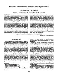
Recent years have seen a major change in how activities of the human gastrointestinal tract are perceived. This has been driven by increased knowledge of the gut microflora composition and activities. This has been helped by a shift away from traditional microbiological culture methods (Figure 1) to the use of molecular markers of culture identity. The colon is the most heavily
estimates that the functional food market in the UK in 2007 will have annual sales worth around £1800m. This shows an exponential rise from the 1996 figure of £134m. Examples of functional foods include organic and inorganic micronutrients, vitamins, anti-oxidants, dietary fibre, some proteins (e.g. lactoferrin), certain bioactive peptides and polyunsaturated fatty acids. The concept has now moved markedly towards gastrointestinal function, in particular the impact of gut bacteria. Possibly this is driven by the ubiquity of gastrointestinal disorders but also the fact that diet is an important controlling factor with regard to indigenous microbiota activities. The gut microflora contains pathogenic, benign and beneficial microbial species. A predominance of the former can lead towards gut upset which can be both acute (e.g. gastroenteritis) and chronic (e.g. inflammatory bowel disease). Functional foods directed towards the gut microbiota would serve to influence the composition of activities towards a more positive metabolism.
populated region of the gastrointestinal tract and, because of this resident microbiota, is one of the most metabolically active organs in the body. The concept of modulating activities directed towards improving gut microbial function has a long history, as diet can have a major effect on the gut microflora activities1. Whilst some indigenous bacteria can be pathogenic (e.g. proteolytic clostridia and
bacteroides), it is also the case that some genera/species may offer health promoting attributes. For example, bifidobacteria and lactobacilli are thought to exert powerful antipathogenic effects and are mainly responsible for ‘colonisation resistance’ in the gut. Moreover, the same genera have been attributed with other beneficial aspects: such as protection from bowel tumours and metabolism of cholesterol and other lipids in the gut1. Whilst many of the health promoting aspects have yet to be definitively proven in humans, it would appear that there is value in eliciting a change away from a gut flora dominated by potentially harmful bacteria towards a more benign, or beneficial, composition. Probiotics The most frequently used dietary method of influencing the gut flora composition is that of probiotics, whereby live microbial additions are made to appropriate food vehicles, usually fermented milks2. The concept was expounded in a scientific note by Metchnikoff2. He hypothesised that longevity in Bulgarian peasants was associated with their elevated intake of ‘soured milks’, i.e. dairy based drinks
Figure 1. Studies on the complex microbial ecology of the intestinal tract have moved away from cultural procedures towards molecular biological methods. Courtesy of Dr Kieran Tuohy (University of Reading).
containing live bacteria. This was the basis of what is now recognised as the probiotic concept. A recent definition of probiotics was given as ‘a live microbial feed supplement that is beneficial to health’3. Over the years, many species of microorganisms have been used. They consist not only of lactic acid bacteria (e.g. lactobacilli, streptococci, enterococci, lactococci, bifidobacteria) but also Bacillus spp., E. coli and
Vol 28 No 2
fungi/yeasts such as Saccharomyces spp. and Aspergillus spp. The most common probiotics belong to the genera Lactobacillus (e.g. L. casei, L. acidophilus, L. rhamnosus, L. johnsonii, L. reuteri) and Bifidobacterium (e.g. B. bifidum, B. longum, B. breve). To be effective, probiotics must be capable of being prepared in a viable manner and on a large scale (e.g. for industrial purposes), whilst during use and under storage the probiotic should remain viable and stable, be able to survive in the intestinal ecosystem and the host should gain beneficially from harbouring the probiotic. The strains used should be generally regarded as safe. Probiotics are marketed as functional foods, whereby they are ingested for their purported positive advantages in the digestive tract and/or systemic areas like the liver, vagina or bloodstream. Consumers should be provided with an independent assessment of physiological, microbial and safety aspects of these live microbial products – especially if they can improve health. Probiotic trials should use the best methodologies available. For probiotics to exert beneficial properties, they must have a high viability in the product and have robust survival properties in the gut, which is their first point of contact4. Moreover, they should not adversely affect immune up-regulation, produce toxins, disrupt colonocyte function or have the ability to transfer antibiotic resistance to the normal gut microflora. Food vehicles include live yoghurts, fermented dairy drinks, freeze-dried supplements (capsules, pills, liquid suspensions, sprays), cheese, fromage frais and fruit juices. Both single and multiple strain products are available.
these carbohydrates have a specific colonic fermentation directed towards bifidobacteria1,5. Bifidobacteria are able to break down and utilise fructo-oligosaccharides due to their possession of a β-fructofuranosidase enzyme, providing a competitive advantage in a mixed culture environment like the human gut11. Galacto-oligosaccharides (GOS) are another class of prebiotics that are manufactured and marketed in Europe as well as Japan. These consist of a lactose core with one or more galactosyl residues linked via β1→3, β1→4 and β1→6 linkages12. They have found application in infant formula foods. Recent documents have suggested that FOS and GOS are accepted prebiotics that fulfil current selection criteria13, 14. A prebiotic dose of 5 grams/day should be sufficient to elicit a positive effect upon the gut
microbiota (in some exceptional cases this may be nearer to 8g/d). A possible side effect of prebiotic intake is intestinal discomfort from gas production. However, bifidobacteria and lactobacilli cannot produce gas as part of their metabolic process. Therefore, at a rational dose, of up to 20g/d, gas distension should not occur. If gas is being generated, then the carbohydrate is not acting as an authentic prebiotic. This is perhaps because dosage is too high and the prebiotic effect is being compromised i.e. bacteria other than the target organisms are becoming involved in the fermentation5. Possible Health Benefits Several different avenues are being explored for pre/probiotics. These are largely mediated by affecting an increase in beneficial bacteria within
Prebiotics An alternative, or additional, approach is the prebiotic concept. A prebiotic is ‘a non-digestible food ingredient that beneficially affects the host by selectively stimulating the growth and/or activity of one or a limited number of bacteria in the colon, that can improve the host health’5. Thus, the prebiotic approach advocates the administration of non-viable entities. Dietary carbohydrates, such as fibres, are candidate prebiotics, but most promise has been realised with non-digestible oligosaccharides, because of their selective metabolism. In particular, the ingestion of fructo-oligosaccharides (FOS) has been shown to stimulate bifidobacteria in the lower gut. As prebiotics exploit non-viable food ingredients, their applicability in diets is wide ranging. A further approach is synbiotics, where probiotics and prebiotics are combined5. The prebiotic activity of fructose-containing oligosaccharides has been confirmed in both laboratory and human trials6-10. This is because
2
Figure 2. Model system of the large intestine used for in vitro studies on probiotic and prebiotic functionality. Courtesy of Dr Kieran Tuohy (University of Reading). The system consists of 3 vessels of increasing size, aligned in series such that a sequential feeding of growth medium occurs. The vessels are pH regulated to reflect in vivo differences. Thus, vessel 1 has a high availability of substrate, bacteria grow quickly and is operated at an acidic pH, similar to events in the proximal colon. In contrast, the final vessel resembles the neutral pH, slow bacterial rate and low substrate availability which is characteristic of more distal regions. After inoculation with faeces, an equilibration period is allowed such that the bacterial profiles respond to their imposed conditions. Then, candidate pro/prebiotics are added and the fermentation profiles monitored.
Vol 28 No 2
the gut flora. At Reading, a model system of the human colon is in operation (Figure 2) whereby probiotic and prebiotic efficacy can be researched before moving onto human studies. The health evidence is variable with the following being examples: ● Improved tolerance to lactose: it is thought
that probiotics may help in this regard, through their β-galactosidase activity. ● Protection from gastroenteritis: the most compelling evidence for the success of probiotics and prebiotics probably lies in their ability to improve resistance to pathogens. Lactic acid excreting microrganisms are known for their inhibitory properties. There are a number of potential mechanisms for probiotic micro-organisms to reduce intestinal infections1. Firstly, metabolic end products such as acids excreted by these micro-organisms may lower the gut pH to levels below those at which pathogens are able to effectively compete. Also, many lactobacilli and bifidobacteria are able to excrete natural antibiotics which can have a broad spectrum of activity. Moreover, there is competition for nutrients and colonisation sites15. This inhibitory effect also has relevance for more chronic diseases thought to have an involvement of pathogens. ● Reduced toxins: stimulating a more beneficial community should reduce toxin levels, perhaps including carcinogens, some of which act systemically as well as locally. In humans, colorectal cancer is thought to have a bacterial origin, with around 10 different carcinogens described that have been attributed to microbial events16. Dietary strategies that lead to a reduced accumulation of such products may be possible. Dietary fibres and resistant starches may be fermented in the large gut to increase faecal bulk and reduce the residence time of such materials in the gut. Moreover, probiotics and prebiotics may modify the activities of enzymes that are involved in carcinogenesis. ● Cholesterol reduction: the lipid hypothesis
purports that dietary saturated fatty acids lead to an increase in blood cholesterol levels. This may have the effect of depositing cholesterol in the arterial wall, leading to atherosclerosis and possibly coronary heart disease. Some studies have hypothesised a role for the lactic microflora in systemically reducing blood lipid values17. It has been suggested that some probiotics can degrade cholesterol in the gut as well as produce metabolites that interfere with its synthesis in
●
●
●
●
●
●
the liver. However, this has not been unequivocally proven, and there are contrasting data from human volunteer trials. Vitamin synthesis: bifidobacteria can synthesise various vitamins, largely of the B group. The physiological value of this in the lower bowel is questionable, however. Irritable bowel syndrome: IBS is a major drain on general practitioners’ time, and some evidence has implicated a ‘dysfunctional’ gut flora. This may be addressed through pro/prebiotics. Evidence is very contradictory, however. Improved digestion and gut function: an active gut flora helps to adequately digest the food that enters the adult colon each day. Food allergy: it has been suggested that gut flora modulation may down-regulate gut inflammation and hypersensitivity that would otherwise lead to atopic eczema. Immune regulation: a stimulation of the nonspecific immune response through nonpathogenic means may help improve resistance to infection. Mineral bioavailability: a reduced pH in the bowel because of a lactic fermentation is thought to better sequester calcium and perhaps magnesium.
Conclusions The incidence of acute and chronic gut disorders continues to rise, with many diseases being untreatable. The functional food industry’s perception of the importance of gut microbiology in human health and nutrition has led to a major increase in probiotic and prebiotic-based products. Not all products will be reliable in terms of their efficacy, however, and it is important that these are not allowed to skew an important area of human health and the functional food concept generally. Moreover, claims on particular products cannot be extrapolated to others, e.g. if one probiotic strain elicits a particular positive effect, it cannot be assumed that this is applicable to others (even of the same species). A further issue is public acceptance, with dietary response to change being weak – it is estimated that only about 8% of UK citizens consume at least 5 pieces of fruit/vegetables per day, and this is a well understood health message. Next, legislation is loose and open to abuse from manufacturers launching untested products. This will be tightened up in time and is needed. However, if food law and claim hurdles are set too high, a degree of reluctance among manufacturers with good products may ensue. For the full value to be realised, it is imperative that developments are based upon sound
scientific principles and research that provide reliable information on efficacy – effect as well as mechanisms involved.
References 1. Gibson, G.R., and Roberfroid, M.B. (eds.) (1999) Colonic Microbiota, Nutrition and Health. Kluwer Academic Publishers, Dodrecht. 2. Fuller, R. (1989) Probiotics in man and animals. Journal of Applied Bacteriology 66, 365–378. 3. Salminen, S., Bouley, C., Boutron-Ruault, M-C., et al. (1998) Functional food science and gastrointestinal physiology and function. British Journal of Nutrition 80, S147–S171. 4. Collins, M.D. and Gibson, G.R. (1999) Probiotics, prebiotics and synbiotics: Dietary approaches for the modulation of microbial ecology. American Journal of Clinical Nutrition 69, 1052–1057. 5. Gibson, G.R. and Roberfroid, M.B. (1995) Dietary modulation of the human colonic microbiota: introducing the concept of prebiotics. Journal of Nutrition 125, 1401–1412. 6. McCartney, A.L. and Gibson, G.R. (1998) The application of prebiotics in human health and nutrition, In: Proceeding Lactic 97. Which Strains? For Which Products? Adria Normandie, pp. 59-73. 7. Wang, X. and Gibson, G.R. (1993) Effects of the in vitro fermentation of oligofructose and inulin by bacteria growing in the human large intestine. Journal of Applied Bacteriology 75, 373–380. 8. Williams, C., Witherly, S.A. and Buddington, R.K. (1994) Influence of dietary neosugar on selected bacterial groups of the human faecal microbiota. Microbial Ecology in Health and Disease 7, 91–97. 9. Kleessen, B., Sykura, B., Zunft, H-J. and Blaut, M. (1997) Effects of inulin and lactose on fecal microflora, microbial activity and bowel habit in elderly constipated persons. American Journal of Clinical Nutrition 65, 1397–1402. 10. Gibson, G.R., Beatty, E.R., Wang, X. and Cummings, J.H. (1995) Selective stimulation of bifidobacteria in the human colon by oligofructose and inulin. Gastroenterology 108, 975–982. 11. Imamura, L., Hisamitsu, K. and Kobashi, K. (1994) Purification and characterization of β-fructofuranosidase from Bifidobacterium infantis. Biological and Pharmacological Bulletin 17, 596–602. 12. Playne, M.J. and Crittenden, R. (1996) Commercially available oligosaccharides. Bulletin International Dairy Foundation 313, 10–22. 13. Gibson, G.R., Probert, H.M., van Loo, J.A.E., et al. (2004) Dietary modulation of the human colonic microbiota: Updating the concept of prebiotics. Nutrition Research Reviews 17, 259–275. 14. Gibson, G.R. and Rastall, R.A. (eds.) (2006) Prebiotics: Development and Application. John Wiley & Sons Ltd., Chichester. 15. Mackey, B.M. and Gibson, G.R. (1997) Escherichia coli 0157 – from farm to fork and beyond. Society for General Microbiology Quarterly 24, 55–57. 16. Reddy, B.S. (1998) Prevention of colon cancer by pre- and probiotics: evidence from laboratory studies. British Journal of Nutrition 80, S219–S223. 17. Gilliland, S.E., Nelson, C.R. and Maxwell, C. (1985) Assimilation of cholesterol by Lactobacillus acidophilus. Applied and Environmental Microbiology 49, 377–381.
3
Vol 28 No 2
Chromogenic media: bacteriology in colour Alistair Brown Senior Research Scientist, Oxoid, UK
Introduction Laboratory managers are often faced with difficult decisions in terms of allocation of resources, not only with regard to staffing and prioritisation of work tasks, but also procurement of the most effective tools for the job within tight budgetary constraints. These decisions can have a significant effect on the efficiency of sample processing and turnaround times to the reporting of results. By its very nature, microbiological bench work is often very labour intensive, time consuming and requires skill and experience. Whilst new, rapid methods of sample analysis, such as real-time PCR and other automated molecular techniques, inevitably generate an air of excitement amongst scientists and represent important advances in scientific technology, the importance and relevance of advances in culture methods in routine microbiology must not be forgotten or ignored. The introduction of new culture media is a crucial factor that can have a significant impact on cost-effective, accurate and timely results in food, water, clinical and industrial laboratories worldwide. Culture media For over a century, since Robert Koch’s early work with “the mixture of nutrient liquid and gelatin”, solid culture media have been used for the cultivation of an ever-expanding array of micro-organisms. It was the work of Walther Hesse (1846-1911) and his wife Fanny (née Eilshemius) (1846-1934) that established the use of agar as a superior gelling agent for solid media1. The main problem with such general purpose nutrient media was the inability to distinguish between pathogenic and nonpathogenic organisms by morphological characteristics alone. At a time when many new strains were being discovered, microbial identification and taxonomy were in their relative infancy and the need to establish biochemical profiles of bacterial species (and thus provide a means of differentiation) became apparent. However, this involved extensive further testing of individual colonies of bacterial growth and was highly labour intensive. Furthermore, the work was highly skilled and experience was in the hands of a few.
4
Gradually, as more became known about the biochemical distinctions between different genera and species (i.e. their ability to degrade certain substances or produce specific biochemical substances as end-points of metabolism), so culture media evolved to incorporate additional components, such as specific carbohydrate sources and a suitable pH indicator, to aid differential identification. Inhibitory agents (e.g. bile salts, certain dyes and other compounds) also became commonly used to reduce or eliminate growth of unwanted organisms. Thus, media could be designed to be selective as well as differential. A classic example of this is MacConkey Agar (Figure 1), which contains lactose and neutral red for the differentiation of lactose fermenting organisms, along with bile salts for the inhibition of bilesensitive species. These properties make it useful in the identification of intestinal pathogens such as salmonellas (of importance in clinical, food and water microbiology), which are generally non-lactose fermenting (NLF), as well as commensals such as Escherichia coli that are able to ferment lactose (a key indicator of faecal contamination, also of importance in food, water and industrial microbiology). In many cases, the specificity of conventional selective media has been improved by the addition of antibiotic supplements to inhibit unwanted organisms. One example of this is mannitol salt agar with oxacillin for the detection and isolation of meticillin-resistant Staphylococcus aureus (MRSA) (Figure 2). Many variants of S. aureus are halophilic and able to ferment mannitol. Only those resistant to oxacillin (the first widely-used surrogate marker for meticillin resistance) are able to survive the presence of this antibiotic. Such organisms appear as yellow colonies with yellow haloes on this medium. However, owing to other, less clinically significant staphylococci (e.g. S. haemolyticus) that also possess these qualities, a significant number of false positives are encountered. This has a significant effect on increasing the volume of confirmatory testing required. Also, there are emerging variants of MRSA that are unable to ferment mannitol, yielding false negative results. Although conventional selective, differential media have effected a reduction in the volume
and extent of confirmatory testing required compared to the original Nutrient Agar, their overall specificity remains comparatively limited. Despite their limitations, many of these types of media remain useful microbiological tools and are still extensively used today. Given the widespread use of fluorogenic and chromogenic substrates in biochemistry, it is somewhat surprising that their application to microbiology did not really take off until the 1980s. The catalyst for research in this field was the desire of water microbiologists to develop a
Figure 1. MacConkey agar inoculated with Escherichia coli (ATCC 25922) and Salmonella enterica subsp. enterica serovar Enteritidis (ATCC 13076). Lactose fermenters (including E. coli and other coliforms) appear as pink to red colonies, while non-lactose fermenters (NLFs) (including Salmonellas and Shigellas) produce colourless or straw colonies with orange to yellow halos.
Figure 2. Mannitol salt agar with oxacillin after 48 hours incubation at 37°C following inoculation with meticillin-resistant Staphylococcus aureus (MRSA) (ATCC 43300).
Vol 28 No 2
rapid screening method for the faecal indicator E. coli 2. Since the work of Feng and Hartman3, who pioneered the use of 4-methylumbelliferone-β, D-glucuronide for the detection of E. coli in water and food samples, an explosion of research and development in the field of chromogenic culture media has ensued. Chromogenic substrates A chromogenic substrate may be defined as “a compound or substance that contains a colour-forming group”4. Commercially synthesised chromogenic substrates (or chromogens, for short) are available for the detection of many hydrolase enzymes, including glycosidases, peptidases, phosphatases and esterases (Table 1). This group of enzymes includes many gene products specific to certain genera (or in some cases species) of bacteria and their detection can often be an invaluable aid to differentiation and identification. This can often significantly reduce the amount of work required to confirm the identity and significance of the suspect colony. Glycosidases exhibit specificity not only for the sugar type, but also for its steric conformation (D- or L-) and the conformation of the glycosidic bond (α- or β-)2. For example, β-D-glucoside chromogens are specific for detecting β-D-
Table 1. Examples of commercially available hydrolase substrates (modified from Bovill and Druggan, 2005). Enzyme Aminopeptidase Esterase (carboxilic) Esterase (inorganic) Glycosidase
Others
Chromogenic substrate A wide variety of substrates are available, containing single amino acids through to longer peptide lengths. A range of substrates containing various fatty acid chain lengths: C2, C4, C6, C9, C10, C12, C14, C15, C16 and C18. Phosphate, phosphodiester, venom phosphodiester, sulphate. α-L-arabinoside, β-D-cellobioside, α- and β-L-fucoside, β-D-fucoside, α- and β-galactosaminide, α- and β-D-galactoside, α- and β-D-glucosaminide, α- and β-D-glucoside, β-D-gluconoride, β-lactoside, α- and β-D-maltoside, α- and β-mannoside, α-L-rhamnoside, β-xyloside. Substrates for lysozyme and phosphoinositol phospholipase C.
Figure 3. Formation of indigo blue by enzymic hydrolysis of indoxyl acetate in the presence of oxygen.
glucosidase activity and their usefulness in bacterial differentiation is well documented5-7. Indoxyl chromophores Detection of specific hydrolase activity can be achieved by attachment of a chromophore (the “colour-forming group”) to the target substrate, such that hydrolysis of the substrate yields a specific colour, dependant on the type of chromophore used. Various types are available, but the most commonly used in solid culture media are derivatives of indoxyl. In its simplest form, indoxyl is a colourless, water soluble compound that rapidly oxidises in air to form indigo blue, a coloured, insoluble, dimeric compound (Figure 3). In practice, indoxyl generally gives a weak colour when used in culture media, but the indoxyl ring may be modified by the addition of one or more halogens at certain locations on the ring. This results in changes in absorbance in the visible spectrum and consequently yields different coloured end-products. Examples of commonly used indoxyl-based chromophores and their respective colours are shown in Figure 4. Furthermore, subtle changes to these colours and their intensities can be achieved by the inclusion of other components in the medium (e.g. cations, peptones, inducers, etc.).
Figure 4. Examples of indoxyl chromophores, showing chemical structures and resultant colour formation. The addition of halogens at specific positions on the indoxyl ring affects the resultant colour formation in colonies of bacterial growth possessing the hydrolase enzyme necessary to release the chromophore from the specific chromogenic substrate.
5
Vol 28 No 2
This technology has also been applied to the differentiation of Candida spp. by the use of indoxyl chromogens to detect the presence of hexosaminidase and alkaline phosphatase. This is illustrated in Figure 7. The solubility of the initial substrates and the insolubility of the end-products are characteristics that make the indoxyl group of chromophores particularly suitable for use in solid culture media. This is because colouration is restricted to the cellular mass, enabling colonies of a species possessing the relevant hydrolase to be easily recognised in a mixed culture. A drawback of the indoxyl chromogens is their reliance on oxidation, making them unsuitable for detection of anaerobic bacteria. Alternative chromogens have been described that overcome this problem, notably the metal chelators (e.g. esculin, 8-hydroxyquinoline, dihydroxyflavones and alizarin)2,8.
Figure 5. Venn diagram of colour reactions.
Other chromophores
Figure 6. Differentiation of organisms commonly isolated from urine samples on a chromogenic medium by their ability to produce either, both or neither β-galactosidase or β-glucosidase.
Other chromophores, such as nitrophenol and nitroaniline, are available in a variety of substrate forms. Indeed, ortho-nitrophenol-β-Dgalactoside (ONPG) is still widely used in biochemical differentiation of bacteria. However, nitrophenol and nitroaniline substrates suffer two drawbacks: they have a low extinction coefficient, often resulting in poor sensitivity due to insufficient colour production; and the endproduct is highly soluble, rendering them better suited to liquid (broth) assays than to solid media. Nitroaniline (and other amine-containing) substrates are particularly suitable for the detection of amino-peptidases when linked to a peptide. However, these require the addition of a developer (usually dimethylaminocinnamaldehyde) to illicit the observed colour reaction (formation of the Schiff base) and this makes them impractical for use in solid culture media.
Figure 7. Enzyme-dependant colour production and consequent differentiation of clinically significant Candida spp. on a chromogenic medium for the detection of hexosaminidase and alkaline phosphatase activity.
Chromogenic Culture Media Incorporation of more than one chromogen in a medium can improve both its specificity and differential properties. A medium containing both 5-bromo-4-chloro-3-indoxyl β-Dglucopyranoside (X-Gluc) and 6-chloro-3-indoxyl β-D-galactopyranoside (red-Gal) is useful for the differentiation of potential urinary tract pathogens, as illustrated in Figures 5 and 6. Organisms that express β-galactosidase cleave the red-Gal substrate to produce pink/red colonies (e.g. E. coli), while expression of β-glucosidase results in cleavage of the X-Gluc 6
to form green colonies (e.g. Enterococcus spp.). Expression of both enzymes results in dark, blue-purple colonies and is indicative of Klebsiella, Enterobacter or Serratia spp. (KES group). Staphylococci (with the main exception of S. saprophyticus) and streptococci do not produce either enzyme and grow as white or colourless colonies. Differentiation of these genera is rapidly established by a catalase test (staphylococci are positive for this enzyme, streptococci negative). Proteus spp. may be differentiated by inclusion of tryptophan, forming tan coloured colonies due to the tryptophan deaminase (TDA) reaction.
Benefits of chromogenic media A wide range of chromogenic media are commercially available for the detection of many organisms of significance in food, water, clinical and industrial microbiology (e.g. Listeria, Salmonella, Bacillus cereus, clostridia, Candida, enterococci, staphylococci, E. coli and coliforms). The main benefits of these over conventional media are their improved sensitivity and specificity. In some cases improved sensitivity may lead to a reduction in incubation time (e.g. chromogenic agars for MRSA detection), allowing a faster turnaround time to reporting of results. These properties also make them ideally suited as high-volume screening media owing to the resultant reduction in
Vol 28 No 2
confirmatory testing required. From a laboratory manager’s perspective, the key benefits of chromogenic media can be summarised, as follows: ● Ease of use and interpretation:
– minimal training required; – allow for more appropriate use of experienced staff; ● Improved performance: – greater confidence in results compared to conventional media; – faster results; – reduced volume of follow-up work; ● Cost effectiveness: – reduced confirmatory testing outweighs extra cost of media; – significantly cheaper than PCR and other automated molecular methods.
certain circumstances, but can be a major disadvantage, especially in many scenarios, where further phenotypical characterisation is required (e.g. antimicrobial sensitivity patterns). The main advantage of molecular systems is a faster time to result. The impact of this in real terms can be measured only by the efficiency of the reporting system itself and in certain cases there may be no time benefit at all. Overall, chromogenic media represent a cost effective way of achieving improved sensitivity and specificity of results without the expense of automated molecular techniques. Further applications of chromogenic technology are being found all the time and are limited only by the quest to find more differentially useful substrates.
Acknowledgements Although molecular techniques are rapidly gaining recognition and credibility for certain applications, owing to their expense they are generally only cost-effective for a very large throughput of samples or where detection is not achievable by conventional means. These techniques do not allow subsequent culturing of the organism. This is not necessarily a problem in
This article is adapted from one previously published by the author in The Biomedical Scientist. I would especially like to thank Marie Blackman, Bryan DeCaux, Stephen Dimmer, Fiona Macrae, Brian Nation, Frances Presland, Jane Ramsay, Peter Stephens and Jane Williams for their assistance and support in preparing this article.
References 1. Bridson, E.Y. The development, manufacture and control of microbiological culture media. Unipath Ltd. 1994. 2. Bovill, R., Druggan, P. The use of chromogenic enzyme substrates in microbial identification. Culture 2005; 26(2): 5–8. 3. Feng, P.C., Hartman, P.A. Fluorogenic assays for immediate confirmation of Escherichia coli. 1982; Appl. Environ. Microbiol. 43: 1320–1329. 4. Azaid, A., Hughes, H.G., Porceddu, E., Nicholas, F. Glossary of biotechnology and genetic engineering. 1999. Series title: FAO Research and Technology Papers – 7. ISBN: 9251043698. Available from: http://www.fao.org/docrep/003/X3910E/X3910E00. HTM, cited 27th April 2007. 5. Bascomb, S. Enzyme tests in bacterial identification. Methods Microbiol. 1987; 19: 105–160. 6. Kaufhold, A., Lütticken, R., Schwien, U. Fewminutes test for the identification of group A streptococci and enterococci with chromogenic substrates. 1989. Zentralbl. Bakteriol. 272: 191–195. 7. Manafi, M., Kneifel, W., Bascomb, S. Fluorogenic and chromogenic substrates used in bacterial diagnostics. Microbiol. Reviews 1991; Sept:335–348. 8. Perry, J., Morris, K., James, A., Oliver, M., Gould, F. Evaluation of novel chromogenic substrates for the detection of bacterial b-glucosidase. Journal Appl. Microbiol. 2007; 102: 410–415.
Help Us to Improve Culture by Completing our Reply Paid Card Title (tick)
Mr
P
Mrs
P
Miss
P
First Name
Ms
P
Dr
P
Other (please state)
Last Name
Company Address
Postcode/Zip
Town/City
Tel
Country
Email Can we contact you by email? (tick)
P NoP
Yes
Would you like to be added to the Culture mailing list? (tick)
P NoP
Yes
Please help us to make Culture better: 1 Do you have any suggestions for article topics for future issues of Culture? If so, please tell us what they are: ____________________________________________________________________________________________
Please tick which industry you work in:
____________________________________________________________________________________________
Clinical
____________________________________________________________________________________________
Food
____________________________________________________________________________________________
Pharmaceutical
P NoP YesP NoP 3 Culture is published twice a year, would you like it to be more frequent? 3 times a yearP 4 times a yearP 6 times a yearP If yes: I would like to receive Culture (tick one only): 4 How do you receive Culture? PostP Oxoid Representative deliversP Pick up at ExhibitionsP YesP NoP 5 Would you like to receive Culture electronically rather than as a printed copy?
Cosmetics
2 Would you like to contribute to Culture?
Yes
Research Government University
P P P P P P P
Vol 28 No 2
The simple answer to a stack of work Salmonella Rapid Culture Method A quick and easy culture method for the detection and differentiation of Salmonella from food in just 2 days: ● ● ● ●
SINGLE 18 HOUR ENRICHMENT BROTH SINGLE SAMPLE TRANSFER SINGLE 24 HOUR PLATE INCUBATION REDUCED TIME TO RESULT
For more details visit: www.oxoid.com
www.oxoid.com Oxoid, Wade Road, Basingstoke, Hants, RG24 8PW, UK. Tel: +44 (0) 1256 841144 Fax: +44 (0) 1256 329728 Email:
[email protected] ©2007, Oxoid Ltd.; copyrights to photographs held separately, contact Oxoid Ltd. for details. Photographs may not be extracted or reproduced in any way Culture is edited by Joe Ridge and peer-reviewed by Dr Eric Y Bridson, Professor Grahame W Gould, Mr David E Post and Dr Peter Stephens
NE PAS AFFRANCHIR
IBRS/CCRI NUMBER: PHQ - D/8277/RG
REPONSE PAYEE GRANDE-BRETAGNE VAL KANE OXOID LIMITED WADE ROAD BASINGSTOKE HAMPSHIRE GREAT BRITAIN RG24 8BR
Folio No. LT 1160A/09/07
D E D I C AT E D T O M I C R O B I O L O G Y