Functional-cosmetic techniques for Rhino surgical planning Abstract: In this work, we present a three-dimensional (3D) surgical simulation system, which can assist surgeons in planning rhinoplasty procedures. A virtual-reality system for surgeons to perform virtual functional-cosmetic rhino surgical planning and prediction functional-cosmetic result of the surgical Intervention. The system consists of several stages: computed tomography (CT) data post-processing and reconstruction, three-dimensional (3-D STI voxel) head model generation, which contains of the upper respiratory tract. the nose correction system ,done by objective geometrical criteria. Also was Boundary conditions of nasal breathing that given by a developed ( TNDA-PRH rhino manometer device). Was reconstruct a numerical-analytical model for nasal cavity to determine the coordinate of aerodynamic resistance. Functional-cosmetic external nose reshaping and correction nasal cavities system was demonstrated. Practice diagnostic and treatment protocol, promoting full aesthetic and functional patient’s rehabilitation with nasal bones fractures with different variants clinical flow of the traumatic process. Was develop an expert system to predict the results of Surgical intervention for improving the connection of air flow through a nasal cavity. The method expand the understanding of the detailed flow phenomena inside the human nasal cavity without any intervention and clinical risk for the patient, the CFD methods simulate the nasal airflow and visualized the results.
Proposed technique: With regard to the aesthetic aspects: In this system, the main processing components include image processing, 3-D volume rendering and reshaping method is proposed, and quantitative measurement of changes in the human nose are described. Fig. 1.3 shows the exact processing scenario in the proposed system. Surgeons follow this scenario to simulate surgeries and simultaneously patients feed back their suggestions about what they would like their noses to look like. First, we obtain 3-D computed tomography (CT) head that contained nasal cavity data of a patient. These CT scans were obtained from a (high-speed) CT scanner. We then apply image processing to this CT head dataset including image enhancement and segmentation. To 3-D different views of the CT head, the CT data is converted to a data structure called semi boundary (SB) [6]. This data structure is a compact voxel surface representation of the structure from CT slides. It represents the boundary of the extracted structure intered in a 3D volume dataset that can be modeled [7] proposed a 3-DVIEWNIX system that was one of the first to demonstrate deferent's surgery simulation and measurement procedures with extremely rapid response time. In this system, the SB method can handle multiple objects that can be made in a translucent fashion. In our
proposed system, we implement a fast SB rendering technique [8] to visualize the CT head. This method achieves an interactive rendering speed for our CT head dataset on PC majority of operating systems. In the proposed system, both the surface of the CT head and three sides’ views of the volume data are observed at the same time. To better understand the head structure, the CT head can be interactively segmented using different sections, and three orthogonal views of the volume data can be viewed slice by slice. In this model, we show the bone structure and Soft tissue and skin, therefore, surgeons can examine the target structure in more detail. This feature is blogger basic information to surgical planning, the system order the 3-D spatial relationship between skin and bone structure to design an appropriate surgical operation. The surgery to reshape the nose is simulated using a 3-D feature-based correction technique. This technique allows an visual simulation of rhino surgery on PC system. In Section (4.1), this correction technique is described in detail. To apply this technique, we need to specify several feature points in 3-D space on the surface of the CT head. The rhino surgical simulation system of nose are described in the section (6). With regard to the functionality aspects: There is a known method of preoperative examining patients with deformities nose (see Nosulya EV, Kim I.A, Preoperative evaluation of patients with deformities nose / / Russian Rhino logy. - 2000. - № 3. - P.36-38) [9] the study is consist of rhino metric studies with endoscopic examination of the nasal cavity, and carrying out functional studies of the upper respiratory tract, and was made performance intro scope survey test for sinuses state, and analysis of the psycho-emotional status of the patient. However, present method does not construct an aerodynamic model of the upper respiratory tract and geometrical surface analysis of face, and that is not allow to prediction the functional results of rhino surgical interventions. Most similar method by the set of similar features, is that planning method of surgical interventions for treatment the air flow conduction in upper respiratory tract (see Pat. Ukraine № 92 395 MBK (2009) A61V 5/087, Published. 26.10.2010, Bull. № 20, 2010) [10 ], which is composed of a rhino metric studies by endoscopic examination of the nasal cavity. And carrying out functional studies of the upper respiratory tract, and was made performance intro scope survey test for sinuses state. And carrying out the reconstructing procedure of a virtual 3-D segmented model of nasal cavity, and was reconstruct aerodynamic model to determine the air flow rate Q by the formula:
Q=
Δp Δp = Δl R ρg S
(1.1)
As well as, was made virtual simulation reshaping anatomical structures, by a the way of displacing bone formation and reducing parts of mucous membrane in the space of segmented 3-D nasal cavity model, to reduce the local aerodynamic resistance in nasal passages, and to determining the aerodynamic parameters of the nasal breathing ,as well as , for surgical intervention result, and that all for improving nasal air conducting by the K = Q / Q 100% formulas: Q 2 1 and K R = R1 / R2 100% ,That characterize percentage coefficients between the current Q1 , R1 and predicted Q2 , R2 parameters of air flow rate and for aerodynamic resistance of upper respiratory tract, respectively. However, this method is not used the common generalized voxel tomography model (CGVT) of the upper respiratory tract, which allows to virtual rhino surgical simulation, and to immediately detect the internal
changes (functional) and external (aesthetic) of anatomical structures of the nose. And was not carried out the prediction process of aesthetic-changes of the external nose structures, which can be performed only by objective geometrical criteria . The aerodynamic model of upper respiratory tract is not allows to calculates the turbulent air flow rate in the nasal cavity, that considered objective information about the forced breathing. The basis objective of this proposed is to provide a technique for functional-aesthetic rhino surgical planning, which allows to use a (CGVT) model of the upper respiratory tract and to calculates the turbulent air flow rate in nasal cavity, and to immediately detect the internal changes (functional) and external (aesthetic) of the anatomical structures of nose, while virtual rhino surgical simulation. And to perform a prediction of aestheticchanges of nose structures by geometrical objective criteria, and that all to improve the accuracy and efficiency of the functional-aesthetic rhino surgical planning methods. The technical of functional-aesthetic rhino surgical planning can be reached if method composed of a rhino metric studies as endoscopic examination of the nasal cavity, and to carry out functional studies about the upper respiratory tract, and to perform intro scope survey test for sinuses state. And to carrying out the procedure of reconstructing a virtual 3-D segmented model of nasal cavity, and to reconstruct the aerodynamic model of the upper respiratory tract for determination the air flow rate Q . As well as ,to virtual reshaping anatomic structures, by displacing bone formation and reducing the mucous membrane in the space of the 3-D nasal airways segmented model, to reduce the local aerodynamic resistance in nasal passages, and to determining the basic aerodynamic parameters of nasal breathing ,and to predict the result of surgical intervention. To improve the K = Q / Q 100%
2 1 nasal air conduction obtained by formulas Q and K R = R1 / R2 100% , That charactering the percentage ratio between the existing Q1 , R1 and predicted Q2 , R2 parameters, of air flow rate, and to deterring the aerodynamic resistance of upper respiratory tract, respectively. According to the invention an by additional rhino metric studies, as the procedure of reconstructing a virtual 3-D model of the segmented nasal airways and virtual shape correction of anatomic structures, by displacing bone formation that carried out by using the (CGVT) model of the upper respiratory tract, as in section (5) .that allows to immediately detecting the internal changes (functional) and external (aesthetic) of anatomical structures of nose. The determination of the air flow rate Q performed by the formula (1.2), which allows to determine the air flow mode, while nasal breathing presses ,the description in section (4.2):
Q=
Δp
N
li
r i 1 S i2
=
Δp R (1.2) (1)
K (2)
P
100%
En Mn Нn Нn and to determine the (n) coefficients K En =| POn PН n | / PН n 100% and , which POn PM n objectively characterize the relative of geometrical characteristics of the aesthetic changes , , for external PН n nose before & after virtual correction, in respectively, with a normal values of the geometrical characteristics of external nose. Thus, and by using (GCVT) model of the upper respiratory tract while turbulent flow mode of air in the nasal cavity, that achieved the possibility of immediately detecting the changes in internal (functional) and external (aesthetic) anatomical structures, of nose while rhino surgical virtual simulation. And the prediction of aesthetic structure change of nose, that calculated by objective geometric criteria. and all that to improve the accuracy and efficiency of the functional-aesthetic rhino surgical planning.
The method, proposed, can be realized as follows:
=| P
|/P
first ,is to make a rhino metric studies including the determination of the anatomic form and size of the external nose as in section (4.1) , and to make assessment for the deformation degree of external nose , by measuring the distance between midline of the nose and the most lateral points of its surface, and measuring the height of the external nose in the frontal and sagittal planes, and this can be reached by the animator of a high-level control of the visual effect by providing natural feature-based specification and interaction. The next step is to make an endoscopic examination of nose, including a review of various parts of the upper respiratory tract, by optical Video endoscope with direct visual evaluation of the deformation degree of the deep-seated location anatomical structures in the nasal cavity. Next ,functional studies of the upper respiratory tract, main of which are the rhino manometric studies(dynamic Active Inferior Rhinomanometry "AIRM") in section (4.2), and to make studies about smelling disorders, that indicates the degree of damage smelling receptors. anterior active Rhinomanometry allows to objectively estimate of nasal breathing parameters (airflow rate during breathing, corresponding air pressure drop and aerodynamic resistance of the upper respiratory tract ). The axial tomography data allows to reconstruct the (CGVT) model of the upper respiratory tract, that contains structured data about the bone location and cartilage objects, as well as nasal mucosa, and the external skin of nose, and nasal airways and paranasal sinuses ,see (FIG. 1, a) And (FIG. 1, b). Next step, and by using the (CGVT) model allows to developed a software program that executed the formation and visualization process of the 3-D of nasal cavity model, which contains the structures contours that surrounded nasal cavity and paranasal sinuses. Further, we reconstruct the aerodynamic 3-D model of the upper respiratory tract, that allows to make a quantitative assessment of the basic aerodynamic parameters that characterizing the air flow presses, through the nasal cavity. In this case, and according to Reynolds number, which is given by formula: (the description in v 4S Re П section (4.2): (1.3) that allows to determine the air flow rate in the upper airways by using the formula(1.4): (the description in Q=
section4.2):
Δp
l i r i 1 S i2 N
=
Δp R
(1.4) 2300
In this case and for quiet breathing mode, Reynolds number is Re 1700 and that less than critical кр .Therefore, the air flow regime is taken as laminar, in other breathing modes (while normal and forced breathing is Re 2500 and Re 5000 , respectively) in this case we select a turbulent air flow mode and made appropriate calculations, based on approximated partitions nasal airways sections, with a circular cross section tube. The total drop p of the air pressure in the upper airway is determined according to the active rhino manometric values, in relation with the respiration mode. The Virtual anatomical nose structures model that given for shape correcting (for both cases, internal and external) is based on (CGVT) model of the upper respiratory tract ,and suggests correction of anatomical structures of the upper respiratory tract – done by displacement and partial removing bone structures and reducing of the mucous membrane ,and that to minimize the local aerodynamic resistance of nasal passages. The calculation of the basic aerodynamic parameters from the simulated nasal cavity model, are used to reconstruct the subsequently-segmented 3-D aerodynamic model of the upper respiratory tract. And according to a the (CGVT) model is achieved the ability to immediately detect the internalchanges (functional) and external (aesthetic) of nasal anatomical structures, and that are used for virtual rhino surgical simulation. Next step, is to perform a functional effectiveness predictors for the aesthetic rhino surgical results, that done by objective indicators data. Improving nasal passages air conduction (functional outcome) is determined by calculation of the coefficients KQ Q2 / Q1 100% and K R R1 / R2 100% , that characterizing the Re
percentage ratio between the existing Q1 , R1 and the predictable, Q2 , R2 , parameters for air flow rate and the aerodynamic resistance of the upper respiratory tract, respectively. And how the features of the nose are specified: For improving the aesthetic form of the external nose is determined by calculating the( n) coefficients of K (1) E n =| POn PН n | / PН n 100%
K (2)
=| P
P
100%
|/P
En Mn Нn Нn and , which objectively characterized the relative POn between the geometrical aesthetic changes characteristics , PM n , external nose Before & After virtual correction, respectively, in respect to normal values PН n geometrical characteristics of external nose. Geometrical characteristics of the external nose are determined by performing additional studies according to the rhino metrical data of the common generalized voxel model of the upper respiratory tract. The number of (n) coefficients is determined individually, and depending on the kind of disease and the type of correction (plastics), and, as generally based on the equation (1.3) linearly or angular characteristics.
FIG. 2 shows an example of defining the objective aesthetic parameters of the external nose in the sagittal projection of the generalized voxel topographic model, in a mode of translucent skin. In this case identifies the length,
aO1
,
aМ 1 K
(1)
and
bO1
=a
,
bМ 1
/b
, the tip of the nose after the virtual correction of nasal structures and calculation of
0,67 / 0,67 100%
K (2)
=a
/b
0,67 / 0,67 100%
E1 E1 O1 O1 М1 М1 coefficients: , , that characterizing percentage a Н 1 / bН 1 deviation from the normal value( = 0,67) before and after surgery. Also used coefficients associated with measurements of the nose height, forms Columella, nose-forehead and nose-Lip and Columelangles. The Use of the generalized voxel topographic model of the upper respiratory tract in translucent skin mode allows clearly identify the bony landmarks and soft tissue of the external nose.An was used the modulation volume ograitham for aesthetic parameters external nose reshaping. The technique is built on A 3-D expansion method to perform 3-D CT volume transformati. Like modulating in 2-D, the surgeons need many feed back from Patient to give many feature line segments on the 3-D CT head surface. These characteristics are used to control the reshaping of the nose. there are two given volume data of equal size, i.e., target and source, where source is preoperative CT volume data and target is postoperative volume data. Each voxel of the target volume is initially assigned zero intensity. Using the 3-D volume morphing technique, we fill each voxel of the target volume data with the correct intensity.
VULUME RESHAPING WITH ONE PARIR OF LINES: FEATURE LINE INTIALIZATION 1.
Given feature line pair AB , A B , we compute their local coordinate system and then let their new x,y,z
X ,Y , Z axes be and X ,Y , Z ,respectively;
Map each V in the target to a V in the source according to AB , A B . 2. For each voxel V (i.e., its coordinate denotes(x,y,z)) in the target volume . i,j,k are normalized length of AV along three axes of the local coordinate system X ,Y , Z .
AV
denotes a vector from a point of A to V (i.e., (x,y,z)) , where A is the starting point of the feature of the line
AB .
3. Compute I equation;
i
AV X , X
2
j
AV X , k AV X 2 2 z Y
extend in [6] ,we find V in the source using identical normalized lengths, i,J,k along thire axes of local coordinate system X ,Y , Z , . Note that A is the starting point of the feature line A B . 4.
V A i X j Y K Z
Assign intensity stored at V in source volume to V in target volume. 5. Target voxel (V) = source voxel(V ) Using this method allows for surgical planning stage a quantitative assessment of the postoperative improvement of functional and aesthetic results of surgical intervention. The method must clinically test in 100 patients in the ENT department of King Abdul Aziz City For Technology Regional Hospital to conduct postoperative rhinomanometry and spiral computed tomography.
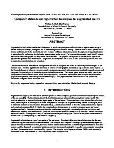
A)
Fіg. 1.1.
B)
Fіg. 1.2.
(FIG. 1.1, a) represented an axial section of the common generalized voxel tomographic model of the upper respiratory tract, at the level of the nasal cavity.( FIG. 1.2,b) shows the external view of anatomical structures of the head model for the upper respiratory tract . (FIG. 1.2) shows an example of determination of the objective parameters of external the nose in sagittal projection voxel topographic model, in translucent skin mode.