Revisión Inmunología Vol. 25 / Núm 2/ Abril-Junio 2006: 101-114
From immune surveillance to tumor-immune escape: the story of an enemy with multiple strategies of resistance and counterattack O.G. Scharovsky1, P. Matar1, M.Z. Fluck1, M.J. Rico1, G.A. Rabinovich2 Institute of Experimental Genetics, School of Medical Sciences, National University of Rosario, Rosario. Division of Immunogenetics, Hospital de Clínicas «José de San Martín», University of Buenos Aires, Buenos Aires, Argentina. 1
2
DE LA INMUNOVIGILANCIA AL ESCAPE TUMORAL: HISTORIA DE UN ENEMIGO CON MÚLTIPLES ESTRATEGIAS DE RESISTENCIA Y CONTRA-ATAQUE Recibido: 21 Abril 2006 Aceptado: 16 Mayo 2006
RESUMEN Los tumores deben evadir la respuesta inmunológica para ser clínicamente detectables en el paciente. A estos fines, las células cancerosas han desarrollado múltiples estrategias para eludir el ataque inmunológico. Estos mecanismos conspiran en estadíos avanzados de un tumor limitando la habilidad del sistema inmune para inhibir el crecimiento tumoral y la efectividad de estrategias de inmunoterapia en cáncer. Desde la biología tumoral a la clínica, en el presente artículo expondremos los más importantes mecanismos utilizados por tumores para evadir la respuesta inmunológica y su potencial impacto en el diseño de estrategias de inmunoterapia.
ABSTRACT Tumors must circumvent the immune response of the host to become clinically detectable. For this purpose, malignant cells have devised multiple strategies to thwart immune attack. These mechanisms are suggested to conspire in advanced stages of cancer to limit the ability of the immune system to restrain the tumor and the effectiveness of immunotherapy strategies to successfully eradicate malignant cells. From tumor biology to cancer immunotherapy and back again, we will summarize here some of the most important mechanisms used by tumors to evade the immune response and their potential impact in the design of cancer immunotherapy strategies.
PALABRAS CLAVE: Escape tumoral/ Presentación antigenica/ Apoptosis/ Contraataque/ Fas ligando/ Galectina-1.
KEY WORDS: Tumor-immune escape/ Antigen presentation/ Apoptosis/ Counterattack/ Fas ligand/ Galectin-1.
A BRIEF INTRODUCTION: FROM IMMUNE SURVEILLANCE TO TUMOR-IMMUNE ESCAPE All living organisms are exposed to different external insults such as viruses, bacteria, fungi, parasites and toxins, among others. To overcome these adverse stimuli, vertebrates have successfully developed a sophisticated system of protection against such foreign pathogens. As a consequence, the immune system displays different and complex innate
and adaptive mechanisms in which several cell types are involved. The first suggestion of a pivotal role of the immune system in recognizing tumor cells as foreign was postulated by Paul Erlich early during the 20th century (1). Erlich postulated the concept that the immune system would protect the organism, namely «the self», detecting and eliminating the «non-self». The idea that tumor cells are
101
FROM IMMUNE SURVEILLANCE TO TUMOR-IMMUNE ESCAPE: THE STORY OF AN ENEMY WITH MULTIPLE STRATEGIES...
considered as «non-self» and can be recognized and eliminated by T-cell mediated mechanisms was controversial for many years. The genetic and biochemical changes that a normal cell undergoes during its transition to a malignant cell can frequently lead to the production of new antigens that are not present in normal cells. These new antigens are called tumor-specific antigens (TSA). However, the first experimental evidence supporting the notion that tumors can induce an immune response emerged four decades later. The original observations of Gross(2), Foley(3), and Prehn(4) demonstrating that different inbred strains of mice can develop resistance to tumor transplantation by preimmunization with the same tumor, were confirmed in different experimental models by several researchers(5, 6). Nevertheless, those and other experimental results, have not received wide acceptance among the community of tumor biologists, as it was believed that MHC and not TSA antigens would be responsible for the observed immune response. In spite of this controversy, Burnet and Thomas provided a re-birth of Erlich’s hypothesis(7, 8) and developed the theory of tumor-immune surveillance. The authors proposed that the majority of the tumors, which appeared frequently, were eliminated efficiently by the immune system, even before they could be clinically detectable. This seminal theory not only induced a great deal of experimental work that meant a significant advance in tumor immunology, but also opened new avenues for immunological intervention in order to eradicate primary tumors and metastases.
THE «GREAT CONTRADICTION»: AN IMMUNOCOMPETENT HOST CANNOT BEAT AN IMMUNOGENIC TUMOR From its very beginning, the immune surveillance theory had strong supporters but also strong detractors. In spite of the evidences obtained from different experimental models, supporting the existence of an immune response against tumors(9), several arguments were posed against this theory(10). The lack of increase of tumor incidence in nude mice, which are congenitally athymic and, therefore, incapable of mounting a T cell response, was the main experimental fact that seemed to defeat Burnet’s theory. Nevertheless, the experimental evidence obtained later finally confirmed Burnet’s hypothesis. It was clearly demonstrated that natural immunity in nude mice could control tumor development. Moreover, it was also shown that nude mice indeed develop extrathymic T cells. After several controversial discussions, this theory is at present in good health, due to the emergence of new experimental
102
VOL. 25 NUM. 2/ 2006
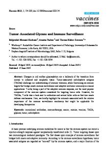
data supporting the concept of immunosurveillance(11-14). However, if we accept that the immune system of an immunocompetent host is able to detect and respond to the new antigens displayed by the tumors, an important contradiction emerges when we confront this situation with the fact that, very frequently, the immune system is not able to cope with the tumor that grows and, eventually, kills the host. Several mechanisms, known as tumor escape mechanisms, appear to be responsible for the lack of an effective immune response against the tumors. Tumors must circumvent the immune response of the host to become clinically detectable. For this purpose, malignant cells have devised multiple strategies to evade or thwart the immune attack. These mechanisms are suggested to conspire in advanced stages of cancer to limit the ability of the immune system to restrain the tumor and the effectiveness of immunotherapy strategies to successfully eradicate malignant cells. In the next sections we will critically discuss some of the most important mechanisms developed by tumors to evade immune recognition and resist immune attack (Figure 1).
TUMOR-IMMUNE ESCAPE STRATEGIES Alterations in the expression and function of Major Histocompatibility Complex (MHC) molecules The success in tumor elimination by CD8+ cytotoxic T lymphocytes (CTLs) relies on the efficient presentation of the tumor antigen-derived peptides by MHC class I molecules. Effector T cells specifically recognize those peptides associated with MHC-I and exert their cytotoxic effect by inducing tumor cell apoptosis. Any alteration of the antigen presentation machinery will alter the generation of an effective immune response. Cancer cells have developed multiple mechanisms aimed at eliminating or, at least, weakening the adaptive immune response. Such a strategy is clearly evidenced by the demonstration of a frequent down-regulation of the expression of MHC class I antigens in several tumor types. The loss of these molecules results in decreased ability to present peptides derived from tumor antigens, thus making these cells resistant to CTLs. Complete loss of class I or loss of single alleles are frequently seen and more than half of all tumors may have one of these alterations. It has been reported by several laboratories that the down-regulation or loss of MHC class I expression prevents the recognition of tumor antigens and the lysis by CTLs. In fact, down-regulation of HLA-A and HLA-B alleles is quite common in different tumor types(15-17). Moreover, downregulation of MHC I
INMUNOLOGÍA
O.G. SCHAROVSKY ET AL.
A) Down-regulation or loss of MHC I molecules
B) Alterations in antigen expression or processing
C) Down-regulation or loss of costimulatory molecules
Host T cell
D) Alterations in signal transduction molecules
Tumor cell
Regulatory T cell
TCR CD3 CD8 MHC I
E) Induction of T cell apoptosis
F) Immunosuppression
antigen
IL-10; TGF-β
ICAM-1 LFA-1 B7-1/2 CD28 LFA-3 CD2
IL-10 Th1
PD-1
Th2
PD-L1 galectin-1 CD95L CD95
Figure 1. Mechanisms of tumor-mediated immune evasion: A) alterations (down-regulation or loss) in MHC expression and function in tumor cells; B) alterations in tumor antigen expression and/or processing; C) alterations in the expression of costimulatory molecules; D) alterations in signal transduction molecules (p56lck; p59fyn; CD3-ζ, and others); E) induction of T cell apoptosis by: CD95/CD95L, PD-1/PD-L1, or galectin-1; F) tumor-induced immunosuppression by regulatory T cells, cytokines or galectin-1.
expression and function has been demonstrated to have an important prognostic value(18, 19). A number of studies have been carried out to assess the different HLA phenotypes and the molecular mechanisms leading to those phenotypes, which are regulated at least in part, through changes in oncogenic factors(20). Complete loss of expression of the HLA class I alleles is a phenotype found in cervical carcinomas(21), bladder carcinomas(22) and melanomas(23, 24), among others. In the majority of the cases the HLA loss is caused by different mutations involving β2-microglobulin genes. It has been proposed that tumor suppressor genes and genes implicated in the immune response could undergo inactivation by the same molecular mechanisms(20). Haplotype loss is the second HLA phenotype that has been described. This alteration is caused by the hemizygous loss of HLAA, -B and –C alleles, and the underlying mechanism is the presence of large deletions in chromosome 6(25, 26). Allelic loss of single HLA alleles defines a third HLA phenotype
that is caused by a wide array of genetic defects including point mutations, frame shifts or deletions. These kind of genetic alterations have been detected in melanomas and colorectal carcinomas(27). It has been well established that cells that express lower levels of HLA-A and HLA-B molecules are more susceptible to lysis by the natural cytotoxicity exerted by NK cells(28). Such a condition revealed another mechanism of evasion by cancer cells involving the expression of non classical HLA molecules such as HLA-G and HLA-E, which belong to the group MHC class Ib. This group of molecules is involved in immunoregulation, acting as inhibitory signals for NK cell cytotoxicity(29). It has been shown that expression of HLA-G by melanoma cells contributes to cell evasion of the immune response by inhibiting NK cell recognition(30). Furthermore, recent evidence indicates that the inhibition of the interactions between NKG2D and its ligands on tumor cells (i.e MICA and MICB proteins) may result in tumor
103
FROM IMMUNE SURVEILLANCE TO TUMOR-IMMUNE ESCAPE: THE STORY OF AN ENEMY WITH MULTIPLE STRATEGIES...
cell evasion of NK cell responses(11, 12). The identification of different strategies used by tumors to avoid immune surveillance and the elucidation of the molecular mechanisms involved in these strategies are crucial for the development of novel therapeutic approaches directed to restore an effective immune response. Alteration in Tumor Antigen Expression and the Antigen Processing Machinery In recent years a great progress has been made in the identification of tumor-specific antigens (TSA), derived from antigens localized on the surface of tumor cells. Tumor antigens can be classified into two types: 1) those that are unique to transformed cells and do not appear in normal cells (TSA) and 2) those called tumor-associated antigens (TAA), which are frequently found on the surface of tumor cells but are also expressed on the surface of normal cells during the embryonic or fetal life (oncofetal antigens), in selected organs after birth, or in normal cells but at much lower concentration than on tumor cells. Research over the past decade has led to the identification of different tumor antigens recognized by T cells and the elucidation of their structure and nature at the molecular level(31-35). The presence of specific antigens is critical for the development of an antitumor immune response. Therefore, if the tumor finds the way of downregulating, modifying or losing its own antigens, it will have found an efficient tactic for avoiding immune recognition. Actually, it has been demonstrated that several of these modifications take place in different tumor models. Independently of the alterations in the expression of HLA class I antigens, a wide array of modifications in the structure and/or the expression of TSA have been observed in different tumor types. Besides the intrinsic heterogeneity of antigen expression among different tumors, a downregulation of antigen expression has been demonstrated, which could range from decreased expression levels to a complete loss of one or more tumor antigens(27, 36-38). Thus, antigen-loss variants that appear or remain within the tumor, particularly after immunotherapy(39, 40) have a selective growth advantage, leading to a more malignant tumor. Melanoma is a prototype tumor in which down-modulation of several differentiation antigens (including Melan-A/MART1 and tyrosinase)(41-43) has been clearly demonstrated. Downregulation of Melan-A/MART-1 antigen expression by different mechanisms (including antigen silencing via its promoter)(44) avoids recognition by Melan-A/MART-1specific T cells and contributes to tumor-immune escape. A second immune escape strategy based on modifications of tumor antigens has recently been demonstrated. Tumors
104
VOL. 25 NUM. 2/ 2006
can take advantage of the mechanism of antigenic drift, like viruses, and generate point mutations that change the amino acid sequence of the antigen(45, 46). Then, cells harboring the new antigen variants are selected by their growth advantage, since the specific T-cell response elicited by the original tumor is directed toward an antigen that is no longer represented on the tumor cell surface. Moreover, even if the expression of HLA I molecules and tumor antigens is conserved, the induction of a specific cytotoxic T cell response may be impaired because of the presence of alterations in the antigen-processing machinery(27). Down-regulation of the transporters of peptides related to antigen processing TAP-1 and TAP-2, has been associated with the malignant potential of murine(47) and human(48) tumors. A retrospective analysis of the classical theory of «genetic instability» described by Peter Nowell in 1976(49) provides support to the different tumor cell variants that will harbor, heterogeneity not only in MHC molecules expression, but also in the expression of tumor antigens. The consequence of such variety of phenotypes is rapidly observed: the success of the patrolling immune cells in eradicating susceptible cancer cells is reverted towards rapid tumor growth. These tumor cell variants will have increased malignancy, tumor progression and, finally, metastatic dissemination. Alterations in the Expression of Costimulatory or Adhesion molecules Naïve T lymphocytes require at least two different extracellular signals in order to activate and differentiate into effector cells. The binding of the MHC-peptide complex to its specific receptor (TCR-CD3 complex) and to the coreceptors CD4 or CD8, provides the first signal required for the development of a specific immune response. However, this signal is not sufficient for full activation of T cells, requiring the participation of a second signal provided by costimulatory molecules (B7-1/CD80 and B7-2/CD86) expressed by antigen presenting cells (APC). However, other molecules are also involved in nonspecific adhesion mechanisms that are required for the interaction and immunological synapse between effector cells and target cells. Some of these interactions involve ICAM-1 (CD54) binding to LFA-1, CD40L binding to CD40, and members of the TNF receptor family(46). The majority of the tumors are derived from parenchymal or mesenchymal cells that do not express B7 molecules. Moreover, cells that normally express B7 can down-regulate its expression during the process of tumor progression(50). Thus, the ability of tumor cells to effectively present tumor antigen derived-peptides might be severely impaired. Consequently, that lack of
INMUNOLOGÍA
costimulation could be an important mechanism of evasion of the immune response, even if tumor cells express normal levels of MHC-I and TSA(51, 52). In this regard it has been recently demonstrated that low surface expression of B71/CD80 represents an immune escape mechanism in colon carcinoma(53). Tirapu and colleagues showed that silencing of CD80 by interfering RNA led to loss of tumorigenicity of CT26 colon carcinoma in immunocompetent, but not immunodeficient Rag-/- mice(53). The differentiation of CTLs is stimulated by activated helper T cells and requires the presence of MHC molecules on APC. Thus, for the induction of effective anti-tumor immune responses, a cross-priming by APCs that express costimulatory and MHC molecules is usually required. In this context, a decrease or lack of an adequate cytotoxic antitumor response would be provoked by APCs that are not able to efficiently present tumor antigens and activate helper T cells. Induction of T cell apoptosis: «Tumor Counterattack» Following the principle that the best defense is a timely attack, cancer cells not only resist immune attack, but have also devised strategies of tumor counterattack. The CD95/CD95L System Apoptosis mediated by Fas (CD95)/Fas L (CD95L) interactions is critical for the regulation of the biology and homeostasis of normal tissues. The Fas receptor belongs to the tumor necrosis factor (TNF) family of receptors involved in proliferation and apoptosis and is characterized by an intracellular domain, called «the death domain». Fas ligand (FasL) is a type II transmembrane protein which functions by inducing apoptosis of Fas-bearing cells(54). In 1995, D.L Vaux observed that FasL-expressing Sertoli cells were not rejected when transplanted into allogeneic mice(55). This observation was supported by the finding of FasL expression in the eye(56), and allowed the conclusion that FasL expression confers immunological privilege to selected tissues under physiological and pathological conditions. A few years later, the interaction of FasL on tumor cells with its specific receptor Fas on T lymphocytes was implicated in tumor cell evasion of immune surveillance(57). Binding of FasL to Fas leads to oligomerization of receptors and recruitment of FADD and the pro-caspase 8; these molecules together form a protein complex called the deathinducing signaling complex (DISC). In the DISC, pro-caspase8 is autocatalytically cleaved and then activates further effector caspases directly or via a mitochondrial pathway, which cleave cellular substrates leading to the typical
O.G. SCHAROVSKY ET AL.
biochemical and morphological changes of apoptosis. The Fas signaling pathway is regulated by many pro- and anti-apoptotic factors, including members of the Bcl-2 family which regulate apoptosis at the mitochondrial level, FLIPs (FLICE-inhibitory proteins) which interfere with the initiation of apoptosis directly at the level of death receptors, and IAPs/ survivin which bind to and inhibit caspases and apoptosome formation(58). Expression of FasL has been reported in solid tumors of nonhematopoietic origin, including colon, hepatocellular carcinoma, melanoma, astrocytoma, lung carcinoma, ovarian carcinoma, esophageal carcinoma, glioblastoma, renal cell carcinoma, and head and neck carcinoma. However, considerable heterogeneity in surface expression of FasL has been detected even within a particular lineage of tumor(59). Although unspecific staining of certain FasL antibodies has been demonstrated(60), it has been shown that FasL-positive tumor cells can kill Fas-positive T cells in vitro, demonstrating functional FasL expression(61). A number of tumors are resistant to apoptosis and express functional FasL constitutively or after chemotherapy. This situation may enable tumor cells to delete anti-tumor lymphocytes and to suppress anti-tumor immune responses, a phenomenon called «tumor counterattack»(62). Several lines of evidence support the involvement of the Fas system in tumor counterattack. Fas/FasL interactions are proposed to be an important mechanism for the maintenance of immune privilege in selected organs including the eye, placenta, testis, and the central nervous system(63). Also, various animal models have been used to demonstrate the ability of FasL expressed on tumors to down-regulate anti-tumor immune responses(64, 65). However, certain experimental evidence seems to contradict the FasL counterattack hypothesis. It has been reported that overexpression of FasL on tumor cells does not confer immune privilege but, instead, induces a strong inflammatory response that accelerates tumor rejection. These reports propose that FasL has a proinflammatory function and that gene transfer of FasL can be used in tumor eradication. It has been suggested that locally expressed FasL acts on surrounding cells to induce the production of IL-8 or other leukocyte chemoattractants(66). Other authors reported that FasL induces the processing and release of IL-1β, which in turn may be responsible for the infiltration by neutrophils(67). The in vivo consequences of FasL expression on tumors are far from clear. So far, many studies of tumor counterattack have been published, but the results are confusing and contradictory because many factors may influence FasL activity in vivo. The different levels and the kinetics of FasL expression on tumor cells may be of particular relevance in
105
FROM IMMUNE SURVEILLANCE TO TUMOR-IMMUNE ESCAPE: THE STORY OF AN ENEMY WITH MULTIPLE STRATEGIES...
tumor-immune system interactions. The sensitivity of T cells to Fas-mediated apoptosis varies considerably depending on the activation status of T cells. Also, T-cell sensitivity to Fas-mediated apoptosis is modulated by many factors, particularly by signaling through various receptors and cytokines(68). The consequences of FasL expression may also depend on the tumor microenvironment. Various site-specific factors may have a critical impact on the physiology of immune cells, on the interaction of immune and tumor cells and on the tumor itself. In addition the release of different type of cytokines, such as TGF-β and IL-10, the oxygen levels (hypoxia or normoxia), the extent of vascularization and the accessibility to immune cell infiltration, may also influence Fas L-mediated immune privilege. A further complicating aspect in tumor-immune escape mediated by FasL is the role of soluble FasL(69). Soluble FasL differs functionally from the membrane-bound form and its biological function is controversial. In some studies soluble FasL was cytotoxic(70), while in other studies Fas L shedding resulted in the absence of killing activity(71). Finally, other death factors, such as TNF and TRAIL, have also been shown to kill T cells(72) and, thus, might contribute to the suppression of anti-tumor immune responses. Taken together, the evidence presented suggest that tumor counterattack may be a powerful mechanism of tumor immune escape. To further understand its relevance under physiological and pathological conditions, it will be essential to use defined experimental systems in which all the above mentioned key elements could be controlled or carefully modulated. Thus, inhibition or blocking of tumor counterattack mechanisms might be beneficial for cancer therapy. Tumor-induced Immunosuppression: Regulatory T cells and Cytokines Recently, a subset of T cells, co-expressing CD4 and CD25 (IL-2Rα chain), with unique immune regulatory properties has been described(73). Regulatory CD4+CD25+ T cells (Treg) contribute to the prevention of autoimmune disorders by controlling the activity of autoreactive T lymphocytes and by inhibition of the proliferation of effector T cells. It has been suggested that CD4+CD25+ regulatory T cells may also contribute to immune tolerance at the tumor microenvironment and that depletion of these regulatory cells might be advantageous for boosting the anti-tumor immune response (74). These cells constitute approximately 5-10% of the total CD4+ T cell population in both humans and rodents, and their removal induces autoimmune disease in target tissues(75). Besides expressing the CD25 receptor, Tregs express other molecules including the cytotoxic T lymphocyte-associated protein-4 (CTLA-
106
VOL. 25 NUM. 2/ 2006
4), a glucocorticoid-induced TNF receptor family-related protein (GITR) and FoxP3 (a forkhead family transcriptional regulator). Although none of them is a distinct marker of the lineage, FoxP3 has turned out to be a useful marker as well as a critical factor for the differentiation and function of these cells(76). Association of these markers with the immunoregulatory activity of T regs has provided insights for the identification and molecular characterization of this particular cell subset. It has been recently demonstrated that Gr-1+ CD115+ immature myeloid suppressor cells can mediate the development of tumor-induced T regulatory cells in tumor-bearing hosts(77). In addition to CD4+ CD25+ FoxP3+ natural Treg, recent studies also report the identification of inducible T regulatory cells (Tr1)(78). The immune regulatory function of Tr1 cells has been attributed to their capacity to secrete immunosuppressive cytokines such as IL-10 and TGF-β(79). In contrast Treg can suppress the effector immune response by cell-cell contact or by release of immune suppressive factors(80). However, besides of being responsible for the suppressive effect of regulatory T cells, the unresponsiveness of the immune system in patients with cancer has been attributed to the secretion of immunosuppressive cytokines by the tumor itself. IL-10 is an anti-inflammatory cytokine that participates in the regulation of the immune response at several levels. IL-10 inhibits cytokine production of activated T and NK cells and of monocytes/macrophages and it blocks the antigen presenting activity of macrophages and dendritic cells, along with many other immunological functions. High IL-10 production was found to be associated with the immunosuppressive activity frequently observed in tumorbearing hosts, and it was speculated that overproduction of IL-10 could be responsible of tumor cell evasion of immune responses(81). TGF-β is another cytokine that inhibits activation, proliferation, and activity of lymphocytes(82). High levels of this immunosuppressive cytokine are often found in several malignancies and have associated with poor prognosis and lack of response to immunotherapy(83). Recent evidence indicates that TGF-β acts on cytotoxic T lymphocytes (CTL) to specifically inhibit the expression of different cytolytic gene products; namely perforin, granzyme A, granzyme B, Fas ligand, and IFN-γ, which are collectively responsible for CTL-mediated tumor cytotoxicity (84). Consistently, blockade of TGF-β signaling allows the generation of a potent antitumor immune response(85). Natural regulatory T cells are found at a higher frequency in peripheral blood of cancer patients and may induce tolerance at the tumor microenvironment, facilitating metastatic spread of cancer cells(86). When Treg were depleted
INMUNOLOGÍA
in mice, transplantable tumors were efficiently rejected by the host immune system(87). Interestingly, Dannull and colleagues recently demonstrated that vaccine-mediated antitumor immunity can be significantly enhanced following depletion of regulatory T cells(88). In addition, administration of anti-CD25 monoclonal antibody and/or anti-CTLA-4 for a limited time period also provoked effective tumor-specific immunity against syngeneic tumor cells(89). It has been postulated that an imbalance in Th1/Th2 cytokine production may also be responsible for the development of cancer, with a shift toward a Th2 response and induction of immunosuppressive cytokines including IL-10, IL-4 and IL-6(90). Cyclophosphamide (Cy) is an alkylating agent widely used in cancer chemotherapy. It has bimodal effect on the immune system, depending of the dose and schedule of administration(91). We have demonstrated that a single low-dose of Cy induced a Th2/Th1 shift in the cytokine profile of syngeneic lymphoma-bearing rats, which may be responsible of its antimetastatic effect. Such a treatment reduced the splenic production of IL-10, TGF-β and nitric oxide, restoring the lymphoproliferative capacity (92-94). Cyclophosphamide has been demonstrated to have selective toxicity to suppressor T cells (95) and, more recently, to CD4+CD25+ regulatory T cells(96), thus promoting tumor growth. Currently, we have preliminary evidence in an experimental lymphoma model supporting the role of Cymediated immunopotentiation through a mechanism of selective toxicity against CD4+CD25+ T cells (unpublished results). T regulatory cells have been proposed as key regulators of antitumor immune responses and their induction is considered a mechanism of tumor-immune escape. The idea that removal and/or inhibition of CD4+CD25+ regulatory T cells or inducible Tr1 cells can abrogate immunological unresponsiveness to syngeneic tumors, has established a novel strategy of evoking tumor immunity that would boost other cancer immunotherapy strategies. Alterations in Signal Transduction Molecules Patients in advanced stages of cancer and mice with large transplantable tumors have a compromised systemic immune response with highly decreased delayed-type hypersensitivity (DTH) responses. In this regard, NK and T cells often exhibit alterations in their proliferative and cytotoxic capacities as well as in cytokine secretion(97). Recent observations showing alterations in signal transduction molecules in T and NK cells from tumor-bearing mice and cancer patients provide a molecular basis to better understand this immune dysfunction. The original observation in mice bearing a mouse colon carcinoma(98) showed an altered
O.G. SCHAROVSKY ET AL.
pattern of protein tyrosinase phosphorylation, a reduction of the protein tyrosine kinases p56lck and p59fyn, and a decreased CD3-ζ chain. These findings have subsequently been confirmed and extended to a variety of human tumors, including renal, colorectal, ovarian, liver, gastric, pancreatic, and cervical carcinomas and melanomas(99). Of particular interest is the correlation observed between CD3-ζ expression and the disease stage in cancer patients(100). The TCR-associated CD3-ζ chain is responsible for transduction of signals delivered through the CD3-TCR complex and, therefore, its expression is important for activation of T cells(101). By quantitative flow cytometry analysis, decreased or absent expression of the CD3-ζ chain in CD4 + or CD8 + T cells as well as in NK cells was demonstrated in patients with malignancies. A similar finding was observed in tumor-infiltrating lymphocytes (TILs). These observations have suggested that the tumor microenvironment has negative effects on immune cells, and that the failure of the host to eliminate tumors might be a consequence of the impaired function of T cells, including the inability of these cells to signal normally upon TCR engagement(102). Interestingly, evidence has been provided for tumor-induced degradation of the CD3-ζ chain. It has been demonstrated that tumor cells can induce activation of intracellular peptidases in T lymphocytes, and that this tumor-induced enzymatic degradation is responsible for decreased or absent expression of signal transducing molecules, including the CD3-ζ chain in activated T cells (103). On the other hand, a computer-assisted analysis of the amino acid sequence of the CD3-ζ chain has revealed the presence of sites sensitive to cleavage by caspases, and it has been demonstrated that the CD3ζ chain is a substrate for proteolysis mediated by intracellular caspases(104). These results indicate that tumor-induced activation of caspases seems to be responsible for low or absent expression of CD3-ζ and perhaps other signaling molecules in T cells. Finally, other mechanisms, including free oxygen radical generation within the tumor microenvironment, have been proposed to account for decreased CD3-ζ expression in cancer(105). Although the mechanisms involved in tumor-induced down-regulation of CD3-ζ expression in T cells are under intense scrutiny, there are evidences indicating that direct contact with tumor or tumor-derived factors might participate in degradation of CD3-ζ and perhaps other signaling molecules. In order to prevent signaling defects of T cells in tumorbearing hosts, it will be necessary to unravel the mechanism(s) involved in tumor-T cell interactions. These studies are of critical importance for the design of future immunotherapy strategies.
107
FROM IMMUNE SURVEILLANCE TO TUMOR-IMMUNE ESCAPE: THE STORY OF AN ENEMY WITH MULTIPLE STRATEGIES...
Novel Mechanisms of Tumor-immune Escape Indoleamine 2,3 Deoxygenase (IDO) An additional mechanism that mediates tumor-induced immunosuppression includes the overexpression of the IFN-γ-inducible enzyme, indoleamine 2,3-dioxygenase (IDO). This enzyme is responsible for the catabolism of tryptophan (the amino acid that is critical for T cell function) and a competitive inhibitor of IDO, 1-methyl-tryptophan (1-MT), can prevent tumor-immune escape(106). Uyttenhove and colleagues demonstrated that expression of IDO by immunogenic mouse tumor cells prevents their rejection by preimmunized mice(106). This effect was accompanied by a lack of accumulation of specific T cells at the tumor site and could be partly reverted by systemic administration of 1-methyl-tryptophan, an inhibitor of IDO. These results suggested that the efficacy of immunotherapy strategies might be improved by concomitant administration of an IDO inhibitor. The STAT3 Pathway Signal transducer and activator of transcription (STAT)family proteins are latent cytoplasmic transcription factors that convey signals from cytokine and growth factor receptors to the nucleus. It has been demonstrated that STAT3 signaling in tumor cells suppresses both innate and adaptive antitumor immune responses, further enhancing tumor progression (107, 108). In addition a critical role has been demonstrated for STAT3 in the generation of immune cell tolerance(109). Proof-of-concept studies in cell culture and animal models have validated STAT3 protein as a promising molecular target for novel cancer therapies, including smallmolecule inhibitors of STAT3 signaling. The PD-1/PD-L1 System and other negative costimulatory signals PD-1 is an inducible receptor expressed on CD4+ and CD8+ T cells following activation. The expression of one ligand for PD-1, designated PD-L1 or B7-H1, on tumor cells of a variety of histological origins has suggested a potential mechanism for tumor-immune escape (110). It has been demonstrated that cancer cell-associated PD-L1 increases apoptosis of antigen-specific human T cell clones in vitro and in vivo(111). Thus, blockade of PD-L1 (B7-H1) might also be a complementary therapy to augment tumor-specific T cell responses(112). In addition, Kryczek and colleagues recently demonstrated that B7-H4, a recently identified B7 family molecule, identifies a novel suppressive macrophage population in human ovarian carcinoma and that depletion of B7-H4+ tumor
108
VOL. 25 NUM. 2/ 2006
macrophages may represent a novel strategy to enhance Tcell mediated immunity in cancer(113). Galectin-1: The «Sweet Escape» Galectins are animal lectins defined by shared consensus amino acid sequences and affinity for β-galactose-containing oligosaccharides(114). Recent evidence indicates that galectin1, a proto-type member of this family, has the potential to inhibit T cell effector functions by regulating T cell apoptosis and the Th1/ Th2 cytokine balance(115). Expression of galectin-1, a prototype member of the galectin family, has been well documented in many different tumor types including astrocytoma, melanoma and prostate, thyroid, colon, bladder and ovary carcinomas(116). Interestingly, in most cases such expression correlates with the aggressiveness of these tumors and the acquisition of metastatic phenotype. The immunoregulatory effects of galectin-1 and the correlation between galectin-1 expression in cancer cells and the aggressiveness of these tumors, prompted us to investigate the role of galectin-1 in tumor-immune escape. We hypothesized that tumor cells may impair T cell effector functions through secretion of galectin-1 and that this mechanism may contribute in tilting the balance toward an immunosuppressive environment at the tumor site. By a combination of in vitro and in vivo experiments using knockdown transfectants, we established a link between galectin-1-mediated immunoregulation and its contribution to tumor-immune escape(117). Blockade of the inhibitory effects of galectin-1 within tumor tissue resulted in reduced tumor mass (an effect which required intact CD4+ and CD8+ T cell responses) and stimulated the generation of a tumor-specific T-cell response in vivo. Our observations support the idea that galectin-1 may contribute to immune privilege of tumors by modulating survival or polarization of effector T cells and suggest a potential molecular target for manipulation of T-cell apoptosis with potential implications in immunotherapy. Recently, Le and collagues confirmed our findings demonstrating a link between tumor hypoxia and galectin-1 expression in head and neck squamous carcinoma cells(118). Interestingly, the authors found a negative correlation between galectin-1 expression and presence of CD3+ T cells in tissue sections(118). Challenges for the future will be to employ potent and selective small inhibitors of galectin-1 and, in fact, molecules with such properties have already been developed for galectin1 or other galectins(119-121). Furthermore, galectin-1 expression can be modulated by chemotherapeutic and anti-metastatic agents including low doses of Cy(122). A current challenge is the design of more specific and potent galectin-1 inhibitors for therapeutic purposes with no or minimal adverse effects. Although galectin-1 still remains elusive in terms of our
INMUNOLOGÍA
understanding of its multifunctional modes of action, we are moving closer to unraveling this mystery at a molecular level and to design new therapeutic approaches directed toward modulating its activities. Other galectins, including galectin-2, galectin-3 and galectin-9 promote immune cell apoptosis and modulate cytokine production in vitro, suggesting their potential role in tumor-immune escape(114). Other Mechanisms of Tumor-immune Escape Other tumor-derived suppressive factors that are able to impair T-cell function systemically include prostaglandin E2 (PGE2), prostate-specific antigen (PSA), sialomucins (MUC1) and RCAS1, a receptor-binding cancer antigen that induces cell cycle arrest and apoptosis of target cells(123).
IMMUNE STIMULATION This phenomenon was first described by Prehn and Lappé(124) who found that intermediate levels of antitumor immune reactivity might often stimulate rather than inhibit the development of certain tumor types(125,126). Shortly after, Stewart and colleagues published several data on human breast cancer(127,128) which agreed with Prehn’s postulation. They found that certain types of human mammary adenocarcinomas, but not other cancer types, are favored in its growth properties by a mild immune response, at least at the first stages of tumor evolution. Several molecules secreted by, or expressed on the surface of effector immune cells may be involved in the phenomenon of immunostimulation. Similar to the cross-talk between different immune cell types, cytokines produced by T cells and the stroma can act as paracrine growth factors stimulating tumor growth and progression. Different experimental evidences support this reasoning. IL-10 and its receptor (IL10R) can stimulate the growth of metastatic cells of a B-cell lymphoma, establishing an autocrine loop(129). Also, increased production of IL-10, induced by CEA, enhances liver metastasis in human colorectal carcinomas(130). Moreover, IL-4 stimulates survival of thyroid cancer(131), head and neck squamous cell carcinoma cell lines(132) and B-CLL cells(133). In addition, other soluble factors released by immune cells and involved in angiogenesis, including vascular endothelial growth factor (VEGF) (134), may also play an important role in immunostimulation. Interestingly, a positive association has been observed between serum levels of VEGF and/or other angiogenic molecules and cancer progression(135). In contrast, other studies report a critical role for VEGF in the suppression of dendritic cell differentiation and maturation(136). Taking into account the aforementioned experimental evidence, the implementation of cancer immunotherapy
O.G. SCHAROVSKY ET AL.
strategies should be preceded by a careful examination of the tumor immunogenicity and the phenomena of tumor escape and immunostimulation. In this regard, recent evidence indicates that inflammatory cells can sculpt the immunogenicity of different tumor types. This phenomenon led to the identification by Schreiber and colleagues of the «Cancer Immunoediting hypothesis»(137). This hypothesis proposes three different mechanisms («The Three Es of immunoediting: elimination, equilibrium and escape»), responsible of eliminating tumors and sculpting the immunogenic phenotypes of tumors that eventually form in immunocompetent hosts(137).
CONCLUDING REMARKS AND PERSPECTIVES Research over the past few years has contributed to increase enormously the available information about the interactions between tumor cells and the immune system. Currently we accept that tumors have smartly developed a number of evasion strategies which, individually or in combination, may enable these cells to thwart immune attack. The identification of the mechanisms employed by each tumor type will hopefully give the hints required to design novel therapeutic strategies aimed at destroying the tumor «fortress». Such therapeutic approaches should be aimed at overcoming several potential mechanisms that individual tumors may develop at different stages to overthrow or avoid innate and adaptive immunity.
ACKNOWLEDGEMENTS Research in authors` laboratories is supported by grants from Fundación Sales (to G.A.R.), The National Agency for Promotion of Science and Technology (to G.A.R PICT 200305-13787), Mizutani Foundation for Glycoscience (to G.A.R.), University of Buenos Aires (M091 to G.A.R.) and National University of Rosario (to O.G.S). G.A.R. is a member of the Scientific Career of CONICET. O.G.S. is a member of the Scientific Career of C.I.U.N.R. The authors thank members of their laboratory for critical discussions and continuous support.
CORRESPONDENCE TO: Gabriel A. Rabinovich, División Inmunogenética Hospital de Clínicas «José de San Martín» Facultad de Medicina. Universidad de Buenos Aires Av. Córdoba 2351. 3er Piso (C1120) Ciudad de Buenos Aires. Argentina Phone: +54-11-5950-8755. Fax: +54-11-5950-8758 E-mail:
[email protected]
109
FROM IMMUNE SURVEILLANCE TO TUMOR-IMMUNE ESCAPE: THE STORY OF AN ENEMY WITH MULTIPLE STRATEGIES...
REFERENCES 1. Erlich P. Ueber den jetzigen Stand der Karzinomforschung. Ned Tijdschr Geneeskd 1909; 5: 273-290. 2. Gross L. Intradermal immunization of C3H mice against a sarcoma that originated in an animal of the same line. Cancer Res 1943; 3: 326-333. 3.
Foley EJ. Antigenic properties of methylcholanthrene-induced tumors in mice of the strain of origen. Cancer Res 1953; 13: 835-837.
4. Prehn RT, Main JM. Immunity to mehylcholanthrene.induced sarcomas. J Natl Cancer Inst 1957; 18: 769-778. 5. Klein G, Sjögren HO, Klein E, Hellström KE. Demostration of resistance against methylcholanthrene-induced sarcomas in the primary autochthonous host. Cancer Res 1960; 20: 1561-1572. 6. Old LJ, Boyse EA, Clarke DA and Carswell EA. Antigenic properties of chemically-induced tumors. Ann NY Acad Sci 1962; 101: 80-106. 7. Burnet FM. The concept of immunological surveillance. Prog Exp Tumor Res 1970; 13: 1-27. 8. Thomas L. On immunosurveillance in human cancer. Yale J Biol Med 1982; 55: 329-333. 9. Pardoll D. Does the immune system see tumors as foreign or self?. Annu Rev Immunol 2003; 21: 807-839. 10. Stutman O. Chemical carcinogenesis in nude mice: comparison between nude mice from homozygous matings and heterozygous matings and effect of age and carcinogen dose. J Natl Cancer Inst 1979; 62: 353-358. 11. Smyth MJ, Godfrey DI, Trapani JA. A fresh look at tumor immunosurveillance and immunotherapy. Nat Immunol 2001; 2: 293-299. 12. Lanier LL. A renaissance for the tumor immunosurveillance hypothesis. Nat Med 2001; 7: 1178-1180.
VOL. 25 NUM. 2/ 2006
downregulation but not complete loss of MHC class I expression correlates with a poor prognosis. Int J Cancer 2006; 118: 6-10. 19. Zia A, Schildberg FW, Funke I. MHC class I negative phenotype of disseminated tumor cells in bone marrow is associated with poor survival in R0M0 breast cancer patients. Int J Cancer 2001; 93: 566-570. 20. Algarra I, García Lora A, Cabrera T, Ruiz-Cabello F, Garrido F. The selection of tumor variants with altered expresión of classical and nonclassical MHC class I molecules: implications for tumor immune escape. Cancer Immunol Immunother 2004; 53: 904910. 21. Keating PJ, Cromme FV, Duggan-Keen M, Snijders PJ, Walboomers JM, Hunter RD et al. Frequency of downregulation of individual HLA-A and –B alleles in cervical carcinomas in relation to TAP1 expression. Br J Cancer 1995; 72: 405-411. 22. Cabrera T, Pedrajas G, Cozar JM, Garrido A, Vicente J, Tallada M et al. HLA class I expression in bladder carcinomas. Tissue antigens 2003; 62: 324-327. 23. Hicklin DJ, Wang Z, Arienti F, Rivoltini L, Parmiani G, Ferrone S. β2- Microglobulin mutations, HLA class I antigen loss, and tumor progression in melanoma. J Clin Invest 1998; 101: 2720-2729. 24. Paschen A, Mendez RM, Jimenez P, Sucker A, Ruiz-Cabello F, Song M et al. Complete loss of HLA class I antigen expression on melanoma cells: a result of succesive mutational events. Int J Cancer 2003; 103: 759-767. 25. Jimenez P, Canton J, Collado A, Cabrera T, Serrano A, Real LM et al. Chromosome loss is the most frequent mechanism contributing to HLA haplotype loss in human tumors. Int J Cancer 1999; 83: 91-97.
13. Dunn GP, Bruce AT, Ikeda H, Old LJ, Schreiber RD. Cancer immunoediting: from immunosurveillance to tumor escape. Nat Immunol 2002; 3: 991-998.
26. Feenstra M, Verdaasdonk M, van der Zwan AW, de Weger R, Slootweg P, Tilanus M. Microsatellite analisis of microdissected tumor cells and 6p high density microsatellite analysis in head and neck squamous cell carcinomas with down-regulated human leukocyte antigen class I expression. Lab Invest 2000; 80: 405-414.
14. Girardi M, Oppenheim DE, Steele CR, Lewis JM, Glusac E, Filler R et al. Regulation of cutaneous malignancy by gammadelta T cells. Science 2001; 294: 605-609.
27. Rivoltini L, Carabba M, Huber V, Castelli C, Novellino L, Dalerba P et al. Immunity to cancer: attack and escape in T lymphocytetumor cell interaction Immunol Rev 2002; 188: 97-113.
15. García-Lora A, Algarra I, Garrido F. MHC class I antigens, immune surveillance, and tumor immune escape. J Cell Physiol 2003; 195: 346-355.
28. Gilboa E. How tumors escape immune destruction and what we can do about it. Cancer Immunol Immunother 1999; 48: 382-385.
16. Rivoltini L, Barrachini KC, Viggiano V, Kawakami Y, Smith A, Mixon A et al. Quantitative correlation between HLA class I expression and recognition of melanoma cells by antigen-specific cytotoxic T-lymphocytes. Cancer Res 1995; 55: 3149-3157. 17. Koopman LA, Corver WE, van der Slikm AR, Giphart MJ, Fleuren GJ. Multiple genetic alterations cause frequent and heterogeneous human histocompatibility leukocyte antigen class I loss in cervical cancer. J Exp Med 2000; 191: 961-975. 18. Watson NF, Ramage JM, Madjd Z, Spendlove I, Ellis IO, Scholefield JH, et al. Immunosurveillance is active in colorectal cancer as
110
29. Rodgers JR, Cook RG. MHC class Ib molecules bridge innate and acquired immunity. Nat Rev Immunol 2005; 5: 459-471. 30. Paul P, Rouas-Freiss N, Khalil-Daher I, Moreau P, Riteau B, Le Gal FA et al. HLA-G expression in melanoma: a way for tumor cells to escape from immunosurveillance. Proc Natl Acad Sci USA 1998; 95: 4510-4515. 31. Boon T, Ceottini JC, Van den Eynde B, van der Bruggen P, Van Pel A. Tumor antigens recognized by T lymphocytes. Annu Rev Immunol 1994; 12: 337-365. 32. Kawakami Y, Fujita T, Matsuzaki Y, Sakurai T, Tsukamoto M, Toda M, et al. Identification of human tumor antigens and its
INMUNOLOGÍA
implications for diagnosis and treatment of cancer. Cancer Sci 2004; 95: 784-791. 33. Jager D, Taverna C, Zippelius A, Knuth A. Identification of tumor antigens as potential target antigens for immunotherapy by serological expression cloning. Cancer Immunol Immunother. 2004; 53: 144-147. 34. Dudley ME, Rosenberg SA. Adoptive-cell-transfer therapy for the treatment of patients with cancer. Nat Rev Cancer 2003; 3: 666675.
O.G. SCHAROVSKY ET AL.
47. Agrawal S, Reemtsma K, Bagiella E, Oluwole SF, Braunstein NS. Role of TAP-1 and/or TAP-2 antigen presentation defects in tumorigenicity of mouse melanoma. Cell Immunol 2004; 228: 130-137. 48. Cresswell AC, Sisley K, Laws D, Parsons MA, Rennie IG, Murray AK. Reduced expression of TAP-1 and TAP-2 in posterior uveal melanoma is associated with progression to metastatic disease. Melanoma Res 2001; 11: 275-281. 49. Nowell PC. The clonal evolution of tumor cell populations. Science 1976; 194: 23-28.
35. Renkvist N, Castelli C, Robbins PF, Parmiani G. A listing of human tumor antigens recognized by T cells. Cancer Immunol Immunother 2001; 50: 3-15.
50. Zheng P, Sarma S, Guo Y, Liu Y. Two mechanisms for tumor evasion of preexisting cytotoxic T-cell responses: lessons from recurrent tumors. Cancer Res 1999; 59: 3461-3467.
36. Sigalotti L, Coral S, Fratta E, Lamai E, Danielli R, Di Giacomo AM et al. Epigenetic modulation of solid tumors as a novel approach for cancer immunotherapy. Semin Oncol 2005; 32: 473-478.
51. Ugurel S, Uhlig D, Pfohler C, Tilgen W, Schadendorf D, Reinhold U. Down-regulation of HLA class II and costimulatory CD86/B72 on circulating monocytes from melanoma patients. Cancer Immunol Immunother 2004; 53: 551-559.
37. Spiotto MT, Rowley DA, Schreiber H. Bystander elimination of antigen loss variants in established tumors. Nat Med 2004; 10: 294-298. 38. Khong HT, Wang OJ, Rosenberg SA. Identification of multiple antigens recognized by tumor-infiltrating lymphocytes from a single patient: tumor escape by antigen loss and loss of MHC expression. J Immunother 2004; 27: 184-190. 39. Lozupone F, Rivoltini L, Luciani, F, Venditti M, Lugini, L, Cova A et al. Adoptive transfer of an anti-MART-1(27-35)-specific CD8+ T cell clone leads to immunoselection of human melanoma antigenloss variants in SCID mice. Eur J Immunol 2003; 33: 556-566. 40. Riker A, Cormier J, Panelli M, Kammula U, Wang E, Abati A et al. Immune selection after antigen-specific immunotherapy of melanoma. Surgery 1999; 126: 112-120. 41. Jager E, Ringhoffer M, Altmannsberger M, Arnd M, Karbach J, Jager D et al. Immunoselection in vivo: independent loss of MHC class I and melanocyte differentiation antigen expression in metastatic melanoma. Int. J Cancer 1997; 71: 142-147. 42. Marincola F, Jaffee E, Hicklin D, Ferrone S. Escape of human solid tumors from T-cell recognition: molecular mechanisms and functional significance. Adv Immunol 2000; 74: 181-273. 43. Rosenberg SA. Progress in human tumor immunology and immunotherapy. Nature 2001; 411: 380-384. 44. Kurnick JT, Ramírez Montagut T, Boyle LA, Adrews DM, Pandolfi F, Durda PJ et al. A novel autocrine pathway of tumor escape from immune recognition: melanoma cell lines produce a soluble protein that diminishes expression of the gene encoding the melanocyte lineage melan-A/MART-1 antigen through down-modulation of its promoter. J Immunol 2001; 167: 1204-1211. 45. Bai XF, Liu J, Li O, Zheng P, Liu Y. Antigenic drift as a mechanism for tumor evasion of destruction by cytolytic T lymphocytes. J Clin Invest 2003; 111: 1487-1496. 46. Ahmad M, Rees RC, Ali SA. Escape from immunotherapy: possible mechanisms that influence tumor regression/progression. Cancer Immunol Immunother 2004; 53: 844-854.
52. Frydecka I, Kosmaczewska A, Bocko D, Ciszak L, Wolowiec D, Kuliczkowski K, et al. Alterations of the expression of T-cell-related costimulatory CD28 and downregulatory CD152 (CTLA-4) molecules in patients with B-cell chronic lymphocytic leukaemia. Br J Cancer 2004; 90: 2042-2048. 53. Tirapu I, Huarte E, Guiducci C, Arina A, Zaratiegui M, Murillo O, Gonzalez A, Berasain C, Berraondo P, Fortes P, Prieto J, Colombo MP, Chen L, Melero I. Low surface expression of B7-1 (CD80) is an immunoescape mechanism of colon carcinoma. Cancer Res 2006; 15: 2442-2450. 54. Peter ME, Krammer PH. Mechanisms of CD95 (APO-1/Fas)mediated apoptosis. Curr Opin Immunol 1998; 10: 545-551. 55. Vaux DL. Immunology: ways around rejection. Nature 1995; 377: 576-577. 56. Griffith TS, Brunner T, Fletcher SM, Green DR, Ferguson TA. Fas ligand-induced apoptosis as a mechanism of immune privilege. Science 1995; 270: 1189-1192. 57. Ryan AE, Shanahan F, O´Connel J, Houston AM. Addressing the «Fas counterattack» controversy: blocking Fas ligand expression suppresses tumor immune evasion of colon cancer in vivo. Cancer Res 2005; 65: 9817-9823. 58. Curtin JF, Cotter TG. Live and let die: regulatory mechanisms in Fas-mediated apoptosis. Cell Signal 2003; 15: 983-992. 59. Gastman BR, Johnson DE, Whiteside TL, Rabinowich H. Tumorinduced apoptosis of T lymphocytes: elucidation of intracellular apoptotic events. Blood 2000; 5: 2015-2023. 60. Strater J, Walczak H, Hasel C, Melzner I, Leithauser F, Moller P. CD95 ligand (CD95L) immunohistochemistry: a critical study on 12 antibodies. Cell Death Differ 2001; 8: 273-278. 61. Igney FH, Behrens CK, Krammer PH. CD95L mediates tumor counterattack in vitro but induces neutrophil-dependent tumor rejection in vivo. Int J Cancer 2005; 113: 78-87. 62. Igney FH, Behrens CK, Krammer PH. Tumor counterattack-concept and reality. Eur J Immunol 2000; 30: 725-731.
111
FROM IMMUNE SURVEILLANCE TO TUMOR-IMMUNE ESCAPE: THE STORY OF AN ENEMY WITH MULTIPLE STRATEGIES...
63. French LE, Hahne M, Viard I, Radlgruber G, Zanone R, Becker K et al. Fas and Fas ligand in embryos and adult mice: ligand expression in several immune privileged tissues and coexpression in adult tissues characterized by apoptotic cell turnover. J Cell Biol 1996; 133: 335-343. 64. Hahne M, Rimoldi D, Schroter M, Romero P, Schreier M, French LE et al. Melanoma cell expression of Fas (APO-1/CD95) ligand: implications for tumor immune escape. Science 1996; 274: 13631366. 65. Nishimatsu H, Takeuchi T, Ueki T, Kajiwara T, Moriyama N, Ishida T et al. CD95 ligand expression enhances growth of murine renal cell carcinoma in vivo. Cancer Immunol Immunother 1999; 48: 56-61. 66. Arai H, Chan SY, Bishop DK, Nabel GJ. Inhibition of the alloantibody response by CD95 ligand. Nat Med 1997; 8: 843-848. 67. Miwa K, Asano M, Horai R, Iwakura Y, Nagata S, Suda T. Caspase 1-independent IL-1b release and inflammation induced by the apoptosis inducer Fas ligand. Nat Med 1998; 4: 1287-1292. 68. Scaffidi C, Kirchhoff S, Krammer PH, Peter ME. Apoptosis signaling in lymphocytes. Curr Opin Immunol 1999; 11: 277-285. 69. Tanaka M, Suda T, Takahashi, T, Nagata S. Expression of the functional soluble form of human Fas ligand in activated lymphocytes. EMBO J 1995; 14: 1129-1135. 70. Suda T, Tanaka M, Miwa K, Nagata S. Apoptosis of mouse naive T cells induced by recombinant soluble Fas ligand and activation-induced resistance to Fas ligand. J Immunol 1996; 167: 3918-3924. 71. Suda T, Hashimoto H, Tanaka M, Ochi T, Nagata S. Membrane Fas ligand kills human peripheral blood T lymphocytes, and soluble Fas ligand blocks the killing. J Exp Med 1997; 186: 20452050. 72. Nagata S. Apoptosis by death factors. Cell 1997; 88: 355-365. 73. Sakaguchi S, Sakaguchi N, Asano M, Itoh M, Toda M. Immunologic self-tolerance maintained by activated T cells expressing IL-2 receptor alpha-chains (CD25). Breakdown of a single mechanism of self-tolerance causes various autoimmune diseases. J Immunol 1995; 155: 1151-1164. 74. Zou W. Regulatory T cells, tumor immunity and immunotherapy. Nat Rev Immunol 2006; 6: 205-307. 75. Sakaguchi S, Sakaguchi N, Shimizu J, Yamazaki S, Sakihama T, Itoh M et al. Immunologic tolerance maintained by CD25+CD4+ regulatory T cells: their common role in controlling autoimmunity, tumor immunity, and transplantation tolerance. Immunol Rev 2001; 182: 18-32. 76. Coffer PJ, Burgering BMT. Forkhead-box transcription factors and their role in the immune system. Nat Rev Immunol 2004; 4: 889899. 77. Huang B, Pan PY, Li Q, Sato A, Levy DE, Bromberg J, Divino CM, Chen SH. Gr-1+ CD115+ immature myeloid suppressor cells mediate the development of tumor-induced T regulatory cells and
112
VOL. 25 NUM. 2/ 2006
T-cell anergy in tumor-bearing hosts. Cancer Res 2006; 66: 11231131. 78. Zhang X, Huang H, Yuan J, Sun D, Hou WS, Gordon J, et al. CD48- dendritic cells prime CD4+ T regulatory 1 cells to suppress antitumor immunity. J. Immunol 2005; 175: 2931-2937. 79. Roncarolo MG, Bacchetta R, Bordignon C, Narula S, Levings MK. Type 1 T regulatory cells. Immunol Rev 2001; 182: 68-79. 80. Sakaguchi S, Sakaguchi N, Shimizu J, Yamazaki S, Sakihama T, Itoh M et al. Immunologic tolerance maintained by CD25+CD4+ regulatory T cells: their common role in controlling autoimmunity, tumor immunity, and transplantation tolerance. Immunol Rev 2001; 182: 18-32. 81. Pawelec G. Tumor escape from the immune response the last hurdle for successful immunotherapy of cancer? Cancer Immunol Immunother 1999; 48: 343-345. 82. Fontana A, Frei K, Bodmer S, Hofer E, Schreier MH, Palladino MA Jr, et al. Transforming growth factor-beta inhibits the generation of cytotoxic T cells in virus-infected mice. J Immunol 1989; 143: 3230-3234. 83. Gorsch SM, Memoli VA, Stukel TA, Gold LI, Arrick BA. Immunohistochemical staining for transforming growth factor beta 1 associates with disease progression in human breast cancer. Cancer Res 1992: 52: 6949-6952. 84. Thomas DA, Massague J. TGF-b directly targets cytotoxic T cell functions during tumor evasion of immune surveillance. Cancer Cell 2005; 8: 369-380. 85. Gorelik L, Flavell RA. Immune-mediated eradication of tumors through the blockade of transforming growth factor-b signaling in T cells. Nat Med 2001; 7: 1118-1122. 86. Liyanage UK, Moore TT, Joo HG, Tanaka Y, Herrmann V, Doherty G et al. Prevalence of regulatory T cells is increased in peripheral blood and tumor microenvironment of patients with pancreas or breast adenocarcinoma. J Immunol 2002; 169: 2756-2761. 87. Shimizu J, Yamazaki S, Sakaguchi S. Induction of tumor immunity by removing CD25+CD4+ T cells: a common basis between tumor immunity and autoimmunity. J Immunol 1999; 163: 5211-5218. 88. Dannull J, Su Z, Rizzieri D, Yang BK, Comenan D, Yancey D, Zhang A, Dahm P, Chao N, Gilboa E, Vieweg J. Enhancement of vaccine-mediated antitumor immunity in cancer patients after depletion of regulatory T cells. J Clin Invest 2005; 115: 26233633. 89. Onizuka S, Tawara I, Shimizu J, Sakaguchi S, Fujita T, Nakayama E. Tumor rejection by in vitro administration of anti-CD25 (interleukin-2 receptor a) monoclonal antibody. Cancer Res 1999; 59: 3128-3133. 90. Kidd P. Th1/Th2 balance: the hypothesis, its limitations, and implications for health and disease. Altern Med Rev 2003; 8: 223-246. 91. Matar P, Scharovsky OG. Cyclophosphamide bimodal effects in antineoplasic therapy. Rev Bras Cancerol 1996; 42: 33-42.
INMUNOLOGÍA
92. Matar P, Rozados VR, González AD, Dlugovitzky DA, Bonfil RD, Scharovsky OG. Mechanism of antimetastatic immunopotentiation by low-dose cyclophosphamide. Eur J Cancer 2000; 36: 1060-1066. 93. Matar P, Rozados VR, Gervasoni SI, Scharovsky OG. Down regulation of T-cell-derived IL-10 production by low-dose cyclophosphamide treatment in tumor-bearing rats restores in vitro normal lymphoproliferative response. Int Immunopharmacol 2001; 1: 307-319. 94. Matar P, Rozados VR, Gervasoni SI, Scharovsky OG. Th2/Th1 switch induced by a single low-dose of cyclophosphamide in a rat metastatic lymphoma model. Cancer Immunol Immunother 2002; 50: 588-596. 95. North, R.J. Cyclophosphamide-facilitated adoptive immunotherapy of an established tumor depends on elimination of tumor-induced suppressor T cells. J Exp Med 1982; 155: 1063-1074. 96. Ghiringhelli F, Larmonier N, Schmitt E, Parcellier A, Cathelin D, Garrido C et al. CD4+CD25+ regulatory T cells suppress tumor immunity but are sensitive to cyclophosphamide which allows immunotherapy of established tumors to be curative. Eur J Immunol 2004; 34: 336-344. 97. Baxevanis CN, Papamichail M. Characterization of the anti-tumor immune response in human cancers and strategies for immunotherapy. Crit Rev Oncol Hematol 1994; 16: 157-179. 98. Mizoguchi H, O’Shea JJ, Longo DL, Loeffler CM, McVicar DW, Ochoa AC. Alterations in signal transduction molecules in T lymphocytes from tumor-bearing mice. Science 1992; 258: 17951798. 99. Whiteside TL. Signaling defects in T lymphocytes of patients with malignancy. Cancer Immunol Immunother 1999; 48: 346-352. 100.Matsuda M, Petersson M, Lenkei R, Taupin JL, Magnusson I, Mellstedt H et al. Alterations in the signal-transducing molecules of T cells and NK cells in colorectal tumor-infiltrating, gut mucosal and peripheral lymphocytes: correlation with the stage of the disease. Int J Cancer 1995; 61: 765-772. 101.Weiss A, Littman DR. Signal transduction by lymphocyte antigen receptors. Cell 1994; 76: 263-274. 102.Reichert TE, Rabinowich H, Johnson JT, Whiteside TL. Immune cells in the tumor microenvironment: mechanisms responsible for signaling and functional defects. J Immunother 1998;21: 295-306. 103.Wieckowski E, Wang G, Gastman BR, Goldstein LA, Rabinowich H. Granzyme B-mediated degradation of T-Cell receptor z chain. Cancer Res 2002; 62: 4884-4889. 104.Gastman BR, Johnson DE, Whiteside TL, Rabinowich H. Caspasemediated degradation of TCR-z chain. Cancer Res 1999; 59: 14221427. 105.Kono K, Salazar-Onfray F, PeterssonM, Hansson J, Masucci G, Wasserman K et al. Hydrogen peroxide secreted by tumor-derived macrophages down-modulates signal-transducing z molecules and inhibits tumor-specific T cell- and natural killer cell-mediated cytotoxicity. Eur J Immunol 1996; 26: 1308-1313.
O.G. SCHAROVSKY ET AL.
106.Uyttenhove C, Pilotte L, Theate I, Stroobant V, Colau D, Parmentier N et al. Evidence for a tumoral immune resistance mechanism based on tryptophan degradation by indoleamine 2,3-dioxygenase. Nat Med 2003; 9: 1269-1274. 107.Yu H, Jove R. The STATs of Cancer- New molecular targets come of age. Nat Rev Cancer 2004; 4: 97-105. 108.Kortylewski M, Kujaski M, Wang T, Wei S, Zhang S, Pilon-Thomas S, et al. Inhibiting Stat3 signaling in the hematopoietic system elicits multicomponent antitumor immunity. Nat Med 2005; 11: 1314-1321. 109.Cheng F, Wang HW, Cuenca A, Huang M, Ghansah T, Brayer J, et al. A critical role for Stat3 signaling in immune tolerance. Immunity 2003; 19: 425-436. 110.Zha Y, Blank C, Gajewski TF. Negative regulation of T-cell function by PD-1. Crit Rev Immunol 2004; 24: 229-237. 111.Dong H, Strome SE, Salomao DR, Tamura H, Hirano F, Flies DB et al. Tumor-associated B7-H1 promotes T-cell apoptosis: a potential mechanism of immune evasion. Nat Med 2002; 8: 787-789. 112.Blank C, Kuball JU, Voelkl S, Wiendl H, Becker B, Walter B et al. Blockade of PD-L1 (B7-H1) augments human tumor-specific T cell responses in vitro. Int J Cancer 2006; 119: 317-327. 113.Kryczek I, Zou L, Rodriguez P, Zhu G, Wei S, Mottram P, et al. B7-H4 expression identifies a novel suppressive macrophage population in human ovarian carcinoma. J Exp Med 2006; 203: 871-881. 114.Liu FT and Rabinovich GA. Galectins as modulators of tumor progression. Nat Rev Cancer 2005; 5: 29-41. 115.Rabinovich GA, Baum LG, Tinari N, Paganelli R, Natoli C, Liu FT, Iacobelli S. Galectins and their ligands: amplifiers, silencers or tuners of the inflammatory response? Trends Immunol 2002; 23: 313-320. 116.Danguy A, Camby I, Kiss R. Galectins and cancer. Biochim Biophys Acta 2002; 1572: 285-293. 117.Rubinstein N, Alvarez M, Zwirner NW, Toscano MA, Ilarregui JM, Bravo A et al. Targeted inhibition of galectin-1 gene expression in tumor cells results in heightened T cell-mediated rejection; A potential mechanism of tumor-immune privilege. Cancer Cell 2004; 5: 241-251. 118.Le QT, Shi G, Cao H, Nelson DW, Wang Y, Chen EY, et al. Galectin1: a link between tumor hypoxia and tumor immune privilege. J Clin Oncol 2005; 23: 8932-8941. 119.Andre S, Pieters RJ, Vrasidas I, Kaltner H, Kuwabara I, Liu FT et al. Wedgelike glycodendrimers as inhibitors of binding of mammalian galectins to glycoproteins, lactose maxiclusters, and cell surface glycoconjugates. Chembiochem 2001; 2: 822-830. 120.Sorme P, Qian Y, Nyholm PG, Leffler H, Nilsson UJ. Low micromolar inhibitors of galectin-3 based on 3'-derivatization of Nacetyllactosamine. Chembiochem 2002; 3: 183-189. 121.Rabinovich GA, Cumashi A, Bianco GA, Ciavardelli D, Iurisci I, D`Egidio M et al. Synthetic lactulose amines: novel class of anticancer
113
FROM IMMUNE SURVEILLANCE TO TUMOR-IMMUNE ESCAPE: THE STORY OF AN ENEMY WITH MULTIPLE STRATEGIES...
VOL. 25 NUM. 2/ 2006
agents that induce tumor cell apoptosis and inhibit galectinmediated homotypic cell aggregation and endothelial cell morphogenesis. Glycobiology 2006; 16: 210-220.
130.Jessup JM, Samara R, Battle P, Laguinge LM. Carcinoembryonic antigen promotes tumor cell survival in liver through an IL-10dependent pathway. Clin Exp Metastasis 2004; 21: 709-717.
122.Rabinovich GA, Rubinstein N, Matar P, Rozados V, Gervasoni S, Scharovsky, OG. The antimetastatic effect of a single low dose of cyclophosphamide involves modulation of galectin-1 and Bcl-2 expression. Cancer Immunol Immunother 2002; 50: 597603.
131.Vella V, Mineo R, Frasca F, Mazzon E, Pandini G, Vigneri R, et al. Interleukin-4 stimulates papillary thyroid cancer cell survival: implications in patients with thyroid cancer and concomitant Graves' disease. J Clin Endocrinol Metab 2004; 896: 2880-2889.
123.Gabrilovich D, Pisarev V. Tumor escape from immune response: mechanisms and targets of activity. Curr Drug Targets 2003; 4: 525-536. 124.Prehn RT, Lappé MA. An immunostimulation theory of tumor development. Transpl Rev 1971; 7: 26-54.
132.Myers JN, Yasumura S, Suminami Y, Hirabayashi H, Lin W, Johnson JT et al. Growth stimulation of human head and neck squamous cell carcinoma cell lines by interleukin 4. Clin Cancer Res 1996; 2: 127-135.
125.Prehn RT, Outzen HC. Immunostimulation of tumor growth. Prog Immunol 1980; 4: 651-675.
133.Dancescu M, Rubio-Trujillo M, Biron G, Bron D, Delespesse G, Sarfati M. Interleukin-4 protects chronic lymphocytic leukemic Bcells from death by apoptosis and upregulates Bcl-2 expression. J Exp Med 1992; 176: 1319-1326.
126.Prehn RT. Stimulatory effects of immune reactions upon the growths of untransplanted tumors. Cancer Res 1994; 54: 908914.
134. Mor F, Quintana FJ, Cohen IR. Angiogenesis-inflammation crosstalk: vascular endothelial growth factor is secreted by activated T cells and induces Th1 polarization. J Immunol 2004; 1727: 4618-4623.
127.Stewart THM. Evidence for immune facilitation of breast cancer growth and for the immune promotion of oncogenesis in breast cancer. Medicina (Buenos Aires) 1996; 56 (Supl I): 13-24.
135.Poon RT, Fan ST, Wong J. Clinical implications of circulating angiogenic factors in cancer patients. J Clin Oncol 2001; 194: 12071225.
128.Stewart TH, Heppner GH. Immunological enhancement of breast cancer. Parasitology 1997; 115 (Suppl):S141-S153.
136.Gabrilovich DI, Chen HL, Girgis KR, Cunningham HT, Meny GM, Nadaf S et al. Production of vascular endothelial growth factor by human tumors inhibits the functional maturation of dendritic cells. Nat Med 1996; 2: 1096-1103.
129.Rico MJ, Matar P, Gervasoni SI, Bonfil RD, Calcaterra N, Scharovsky OG. The transition to the metastatic phenotype of rat lymphoma cells involves up-regulation of IL-10 receptor expression and IL10 secretion. Clin Exp Metastasis 2005; 22: 127-135.
114
137.Dunn GP, Old LJ, Schreiber RD. The three Es of cancer immunoediting. Annu Rev Immunol 2004; 22: 329-360.