Indian Journal of Experimental Biology Vol 48, May 2010, pp. 425-435
Review Article
Free radicals: Their beneficial and detrimental effects on sperm function Shiva Kothari1, Aaron Thompson1, Ashok Agarwal1* & Stefan S du Plessis2 1
Center for Reproductive Medicine, Cleveland Clinic, 9500 Euclid Avenue, Desk A19.1, Cleveland, Ohio 44195, USA 2 Division of Medical Physiology, Faculty of Health Sciences, University of Stellenbosch, South Africa
Free radicals are molecules with one or more unpaired electron(s) commonly found in seminal plasma. Physiologically, free radicals control sperm maturation, capacitation and hyperactivation, the acrosome reaction, and sperm-oocyte fusion. Pathologically, free radicals induce lipid peroxidation, DNA damage and apoptosis of spermatozoa. The present review deals with both the beneficial and detrimental effects of free radicals on sperm function. Keywords: Apoptosis, Acrosome reaction, Capacitation, DNA damage, Free radical, Sperm function
Free radicals are molecules with one or more unpaired electron(s)1. These highly reactive molecules attack the nearest stable molecule to obtain an electron. Subsequently, the targeted molecule becomes a free radical itself and initiates a cascade of events that can ultimately lead to cellular damage2. However, at physiological levels, free radicals also help preserve homeostasis by acting as signal transducers3. There are two common forms of free radicals namely, reactive oxygen species (ROS) and reactive nitrogen species (RNS). Examples of ROS include the superoxide anion (O2-), hydrogen peroxide (H2O2), the extremely reactive hydroxyl radical (OH.) and the peroxyl radical (HO2-)4. RNS are often considered to be a subclass of ROS 4 and include nitric oxide (NO), nitrous oxide (N2O), peroxynitrite (NO3-), nitroxyl anion (HNO) and peroxynitrous acid (HNO3)1,5. Antioxidants act as free radical scavengers that help to keep free radicals at homeostatic levels in order to maintain physiologic function and prevent pathological effects due to development of oxidative stress (OS)6. OS is a state when there is an imbalance between ROS and the scavenging properties of antioxidants. Under these circumstances ROS outnumber antioxidants due to either increased ROS levels, decreased antioxidant properties, or a combination of both. Physiologically, free radicals control sperm maturation, capacitation and hyperactivation, the _________________ *Correspondent author Telephone: 216-444-9485 Fax: 216-445-6049 E-mail:
[email protected]
acrosome reaction (AR), and sperm-oocyte fusion. Pathologically, free radicals induce lipid peroxidation (LPO), DNA damage and apoptosis. The pathological effects of free radicals on sperm function have been described extensively in the literature, but a discussion physiological roles of free radicals in sperm function is required for better understanding of the phenomena. It is, therefore, the aim of the present review article is to explore both the beneficial and detrimental role of free radicals on sperm function. Sources of ROS in seminal plasma ROS that ultimately end up in the seminal plasma and exert their effects on spermatozoa can be generated from or originate from various sources that can either be endogenous or exogenous. Endogenous sources of ROS
Semen consists of different types of cells such as mature and immature spermatozoa, round cells from different stages of spermatogenesis, leukocytes and epithelial cells. Of these, leukocytes (neutrophils and macrophages) and immature spermatozoa are the two main sources of ROS7,8. Peroxidase positive leukocytes — Peroxidase positive leukocytes include polymorphonuclear leukocytes (PMN) (50-60% of all leukocytes) and macrophages (20-30% of all leukocytes)9,10. These peroxidase positive leukocytes originate from the prostate and seminal vesicles11. As one of the main mechanisms to combat pathogens, leukocytes produce high levels of ROS12. More specifically, when activated during disease states, peroxidase positive leukocytes produce ROS through the nicotinamide
426
INDIAN J EXP BIOL., MAY 2010
adenine dinucleotide phosphate (NADPH) system13,14. In addition, an increase in proinflammatory cytokines such as interleukin-8 and a decrease in the antioxidant superoxide dismutase (SOD) lead to increased levels of OS13. During infection, there are increased levels of leukocytes subsequently leading to more ROS production with detrimental effects on sperm function15. Sperm are particularly susceptible to infections in the testis and epididymis because of their lack of adequate antioxidant defense mechanisms16. More specifically, patients with genito-urinary tract infection have decreased sperm motility and decreased fertilization capacity17,18. Leukocytes, rather than immature spermatozoa, are the predominant source of ROS during sperm preparation19. In a study by Plante et al. 20 increased levels of PMN, not abnormal spermatozoa, led to decreased sperm motility. It has also been shown that leukocytes produce 1000 times more ROS than spermatozoa in human semen20,21. In addition, levels of ROS were correlated to the degree of leukocyte contamination22. In vivo, seminal plasma contains powerful antioxidants to scavenge the potentially dangerous ROS in the external environment23. However, during the in vitro preparation of sperm for assisted reproductive techniques, these antioxidant seminal plasma protective capacities are removed and it leaves sperm particularly susceptible to OS damage24. Immature spermatozoa — Normally, during spermatogenesis, developing sperm extrude their cytoplasm in preparation for fertilization. However, impaired spermatogenesis does not allow for the proper extrusion of cytoplasm. If excess cytoplasm exists, these sperm are deemed immature and functionally defective25,26. Excess cytoplasm (cytoplasmic droplets) activates the NADPH system, which provides electrons for free radicals through a cascade of events that ultimately results in the activation of the enzyme NADPH oxidase26. This production of NADPH is mediated through the enzyme glucose-6-phosphate dehydrogenase27. This cascade of events ultimately increases ROS production. Overall, there are two mechanisms by which spermatozoa produce ROS—(1) NADPHoxidase system at the plasma membrane level; and (2) NADH-dependent oxidoreductase system at the mitochondrial level28. The mitochondrial system has been shown to be the primary source of ROS in infertile men20.
Exogenous sources of ROS
Along with the endogenous sources of ROS, there are numerous exogenous sources of ROS, which produce ROS in pathological amounts. These sources include industrial compounds, smoking, alcohol, spinal cord injury and varicocele. Industrial compounds — Industrial compounds have been well documented to cause adverse health effects. In male reproduction, many studies have described the negative effects of various chemicals and compounds. Environmental pollutants have shown to increase ROS such as O2- and H2O2 in male testes. More specifically, phthalate, a compound commonly found in plastics and beauty products, induces sperm DNA damage and impairs spermatogenesis29,30. Heavy metals like lead, pesticides, and sulfur dioxide, a widespread food preservative, all increase OS in the male testes31-33. Smoking — Like industrial compounds, cigarette smoking has been shown to cause widespread harmful effects on health. Specific components of cigarettes such as nicotine are associated with free radical production34. In sperm, cigarette smoke increases ROS production while decreasing antioxidant capacity. Cigarette smoke decreases sperm parameters such as sperm motility, morphology and concentration35,36. In a study by Saleh et al.35, in smokers, seminal ROS production increased as measured by ROS-total antioxidant capacity (TAC) score. Smoking has also been shown to increase seminal leukocyte concentration, a potential source of ROS. Alcohol — Ethanol metabolism increases ROS, while it simultaneously decreases antioxidants. These effects result in biomolecule damage including damage to proteins, lipids and DNA37. In addition, consumption of alcohol has been shown to increase ROS, if it is consumed by a person eating low nutrition diet38. Spinal cord injury — Over 90% of men with spinal cord injury are infertile39. It is thought that these men have elevated ROS levels in sperm. These high ROS levels correlate with poor sperm motility and morphology that may lead to infertility40,41. In addition, men with spinal cord injury have increased polymorphonuclear leukocytes (producers of ROS) in their semen5. Varicocele — Varicocele is the uncharacteristic dilation and tortuosity of veins in pampiniform plexus around the human male spermatic cord42. Varicocele appears to be associated with higher levels of ROS8.
KOTHARI et al.: BENEFICIAL AND DETRIMENTAL ROLE OF FREE RADICALS
Increased levels of xanthine oxidase (a source of the O2-) and NO appear to increase ROS, thereby impairing sperm function43. High levels of NO have been reported in infertile men with varicocele compared to fertile men as well as infertile men without varicocele44. In addition, increased ROS levels in semen have been correlated with a higher varicocele grade. Those patients with varicocele grade 2 or 3 have also been shown to have higher levels of ROS in their semen45. In varicocele patients there are more markers of OS such as ROS induced LPO and presence of DNA damage46,47. Finally, those patients receiving a varicocelectomy have shown a decrease in ROS levels and increase in levels of TAC48. Physiological role of ROS Besides the well-known pathological effects that are a consequence of excess ROS production, physiological levels are actually essential for proper sperm function. ROS plays an important role in transducing signals in the complex biochemical cascades of the spermatozoa. Positive effects on maturation, hyperactivation, capacitation, AR and sperm-oocyte fusion have all been well documented. The specific ROS involved in the activation of these processes is still somewhat controversial and may be species specific3 with NO being identified as the RNS central to mediating sperm function5 (Fig. 1). Maturation — Spermatozoa have a characteristically tight packaging of chromatin, a process largely the result of ROS. During spermiogenesis, histone beads are replaced by smaller proteins called protamines. The majority of cysteine residues in protamines, under the slight oxidative conditions of the caudal epididymis, form disulfide bonds that result in condensed, stabilized nuclear DNA49-51. Because spermatozoa lack the preventative and repair systems of somatic cells or the oocyte, the enhanced stability of sperm chromatin is required for genetic integrity52. In addition, ROS induce changes in the mitochondrial membrane that confer protection to the major energy producer of the cell. Mature spermatozoa contain mitochondria with a keratinouslike protective coat called the mitochondrial capsule53. During spermatogenesis, peroxides such as H2O2 may induce the formation of this coat through the oxidation of phospholipid hydroperoxide glutathione peroxidase (PHPGx). Once oxidized, PHGPx, a selenoenzyme, forms an intermediate that can react with reduced protein thiol groups of the capsule to
427
form a selenadisulfide bond. Inactivation of PHGPx and oxidation of thiol groups results in the complex, yet stable protein network of the mitochondrial capsule54. Capacitation and hyperactivation — Capacitation is the poorly understood maturation process that spermatozoa undergo in the female genital tract. It includes the attainment of hyperactive motility and potential to undergo acrosome reaction (AR), both of which are necessary for successful fertilization to occur. The process includes an array of molecular changes, most notably are— an efflux of cholesterol; fluxes of bicarbonate, calcium (Ca2+) and other small ions that cause membrane hyperpolarization; and increase in pH, cAMP, and protein phosphorylation55. Hyperactivation, an essential process for successful fertilization, is considered a sub-category of capacitation. In non-hyperactive spermatozoa, lowamplitude flagellar movements are associated with slow, linear movements of the cell. Alternately, hyperactive spermatozoa exhibit high amplitude, asymmetric flagellar movement, significant side-toside displacement of the sperm head, and an overall non-linear motility. In the female genital tract, spermatozoa with hyperactivated motility exhibit less stagnation in the oviductual epithelium and the surrounding viscous mucus. In addition, hyperactive motility provides spermatozoa with the necessary propulsion to penetrate the cumulus cells—which via secretions of progesterone may actually initiate hyperactivation at the zona pellucida (ZP) of the oocyte56. Results supporting the role of ROS in the initiation of hyperactivation are well accepted. Generation of ROS can be performed by a variety of methods, including the addition of xanthine + xanthine-oxidase (X + XO), glucose oxidase, potassium superoxide, H2O2, NADPH, fetal cord serum (FCS) and fetal cord serum ultrafiltrate (FCSU). The results from in vitro experiments using such ROS generating techniques has led to the suggestion that O2- is crucial to hyperactive motility. In hamster sperm epididymis, NO was also shown to regulate hyperactivation57. Hyperactivation initiated by the biological fluids FCS and FCSU was inhibited in the presence of SOD, indicating the potential activity of O2- in vivo58. H2O2 has been observed to induce a positive effect, but the delicate balance between motility activation and LPO induced by H2O2 may be a reason for the variable reports3.
428
INDIAN J EXP BIOL., MAY 2010
Whereas O2- is speculated to regulate hyperactivation, H2O2 is proposed to be central to the remaining processes of capacitation59. However, at low concentrations, NO is also thought to be involved in this process. Catalase prevents NO-induced capacitation, an indication that NO effects are a result of a complex mechanism involving H2O260. In fact, the presence of ROS scavengers and nitric oxide synthase (NOS) inhibitors decreases cAMP, a well known molecule necessary for capacitation61. In many
ways, the molecular events accompanying capacitation are indistinguishable from those of the AR, except that the process is reversible and the time frame is significantly longer. Because of the similarities, few techniques are available that definitively distinguish between the two processes. As such, the two most common methods to assess the status of capacitation include the measurement of tyrosine phosphorylation and the ability to undergo the AR in response to external stimuli. Capacitation,
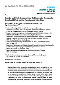
Fig. 1 — Physiological roles of ROS include maturation, capacitation, hyperactivation, the acrosome reaction and sperm-oocyte fusion. Pathological roles of ROS include lipid peroxidation, DNA damage and apoptosis.
KOTHARI et al.: BENEFICIAL AND DETRIMENTAL ROLE OF FREE RADICALS
the final maturation process, ensures that only fertile spermatozoa are able to reach, bind to, and penetrate the oocyte55. The general biochemical scheme involves a protein kinase A (PKA)-dependent mechanism that may be regulated by ROS. Current evidence suggests that the process is initiated by an influx of Ca2+ and HCO3that activates the amplifier enzyme adenyl cyclase (AC). AC converts ATP to cAMP, which then activates PKA that eventually leads to tyrosine phosphorylation associated with capacitation. ROS promote capacitation by stimulating AC, inhibiting phosphotyrosine phosphatases (PTPases) and activating tyrosine kinases (TKs). By doing so, ROS thereby increase the amount of tyrosine phosphorylation, one of the final steps in the biochemical scheme. Specifically, O2- may activate AC via oxidation of an essential thiol group. H2O2, on the other hand, is hypothesized to promote tyrosine phosphorylation more indirectly through activation of TKs and inhibition of PTPases. The intermediate steps between PKA activation and tyrosine phosphorylation are unknown, although several different, interconnected biochemical pathways may ultimately regulate capacitation61. Acrosome reaction (AR) — Once hyperactivated motility has propelled the spermatozoa past the cumulus oophorus, a capacitated sperm may bind to ZP and initiate an exocytotic release of proteolytic enzymes. AR, as the event is termed, creates a pore in the extracellular matrix of ZP and allows spermatozoa to penetrate this physical barrier and fuse with the oocyte. As noted above, the molecular events of AR share considerable overlap with those of capacitation. Included in these similarities is phosphorylation of similar tyrosine proteins, an influx of Ca2+, and activity of AC, cAMP, and PKA62. In vivo, the process is initiated upon binding to ZP. ROS have been shown to increase sperm affinity for ZP, possibly through the phosphorylation of three plasma membrane proteins at the apex of the sperm head, fertilin beta (ADAM2), spermadhesin family (includes AZN-3 and others), and P47 (the porcine homologue of SED1)63. In vitro, activation of AR has been observed upon addition of ROS59,64. At low concentrations, O2-, H2O2, and NO have all been shown to have positive effects on the AR65-68. NOS inhibitors have inhibited AR in human sperm, further evidence that NO is integral in AR. Similar to capacitation, ROS stimulates AR by activating AC,
429
which triggers downstream molecules (cAMP, PKA, etc) to initiate the exocytotic event61. Sperm-oocyte fusion — After bypassing the barrier of ZP, a spermatozoon may fuse with the oocyte and result in successful fertilization69. A high degree of sperm membrane fluidity is necessary for proper fusion to occur. Some degree of membrane fluidity is assured by the high poly-unsaturated fatty acid (PUFA) content of the sperm plasma membrane70. In studies of human spermatozoa, ROS have been shown to increase the rates of sperm-oocyte fusion71. The mechanism through which ROS increase membrane fluidity actually occurs during the biochemical cascade of capacitation and AR. During capacitation, by inhibiting PTPase activity, ROS prevent the dephosphorylation and deactivation of phospholipase A2 (PLA2) which cleaves the secondary fatty acid from the triglycerol backbone of the membrane phospholipid and increases membrane fluidity72. In addition, by activating AC and downstream molecules during the AR, ROS may activate PLA2. Protein kinase C is also known to phosphorylate and therefore activate PLA272. Pathological roles of ROS Physiologically, ROS are integral as signaling molecules for many processes involved in fertilization. However, when ROS levels overwhelm the antioxidant defense systems, pathological effects result. Because ROS are highly reactive, when they reach pathological levels they exert significant damage on biomolecules, such as proteins, lipids and nucleic acids73. The extent of this damage depends on many factors including duration of exposure, type of ROS and concentration as well as external factors like temperature, oxygen tension and antioxidant levels74 (Fig. 1). Lipid peroxidation (LPO) — The sperm plasma membrane is largely composed of PUFAs which are susceptible to oxidative damage due to the existence of double bonds75. The sperm membrane contains almost 50 per cent decosahexanoic acid, which contains six unsaturated double bonds in every molecule. As the LPO cascade proceeds in sperm, almost 60% of the fatty acid is lost from the membrane. LPO affects membrane structure and function such as fluidity, ion gradients, receptor transduction, transport processes and membrane enzymes9. As a result, functions that are necessary for normal fertilization are impaired73. Because LPO has
430
INDIAN J EXP BIOL., MAY 2010
been implicated in infertility, it is important to understand the processes underlying LPO76. LPO can be divided into three phases, the initiation phase, the propagation phase and the termination phase. Before any of these phases commence, ROS generation is necessary. O2- is generated either through NADPH system, an intracellular source, or through leukocytes, an extracellular source. O2- can be directly protonated to form hydroperoxyl radical (HO2) or it can be converted into H2O2 via SOD. H2O2 can then be converted into OH•. via Fenton reaction involving ferrous ion. These two ROS molecules, OH• and HO2, mark the beginning of initiation stage77. During the initiation phase, OH• or HO2 initiates LPO77. O2- and H2O2 are not energetic enough to initiate LPO directly13. As the initiation stage proceeds, unsaturated lipids undergo hydrogen abstraction to form lipid radicals. Presence of a double bond next to a methylene group makes the methylene carbon-hydrogen bonds weaker and more vulnerable to abstraction. Lipid radical then reacts with oxygen to form lipid HO2-, which eventually converts into lipid peroxide through antioxidant defense mechanisms. This lipid peroxide actually stabilizes the sperm plasma membrane. However, during the propagation phase, in the presence of a transition metal ion, the lipid peroxide gets broken down into alkoxyl radical and HO2-. These radicals subsequently act on additional lipids until lipid damage is widespread and irreversible73,77. In the termination phase two radicals react with each other to form a stable product and LPO ceases76,78. LPO damage can be quantified by measuring malondialdehyde (MDA), a stable by-product of LPO79,80. MDA is measured using the spectrophotometric thiobarbituric acid test. Another biological by-product of LPO is 4-hydroxynoneal, which is formed from low density lipoproteins. This compound can act on protein and DNA levels, causing significant damage. Moreover, hydroxynoneal can act as a chemical signal for neutrophils, preventing cell growth and exerting pathological effects76. There are many pathological effects of LPO on sperm function. Overall, LPO damages DNA and proteins through oxidation from lipid peroxyl or alkoxyl radicals. DNA damage by LPO can occur via base modifications, strand breaks or cross-linking4. In addition, LPO can result in a loss in membrane fluidity and as a result decreased sperm motility and
sperm-oocyte fusion81-83. NO has been shown as a potential ROS involved in decreasing sperm motility in humans84,85. The degree to which sperm motility and sperm-oocyte fusion are impaired is related to the amount of MDA81,86. In LPO, ROS initiate a cascade of events involving X + XO system and depleted ATP production that ultimately results in sperm immobilization87. In a study by de Lamirande et al88., increased levels of H2O2 induced LPO that eventually led to death of spermatozoa. Recent studies of LPO have shown two trends. The first is that those patients with abnormal semen parameters have increased LPO. In a study by Khosrowbeygi et al89., patients with asthenozoospermia, asthenoteratozoospermia, and oligoasthenoteratozoospermia sperm all had higher levels of PUFA and therefore, were more vulnerable to LPO caused by ROS. Another study has shown similar results, as patients with asthenozoospermia and oligoasthenoteratozoospermia also show higher MDA levels90. Furthermore, LPO as measured via MDA levels correlated inversely to in vitro fertilization rates in infertile men91. Secondly increases in inflammation and infection have also been reported in LPO. In a study by Fraczek et al92., proinflammatory cytokines propagate an increase in ROS levels, causing OS leading to increased LPO and as such, altering sperm function. Another study has demonstrated that the presence of various bacteria strains in semen, for instance Escherichia coli, increases white blood cells, thereby causing OS and increased LPO93. DNA damage — Tight packing of DNA combined with the antioxidant defense mechanisms of semen protect DNA from damage94. It is perhaps for this reason that sperm are particularly resistant to DNA damage compared to many cell lines including germ cells and myoblastoid cells95. However, the fact still remains that sperm undergoes DNA damage and it can be a possible etiology of infertility. ROS as well as apoptosis are proposed mechanisms that can induce DNA damage96. In a recent study by Amiri et al97., NO has been correlated with increased DNA damage. In a study by Irvine et al.98, infertile men have shown increased DNA fragmentation, decreased semen parameters and increased levels of ROS. In sperm, the mitochondrial genome is more susceptible to DNA damage than nuclear genome via H2O295. In addition, due to Y chromosome’s structure and the fact that it cannot repair double strand breaks, it is especially
KOTHARI et al.: BENEFICIAL AND DETRIMENTAL ROLE OF FREE RADICALS
susceptible to DNA damage16. High levels of ROS have also been correlated with DNA single and double strand breaks99. In a state of OS, DNA damage occurs due to oxidation of bases, strand breaks, crosslinking, deletions, frame shifts and rearrangement of chromosomes9,100. During intrauterine insemination and in vitro fertilization sperm with increased levels of DNA damage is not a cause for alarm as the concurrent LPO damage by the ROS eliminate the possibility for fertilization74. However, during intracytoplasmic sperm injection natural selection is bypassed and sperm with significant amounts of DNA damage have the opportunity to fertilize the oocyte52. Despite the fact that the sperm can have significant DNA damage, it still retains the ability to fertilize the egg, which poses significant risk to the embryo14. Increased DNA damage has been associated with decreased fertilization rates in vitro as well as increased early embryo death101,102. Unfortunately, there is no established clinical treatment for DNA damage16. Apoptosis — Often, when cellular components undergo damage, apoptosis or programmed cell death is initiated103. In male reproduction, abnormal spermatozoa may be eliminated via apoptosis102. The mechanisms of action of apoptosis in male reproduction are not well studied. However, preliminary studies have discussed that ROS serves as a stimulus that activates the mitochondria to release the signaling molecule cytochrome c104,105. Cytochrome c initiates a cascade of events that involve several caspases including caspase 3 and 9 that eventually leads to sperm apoptosis. In fact, ROS is associated with apoptotic caspases, including caspase 3 and 9105. Fas-protein may also be integral in the apoptotic pathway. When Fas-ligand or anti-Fas antibody binds to Fas, apoptosis is initiated106. An additional mechanism for apoptosis involves the inflammatory production of ROS, more specifically, hypochlorous acid (HOCl). HOCl is formed from H2O2 and chloride ion. HOCl oxidizes many cellular components, thus causing apoptosis. In a study by Said et al.107, have reported that HOCl is associated with elevated levels of apoptosis in spermatozoa. Many studies have outlined apoptosis in spermatozoa. In a study, increased ROS levels and markers for apoptosis, as measured by annexin-V staining, have been correlated in mature spermatozoa. ROS concentrations have been correlated with increased levels of DNA damage and
431
apoptosis105,108-110. In deer spermatozoa it has been shown that ROS-specifically H2O2-induces apoptosis, while O2- and OH• do not induce apoptosis111. Meanwhile studies in primate, murine and boar spermatozoa indicate that NO is correlated with apoptosis perhaps through caspase activation112-114. Recent literature has explored the potential for antioxidants to treat the pathological effects of ROS, including apoptotic cell death. In a study, in vivo antioxidant therapy has been shown to decrease sperm DNA damage, seminal levels of OS and apoptosis115. Association between these elements can underscore a possible etiology of male infertility. However, further studies need to be conducted to understand the mechanisms of action behind this association. In another study, taurine, a known antioxidant, reduces levels of arsenic-induced apoptosis in sperm both through mitochondrial dependent and independent pathway116. Melatonin, a known endogenous antioxidant, counteracts the 2-bromopropane (2-BP)induced apoptotic damage as measured by caspase 3 activation in the testis. In addition, microcystin-LR, a bacterial toxin increases apoptotic DNA damage and a proposed mechanism is through an increase in ROS. New research has also explored the role of external sources of ROS production to mediate apoptosis. In a study by Kumar et al.109, a cell extract with known pathologic properties induces apoptosis through ROS cell damage. Furthermore, the herbicide, alachlor, potentially induces apoptotic cell death via ROS117. Finally, increased temperature in the male reproductive system is associated with a release of ROS that eventually causes sperm death. In this pathway, ROS has been proposed to directly signal cell death118. Physiological and pathological levels of ROS Much literature, both current and past, fails to really elaborate on the exact numerical values of “physiological” levels of ROS when compared to “pathological” levels of ROS. It is hard to understand the subject completely without some numbers quantifying physiological versus pathological levels of ROS. There have been several recent attempts to elucidate this matter. Three different studies have reported three relatively similar cutoff values of ROS between low (physiological) and high (pathological) levels of ROS. Desai et al.119 have reported the cutoff value to be 0.0185 × 10⁶ counted photons per minute
432
INDIAN J EXP BIOL., MAY 2010
(cpm)/20 × 106 sperm, Agarwal et al.120 have reported the cutoff value to be 0.0125 × 10⁶ cpm/20 × 106 sperm, and Allameni et al.121 have reported the cutoff value to be 0.0145 × 10⁶ cpm/20 × 106 sperm. In general, values below the cutoff are low levels of ROS, while values above the cutoff are high levels of ROS. However, these experiments were not consistent with respect to samples used and subjects tested, among other things. For example, Agarwal et al.120, included only infertile patients, while Desai et al.119 included infertile and fertile patients in their study. Therefore it would be hard to accurately compare results. It is at least a start in the right direction to having a tangible number when referring to physiological and pathological levels of ROS. Conclusion This paper has discussed both the physiological and pathological roles that free radicals serve in the male reproductive system. An important perspective is that free radicals are not exclusively beneficial or exclusively detrimental. Rather, they need to be maintained at appropriate levels to ensure physiological function, while preventing pathological damage. Antioxidants represent a potential treatment for pathological levels of free radicals. However, despite the lack of clinical consensus on the efficacy of antioxidants as a potential therapy, if used, antioxidants need to keep free radicals at levels that are physiologically appropriate. Even though this paper elucidates many mechanisms dealing with free radicals and sperm, further research needs to be undertaken in order to understand the mechanisms of action behind free radicals. References 1 Agarwal A, Prabakaran S & Allamaneni S, What an andrologist/urologist should know about free radicals and why, Urology, 67 (2006) 2. 2 Agarwal A CM, Abdelrazik H & Sharma RK, Oxidative stress management in patients with male or female factor infertility, Handbook of Chemiluminescent Methods in Oxidative Stress Assessment. 2008 195 3 de Lamirande E, Jiang H, Zini A, Kodama H & Gagnon C, Reactive oxygen species and sperm physiology, Rev Reprod, 2 (1997) 48. 4 Sikka SC, Relative impact of oxidative stress on male reproductive function, Curr Med Chem, 8 (2001) 851. 5 Sharma RK & Agarwal A, Role of reactive oxygen species in male infertility, Urology, 48 (1996) 835. 6 Agarwal A, Nallella KP, Allamaneni SS & Said TM, Role of antioxidants in treatment of male infertility: an overview of the literature, Reprod Biomed Online, 8 (2004) 616.
7 Aitken RJ & West KM, Analysis of the relationship between reactive oxygen species production and leucocyte infiltration in fractions of human semen separated on Percoll gradients, Int J Androl, 13 (1990) 433. 8 Hendin BN, Kolettis PN, Sharma RK, Thomas AJ, Jr. & Agarwal A, Varicocele is associated with elevated spermatozoal reactive oxygen species production and diminished seminal plasma antioxidant capacity, J Urol, 161 (1999) 1831. 9 Sikka SC, Rajasekaran M & Hellstrom WJ, Role of oxidative stress and antioxidants in male infertility, J Androl, 16 (1995) 464. 10 Wolff H & Anderson DJ, Immunohistologic Characterization and Quantitation of Leukocyte Subpopulations in HumanSemen, Fertility and Sterility, 49 (1988) 497. 11 Wolff H, The biologic significance of white blood cells in semen, Fertil Steril, 63 (1995) 1143. 12 Tremellen K, Oxidative stress and male infertility - a clinical perspective, Human Reproduction Update, 14 (2008) 243. 13 Blake DR, Allen RE & Lunec J, Free radicals in biological systems--a review orientated to inflammatory processes, Br Med Bull, 43 (1987) 371. 14 Aitken RJ & Baker MA, Oxidative stress and male reproductive biology, Reprod Fertil Dev, 16 (2004) 581. 15 Ochsendorf FR, Infection and reactive oxygen species, Andrologia, 30 Suppl 1 (1998) 81. 16 Cocuzza M, Sikka SC, Athayde KS & Agarwal A, Clinical relevance of oxidative stress and sperm chromatin damage in male infertility: an evidence based analysis, Int Braz J Urol, 33 (2007) 603. 17 Wolff H, Politch JA, Martinez A, et al, Leukocytospermia is associated with poor semen quality, Fertil Steril, 53 (1990) 528. 18 Maruyama DK, Jr., Hale RW & Rogers BJ, Effects of white blood cells on the in vitro penetration of zona-free hamster eggs by human spermatozoa, J Androl, 6 (1985) 127. 19 Whittington K & Ford WC, Relative contribution of leukocytes and of spermatozoa to reactive oxygen species production in human sperm suspensions, Int J Androl, 22 (1999) 229. 20 Plante M, de Lamirande E & Gagnon C, Reactive oxygen species released by activated neutrophils, but not by deficient spermatozoa, are sufficient to affect normal sperm motility, Fertil Steril, 62 (1994) 387. 21 Kessopoulou E, Tomlinson MJ, Barratt CL, Bolton AE & Cooke ID, Origin of reactive oxygen species in human semen: spermatozoa or leucocytes?, J Reprod Fertil, 94 (1992) 463. 22 Aitken RJ, Buckingham DW, Brindle J, et al, Analysis of sperm movement in relation to the oxidative stress created by leukocytes in washed sperm preparations and seminal plasma, Hum Reprod, 10 (1995) 2061. 23 Zini A, de Lamirande E & Gagnon C, Reactive oxygen species in semen of infertile patients: levels of superoxide dismutase- and catalase-like activities in seminal plasma and spermatozoa, Int J Androl, 16 (1993) 183. 24 Sukcharoen N, Keith J, Irvine DS & Aitken RJ, Predicting the fertilizing potential of human sperm suspensions in vitro: importance of sperm morphology and leukocyte contamination, Fertil Steril, 63 (1995) 1293. 25 Huszar G, Sbracia M, Vigue L, Miller DJ & Shur BD, Sperm plasma membrane remodeling during spermiogenetic
KOTHARI et al.: BENEFICIAL AND DETRIMENTAL ROLE OF FREE RADICALS
26
27
28
29
30
31
32 33
34
35
36 37 38
39 40
41
42
43
maturation in men: relationship among plasma membrane beta 1,4-galactosyltransferase, cytoplasmic creatine phosphokinase, and creatine phosphokinase isoform ratios, Biol Reprod, 56 (1997) 1020. Aitken RJ, Fisher HM & Fulton N, et al, Reactive oxygen species generation by human spermatozoa is induced by exogenous NADPH and inhibited by the flavoprotein inhibitors diphenylene iodonium and quinacrine, Mol Reprod Dev, 47 (1997) 468. Richer S, Whittington K & Ford WCL, Confirmation of NADPH oxidase activity in human sperm, Journal of Reproductin and Fertility, 21 (1998) 118. Aitken RJ, Buckingham DW & West KM, Reactive oxygen species and human spermatozoa: analysis of the cellular mechanisms involved in luminol- and lucigenin-dependent chemiluminescence, J Cell Physiol, 151 (1992) 466. Agarwal DK, Maronpot RR, Lamb JCt & Kluwe WM, Adverse effects of butyl benzyl phthalate on the reproductive and hematopoietic systems of male rats, Toxicology, 35 (1985) 189. Kasahara E, Sato EF, Miyoshi M, et al, Role of oxidative stress in germ cell apoptosis induced by di(2ethylhexyl)phthalate, Biochem J, 365 (2002) 849. Acharya UR, Acharya S & Mishra M, Lead acetate induced cytotoxicity in male germinal cells of Swiss mice, Ind Health, 41 (2003) 291. Meng Z & Bai W, Oxidation damage of sulfur dioxide on testicles of mice, Environ Res, 96 (2004) 298. Chitra KC, Sujatha R, Latchoumycandane C & Mathur PP, Effect of lindane on antioxidant enzymes in epididymis and epididymal sperm of adult rats, Asian J Androl, 3 (2001) 205. Traber MG, van der Vliet A, Reznick AZ & Cross CE, Tobacco-related diseases. Is there a role for antioxidant micronutrient supplementation?, Clin Chest Med, 21 (2000) 173. Saleh RA, Agarwal A, Sharma RK, Nelson DR & Thomas AJ, Jr., Effect of cigarette smoking on levels of seminal oxidative stress in infertile men: a prospective study, Fertil Steril, 78 (2002) 491. Vine MF, Smoking and male reproduction: a review, Int J Androl, 19 (1996) 323. Das SK & Vasudevan DM, Alcohol-induced oxidative stress, Life Sci, 81 (2007) 177. Koch OR, Pani G, Borrello S, et al, Oxidative stress and antioxidant defenses in ethanol-induced cell injury, Mol Aspects Med, 25 (2004) 191. Linsenmeyer TA & Perkash I, Infertility in men with spinal cord injury, Arch Phys Med Rehabil, 72 (1991) 747. Padron OF, Brackett NL, Sharma RK, et al, Seminal reactive oxygen species and sperm motility and morphology in men with spinal cord injury, Fertil Steril, 67 (1997) 1115. de Lamirande E, Leduc BE, Iwasaki A, Hassouna M & Gagnon C, Increased reactive oxygen species formation in semen of patients with spinal cord injury, Fertil Steril, 63 (1995) 637. Agarwal A, Sharma RK, Desai NR, et al, Role of oxidative stress in pathogenesis of varicocele and infertility, Urology, 73 (2009) 461. Mitropoulos D, Deliconstantinos G, Zervas A, et al, Nitric oxide synthase and xanthine oxidase activities in the spermatic vein of patients with varicocele: a potential role for
44
45
46
47
48
49
50
51
52
53 54 55 56
57 58
59
60
61
62
433
nitric oxide and peroxynitrite in sperm dysfunction, J Urol, 156 (1996) 1952. Mehraban D, Ansari M, Keyhan H, et al, Comparison of nitric oxide concentration in seminal fluid between infertile patients with and without varicocele and normal fertile men, Urol J, 2 (2005) 106. Allamaneni SS, Naughton CK, Sharma RK, Thomas AJ, Jr. & Agarwal A, Increased seminal reactive oxygen species levels in patients with varicoceles correlate with varicocele grade but not with testis size, Fertil Steril, 82 (2004) 1684. Agarwal A, Prabakaran S & Allamaneni SS, Relationship between oxidative stress, varicocele and infertility: a metaanalysis, Reprod Biomed Online, 12 (2006) 630. Saleh RA, Agarwal A, Sharma RK, et al, Evaluation of nuclear DNA damage in spermatozoa from infertile men with varicocele, Fertil Steril, 80 (2003) 1431. Mostafa T, Anis TH, El-Nashar A, Imam H & Othman IA, Varicocelectomy reduces reactive oxygen species levels and increases antioxidant activity of seminal plasma from infertile men with varicocele, Int J Androl, 24 (2001) 261. Saowaros W & Panyim S, The formation of disulfide bonds in human protamines during sperm maturation, Experientia, 35 (1979) 191. Rousseaux J & Rousseaux-Prevost R, Molecular localization of free thiols in human sperm chromatin, Biol Reprod, 52 (1995) 1066. Aitken RJ, Ryan AL, Baker MA & McLaughlin EA, Redox activity associated with the maturation and capacitation of mammalian spermatozoa, Free Radic Biol Med, 36 (2004) 994. Twigg JP, Irvine DS & Aitken RJ, Oxidative damage to DNA in human spermatozoa does not preclude pronucleus formation at intracytoplasmic sperm injection, Hum Reprod, 13 (1998) 1864. Roveri A, Ursini F, Flohe L & Maiorino M, PHGPx and spermatogenesis, Biofactors, 14 (2001) 213. Baker MA & Aitken RJ, The importance of redox regulated pathways in sperm cell biology, Mol Cell Endocrinol, 216 (2004) 47. O'Flaherty C, de Lamirande E & Gagnon C, Positive role of reactive oxygen species in mammalian sperm capacitation: triggering and modulation of phosphorylation events, Free Radic Biol Med, 41 (2006) 528. Suarez SS, Control of hyperactivation in sperm, Hum Reprod Update, 14 (2008) 647. Yeoman RR, Jones WD & Rizk BM, Evidence for nitric oxide regulation of hamster sperm hyperactivation, J Androl, 19 (1998) 58. de Lamirande E & Gagnon C, A positive role for the superoxide anion in triggering hyperactivation and capacitation of human spermatozoa, Int J Androl, 16 (1993) 21. Griveau JF, Renard P & Le Lannou D, An in vitro promoting role for hydrogen peroxide in human sperm capacitation, Int J Androl, 17 (1994) 300. Zini A, De Lamirande E & Gagnon C, Low levels of nitric oxide promote human sperm capacitation in vitro, J Androl, 16 (1995) 424. de Lamirande E & O'Flaherty C, Sperm activation: role of reactive oxygen species and kinases, Biochim Biophys Acta, 1784 (2008) 106.
434
INDIAN J EXP BIOL., MAY 2010
63 Breitbart H & Naor Z, Protein kinases in mammalian sperm capacitation and the acrosome reaction, Rev Reprod, 4 (1999) 151. 64 Gadella BM, Tsai PS, Boerke A & Brewis IA, Sperm head membrane reorganisation during capacitation, Int J Dev Biol, 52 (2008) 473. 65 Leclerc P, de Lamirande E & Gagnon C, Interaction between Ca2+, cyclic 3',5' adenosine monophosphate, the superoxide anion, and tyrosine phosphorylation pathways in the regulation of human sperm capacitation, J Androl, 19 (1998) 434. 66 Herrero MB, de Lamirande E & Gagnon C, Nitric oxide is a signaling molecule in spermatozoa, Curr Pharm Des, 9 (2003) 419. 67 de Lamirande E, Tsai C & Harakat A, Gagnon C, Involvement of reactive oxygen species in human sperm arcosome reaction induced by A23187, lysophosphatidylcholine, and biological fluid ultrafiltrates, J Androl, 19 (1998) 585. 68 O'Flaherty CM, Beorlegui NB & Beconi MT, Reactive oxygen species requirements for bovine sperm capacitation and acrosome reaction, Theriogenology, 52 (1999) 289. 69 Revelli A, Soldati G, Costamagna C, et al, Follicular fluid proteins stimulate nitric oxide (NO) synthesis in human sperm: a possible role for NO in acrosomal reaction, J Cell Physiol, 178 (1999) 85. 70 Aitken RJ, The cell biology of fertilization, Adv Exp Med Biol, 424 (1997) 291. 71 Wathes DC, Abayasekara DR & Aitken RJ, Polyunsaturated fatty acids in male and female reproduction, Biol Reprod, 77 (2007) 190. 72 Aitken RJ, Paterson M, Fisher H, Buckingham DW & van Duin M, Redox regulation of tyrosine phosphorylation in human spermatozoa and its role in the control of human sperm function, J Cell Sci, 108 ( Pt 5) (1995) 2017. 73 Riffo MS & Parraga M, Study of the acrosome reaction and the fertilizing ability of hamster epididymal cauda spermatozoa treated with antibodies against phospholipase A2 and/or lysophosphatidylcholine, J Exp Zool, 275 (1996) 459. 74 Agarwal A AS, Oxidative stress and human reproduction, Oxidative Stress, Disease and Cancer. Roswell Park Cancer Institute (New York) 2006 687 75 Agarwal A & Saleh RA, Role of oxidants in male infertility: rationale, significance, and treatment, Urol Clin North Am, 29 (2002) 817. 76 Jones R, Mann T & Sherins R, Peroxidative breakdown of phospholipids in human spermatozoa, spermicidal properties of fatty acid peroxides, and protective action of seminal plasma, Fertil Steril, 31 (1979) 531. 77 Sanocka D & Kurpisz M, Reactive oxygen species and sperm cells, Reprod Biol Endocrinol, 2 (2004) 12. 78 Aitken J & Fisher H, Reactive Oxygen Species Generation and Human Spermatozoa - the Balance of Benefit and Risk, Bioessays, 16 (1994) 259. 79 Agarwal A & Prabakaran SA, Mechanism, measurement, and prevention of oxidative stress in male reproductive physiology, Indian J Exp Biol, 43 (2005) 963. 80 Aitken RJ, Clarkson JS & Fishel S, Generation of reactive oxygen species, lipid peroxidation, and human sperm function, Biol Reprod, 41 (1989) 183.
81 Aitken RJ, Harkiss D & Buckingham DW, Analysis of lipid peroxidation mechanisms in human spermatozoa, Mol Reprod Dev, 35 (1993) 302. 82 Aitken RJ, Harkiss D & Buckingham D, Relationship between Iron-Catalyzed Lipid-Peroxidation Potential and Human Sperm Function, Journal of Reproduction and Fertility, 98 (1993) 257. 83 de Lamirande E & Gagnon C, Reactive oxygen species and human spermatozoa. II. Depletion of adenosine triphosphate plays an important role in the inhibition of sperm motility, J Androl, 13 (1992) 379. 84 Aitken RJ & Sawyer D, The human spermatozoon--not waving but drowning, Adv Exp Med Biol, 518 (2003) 85. 85 Weinberg JB, Doty E, Bonaventura J & Haney AF, Nitric oxide inhibition of human sperm motility, Fertil Steril, 64 (1995) 408. 86 Lampiao F & du Plessis SS, TNF-alpha and IL-6 affect human sperm function by elevating nitric oxide production, Reprod Biomed Online, 17 (2008) 628. 87 Gomez E, Irvine DS & Aitken RJ, Evaluation of a spectrophotometric assay for the measurement of malondialdehyde and 4-hydroxyalkenals in human spermatozoa: relationships with semen quality and sperm function, Int J Androl, 21 (1998) 81. 88 de Lamirande E & Gagnon C, Reactive oxygen species and human spermatozoa. I. Effects on the motility of intact spermatozoa and on sperm axonemes, J Androl, 13 (1992) 368. 89 de Lamirande E & Gagnon C, Impact of reactive oxygen species on spermatozoa: a balancing act between beneficial and detrimental effects, Hum Reprod, 10 Suppl 1 (1995) 15. 90 Khosrowbeygi A & Zarghami N, Fatty acid composition of human spermatozoa and seminal plasma levels of oxidative stress biomarkers in subfertile males, Prostaglandins Leukot Essent Fatty Acids, 77 (2007) 117. 91 Ben Abdallah F, Dammak I, Attia H & Hentati B, AmmarKeskes L, Lipid peroxidation and antioxidant enzyme activities in infertile men: correlation with semen parameter, J Clin Lab Anal, 23 (2009) 99. 92 Jedrzejczak P, Fraczek M, Szumala-Kakol A, et al, Consequences of semen inflammation and lipid peroxidation on fertilization capacity of spermatozoa in in vitro conditions, Int J Androl, 28 (2005) 275. 93 Fraczek M, Sanocka D, Kamieniczna M & Kurpisz M, Proinflammatory cytokines as an intermediate factor enhancing lipid sperm membrane peroxidation in in vitro conditions, J Androl, 29 (2008) 85. 94 Fraczek M, Szumala-Kakol A, Jedrzejczak P, Kamieniczna M & Kurpisz M, Bacteria trigger oxygen radical release and sperm lipid peroxidation in in vitro model of semen inflammation, Fertil Steril, 88 (2007) 1076. 95 Twigg J, Irvine DS, Houston P, et al, Iatrogenic DNA damage induced in human spermatozoa during sperm preparation: protective significance of seminal plasma, Mol Hum Reprod, 4 (1998) 439. 96 Sawyer DE, Mercer BG, Wiklendt AM & Aitken RJ, Quantitative analysis of gene-specific DNA damage in human spermatozoa, Mutat Res, 529 (2003) 21. 97 Agarwal A & Said TM, Oxidative stress, DNA damage and apoptosis in male infertility: a clinical approach, BJU Int, 95 (2005) 503.
KOTHARI et al.: BENEFICIAL AND DETRIMENTAL ROLE OF FREE RADICALS
98 Amiri I, Sheikh N & Najafi R, Nitric oxide level in seminal plasma and its relation with sperm DNA damages, Iran Biomed J, 11 (2007) 259. 99 Irvine DS, Twigg JP, Gordon EL, et al, DNA integrity in human spermatozoa: relationships with semen quality, J Androl, 21 (2000) 33. 100 Aitken RJ & Krausz C, Oxidative stress, DNA damage and the Y chromosome, Reproduction, 122 (2001) 497. 101 Duru NK, Morshedi M & Oehninger S, Effects of hydrogen peroxide on dna and plasma membrane integrity of human spermatozoa, Fertil Steril, 74 (2000) 1200. 102 Sun JG, Jurisicova A & Casper RF, Detection of deoxyribonucleic acid fragmentation in human sperm: correlation with fertilization in vitro, Biol Reprod, 56 (1997) 602. 103 Sakkas D, Mariethoz E, Manicardi G, et al, Origin of DNA damage in ejaculated human spermatozoa, Rev Reprod, 4 (1999) 31. 104 Agarwal A AS, Oxidants and antioxidants in human fertility, Middle East Fertility Society Journal, 9 (2004) 187. 105 Agarwal A, Makker K & Sharma R, Clinical relevance of oxidative stress in male factor infertility: an update, Am J Reprod Immunol, 59 (2008) 2. 106 Wang X, Sharma RK, Sikka SC, et al, Oxidative stress is associated with increased apoptosis leading to spermatozoa DNA damage in patients with male factor infertility, Fertil Steril, 80 (2003) 531. 107 Agarwal A, Significance of Oxidative Stress and Sperm Chromatin Damage in Male Infertility, Male Fertility and Lipid Metabolism. AOSC Press (Champaign) 2003 157 108 Said TM, Paasch U, Glander HJ & Agarwal A, Role of caspases in male infertility, Hum Reprod Update, 10 (2004) 39. 109 Tunc O, Thompson J & Tremellen K, Development of the NBT assay as a marker of sperm oxidative stress, Int J Androl, (2008). 110 Kumar S, Chatterjee R, Dolai S, et al, Chenopodium album seed extract-induced sperm cell death: exploration of a plausible pathway, Contraception, 77 (2008) 456. 111 Shamsi MB, Kumar R & Dada R, Evaluation of nuclear DNA damage in human spermatozoa in men opting for assisted reproduction, Indian J Med Res, 127 (2008) 115.
435
112 Martinez-Pastor F, Aisen E, Fernandez-Santos MR, et al, Reactive oxygen species generators affect quality parameters and apoptosis markers differently in red deer spermatozoa, Reproduction, 137 (2009) 225. 113 Guo J, Jia Y, Tao SX, et al, Expression of nitric oxide synthase during germ cell apoptosis in testis of cynomolgus monkey after testosterone and heat treatment, J Androl, 30 (2009) 190. 114 Johnson C, Jia Y, Wang C, et al, Role of caspase 2 in apoptotic signaling in primate and murine germ cells, Biol Reprod, 79 (2008) 806. 115 Moran JM, Madejon L, Ortega Ferrusola C & Pena FJ, Nitric oxide induces caspase activity in boar spermatozoa, Theriogenology, 70 (2008) 91. 116 Tunc O, Thompson J & Tremellen K, Improvement in sperm DNA quality using an oral antioxidant therapy, Reprod Biomed Online, 18 (2009) 761. 117 Das J, Ghosh J, Manna P, Sinha M & Sil PC, Taurine protects rat testes against NaAsO(2)-induced oxidative stress and apoptosis via mitochondrial dependent and independent pathways, Toxicol Lett, 187 (2009) 201. 118 Grizard G, Ouchchane L, Roddier H, et al, In vitro alachlor effects on reactive oxygen species generation, motility patterns and apoptosis markers in human spermatozoa, Reprod Toxicol, 23 (2007) 55. 119 Ishii T, Matsuki S, Iuchi Y, et al, Accelerated impairment of spermatogenic cells in SOD1-knockout mice under heat stress, Free Radic Res, 39 (2005) 697. 120 Desai N, Sharma R, Makker K, Sabanegh E & Agarwal A, Physiologic and pathologic levels of reactive oxygen species in neat semen of infertile men, Fertil Steril, 92 (2009) 1626. 121 Agarwal A, Sharma RK, Nallella KP, et al, Reactive oxygen species as an independent marker of male factor infertility, Fertil Steril, 86 (2006) 878. 122 Allamaneni SS, Agarwal A, Nallella KP, et al, Characterization of oxidative stress status by evaluation of reactive oxygen species levels in whole semen and isolated spermatozoa, Fertil Steril, 83 (2005) 800.