CLINICAL MICROBIOLOGY REVIEWS, Jan. 2000, p. 16–34 0893-8512/00/$04.00⫹0 Copyright © 2000, American Society for Microbiology. All Rights Reserved.
Vol. 13, No. 1
Exotoxins of Staphylococcus aureus MARTIN M. DINGES, PAUL M. ORWIN,
AND
PATRICK M. SCHLIEVERT*
Department of Microbiology, University of Minnesota School of Medicine, Minneapolis, Minnesota 55455
and the diseases caused by them. Next, we consider in detail the molecular structures of the PTSAgs, since knowledge of these structures has been instrumental in understanding their biological functions. In later sections, we review the structure and function studies of the hemolysins and leukocidin. The reader is referred to references 91 and 135 for a review of the structural biology of the exfoliative toxins and their relationship to staphylococcal scalded skin syndrome.
INTRODUCTION Staphylococcus aureus produces a wide variety of exoproteins that contribute to its ability to colonize and cause disease in mammalian hosts. Nearly all strains secrete a group of enzymes and cytotoxins which includes four hemolysins (alpha, beta, gamma, and delta), nucleases, proteases, lipases, hyaluronidase, and collagenase. The main function of these proteins may be to convert local host tissues into nutrients required for bacterial growth. Some strains produce one or more additional exoproteins, which include toxic shock syndrome toxin-1 (TSST-1), the staphylococcal enterotoxins (SEA, SEB, SECn, SED, SEE, SEG, SEH, and SEI), the exfoliative toxins (ETA and ETB), and leukocidin. Each of these toxins is known to have potent effects on cells of the immune system, but many of them have other biological effects as well. Their primary function in vivo may be to inhibit host immune responses to S. aureus. TSST-1 and the staphylococcal enterotoxins are also known as pyrogenic toxin superantigens (PTSAgs). Two former names for TSST-1 were staphylococcal pyrogenic exotoxin C and staphylococcal enterotoxin F. This review addresses the structure and biological functions of the exotoxins and hemolysins secreted by S. aureus. We first describe the general properties of the staphylococcal PTSAgs
PTSAg FAMILY OF EXOTOXINS The PTSAgs are a group of exotoxins secreted by either S. aureus or Streptococcus pyogenes that have been grouped together because they share several important biological characteristics (24, 26). The family of PTSAgs presently includes TSST-1, most of the staphylococcal enterotoxins (SEA, SEB, SECn, SED, SEE, and SEH), and the streptococcal pyrogenic exotoxins (SPE A, B, C, F, G, H, and J and streptococcal superantigen) (10, 12, 24, 26). Each of these exotoxins exhibits at least three biological properties: pyrogenicity, superantigenicity, and the capacity to enhance the lethality of endotoxin in rabbits up to 100,000-fold (24, 26, 105). Some PTSAgs possess additional properties. For example, the staphylococcal enterotoxins (SEs) are potent emetic agents whereas the other PTSAgs are not (13, 24, 27). Also, TSST-1 is unique in its ability to cross mucosal surfaces (27, 66, 107) and is the only PTSAg known to reactivate bacterial cell wall-induced arthritis (155). The SPEs are exceptional in that they induce highly significant myocardial necrosis (156). Furthermore, SPE A is
* Corresponding author. Mailing address: Department of Microbiology, University of Minnesota School of Medicine, 420 Delaware St. SE, Minneapolis, MN 55455. Phone: (612) 624-9471. Fax: (612) 6260623. E-mail:
[email protected]. 16
Downloaded from http://cmr.asm.org/ on June 7, 2018 by guest
INTRODUCTION .........................................................................................................................................................16 PTSAg FAMILY OF EXOTOXINS ............................................................................................................................16 ROLE OF PTSAgs IN HUMAN DISEASE ...............................................................................................................17 PTSAgs AND STAPHYLOCOCCAL FOOD POISONING.....................................................................................17 Clinical Aspects.........................................................................................................................................................17 Pathogenesis ..............................................................................................................................................................17 STAPHYLOCOCCAL TOXIC SHOCK SYNDROME.............................................................................................18 Epidemiology .............................................................................................................................................................18 Immunity....................................................................................................................................................................18 Clinical Manifestations ............................................................................................................................................18 Tampons and Menstruation-Associated TSS........................................................................................................19 Pathogenesis ..............................................................................................................................................................20 PTSAg-induced cytokine release .........................................................................................................................20 PTSAg-induced hypersensitivity to endotoxin...................................................................................................21 Direct effects of PTSAgs on endothelial cells ...................................................................................................21 Animal models.......................................................................................................................................................21 STRUCTURAL BIOLOGY OF PTSAgs ....................................................................................................................21 Toxic Shock Syndrome Toxin 1 ..............................................................................................................................21 Staphylococcal Enterotoxins....................................................................................................................................24 STRUCTURAL BIOLOGY OF THE HEMOLYSINS AND LEUKOCIDIN ........................................................26 Alpha-Hemolysin (Alpha-Toxin) .............................................................................................................................26 Beta-Hemolysin (Sphingomyelinase C)..................................................................................................................28 Gamma-Hemolysin and PV-Leukocidin (Two-Component Toxins) ...................................................................28 Delta-Hemolysin (Delta-Lysin, Delta-Toxin) ........................................................................................................29 FUTURE STUDIES ......................................................................................................................................................30 ACKNOWLEDGMENTS .............................................................................................................................................30 REFERENCES ..............................................................................................................................................................30
VOL. 13, 2000
ROLE OF PTSAgs IN HUMAN DISEASE The staphylococcal PTSAgs cause or have been implicated in the pathogenesis of several acute or chronic human disease states (87, 149). For example, TSST-1 has been found in the kidneys of 18% of victims of sudden infant death syndrome (115). TSST-1-producing S. aureus has been isolated from over 60% of patients with Kawasaki syndrome (100), which is the leading cause of acquired heart disease among children in the United States. By activating autoreactive T-cell clones, PTSAgs could theoretically induce autoimmune disease in humans. Although a firm mechanistic link between PTSAg exposure and autoimmunity has not been established, a skewed distribution of T-cell receptor (TCR) beta-chain variable regions (V) has been detected in T-cell populations isolated
17
from the joints of patients with rheumatoid arthritis (121). This V skewing of T cells is not typically observed in normal immune responses but is consistent with oligoclonal expansion of PTSAg-stimulated cells. In addition, TSST-1 reactivates bacterial cell wall-induced arthritis in rats (155), suggesting that this toxin may play a role in recurrence of reactive arthritides. We now address two human diseases that are definitely caused by PTSAgs elaborated by S. aureus: staphylococcal food poisoning (SFP) and staphylococcal toxic shock syndrome (TSS). PTSAgs AND STAPHYLOCOCCAL FOOD POISONING Clinical Aspects S. aureus colonization of food has long been associated with a form of gastroenteritis that is manifested clinically as emesis with or without diarrhea. This condition is called staphylococcal food poisoning (SFP) and results from ingestion of one or more preformed SEs on food that has been contaminated with S. aureus. Signs of systemic toxicity, such as fever and hypotension, are rarely observed in cases of SFP. Furthermore, SFP is a self-limited condition that typically resolves within 24 to 48 h of onset. The incidence of SFP is unknown, but it is probably the most common cause of food poisoning in the United States. It is not clear whether long-lived immunity to SFP develops in humans. However, antibodies to one SE would not necessarily confer immunity to SFP, because multiple SEs are capable of inducing disease. In some instances, antibodies produced against one SE may confer cross-protection against another SE. For example, heterologous antibodies to SEB may confer cross-protection against SEC because these two SEs share antibody binding epitopes (162). All of the SEs cause emesis when administered orally to primates. Ingestion of SEs does not result in measurable enterotoxemia unless extremely high doses are consumed. In contrast to the SEs, orally administered TSST-1 does not cause emesis in monkeys but instead causes the systemic symptoms of TSS when given orally to rabbits (27). Despite its oral toxicity, TSST-1 has not been recognized as a medically significant food poison. TSST-1 may not be emetic for several reasons. First, TSST-1 is susceptible to cleavage by pepsin and may therefore be less stable in the gut than the SEs (53). If TSST-1 remains sufficiently stable in the gut, it may lack the structural determinants shared by the SEs that are responsible for induction of emesis. Alternatively, it is possible that most or all of the orally administered TSST-1 rapidly enters the systemic circulation. As previously noted, TSST-1 is exceptional among the PTSAgs in its ability to cross mucosal surfaces efficiently. SE-induced gastroenteritis is further defined by a characteristic set of histological abnormalities (71). Inflammatory changes are observed in several areas of the gastrointestinal tract, but the most severe lesions appear in the stomach and the upper part of the small intestine (83). These areas exhibit a hyperemic mucosa with neutrophilic infiltrates in the epithelium and the underlying lamina propria. A mucopurulent exudate is observed in the lumen of the duodenum. In the jejunum, there is crypt extension and disruption or loss of the brush border. Extensive infiltrates of neutrophils and macrophages appear in the lamina propria of the jejunum. Pathogenesis It is known that the target for SEs responsible for initiating the emetic reflex is located in the abdominal viscera, where
Downloaded from http://cmr.asm.org/ on June 7, 2018 by guest
toxic to immune system cells in the presence of endotoxin and binds tightly to endotoxin (99), while TSST-1 increases the lethal effects of endotoxin on renal tubular cells (82). These last two interactions with endotoxin have not been examined with other PTSAgs. The best-characterized property of the PTSAgs is superantigenicity, which refers to the ability of these exotoxins to stimulate proliferation of T lymphocytes without regard for the antigen specificity of these cells (55, 105, 138). Much less is known about the molecular mechanisms underlying other shared or unique PTSAg properties. Other staphylococcal enterotoxin proteins (SEG, SEH, and SEI) or genes (sej and sek) may also be PTSAgs or encode PTSAgs because they exhibit superantigenic activity or sequence homology to known PTSAgs (26, 102, 182). In addition to their functional similarities, the staphylococcal PTSAgs share a number of genetic and biochemical characteristics. Like most proteins secreted by S. aureus, they are produced primarily in the postexponential phase of growth. The genes for these toxins are carried by plasmids, bacteriophages, or heterologous genetic elements, referred to as pathogenicity islands (24, 102, 182). Their expression is controlled by at least three global regulatory systems designated accessory gene regulator (agr) (137), staphylococcal accessory gene regulator (sar) (37), and a catabolite repression system. Each toxin is translated into a precursor protein containing an aminoterminal signal sequence, which is cleaved during export from the cell. The mature PTSAgs are small, nonglycosylated, polypeptide molecules with molecular weights ranging from 20,000 to 30,000. They are moderately stable to chemical inactivation, proteolysis, and denaturation by boiling. Comparison of the amino acid sequence of each PTSAg to other PTSAgs reveals 22 to 80% identity (153). However, primary sequence homology among PTSAgs has not been predictive of homology in their tertiary structures: TSST-1, SEA, SEB, and SEC fold into highly similar three-dimensional structures (1, 70, 122, 130, 142, 168). All staphylococcal PTSAgs are easily prepared from S. aureus original source strains or from S. aureus clones containing plasmid-encoded toxins. In our laboratory the organisms are cultured until stationary phase in dialyzable beef heart medium, with high aeration, at 37°C and at pHs from 7 to 8. Cultures (cells and fluid) are treated with 4 volumes of 4°C ethanol to precipitate toxins. Finally, the toxins are resolubilized in pyrogen-free water and subjected to preparative thinlayer isoelectric focusing, first in a pH gradient of 3 to 10 and then in a narrow gradient encompassing the isoelectric point (pI) of the toxin. The toxins are eluted from the gel, and ampholytes are removed by dialysis. Typically, 1 to 20 mg of a PTSAg per liter can be obtained by use of this method, depending on the toxin.
S. AUREUS EXOTOXINS
18
DINGES ET AL.
CLIN. MICROBIOL. REV.
STAPHYLOCOCCAL TOXIC SHOCK SYNDROME Epidemiology The Centers for Disease Control and Prevention (CDC) case definition of TSS is shown in Table 1, along with several laboratory findings that are considered pathognomic for TSS (32, 124). TSS is an acute and potentially fatal illness that is characterized by a high fever, diffuse erythematous rash, desquamation of the skin 1 to 2 weeks after onset (if not fatal before this time), hypotension, and involvement of three or more organ systems (33, 43, 44, 140, 158, 171). The illness was initially brought to the attention of the medical community in 1978 by Todd et al. (171), who recognized TSS as a major systemic illness associated with noninvasive S. aureus infections in children. The illness was also designated TSS by these investigators. In the early 1980s, an epidemic of TSS occurred among young women in the United States. Nearly all of these cases of TSS were associated with menstruation, the use of tampons (particularly those of higher absorbency), and the presence of S. aureus localized to cervical or vaginal colonization (4, 43, 120, 139, 158). The absence of detectable bacteremia in these patients suggested that TSS resulted from intoxication with products elaborated by S. aureus. TSST-1 was the first marker toxin identified for TSS (11, 13, 154), and this toxin is currently accepted as the cause of 100% of menstruationassociated TSS cases. TSST-1 is the only PTSAg known to cause TSS from intravaginal sources; this is presumably due to its unique capacity to cross mucosal surfaces. Of the S. aureus
strains isolated in TSS cases not associated with tampon usage, approximately 50% produce TSST-1, 47% produce SEB, and 3% produce SEC (24, 147). Coagulase-negative staphylococci have not been shown to cause TSS. The incidence of staphylococcal TSS in the United States has decreased significantly since the 1980s, primarily because of increased public awareness of the role of tampons in menstruation-associated TSS (34). In 1997, the incidence of staphylococcal TSS in the United States was estimated to be 6,000 cases per year (150). This incidence was based on the estimated incidence of staphylococcal TSS in Minnesota for 1997. The incidence rates of both menstruation-associated TSS and nonmenstruation-associated TSS decreased in Minnesota from 1996 to 1997 (150). This decrease is presumably due to ongoing prevention efforts targeting tampon usage and management of primary surgical wounds, which are common sites of S. aureus infection. The overall case fatality rate for staphylococcal TSS has remained at 5%. For reasons that are often unclear, patients frequently show many clinical features consistent with PTSAg intoxication but fail to meet the CDC case criteria for the diagnosis of TSS. Many of these persons may be misdiagnosed or diagnosed as having “probable” or “possible” TSS if they fail to exhibit one or two of the case criteria, respectively. In view of this problem, Parsonnet (124) recently proposed that the clinical case definition for TSS be revised to include the laboratory findings listed with the CDC criteria in Table 1. These findings could be used to confirm or disprove a diagnosis of TSS, and thereby improve both the specificity and sensitivity associated with the identification of TSS cases. Immunity As indicated in the revised definition of TSS, a lack of detectable antibodies to TSS-associated PTSAgs in serum is predictive of susceptibility to TSS. In a study of Wisconsin residents (176), antibody titers considered protective against TSST-1 (⬎100) were detected in 30% of 2-year-old infants and in over 90% of men and women 25 years of age. Another study measured low or negative titers of TSST-1-specific antibodies (⬍5) in acute-phase serum samples from 90.5% of patients with menstruation-associated TSS (163). Decreased antibody titers to the SEs were also observed among TSS patients (11). In addition, it was reported that less than half of patients with menstruation-associated TSS developed seropositivity to TSST-1 within 2 months of their illness (163), and some individuals were found to remain serosusceptible even after repeated episodes of TSS (11). The failure of some individuals to make antibodies against TSST-1 may be analogous to the observation that TSST-1 is a poor immunogen in rabbits. Up to 50% of rabbits hyperimmunized with TSST-1 failed to develop antibodies against this toxin, even though the humoral immune responses of these rabbits to other antigens remained intact (146). However, while the immunoglobulin response to TSST-1 was impaired in rabbits, delayed-type hypersensitivity reactions to other antigens were enhanced in the presence of TSST-1 (146). The use of toxoid vaccines may overcome this mechanism of resistance in individuals who fail to develop immunity to TSS. Clinical Manifestations TSS is characterized by a broad spectrum of clinical and histopathological findings (33, 92, 123, 140). Like endotoxinmediated shock, TSS is considered a capillary leak syndrome that is manifested clinically as hypotension, hypoalbuminemia, and generalized nonpitting edema. Many of the signs and
Downloaded from http://cmr.asm.org/ on June 7, 2018 by guest
putative cellular receptors for SEs exist (165). Since these receptors have not yet been identified, there remains much uncertainty regarding the early events in the pathogenesis of SFP. A leading hypothesis is that emesis occurs in response to SE-induced inflammation. The symptoms of SFP are highly correlated with the generation of a number of inflammatory mediators, including prostaglandin E2, leukotriene B4, and 5-hydroxyeicosatetraenoic acid (77). Cysteinyl leukotrienes, such as leukotriene E4, have also been implicated as critical mediators in SFP (143). It is unclear whether these mediators are generated directly or indirectly in response to SEs. Ultimately, the emetic response to the SEs is dependent on activation of the medullary emetic center in the brain stem, which is stimulated by impulses transmitted through the vagus and sympathetic nerves. Several groups have proposed that mast cells are a primary source of inflammatory mediators released during SFP (85, 136). One current hypothesis is that the SEs trigger degranulation of mast cells via direct binding to receptors on these cells rather than through the typical immunoglobulin E-mediated process of mast cell activation (77). Although evidence has been provided for the existence of an SE receptor on mast cells (85, 136), Alber et al. found that SEB did not directly induce monkey mast cells to release inflammatory mediators (3). These findings indicate that mast cell activation in vivo requires both SE binding and additional costimulatory signals. Alternatively, a neurogenic model in which mast cells are stimulated by neuropeptides released from sensory nerves has been proposed. Substance P is one putative mast cell-activating peptide that has been implicated in SEB-induced toxicity (61). However, Beery et al. (7) did not detect binding of SEA to nervous tissue in the rat gastrointestinal tract, suggesting that direct induction of neuropeptide release by SEs is unlikely. In conclusion, if the role of mast cells in SFP is confirmed, the mechanisms through which the SEs promote mast cell degranulation remain to be further elucidated.
S. AUREUS EXOTOXINS
VOL. 13, 2000
19
TABLE 1. Clinical case definition of TSS Symptom or criterion
Description
Fever
Temperature of ⱖ38.9°C (102.0°F)
Rash
Diffuse macular erythroderma
Desquamation
1–2 wk after onset of illness, particularly on the palms and soles
Hypotension
Systolic blood pressure of ⱕ90 mm Hg for adults or less than 5th percentile by age for children younger than 16 yr; orthostatic drop in diastolic pressure of ⱖ15 mm Hg from lying to sitting, orthostatic syncope or orthostatic dizziness
Multisystem involvementa
Case classification
Additional laboratory findings pathognomic for TSS,b but presently not included in the case definition
a b
Negative results on the following tests, if performed: Blood, throat, or cerebrospinal fluid cultures (blood culture may be positive for S. aureus) Rocky Mountain spotted fever, leptospirosis, or measles
Confirmed: a case in which all six of the clinical findings described above are present Probable: a case with five of the six clinical findings described above are present
Isolation of S. aureus from a mucosal or normally sterile body site Production by an incriminated staphylococcal isolate of TSST-1 or an alternative toxin known to cause TSS Serologic susceptibility to the relevant toxin at the time of acute illness Development of antibody to the relevant toxin during convalescence
Three or more of these systems must be involved. Proposed by Parsonnet (124) as additional criteria to be included in the case definition.
symptoms of TSS appear to result from severe hypotension, but some sequelae appear to involve other pathogenic processes. Examples of findings not explained by hypotension alone include the rash, diarrhea, intrahepatic cholestasis, intrinsic renal dysfunction, coagulopathy, thrombocytopenia, hypophosphatemia, and hypocalcemia (33, 35, 36, 64, 92, 123). The acute respiratory distress syndrome and disseminated intravascular coagulation are common and potentially lifethreatening complications of TSS (92, 140). Most histologic abnormalities in fatal cases reflect prolonged hypovolemic shock, yet some types of microscopic lesions associated with TSS are considered idiosyncratic to this disease. These include systemic lymphocytic perivasculitis, hepatic periportal triaditis and fatty change, extensive erythrophagocytosis by reticuloendothelial cells, and ulcerative vaginitis in menstruation-associated cases (98, 140). These lesions distinguish TSS from other forms of septic shock. In addition to menstruation- and wound-associated forms of TSS, several other categories of TSS are recognized today. For example, there are cases associated with the use of contracep-
tive diaphragms and sponges and cases associated with childbirth. It has also been proposed that these cases of TSS result from processes similar to those described for tampon-associated TSS, but the exact mechanism of disease induction remains unclear. There are also important nonmenstrual subsets of TSS, including (i) influenza-associated cases, in which S. aureus superinfects tracheal lesions causes by influenza virus (104); (ii) recalcitrant erythematous desquamating syndrome (41), in which patients with AIDS develop an unrelenting course of TSS for 70 days or more; (iii) postsurgical TSS, in which S. aureus induces TSS from a wound site but infection is not detected because the organisms typically fail to induce visible signs of inflammation; (iv) atopic dermatitis-like illnesses; and (v) scalded skin syndrome-like illnesses. Tampons and Menstruation-Associated TSS The role of tampons in the development of TSS has been the subject of much debate. A large number of theories have been put forward to explain their association with TSS, but only two
Downloaded from http://cmr.asm.org/ on June 7, 2018 by guest
Laboratory criteria
Gastrointestinal: vomiting or diarrhea at onset of illness Muscular: severe myalgia, or creatinine phosphokinase level at least twice the upper limit of normal Mucous membrane: vaginal, oropharyngeal, or conjuctival hyperemia Renal: blood urea nitrogen or creatinine at least twice the upper limit of normal for laboratory or urinary sediment with pyuria (ⱖ5 leukocytes per high-power field) in the absence of urinary tract infection Hepatic: total bilirubin, alanine aminotransferase, or aspartate aminotransferase levels at least twice the upper limit of normal for laboratory Hematologic: platelet count less than 100,000/mm3 Central nervous system: disorientation or alterations in consciousness without focal neurologic signs when fever and hypotension are absent
20
DINGES ET AL.
CLIN. MICROBIOL. REV. TABLE 2. Biological properties of PTSAgs that may cause hypotension in TSS
PTSAg property
Proposed pathogenesis of hypotension
Superantigenicity ......................................................................................PTSAgs induce cytokine-mediated capillary leak Enhancement of host susceptibility to endotoxin ................................PTSAgs induce endotoxin hypersensitivity or delay clearance of endogenous endotoxin, resulting in TNF-␣-mediated capillary leak Direct effects on endothelial cells ..........................................................PTSAgs bind to endothelial cells, resulting in endothelial cell death, activation and/or hypersensitivity to endotoxin
bency to control menstrual blood flow for women choosing to use tampons. Pathogenesis Among the shared properties of the PTSAgs is their capacity to induce lethal shock in susceptible animal hosts (8, 11, 24, 75, 154). For example, TSST-1 and SPE A each produce a lethal disease resembling TSS when administered as continuous infusions to rabbits over a 7-day period (94, 125). Administration of combinations of PTSAgs to rabbits results in greatly increased rates of mortality compared to administration of an equivalent mass of each toxin given alone (P. M. Schlievert, unpublished data). Anecdotal evidence from clinical accidents suggests that these toxins are highly lethal to humans, in which the lethal dose of an injected PTSAg may be as low as 1 to 2 g. Intravenous fluid replacement is effective in the treatment of TSS and was found to be completely protective in the rabbit model of TSS (95). Thus, the fundamental cause of death in TSS appears to be hypovolemic shock leading to multiorgan failure. Three mechanisms have been proposed to account for the ability of PTSAgs to cause hypotension, the most serious symptom of TSS. These are summarized in Table 2. PTSAg-induced cytokine release. PTSAg-mediated activation of T cells is generally regarded as an important cause of lethal shock in patients with TSS (38, 106, 110). As superantigens, these toxins differ from conventional T-cell antigens in several important ways. First, PTSAgs are presented to T cells on host antigen-presenting cells without first being internalized and processed by these cells. PTSAgs bind directly to the invariant regions of major histocompatibility class II molecules (MHC II) and are known to complex with many different MHC II products (although preferential binding has been observed with some PTSAgs). Second, stimulation of T cells by a PTSAg is dependent not on the antigen specificity of the TCR but, rather, on the composition of the variable part of the TCR  chain (V). Despite having different antigen specificities, large subsets of T cells express the same V region in their TCR. Whereas a typical antigenic peptide-MHC class II complex stimulates host T cells at a frequency of approximately 1/10,000, a superantigen such as TSST-1 stimulates human T cells that express V2, which may represent 5 to 30% of all host T cells. Finally, PTSAgs induce the V-specific expansion of both CD4and CD8-positive subsets of T lymphocytes (55, 138). The mitogenic potential of a given PTSAg or PTSAg mutant protein has been shown to depend on the species from which the target lymphocytes have been isolated (73, 114, 129). This finding presumably reflects the dependence of superantigenicity on PTSAg engagement of MHC class II and TCR molecules. Significant differences exist in the sequences of MHC class II alleles and TCR V elements expressed by different species, and these differences probably have important effects on the interaction of a PTSAg with MHC class II and TCR molecules. A potentially fatal consequence of immune system cell acti-
Downloaded from http://cmr.asm.org/ on June 7, 2018 by guest
of these appear viable today. In 1983, we proposed that the association of tampons with TSS resulted from tampon-mediated introduction of oxygen into the normally anaerobic vagina (152). In this study it was shown that S. aureus required certain conditions for production of TSST-1, including animal protein, low levels of glucose (high glucose served as a catabolite repressor to TSST-1 production), a temperature of 37 to 40°C, a pH of 6.5 to 8, and oxygen. All of these requirements except oxygen are present in the human vagina during menstruation in the absence of tampons. Wagner et al. later showed that tampons significantly oxygenated the vagina during menstruation (178). It is hypothesized that the higher-absorbency tampons are most closely associated with TSS because they introduce greater amounts of oxygen into the vagina during menstruation, presumably allowing greater production of TSST-1. Although generally accepted as the reason for tampon association with TSS, this “oxygen” theory needs to be reconciled with two observations: (i) a small percentage of women who have not used tampons develop menstrual TSS, and (ii) numerous women develop recurrent menstruation-associated TSS despite not using tampons just prior to the onset of an episode of TSS. It has been demonstrated that strains of S. aureus causing menstrual forms of TSS make high levels of proteases (172). Thus, proteolytic cleavage of menstrual blood may release sufficient oxygen to induce the production of TSST-1 in women who develop TSS or recurrent TSS in the absence of tampon use. Finally, as a second possible reason for the association of certain high-absorbency tampons with TSS, Kass et al. (80) proposed that some tampons bind sufficient magnesium such that the intravaginal growth kinetics of toxigenic S. aureus are altered, leading to greater toxin production. However, when we tested this model of TSST-1 production in vitro, toxin production generally occurred at times well beyond the time women kept tampons in the vagina (81). Most recently, there has been debate over whether “allcotton” tampons are safer than the cotton-rayon blend tampons that are the major types available today. In one study it was proposed that all-cotton tampons prevent the production of TSST-1 whereas cotton-rayon tampons support high levels of toxin production (170). Furthermore, it was proposed that if small amounts of TSST-1 were made in the presence of allcotton tampons, TSST-1 would be irretrievably bound by the cotton fibers and would therefore be unavailable for causation of TSS. This study has been contradicted by two subsequent studies, both of which showed no significant difference in toxin production among all-cotton, cotton-rayon, and rayon tampons (126, 148). Furthermore, there was no demonstrable binding of TSST-1 by cotton fibers. It was concluded from the latter studies that the important risk factor for TSS, as provided by tampons, depended on absorbency rather than on tampon composition. Therefore, as tampon absorbency increases, the risk for the development of TSS also increases. It remains prudent, therefore, to recommend tampons of lowest absor-
VOL. 13, 2000
21
endothelial cells and causing endothelial cell death, intercellular gap formation, and/or endothelial cell hypersensitivity to endotoxin. Animal models. There is clearly a need for further research on the lethal effects of the PTSAgs in appropriate animal models. The majority of studies on the lethal effects of PTSAgs in vivo have been conducted with mice, which are highly resistant to the lethal effects of PTSAgs. For example, many strains of mice do not develop a disease resembling TSS even after high-dose injections (4 mg/mouse) or continuous infusions (500 g/mouse) of TSST-1, although they may develop massive splenomegaly (47; M. M. Dinges and P. M. Schlievert, unpublished data). Potent sensitizing agents such the hepatotoxin D-galactosamine have often been given to render mice more susceptible to a PTSAg. However, mice given a PTSAg in conjunction with D-galactosamine develop fulminant liver failure, which appears to be caused by extensive TNF-␣-mediated hepatocellular apoptosis (98, 113). A similar condition has not been reported in patients with TSS, and this discrepancy has raised serious questions about the validity of murine models of TSS (47). Rabbits are also resistant to high doses (⬎1 mg/kg) of PTSAgs when these toxins are given intravenously, but rabbits challenged with low doses of PTSAgs by means of continuous infusion develop a lethal disease that is highly similar to TSS (125). Rabbits have therefore become an accepted animal model for research on TSS. STRUCTURAL BIOLOGY OF PTSAgs By comparing the three-dimensional structure of TSST-1 to those of SEA, SEB, and SEC, it is evident that each of these proteins is folded into a highly prototypical structure (1, 70, 122, 130, 142, 168). The basic structural features exemplified by TSST-1 are also seen in the other PTSAgs, although these toxins have additional features. This high level of structural homology is not surprising in view of their functional relatedness. Of considerable interest are the molecular structures of PTSAgs in complex with MHC class II molecules or the  chain of the TCR. In conjunction with mutational analysis, these structures have provided a highly detailed picture of how PTSAgs activate both T and antigen-presenting cells. Toxic Shock Syndrome Toxin 1 TSST-1 is encoded by tstH (where H refers to human isolate), which is present on the bacterial chromosome within a 15.2-kb mobile genetic element called staphylococcal pathogenicity island 1 (21, 102, 117, 124). TSST-1 is translated as a precursor protein with 234 amino acids and secreted after cleavage of a 40-amino-acid signal sequence located at its amino terminus. The mature protein is a single polypeptide chain with a molecular weight of 22,000 and an isoelectric point (pI) of 7.2. Another form of TSST-1 exhibits a slightly different pI, attributed to microheterogeneity, but is encoded by the same gene and has the same biological properties (22). TSST-1 contains a high percentage of hydrophobic amino acids, yet it is highly soluble in water. There are no cysteine residues, and the toxin is generally resistant to heat and proteolysis. For example, TSST-1 can be boiled for more than 1 h without detectable loss of biological activity, and it is not cleaved after prolonged exposure to trypsin. TSST-1 is antigenically distinct from other PTSAgs and does not have significant primary sequence homology to other known proteins, including other PTSAgs. The ability to obtain diffraction-quality crystals from highly purified TSST-1 (23, 24, 51, 152) has led to the determination
Downloaded from http://cmr.asm.org/ on June 7, 2018 by guest
vation by a PTSAg is the release of large quantities of monokines and lymphokines from host macrophages and T cells. However, experiments in rabbits indicate that the superantigenic effects of the PTSAgs may not be required for their lethality. For example, cyclosporin A, a compound known to inhibit T-cell activation, failed to prevent death in rabbits receiving the 100% lethal dose (LD100) of TSST-1 by a continuous infusion method (95). In addition, a recombinant form of TSST-1 (described below) exhibited complete lethal activity in rabbits despite a significant lack of superantigenic activity (111). Based on these findings, it has been proposed that the PTSAgs have at least one other function that contributes to their lethal activity (111). PTSAg-induced hypersensitivity to endotoxin. The endotoxin enhancement activity of the PTSAgs may also be responsible for their lethal effects. An injection of PTSAg dramatically increases the susceptibility of rabbits to the lethal effects of endotoxin (which is usually given as a second injection several hours later). For example, a 50-g/kg intravenous injection of TSST-1 into rabbits lowers the LD50 of endotoxin in rabbits from 500 to 0.01 g/kg, which represents a 50,000-fold enhancement in susceptibility to endotoxin (145). Since the lethal dose of endotoxin in humans may be as low as 1 to 2 g (141), an equivalent amplification of endotoxicity in humans would reduce the lethal dose of endotoxin into the picogram range. PTSAg-induced endotoxin hypersensitivity may result from impaired hepatic clearance of circulating endotoxins (59, 145, 151), which are hypothesized to induce the release of lethal amounts of monokines, notably tumor necrosis factor alpha (TNF-␣), from host macrophages. Endotoxemia arising from endogenous sources was detected in rabbits injected with TSST-1, as well as in humans during the acute phase of TSS (164). PTSAgs may impair liver endotoxin clearance functions through direct cytotoxic effects on liver cells (31, 151). Although the endotoxin enhancement activity of TSST-1 mutants has been highly predictive of their lethal activity in a rabbit infusion model of TSS (111), three independent investigations reported that polymyxin B, an antibiotic which neutralizes endotoxins, failed to protect rabbits from a lethal infusion of TSST-1 (95, 108, 125). It may be noteworthy that death was significantly delayed in one of these studies (95), but these findings indicated that endotoxin is probably not an essential mediator of TSS. Direct effects of PTSAgs on endothelial cells. In addition to causing the release of vasoactive mediators such as TNF-␣ from host leukocytes, PTSAgs may cause hypotension by binding directly to uncharacterized receptors located on endothelial cells. The interaction between TSST-1 and human umbilical vein endothelial cells was investigated in segments of human umbilical veins perfused with solutions containing radiolabeled TSST-1 (90). TSST-1 was shown to bind to human umbilical vein endothelial cells with high affinity. Immunogold staining techniques further revealed that TSST-1 was present on the luminal surface of endothelial cells, within the cytoplasmic compartment of endothelial cells, and within the perivascular spaces (90). A subsequent study demonstrated high-affinity binding of TSST-1 to porcine endothelial cells (97). In this study, TSST-1 exhibited dose-dependent cytotoxic effects on endothelial cells. At lower concentrations, TSST-1 was not significantly cytotoxic but caused leakage of albumin across porcine endothelial cell monolayers. SEB also caused barrier dysfunction and cytotoxic injury to monolayers of bovine or human pulmonary artery endothelial cells (30). MHC class II molecules have been implicated as the receptors on endothelial cells that interact with TSST-1 or SEB (5, 88, 173). Theoretically, PTSAgs could cause capillary leakage by binding to
S. AUREUS EXOTOXINS
22
DINGES ET AL.
CLIN. MICROBIOL. REV.
of its three-dimensional structure (1, 130). As illustrated in the ribbon diagram in Fig. 1, TSST-1 is composed of two adjacent domains. Domain A (residues 1 to 17 and 90 to 194) contains a long central ␣-helix (residues 125 to 140), which is surrounded by a five-strand -sheet. A short-amino terminal helix approaches the top of this central helix in domain A. Domain B (residues 18 to 89) is composed of a barrel (claw) motif made up of five -strands. The central helix is located at the base of two grooves, referred to as the front- and back-side grooves. The walls of these grooves are defined by the amino-terminal ␣-helix on the back side of the molecule and by several loops on the front side, as viewed from the front of the ribbon diagram. The back-side groove is larger and more exposed than the front-side groove. In addition, one crystal form of TSST-1 contains a zinc molecule bound between two symmetry-related TSST-1 molecules, but the relevance of zinc to the biological activity of TSST-1 is questionable (50, 131). Finally, TSST-1 forms homodimers in most of its known crystal forms (52), but these dimeric interactions are not observed in the crystal structure of the TSST-1–MHC class II complex (84) (see below). Initial studies of TSST-1 mutants revealed that residues on the back side of the central ␣-helix were required for the superantigenic activity of TSST-1 (20). For example, changing the histidine at position 135 to an alanine (H135A) resulted in the complete inactivation of TSST-1: H135A was neither lethal nor superantigenic (28, 29). Murray et al. (111) further demonstrated that mutation of residues proximal to H135A also led to reductions in the lethality and superantigenicity of TSST-1. Most of these mutations, however, did not abrogate the antigenicity of TSST-1 as measured by reactivity to TSST1-specific antibodies. Accordingly, when the effects of single amino acid changes on the structure of TSST-1 were visualized in the crystal forms of H135A, T128A, Q136A, Q139K, and
I140T, each mutant exhibited structural changes that were highly localized to the area of the mutated residue (52). Since TSST-1 H135A completely lacks toxicity, it can be considered a toxoid. A decrease in superantigenicity of TSST-1 may result from decreased binding to the TCR, the MHC class II molecules, or both. Hurley et al. (73) constructed a large set of TSST-1 molecules containing single or double amino acid mutations. These TSST-1 mutant proteins were then tested for their capacity to bind to MHC class II molecules and to stimulate T cells. Mutations having functional consequences were mapped to three regions of TSST-1. One cluster of mutations abrogated the MHC class II binding of TSST-1 and was located within the -claw motif of TSST-1. Mutations in this cluster also abrogated the ability of TSST-1 to stimulate T cells, indicating that MHC class II binding was required for the superantigenic effects of TSST-1. In contrast, mutations directed to the two other regions of TSST-1 were associated with significant reductions in T-cell mitogenicity but had no effect on MHC class II binding activity. It was concluded that these last two regions defined the TCR binding site of TSST-1. Residues in these regions mapped to the major groove of the central ␣-helix or the short amino-terminal ␣-helix. A model of the TSST-1–MHC class II–TCR complex is presented in Fig. 2. This model is based on mutational analysis of TSST-1 and on the published crystal structure of TSST-1 in complex with HLA-DR1 (84). Residues in the -claw motif of TSST-1 are known to interact primarily with residues within the invariant region of the ␣-chain of this MHC class II molecule. Residues forming minor contacts with TSST-1 were also identified in the HLA-DR1 -chain, as well as in the antigenic peptide located in the interchain groove. This arrangement of TSST-1 with respect to the MHC class II molecule imposes steric constraints on the subsequent formation of the ternary
Downloaded from http://cmr.asm.org/ on June 7, 2018 by guest
FIG. 1. Ribbon diagram of TSST-1 in the standard view, showing both domains A and B.
S. AUREUS EXOTOXINS
VOL. 13, 2000
23
complex composed of TSST-1, MHC class II, and the TCR. In particular, the binding sites for the TCR on the MHC class II–peptide complex are completely or partially covered by TSST-1, which may prevent subsequent interactions between the MHC class II molecule and the TCR. Mutational analysis has mapped the putative TCR binding region of TSST-1 to a site located within the back-side groove. If the TCR occupies this site, as shown in the model, the amino-terminal ␣-helix forms a large wedge between the TCR and the MHC class II molecule. This wedge would physically separate the TCR from the MHC class II molecule. The biological characteristics of three TSST-1 proteins containing amino acid mutations indicate that the superantigenic and lethal properties of TSST-1 are separable. Two of these TSST-1 mutants exhibited complete lethal activity when tested in rabbits but lacked significant superantigenic activity when
incubated with rabbit splenocytes in vitro. The first of these was derived from TSST-1 ovine (TSST-O), a nonlethal variant of TSST-1 produced by sheep mastitis isolates of S. aureus (96). TSST-O differs from TSST-1 at residues 19, 55, 57, 69, 80, 132, and 140 (96, 112) and is also not superantigenic. When Lys132 in TSST-O was changed to a Glu, the residue at this position in TSST-1, the resulting mutant protein became completely lethal but not superantigenic (112). TSST-1 Gly16Val was also found to be lethal but not superantigenic (73, 144). Conversely, the mutant TSST-1 Gln136Ala retained significant superantigenicity, but was not lethal to rabbits even when infused into rabbits at doses 20 times the LD100 (111). Although residues Gly16, Glu132, and Gln136 are located in the back-side groove, the putative TCR binding region of TSST-1, Murray et al. (111) have proposed that they are also part of a second and functionally lethal site in TSST-1.
Downloaded from http://cmr.asm.org/ on June 7, 2018 by guest
FIG. 2. Ribbon diagram of the modeled structure of the MHC class II–TSST-1–TCR complex.
24
DINGES ET AL.
CLIN. MICROBIOL. REV.
Staphylococcal Enterotoxins The SEs, SEA through SEI except SEF, are produced by various coagulase-producing staphylococci. Although known for many years as the cause of SFP, they have only recently been shown to be superantigenic, and in particular SEB and SEC have been implicated in nonmenstrual TSS. All SEs thus far characterized have the immunomodulatory properties of superantigens, and several have been demonstrated to be lethal in the rabbit model of TSS. Molecular studies of the SEs have shown that superantigenicity and the capacity for causing SFP are determined by separate parts of the protein (2, 67). Several reviews of SEs are available, and other sections of this review provide background on toxin genetics and TSS pathogenesis. All toxins thus far identified share a number of important properties (10, 12, 24, 26), including (i) an ability to cause emesis and gastroenteritis in a primate model, (ii) superantigenicity, (iii) intermediate resistance to heat and pepsin digestion, and (iv) tertiary structural similarity (where known) including an intramolecular disulfide bond. Eight major toxins have been identified, two very recently (SEG and SEI), and various degrees of investigation into their structures and functions have been undertaken. SEC can be further divided into three major antigenic subtypes, SEC1 through SEC3. The primary peptide sequences of all of these toxins are known, and a significant amount of similarity has been observed. Interestingly, the two toxins recently discovered conform well to the previously defined consensus, in terms of conserved residues in the primary sequence. Overall, 15% of the residues are entirely conserved throughout the known SEs. Most of these residues are located either centrally or at the C terminus. Prior to the
discovery of SEG and SEI, the SEs could be divided into two groups, one containing SEB and the SECs and the other containing SEA, SEE, SED, and SEH. The second group is of some interest because SEA and SEE are 84% identical while SED and SEH are more distantly similar. The additional toxins, SEG and SEI, seem to fall into groups I (37 to 40% identical to SEB and SEC) and II (26 to 28% identical to SEA, SED, and SEE; 20.6% identical to SEH), respectively. The antigenic properties of the SEs have not been as useful in identifying important variants in function as they were for toxin differentiation. An example of this is that of the variants of SEC3 produced in bacterial strains FRI909 and FRI913. These two molecules are antigenically indistinguishable despite differing by nine residues. Several SEC variants isolated from ovine and bovine mastitis which are immunologically identical are drastically altered in function. Three amino acid differences between the two molecules result in a hostdependent superantigenicity. It is likely that these differences represent fine-tuning of the toxins to the host organisms. It is not known whether similar heterogeneity will be found in the other SEs. A great deal of interesting data on the structures of the SEs has been derived from recent crystallographic studies. The first to be published was the crystal structure of SEB, by Swaminathan et al. (168). In this study it was also proposed that all SEs conform to a common protein fold. The three-dimensional structures of SEA and SEC have verified this assertion. Figure 3, using SEC3 as an example, illustrates the general characteristics of SE structure. The overall shape of SE molecules is ellipsoid, and they contain two unequal domains. The secondary structure is a mixture of ␣-helix and -sheet components.
Downloaded from http://cmr.asm.org/ on June 7, 2018 by guest
FIG. 3. Ribbon diagram of SEC3 in the standard view, showing both domains A and B.
VOL. 13, 2000
25
have focused primarily on the binding of PTSAgs to the MHC class II and the TCR. Based on several mutagenic studies, a general mode of TCR binding has been elucidated. This is in contrast to the well-known V specificity of these molecules. The binding of SEA, SEB, and SEC to the TCR was shown to occur through the shallow cavity at the top of the molecule. This cavity appears to interact with all three loops of the V (CDR1, CDR2, and HV4). Recently, SEB and SEC3 (54, 101) were crystallized in complex with the TCR -chain, and this complex verified the previously hypothesized contact residues. The contact residues were shown both mutagenically and crystallographically to be from three distantly spaced regions of the primary sequence, which are brought into proximity with each other and the TCR by the protein fold. These amino acids are not highly conserved across the SEs, and these differences may account for the SE specificity of V interactions. An example is that the nearly half of the amino acid differences between SEC1 and SEC2 are in the contact residue regions. It has been possible to model the TCR-SEB-MHC interaction by using the TCR-SEB crystallographic data coupled with the previously solved SEB–HLA-DR1 structure, determined by Jardetzky et al. (76). This model contains significant differences from that of the modeled TCR–TSST-1–MHC ternary complex. The most significant of these is that TSST-1 appears to insert much more into the peptide groove of the MHC, occluding many of the contact sites between the MHC class II and the TCR. Additionally, the TCR contact residues on TSST-1 are on the top back of the molecule. SEB and SEC, in contrast, make contact with the TCR through residues on the top front of the toxin and are hypothesized to bind predominantly outside the binding cleft, allowing for at least some interaction between the TCR and the peptide-MHC complex. It is also of note that all of the contacts between the TCR and SEC2 and SEC3 are through hydrogen bonding with the backbone atoms of the V, rather than with the residual atoms themselves. Interestingly, the SEs have evolved several distinct modes of interaction with the MHC class II molecules. This is particularly evident in their different (i) affinities for a given MHC molecule, (ii) repertoires of compatible MHCs, (iii) regions of the MHC molecule to which they bind, and (iv) requirement for a metal atom. The best information on the SE-MHC interaction was provided, as mentioned above, by the crystal structure of SEB complexed with HLA-DR1 (Fig. 4). Of the four main regions of SEB involved in the interaction, three are in the small domain near or within the cystine loop. In this orientation, the C-terminal region of the toxin is pointed up and away from the ␣1-chain of HLA-DR1. The 19 residues of SEB and the 21 residues of the MHC molecule involved in the interaction are a mixture of polar and nonpolar amino acids. The overall orientation of SEB in the complex is similar to that of TSST-1 bound to HLA-DR1, except that, as mentioned above, TSST-1 protrudes more into the putative MHC-peptide-TCR binding groove. Additional detailed binding studies of SEB and SEC interacting with the TCR and MHC II have been done by Li et al. (101) with interesting and somewhat unexpected results. Mutations in individual residues responsible for contact between SEC3 and the TCR were made to understand the relationship between TCR binding affinity and mitogenicity. It has been observed that SEB possesses about 10-fold-greater ability to stimulate T cells than SEC3 does in a mitogenicity assay. Interestingly, however, binding assays with wild-type toxin and the 14.3 -chain of the TCR showed that SEB actually has a lower affinity for V than SEC3 does. Mutational studies were done on SEC3, to examine how alanine substitutions in the TCR binding sites of both toxins affected mitogenicity. It was
Downloaded from http://cmr.asm.org/ on June 7, 2018 by guest
The smaller domain (domain B), has an O/B fold, common to staphylococcal nuclease and several other exotoxins. Domain B contains residues near but not including the N terminus of the mature protein, in the fold of a Greek-key -barrel capped by an ␣-helix. This domain structure has been associated with binding to carbohydrates or nucleic acids in other proteins. In this case, however, neither of those two functions has been observed as part of SE activity. The internal -barrel region is richly hydrophobic, and the external surface is covered by a number of hydrophilic residues. The characteristic SE disulfide bond is located at the end of domain B, opposing the ␣-helical cap. The resulting loop structure is flexible, although this seems to vary among the SEs, depending on the length of the loop. Domain A, the larger of the two, contains both the amino and carboxyl termini, as well as a -grasp motif. In SEC, this is composed of a five-strand antiparallel -sheet wall. The amino-terminal residues drape over the edge of the -sheet in a loosely attached structure. The interfaces between the A and B domains are marked by a set of ␣-helices, which form a long groove in the back side of the molecule and a shallow cavity at the top. The long deep groove is similar to TSST-1, and these two structures in SEC3 are the ␣5-groove and ␣3-cavity, respectively. Although there are certainly some differences in SE structures, the similarities are quite remarkable. Also, despite a lack of primary sequence identity, SEs are similar in structure to TSST-1. SE structure is more complex, however, and contains four features that are not observed in TSST-1: (i) the lengthy amino terminus that drapes over the top of domain A, (ii) a second ␣-helix in the long domain-separating groove, (iii) the cysteine loop in domain B, and (iv) an ␣-helix at the bottom of domain B. These structural findings have led to the supposition that TSST-1 contains the minimal structural requirements for PTSAg activity. The extra structural characteristics of the SEs may result in their ability to induce SFP and their resistance to breakdown in the gastrointestinal tract, qualities which TSST-1 lacks. Although the lack of an inexpensive animal model of SFP has slowed efforts to examine the structure-function relationship in SE pathogenesis, recent crystallographic and sequence data have allowed some significant advances. These efforts have facilitated rational mutagenesis studies, in which alteration of specific SE structural motifs can be evaluated. One feature which has been of great interest is the intramolecular cysteine loop. Several mutagenic studies have been undertaken, with great emphasis on changing the emetic properties of SEC. An interesting finding with mutants of SEC1 has been that replacement of one or both cysteines with serine (C93S and C110S) does not eliminate emetic activity. In contrast, similar alanine substitutions completely abrogate the ability of the toxin to induce emesis (72). The most likely explanation of this difference is the ability of serine hydrogen bonds to substitute for the disulfide bond. In this instance, the stabilization of the tertiary structure by the serine hydrogen bonds maintains the integrity of the motif, keeping key residues in place to provide emetic activity. Interestingly, however, the cystine loop itself is not a likely candidate to contain the key residues for emesis, because it is not highly conserved among the SEs. Thus, it is still not known what the key emetic residues are and how they are affected by the disulfide linkage. It has been observed by Iandolo (74) that there is a well-conserved stretch of amino acids directly downstream of the cystine loop. These residues, in the 5-strand of SEC1, might well be influenced by the nearby disulfide linkage. A number of studies have also been undertaken to understand the structural basis for superantigenic activity. These
S. AUREUS EXOTOXINS
26
DINGES ET AL.
CLIN. MICROBIOL. REV.
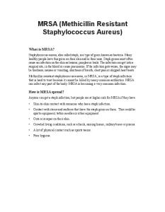
FIG. 4. Representation of formation of the heptameric pore in a eukaryotic cell membrane by staphylococcal alpha-hemolysin. The rim domain of the toxin adheres to the membrane, and the intertwined stem regions are responsible for the formation of a pore, with an exclusionary radius of 14 Å.
(minor) binding site on SEA is analogous to the site described above for SEB, which is not zinc dependent in either case. It is not clear whether this site is physiologically relevant in terms of MHC binding. It may be that cooperation between the two binding sites is responsible for the high affinity of SEA for HLA-DR. It may be that this results in a trimer containing the toxin and two bound MHC class II molecules. The remaining SEs are less well understood, some of them (SEG and SEI) having only recently been isolated. It has been observed that SED has the potential to form a zinc-dependent binding site similar to that in SEA (56). Although the SEC– TCR–pep-MHC complex has been modeled based on homology between SEC and SEB, SEC could have an alternative binding site in the interdomain groove (70). A zinc binding site has been identified in the refined (1.9-Å resolution) structure of SEC3 (25), near the base of this groove, which could be involved in this alternate site. In this toxin, the zinc-coordinating residues are Asp83, His118, and His122. Unlike in SEA, this zinc site is composed of an H-E-X-X-H motif most often found in thermolysin-like metalloproteases. It is not clear what the significance of this structure is in SEC, because it is not in the same context as in the proteases. This motif is usually found in an ␣-helix in the metalloproteases, whereas it occurs in an extended loop in SEC. While other toxins such as Bacillus anthracis lethal factor and botulinum toxin have also been found to be proteases and to contain this zinc binding site, no enzymatic activity has been found associated with SEC. It is also interesting that no such site is predicted by the structure of SEB. STRUCTURAL BIOLOGY OF THE HEMOLYSINS AND LEUKOCIDIN S. aureus produces a number of cytotoxic molecules, which can be divided into four classes which include the four hemolysins (alpha, beta, delta, and gamma) and Panton-Valentine leukocidin, also known as PV-leukocidin. Each class is discussed individually below. Alpha-Hemolysin (Alpha-Toxin) Alpha-hemolysin is by far the most carefully examined of the S. aureus cytotoxins. A high percentage of strains make this toxin, and it is toxic to a wide range of mammalian cells. It is particularly active against rabbit erythrocytes, and it is also dermonecrotic and neurotoxic. Levels of alpha-toxin as low as 1 g are lethal when injected into rabbits intravenously. Extensive reviews of alpha-toxin have been published (18, 57). This paper deals primarily with recent advances.
Downloaded from http://cmr.asm.org/ on June 7, 2018 by guest
observed that mutations in the TCR binding site of this toxin were capable of sharply reducing mitogenicity. In particular, N23A, Q210A, and F176A had a drastic effect on the mitogenic activity of the toxin, and all of the other contact residues were shown to affect mitogenicity as well. It was hypothesized that by changing the residues in the SEB TCR binding site to be the same as those in SEC3, the affinity of SEB for the TCR might be enhanced. Only three contact residues were found to differ, at positions 20, 26, and 91 (Leu, Val, and Tyr in SEB; Thr, Tyr, and Val in SEC3). Each of these residues in SEB was mutated to the corresponding SEC3 residue, and each single mutant was assessed. In only one case, V26Y, was the binding affinity significantly enhanced, with a corresponding increase in mitogenic activity. The triple mutant was found to have severely decreased binding to the TCR and concomitant low mitogenic activity. None of these mutants were able to replicate the binding of SEC3 to the TCR. By using the crystal structure of the -chain complexed with SEC3 (101), attempts were made to design mutants to enhance the binding of this mutant to the TCR. It was expected that mutants Y26W, Y90W, or V91I might enhance van der Waals interactions with the -chain contact residues. Additional mutations were made in an attempt to create salt bridges between the two molecules. None of these attempts created a toxin molecule with enhanced binding affinity to the TCR, and some even had decreases of up to 10-fold in the strength of the interaction. Finally, binding of these two toxins to the MHC-peptide complex was examined by using soluble HLA-DR1 loaded with hemagglutinin peptide HA 306–318. It was found that SEB had a considerably higher affinity for the MHC class II molecule, which may well explain the difference in activity. It was also demonstrated for several of the mutants with mutations in the SEC3 TCR binding site that they had no effect on HLA-DR1 binding, indicating that there were no overall structural effects of the mutagenesis. No other SEs have been crystalized in complex with MHC class II. It is possible, though, to predict some aspects of the interaction between the MHC and SEA or SEE. These predictions are based on mutagenesis studies and on the finding that zinc is required for the interaction. SEA contains two MHC class II binding sites. The zinc-dependent site is the major interaction region, and several important residues (His187, His225, and Asp227) were identified by mutagenesis. It is presumed that this binds the MHC  chain. These findings were confirmed by Schad et al. (142), who solved the crystal structure of SEA and showed that Ser1 is also involved in zinc binding. This major MHC binding site is located in domain B, near the amino terminus. This is on the opposite side of the molecule from the MHC binding site on SEB. The second
VOL. 13, 2000
27
type. The kinetics of assembly were studied as well, and the N-terminal mutant formed heat- and sodium dodecyl sulfate (SDS)-stable oligomers, albeit not as rapidly as the wild-type alpha-toxin did. The mutant toxin was functional but slower to produce erythrocyte lysis, and two- to threefold-larger amounts of mutant alpha-toxin were required to produce 50% lysis compared to the amount of wild type needed. The deletion mutant was able to inhibit lysis by wild-type alpha-toxin, which supports the argument that interactions between the N termini of the monomers are required for pore formation. These competition experiments also showed that homoheptamers of mutant and wild-type alpha-toxin, as well as hybrid oligomers, were capable of forming and presumably of causing lysis, although with differing efficiencies. In contrast, C-terminal deletion mutants thus far examined have been unstable in the monomeric form. It was concluded from these studies that the N terminus of alpha-toxin is important in the opening of the hemolytic pore after oligomerization. The resultant pore allows rapid efflux of K⫹ and other small molecules and influx of Na⫹, Ca2⫹, and small molecules with molecular weights less than 1,000. Osmotic swelling of the erythrocyte finally results in rupture. These pores are not necessarily found on all susceptible cell types, however. Smaller pores appear to form on keratinocytes and lymphocytes and permit only monovalent ions to pass (78, 177). Some cells, fibroblasts in particular, are able to repair membrane damage done by low doses of alpha-toxin. Toxin expression has been a subject of some interest, and outside of agr regulation, effects of environmental factors appear to play a role in alpha-hemolysin expression. Ohlsen et al. (118) explored the transcriptional regulation of hla by using the alpha-toxin promoter fused to a lacZ gene from Escherichia coli. Subsequent analysis of expression revealed that a number of environmental factors impact on alpha-toxin production. Expression was found to be temperature dependent, with maximal expression at 42°C. Additionally, this increased expression was observed to be decoupled from agr RNAIII production, indicating that this altered expression might not be directed by that regulatory system. The expression maximum at 42°C was observed in mid-exponential phase, as opposed to the later expression usually seen in agr-controlled systems. It was found additionally that high osmolarity repressed hla transcription, whereas CO2 stimulated activity. Oxygen was found to be essential for expression, and growth on solid medium was found to be more conducive to expression of the fusion gene. As mentioned above, alpha-hemolysin is dermonecrotic and neurotoxic and can be lethal in a variety of animal systems. These factors contribute to the consensus among researchers that this toxin is important in several disease states caused by S. aureus infection. The significance of alpha-toxin in human disease has not been irrefutably established, however. The toxin has a number of effects on the host, largely due to the formation of unregulated pores for ion transmission across the membranes of a variety of cell types (18). An example is the formation of pores in the membranes of endothelial cells, resulting in arachidonic acid metabolism due to Ca2⫹ influx (157, 167). Formation of thromboxane and prostacyclin results from activation of this metabolic pathway, leading to vasoconstriction. In addition, osmotic swelling causes a breakdown in cell integrity, with the effect of increasing vascular permeability. The final effect on a host is pulmonary edema or adult respiratory distress syndrome. Additionally, alpha-toxin effects on platelets can lead to release of procoagulation factors via Ca2⫹ influx (17). Another mechanism by which alpha-toxin can lead to edema is via endothelial cell contraction caused by leakage of ATP together with calcium ion influx, which results
Downloaded from http://cmr.asm.org/ on June 7, 2018 by guest
The gene that encodes alpha-hemolysin, hla, was cloned from the S. aureus chromosome and sequenced in 1984 by Gray and Kehoe (65). The first 26 residues of the translated polypeptide comprise a signal sequence which is cleaved during secretion. The mature protein contains 293 residues and has a molecular weight of 33,000 and a pI of about 8.5. There are no cysteines in the mature protein, which is composed primarily of -sheets (65%), with ␣-helical structures making up a much smaller percentage (10%) of the secondary structure. This toxin is under control of the accessory gene regulator (agr) and is therefore made during the late exponential phase of growth in a batch culture. It was observed by O’Reilly et al. (119) that some strains contain the hla gene but do not produce detectable alpha-toxin. This occurs particularly often in TSS isolates. In some cases, it was determined that this was due to mutations in the structural gene which prevented the protein from being translated. A number of methods to purity alpha-toxin have been described. The most commonly used medium has been the yeast extract dialysate of Bernheimer and Schwartz (15), and Wood 46 is the strain most commonly used for toxin production. Very straightforward methods for obtaining highly purified alpha-toxin have recently been described by Six and Harshman (160) and Harshman et al. (68). The first of these involved preparative gel electrophoresis, while the second was a selective adsorption and elution from glass-pore beads followed by gel filtration. This latter procedure is quick and simple and yields large amounts of pure toxin (10 to 20 mg/liter). Alpha-hemolysin monomers are secreted by S. aureus and integrate into the membrane (Fig. 4) of a target cell, where they form cylindrical heptamers (63, 174). It is this oligomeric form which is capable of lysing eukaryotic cells. The defining characteristic of alpha-toxin is its ability to lyse erythrocytes. In particular, rabbit erythrocytes are extraordinarily susceptible to hemolysis by alpha-toxin, at least 100 times more so than other mammals and 1,000 times more than human erythrocytes (15, 16). Once the cylindrical heptamer has formed in the cell membrane, a 1- to 2-nm pore is formed (9, 109). This pore forms via a stepwise process (174). The toxin monomer initially binds and incorporates into the target cell membrane. It has been observed that this binding can occur in two different ways (69). When alpha-toxin is present at low concentrations, a specific receptor binds the protein. The identity of this cell surface receptor is not known. However, at higher concentrations, alpha-hemolysin nonspecifically adheres to the cell membrane (69). The monomer, whose structure has been deduced from crystallographic studies on the detergent-solubilized heptamer (62), is capable of penetrating the lipid bilayer. Diffusion of the monomers through the lipid bilayer results in subsequent formation of the heptameric pore (174). Although it is not thought that major structural changes occur in the toxin after oligomerization, some studies of N-terminal deletion mutants suggest that some conformational change is necessary for pore activation (62, 175). Previous studies had shown that the N terminus was important in pore formation, but the way in which it played its role and whether its importance was in oligomer formation of pore function was unclear. Vandala et al. (174) constructed a mutant with a deletion of the first 4 amino acids and analyzed the kinetics of oligomerization and the function of the intact mutant heptamer. These studies were contrasted with results for previously constructed 2- and 11amino-acid deletion mutants, which failed to proceed from a nonlytic to a lytic heptameric pore. Transmission electron microscopic studies were also done to examine the physical structure of the pore, which appeared the same as that of the wild
S. AUREUS EXOTOXINS
28
DINGES ET AL.
in leaky gaps between cells. Finally, the calcium flux may trigger cellular nucleases, leading to apoptotic cell death (78). The neurotoxicity of alpha-toxin has been observed in rabbits and mice, in the destruction of myelin sheaths surrounding rabbit nerves and in the murine cerebral cortex (18). An effect on macrophages has also been observed, resulting in increased secretion of interleukin-1. These widespread effects, and possibly many others, are due primarily to the alteration of the ion balance within the host. The breadth of the effect is consistent with the ability of the toxin to interact with specific receptors on the cell surface as well as nonspecifically with the membrane. Beta-Hemolysin (Sphingomyelinase C)
hemolysis was put forward by Low and Freer (103) in 1977. At 20°C, cohesive forces within the membrane are capable of keeping the hemolytic products together, but upon cooling, phase separation occurs, causing bilayer collapse. The role of beta-hemolysin in disease is not clearly understood. The high level of expression in animal strains indicates that -hemolysin producers garner some selective advantage from toxin secretion. The only evidence to support this, however, is a recent study which shows that toxin production enhances S. aureus growth in the murine mammary glands compared to an isogenic hlb knockout organism. In diagnostic microbiology, a presumptive test for group B streptococci involves beta-hemolysin. This test, the CAMP test, is performed by either dropping pure beta-hemolysin or culture fluid contain toxin on a lawn of presumptive group B streptococci and looking for enhanced hemolytic activity on blood agar after incubation at 35°C for 30 min to a few hours. Alternatively, the presumptive group B streptococci can be streaked perpendicular to, but not touching, a beta-hemolysin producing S. aureus strain on a blood agar plate. A zone of enhanced beta-hemolytic activity can be seen between the two organisms. Gamma-Hemolysin and PV-Leukocidin (Two-Component Toxins) Two types of bicomponent toxins are made by S. aureus, gamma-hemolysin and Panton-Valenine (PV) leukocidin. Each of these toxins is made as two nonassociated secreted proteins, referred to as S and F components (for slow- and fast-eluting proteins in an ion-exchange column) (179, 180). Gamma-hemolysin is made by virtually every strain of S. aureus, while PV leukocidin is made by 2 to 3% of strains. The toxins affect neutrophils and macrophages, and gamma-hemolysin is additionally able to lyse many varieties of mammalian erythrocytes. Gamma-hemolysin is not identifiable on blood agar plates, however, due to the inhibitory effect of agar on toxin activity (132). The physicochemical and biological properties of these two toxins and their relationship to one another have been difficult to study, and the literature is quite confusing. Recently, however, some clarifications have been provided by Prevost et al. (133). Studies by Prevost et al. (133) and others have shown that the components of these proteins arise from two distinct loci within the S. aureus genome, and the two toxins result. In strains containing both cytotoxins, three S components (HlgA, HlgC, and LukS-PV) and two F components (HlgB and LukFPV) are available. A high level of sequence identity is present among these gene products. The genes for gamma-hemolysin are transcribed from a single locus, located on a 4.5-kb ScaI-digested chromosomal fragment. Extracts from a clone containing this fragment were hemolytic and leukotoxic. The open reading frames, in order, are named hlgA, hlgC, and hlgB, and they correspond to previously identified gamma-hemolysin genes (40, 42, 79). hlgC and hlgB are transcribed on a single mRNA, while hlgA is separately expressed. The three encoded proteins are all translated with a single sequence, which is cleaved to form the mature, secreted subunit protein. The mature products have molecular weights of 32,000 for HlgA, 32,500 for HlgC, and 34,000 for HlgB; their pIs are estimated at 9.4, 9.0, and 9.1, respectively. HlgA has previously been termed the ␥1 component of gamma-hemolysin, and HlgC has been termed the ␥2 component. HlgB and HlgC together constitute what was previously known as gam-
Downloaded from http://cmr.asm.org/ on June 7, 2018 by guest
S. aureus beta-hemolysin, first identified in 1935 by Glenny and Stevens (60), was also shown by them to be highly hemolytic for sheep but not rabbit erythrocytes. It is not dermonecrotic in guinea pigs, and it is not lethal in mice. Glenny and Stevens also noted that the hemolytic activity of this toxin was enhanced by incubation below 10°C after treatment at 37°C, leading to the appellation “hot-cold” hemolysin. Beta-hemolysin is secreted into the culture medium as an exotoxin with a molecular weight of 35,000. The gene was cloned by Projan et al. (134) and Coleman et al. (39), and the nucleotide sequence was published by Projan et al. (134). The gene, named hlb, is chromosomally located on a 4-kb ClaI DNA fragment and encodes a 330-amino-acid polypeptide with a predicted molecular weight of 39,000. A 200-residue region of homology has been found to Bacillus cereus sphingomyelinase (55.7% similarity [181]). Unpublished studies in our laboratory have allowed us to unambiguously establish the N terminus of the mature protein. The N-terminal sequence of secreted beta-hemolysin is Glu Ser Lys Lys Asp Asp Thr Asp Leu Lys, corresponding precisely to residues 35 to 44 of the sequence published by Projan et al. (134). The first 34 residues of the polypeptide comprise a signal sequence, cleaved upon secretion, resulting in a mature protein with molecular weight of approximately 35,000, as previously determined by SDS-polyacrylamide gel electrophoresis (PAGE). This protein is extremely basic, with an isoelectric point above pH 9. Beta-hemolysin is produced in large quantity by a number of S. aureus strains, particularly animal isolates. Thorough reviews of the conditions required for toxin production are provided elsewhere (14, 57). Large amounts of toxin have been produced by using yeast dialysate medium, as with alpha-hemolysin, or with beef heart dialysate medium, as we used in our laboratory. Like most other exoproteins in S. aureus, betahemolysin is regulated after the exponential phase. Similarly to the growth conditions, preparative methods for purification of beta-hemolysin have been extensively reviewed; they include gel filtration, precipitation, ion-exchange chromatography, gel electrophoresis, and isoelectric focusing. Others have reported yields of 1 to 2 mg of purified toxin per liter, but our laboratory has been able to obtain 15 to 20 mg per liter of culture by precipitation with ethanol followed by successive isoelectric focusing in pH gradients of 3.5 to 10 and 7 to 9. The homogeneity of the purified protein was shown by SDS-PAGE and N-terminal sequencing. Beta-hemolysin was found in 1963 to have phosphorylase c activity (48). This activity requires the presence of Mg2⫹ and is limited with specificity for sphingomyelin and lyso-phosphatidylcholine. The differing susceptibility of erythrocytes to betahemolysin may be due to the differing sphingomyelin contents of erythrocytes (49). A mechanism to explain the hot-cold
CLIN. MICROBIOL. REV.
VOL. 13, 2000
29
ing that a severe inflammatory response could be elicited by these bicomponent toxins was governed primarily by the potency of the S subunit. A working hypothesis is that the S component is responsible for initial membrane binding, making it more critical of the two subunits in determining activity (159). Despite this research into the details of the functions of the bicomponent toxins, their role in the progression of staphylococcal disease remains unclear. Although Prevost and others have speculated on the mechanism of toxin action, no clear molecular activity has been elucidated. It has also been reported that Staphylococcus intermedius strains make a related bicomponent toxin, which has yet to be studied extensively. Delta-Hemolysin (Delta-Lysin, Delta-Toxin) Delta-hemolysin, a 26-amino-acid peptide, is capable of causing membrane damage in a variety of mammalian cells. Toxin from human isolates of S. aureus contains nine amino acid differences from the canine strain-derived delta-hemolysin, although both are 26 residues long and have molecular weights of approximately 3,000. These different hemolysins have conserved charge properties, and both are hemolytically active, but only partial serological identity is observed. This protein is made by 97% of S. aureus strains, and an immunologically related protein is made by Staphylococcus haemolyticus strains. A 514-nucleotide transcript is encoded by the hld gene, which eventually produces delta-hemolysin (127). The first 160 nucleotides of this transcript contain a 45-codon open reading frame, of which the delta-hemolysin structural gene makes up the 3⬘ end (6, 127). It has been observed in detail that the large transcript plays an important regulatory role in the accessory gene regulator (agr) system, which controls the production of cell-associated and secreted virulence factors. Delta-hemolysin is itself controlled by agr, and the highest expression in broth culture is found in the late-logarithmic (postexponential) phase. In this respect, delta-hemolysin is like many other staphylococcal exoproteins. This toxin, however, is secreted without a signal peptide, and in fact it has been suggested that delta-hemolysin itself would make an efficient signal peptide. Today, delta-hemolysin is made by two major techniques: (i) peptide synthesis and (ii) purification from S. aureus cultures deficient in alpha-hemolysin, such as NCTC 10345 and 186X (19, 45). Excellent yields of toxin from these strains, ⬃300 g/liter have been obtained by using the yeast extract dialysate medium of Bernheimer and Schwartz (15). Toxin has been successfully purified from hydroxyapatite columns and found to be highly pure. In an SDS-PAGE gel (15% polyacrylamide) the delta-hemolysin band migrates at a molecular weight of about 6,000, approximately twice the predicted value. This is probably due to the tendency of delta-toxin to aggregate, and the band in the gel is probably a dimer; alternatively, the alteration may be due to charge or shape properties of the peptide monomer itself. Automated or manual peptide synthesis techniques can yield gram quantities. Delta-hemolysin which lacked an amino formyl-methionine without loss of the toxin characteristics was synthesized by Dhople and Nagaraj (45). As mentioned above, delta-hemolysin is produced by 97% of S. aureus strains. It is also hypothesized that 50 to 70% of coagulase-negative staphylococci produce this toxin. Although delta-toxin causes a wide range of cytotoxic effects, its importance in disease etiology remains unclear. Delta-hemolysin is capable of lysing erythrocytes and other mammalian cells, as well as subcellular structures such as membrane-bound or-
Downloaded from http://cmr.asm.org/ on June 7, 2018 by guest
ma-hemolysin. The hlg locus is nearly identical to the lukR locus, encoding leukocidin R (166). A few nucleotide differences are present between the two regions, but only one protein, HlgA, contained any amino acid differences (Arg258Lys and His248Pro). The earliest description of gamma-hemolysin was in 1938 by Smith and Price (161). A variety of subsequent reviews concerning its production have been published, the most significant by Plommet (128). S. aureus 5R does not produce any alpha-, beta-, or delta-toxin and is therefore a good source of gamma-hemolysin. The review by Plommet makes three suggestions for production of this hemolysin: (i) use of the specific S. aureus strain, (ii) growth in yeast extract dialysate medium, and (iii) aeration with an 80:20 mix of oxygen and carbon dioxide. Gamma-hemolysin may be purified by batch adsorption onto hydroxyapatite and elution with potassium phosphate (128). The S and F components may be separated by eluting with two different potassium phosphate buffers. The genes encoding PV-leukocidin were cloned and sequenced by Prevost et al. (133) and named lukS-PV and lukFPV. A 6.5-kb EcoRV fragment of the S. aureus chromosome contains the entire luk-PV locus, consisting of two separate reading frames. Protein extracted from the clone were leukotoxic but exhibited no hemolytic activity. The lukS-PV gene encodes a 312-amino-acid polypeptide, including a 28-residue signal sequence which is cleaved to produce the mature protein with a molecular weight of 32,000. The LukS-PV protein has an isoelectric point of 9.0. The LukF-PV protein is a 325amino-acid protein which similarly contains a 24-residue signal peptide. The mature protein has a pI of 9 and a molecular weight of 34,000. Noda and Kato (116) illustrated the techniques commonly used in PV-leukocidin production. The V8 strain of S. aureus is useful for this purpose, and it is cultured in yeast extractCasamino Acids medium. Purification of PV-leukocidin is complex, involving successive precipitation with ZnCl2 and ammonium sulfate followed by chromatographic and electrophoretic techniques. Both components of PV-leukocidin have been crystallized. There are six possible forms of gamma-hemolysin/PV-leukocidin, due to the presence of three potential S and two potential F units, all of which may combine (S⫹F) to form a toxin molecule. Konig et al. (86) found that these subunits lacked hemolytic and leukotoxic activity when assayed alone. When added as pairs to erythrocytes, however, different activity levels were noted. HlgA–LukF-PV, HlgA-HlgB, and HlgCHlgB had high hemolytic activity, with HlgA-HlgB having the highest among these. The remaining combinations had much lower hemolytic activity. All six possible combinations, however, were able to efficiently lyse leukocytes (86). Inflammatory responses due to the bicomponent toxins have also been observed, including the ability of PV-leukocidin to induce granule secretion in polymorphonuclear leukocytes and the release of inflammatory mediators (159). It is not clear whether gamma-hemolysin is likewise capable of inducing these responses. Through detailed study of the stimulation of human granulocytes in vitro by all possible bicomponent toxin combinations, Siqueira et al. (159) were able to assess the immunomodulatory activities of these toxins. They found that gamma-hemolysin was less able to induce a response from polymorphonuclear leukocytes than was PV-leukocidin but that the S subunit was the important one in this respect, rather than the F subunit. Further experiments were done with regard to the effects of these toxins in a rabbit corneal model (159). The in vitro data were supported by this rabbit model, indicat-
S. AUREUS EXOTOXINS
30
DINGES ET AL.
FUTURE STUDIES Disease causation by S. aureus is a very complex process and probably involves large numbers of factors, both cell associated and secreted. In this review, we have examined many of the important secreted virulence factors of S. aureus. It is clear from these studies that the major effects of the toxins are to inactivate cells of the host immune system, whether through superantigen-mediated release of cytokines or direct cytotoxicity, as is the case with the action of hemolysins. However, much remains to be learned about how the toxins function. For example, in a number of studies, it appears that the PTSAg properties of superantigenicity, lethality, and emesis (for SEs) are independent. The question then arises of which PTSAg effects are required for TSS induction. Furthermore, PTSAgs are typically secreted in the postexponential phase of growth, being made in the greatest amounts when the bacterial cells are present in the highest concentration. It is reasonable to speculate, then, that the PTSAgs are important in the early events that lead to establishment of an infection site in a new host, which would most successfully occur after transmission of bacteria that were present at high cell densities. Finally, new PTSAgs arise or are recognized every few years, creating the possibility for newly emergent diseases. In the past, S. aureus strains have cycled in roughly 10-year periods, as exemplified by the 80/81 S. aureus strains isolated from hospital populations in the 1950s and by the 52/52A strains isolated in the 1960s. Clearly, TSST-1-positive strains emerged in the 1970s as significant pathogenic strains of S. aureus. Several questions arise. (i) Are new PTSAg-mediated diseases arising at present? (ii) What are the genetic events that lead to production of new PTSAgs? (iii) With the presence of more antibiotic-resistant strains, are there novel ways that we can target S. aureus virulence factors, thereby preventing their production and thus ameliorating disease? ACKNOWLEDGMENTS This work was supported by USPHS research grant AI22159 from the National Institute of Allergy and Infectious Diseases. Martin
Dinges is supported by USPHS training grant AI 07421. Paul Orwin is supported by an NSF fellowship. Gregory Vath and Douglas Ohlendorf are gratefully acknowledged for providing structures of PTSAgs alone and in complexes. Melodie Bahan is acknowledged for help in preparing the manuscript. REFERENCES 1. Acharya, K. R., E. F. Passalacqua, E. Y. Jones, K. Harlos, D. I. Stuart, R. D. Brehm, and H. S. Tranter. 1994. Structural basis of superantigen action inferred from crystal structure of toxic-shock syndrome toxin-1. Nature 367:94–97. 2. Alber, G., D. Hammer, and B. Fleischer. 1990. Relationship between enterotoxic- and T lymphocyte-stimulating activity of staphylococcal enterotoxin B. J. Immunol. 144:4501–4506. 3. Alber, G., P. H. Scheuber, B. Reck, B. Sailer-Kramer, A. Hartmann, and D. K. Hammer. 1989. Role of substance P in immediate-type skin reactions induced by staphylococcal enterotoxin B in unsensitized monkeys. J. Allergy Clin. Immunol. 84:880–885. 4. Altemeier, W. A., S. A. Lewis, P. M. Schlievert, M. S. Bergdoll, H. J. Bjornson, J. L. Staneck, and B. A. Crass. 1982. Staphylococcus aureus associated with toxic shock syndrome: phage typing and toxin capability tested. Ann. Intern. Med. 96:978–982. 5. Araake, M., T. Uchiyama, K. Imanishi, and X. J. Yan. 1991. Activation of human vascular endothelial cells by IFN-gamma: acquisition of HLA class II expression, TSST-1-binding activity and accessory activity in T cell activation by the toxin. Int. Arch. Allergy Appl. Immunol. 96:55–61. 6. Arvidson, S., L. Janzon, and S. Lofdahl. 1990. The role of the ␦-hemolysin gene (hld) in the agr-dependent regulation of exoprotein synthesis in Staphylococcus aureus, p. 419–431. In R. P. Novick (ed.), Molecular biology of the staphylococci. VCH Publishers Inc., New York, N.Y. 7. Beery, J. T., S. Taylor, L. R. Schlunz, R. C. Freed, and M. S. Bergdoll. 1994. Effects of staphylococcal enterotoxin A on the rat gastrointestinal tract. Infect. Immun. 44:234–240. 8. Beisel, W. R. 1972. Pathophysiology of staphylococcal enterotoxin, type B, (SEB) toxemia after intravenous administration to monkeys. Toxicon 10: 433–440. 9. Belmonte, G., L. Cescatti, B. Ferrari, T. Nicolussi, M. Ropele, and G. Menestrina. 1987. Pore-formation by Staphylococcus aureus alpha-toxin in lipid bilayers: temperature and toxin concentration dependence. Eur. J. Biophys. 14:349–358. 10. Bergdoll, M. S. 1983. Enterotoxins, p. 559–598. In C. S. F. Easmon and C. Adlam (ed.), Staphylococci and staphylococcal infections. Academic Press, Ltd., London, United Kingdom. 11. Bergdoll, M. S., B. A. Crass, R. F. Reiser, R. N. Robbins, and J. P. Davis. 1981. A new staphylococcal enterotoxin, enterotoxin F, associated with toxic shock syndrome Staphylococcus aureus isolates. Lancet i:1017–1021. 12. Bergdoll, M. S., R. N. Robbins, K. Weiss, C. R. Borja, I. Y. Huang, and F. S. Chu. 1973. The staphylococcal enterotoxins: similarities. Contrib. Microbiol. Immunol. 1:390–396. 13. Bergdoll, M. S., and P. M. Schlievert. 1984. Toxic-shock syndrome toxin. Lancet ii:691. 14. Bernheimer, A. W., L. S. Avigad, and K. S. Kim. 1974. Staphylococcal sphingomyelinase (beta hemolysin). Ann. N. Y. Acad. Sci. 236:292–306. 15. Bernheimer, A. W., and L. L. Schwartz. 1963. Isolation and composition of staphylococcal alpha toxin. J. Gen. Microbiol. 30:455. 16. Bhakdi, S., M. Muhly, and R. Fussle. 1984. Correlation between toxin binding and hemolytic activity in membrane damage by staphylococal ␣-toxin. Infect. Immun. 46:318–323. 17. Bhakdi, S., M. Muhly, U. Mannhardt, F. Hugo, K. Klapettek, C. MullerEckhardt, and L. Roka. 1988. Staphylococcal ␣-toxin promotes blood coagulation via attack on human platelets. J. Exp. Med. 168:527–542. 18. Bhakdi, S., and J. Tranum-Jensen. 1991. Alpha-toxin of Staphylococcus aureus. Microbiol. Rev. 55:733–751. 19. Birbeck, T. H., and J. H. Freer. 1988. Purification and assay of staphylococcal ␦-lysin. Methods Enzymol. 165:16–22. 20. Blanco, L., E. M. Choi, K. Connolly, M. R. Thompson, and P. F. Bonventre. 1990. Mutants of staphylococcal toxic shock syndrome toxin 1: mitogenicity and recognition by a neutralizing monoclonal antibody. Infect. Immun. 58:3020–3028. 21. Blomster-Hautamaa, D. A., B. N. Kreiswirth, J. S. Kornblum, R. P. Novick, and P. M. Schlievert. 1986. The nucleotide and partial amino acid sequence of toxic-shock syndrome toxin-1. J. Biol. Chem. 261:15783–15786. 22. Blomster-Hautamaa, D. A., B. N. Kreiswirth, R. P. Novick, and P. M. Schlievert. 1986. The resolution of highly purified toxic-shock syndrome toxin-1 (TSST-1) into two distinct proteins by isoelectric focusing. Biochemistry 25:54–59. 23. Blomster-Hautamaa, D. A., and P. M. Schlievert. 1988. Purification of toxic-shock syndrome toxin-1. Methods Enzymol. 165:37–43. 24. Bohach, G. A., D. J. Fast, R. D. Nelson, and P. M. Schlievert. 1990. Staphylococcal and streptococcal pyrogenic toxins involved in toxic shock syndrome and related illnesses. Crit. Rev. Microbiol. 17:251–272. 25. Bohach, G. A., L. M. Jablonski, C. F. Deobald, Y. I. Chi, and C. V. Stauf-
Downloaded from http://cmr.asm.org/ on June 7, 2018 by guest
ganelles, spheroplasts, and protoplasts (58). Also, a dermonecrotic activity has been observed, as well as lethality in experimental animals when used in high concentrations. It remains possible, however, that these activities result from contamination with very small amounts of alpha-toxin. The presence of phospholipids inhibits delta-hemolysin activity. The mechanism of delta-hemolysin action has been the subject of great interest. An ␣-helix is formed by delta-toxin (58, 93) with hydrophobic and hydrophilic domains on opposite sides. Based on this information, it is proposed that deltahemolysin acts as a surfactant to disrupt the cell membrane. The speed with which the target cell is lysed supports this hypothesis. Low-molecular-weight particles are released first, and prolonged exposure subsequently results in release of large molecules. Structural examination by Thelastam and Mollby (169) showed that delta-toxin is similar in both structure and proposed mechanism to the bee venom mellitin. Other members of this family have been found, including some with antimicrobial activity. This activity has not been observed in delta-hemolysin, however. It has been proposed that this family of molecules may be able to aggregate to form channels on a lipid bilayer (89). Mellor et al. (109) showed that deltahemolysin can be made to form cation-selective channels in the membrane. The authors proposed that a channel is formed by six aggregated ␣-helical toxin molecules in a hexagonal arrangements.
CLIN. MICROBIOL. REV.
S. AUREUS EXOTOXINS
VOL. 13, 2000
26.
27.
28.
29.
31. 32. 33. 34. 35. 36.
37. 38. 39.
40.
41.
42. 43. 44.
45. 46. 47.
48.
49. 50. 51. 52.
53.
54.
55.
56.
57. 58. 59. 60. 61.
62. 63.
64. 65. 66. 67.
68. 69. 70.
71. 72.
73.
A phospholipase in staphylococcal toxin which hydrolizes sphingomyelin. Nature (London) 198:1091–1093. Doery, H. M., B. J. Magnuson, J. Galasekharam, and J. E. Pearson. 1965. The properties of phospholipase enzymes in staphylococcal toxins. J. Gen. Microbiol. 40:283–296. Driessen, C., K. Hirv, H. Kircherner, and L. Rink. 1995. Zinc regulates cytokine induction by superantigens and lipopolysaccharide. Immunology 84:272–277. Earhart, C. A., G. S. Prasad, D. L. Murray, R. P. Novick, P. M. Schlievert, and D. H. Ohlendorf. 1993. Growth and analysis of crystal forms of toxic shock syndrome toxin-1. Proteins 17:329–334. Earhart, C. A., D. T. Mitchell, D. L. Murray, D. M. Pinheiro, M. Matsumara, P. M. Schlievert, and D. H. Ohlendorf. 1998. Structures of five mutants of toxic shock syndrome toxin-1 with reduced biological activity. Biochemistry 37:7194–7202. Edwin, C., and E. H. Kass. 1989. Identification of functional antigenic segments of toxic shock syndrome toxin 1 by differential immunoreactivity and by differential mitogenic responses of human peripheral blood mononuclear cells using active toxin fragments. Infect. Immun. 57:2230–2236. Fields, B. A., E. L. Malchiodi, L. X. Y. Hongmin, C. V. Stauffacher, P. M. Schlievert, K. Karjalainen, and R. A. Mariuzza. 1996. Crystal structure of a T-cell receptor B-chain complexed with a superantigen. Nature 384:188– 192. Fleischer, B., and H. Schrezenmeier. 1988. T cell stimulation by staphylococcal enterotoxins. Clonally variable response and requirement for major histocompatibility complex class II molecules on accessory or target cells. J. Exp. Med. 167:1697–1707. Fraser, J. D., S. Lowe, M. J. Irwin, N. R. J. Gascoigne, and K. R. Hudson. 1993. Structural model of staphylococcal enterotoxin A interaction with MHC class II antigens, p. 7–29. In B. T. Huber and E. Palmer (ed.), Superantigens: a pathogen’s view of the immune system. Cold Spring Harbor Laboratory Press, Plainview, N.Y. Freer, J. H., and J. P. Arbuthnott. 1983. Toxins of Staphylococcus aureus. Pharmacol. Ther. 19:55–106. Freer, J. H., and T. H. Birbeck. 1982. Possible conformation of delta-lysin, a membrane-damaging peptide of Staphylococcus aureus. J. Theor. Biol. 94:535–540. Fujikawa, H., H. Igarashi, H. Usami, S. Tanaka, and H. Tamura. 1986. Clearance of endotoxin from blood of rabbits injected with staphylococcal toxic shock syndrome toxin-1. Infect. Immun. 52:134–137. Glenny, A. T., and N. F. Stevens. 1935. Staphylococcal toxins and antitoxins. J. Pathol. Bacteriol. 40:201–210. Gottfried, A., P. H. Scheuber, B. Reck, B. Sailer-Kramer, A. Hartmann, and D. K. Hammer. 1989. Role of substance P in immediate-type skin reactions induced by staphylococcal enterotoxin B in unsensitized monkeys. J. Allergy Clin. Immunol. 84:880–885. Gouaux, E. 1998. ␣-Hemolysin from Staphylococcus aureus: an archetype of -barrel, channel forming toxins. J. Struct. Biol. 121:110–122. Gouaux, J. E., O. Braha, M. R. Hobaugh, L. Song, S. Cheley, C. Shustak, and H. Bayley. 1994. Subunit stoichiometry of staphylococcal alpha-hemolysin in crystals and on membranes: a heptameric transmembrane pore. Proc. Natl. Acad. Sci. USA 91:12828–12831. Gourley, G. R., P. J. Chesney, J. P. Davis, and G. B. Odell. 1981. Acute cholestasis in patients with toxic shock syndrome. Gastroenterology 81:928– 931. Gray, G. S., and M. Kehoe. 1984. Primary sequence of the ␣-toxin gene from Staphylococcus aureus Wood 46. Infect. Immun. 46:615–618. Hamad, A. R., P. Marrack, and J. W. Kappler. 1997. Transcytosis of staphylococcal superantigen toxins. J. Exp. Med. 185:1447–1454. Harris, T. O., D. Grossman, J. W. Kappler, P. Marrack, R. R. Rich, and M. J. Betley. 1993. Lack of complete correlation between emetic and T-cellstimulatory activities of staphylococcal enterotoxins. Infect. Immun. 61: 3175–3183. Harshman, S., N. Sugg, and P. Cassiday. 1988. Preparation and purification of staphylococcal alpha toxin. Methods Enzymol. 165:3–7. Hildebrand, A., M. Roth, and S. Bhakdi. 1991. Staphylococcus aureus alphatoxin: dual mechanisms of binding to target cells. J. Biol. Chem. 266:17195– 17200. Hoffmann, M. L., L. M. Jablonski, K. K. Crum, S. P. Hackett, Y. I. Chi, C. V. Stauffacher, D. L. Stevens, and G. A. Bohach. 1994. Predictions of T-cell receptor and major histocompatibility complex binding sites on staphylococcal enterotoxin C1. Infect. Immun. 62:3396–3407. Holmberg, S. D., and P. A. Blake. 1984. Staphylococcal food poisoning in the United States. JAMA 251:487–489. Hovde, C. J., J. C. Marr, M. L. Hoffmann, S. P. Hackett, Y. I. Chi, K. K. Crum, D. L. Stevens, C. V. Stauffacher, and G. A. Bohach. 1994. Investigation of the role of the disulfide bond in activity and structure of staphylococcal enterotoxin C1. Mol. Microbiol. 13:897–909. Hurley, J. M., R. Shimonkevitz, A. Hanagan, K. Enney, E. Boen, S. Mahmstrom, B. L. Kotzin, and M. Matsumura. 1995. Identification of class II major histocompatibility complex and T cell receptor binding sites in the superantigen toxic shock syndrome toxin 1. J. Exp. Med. 181:2229–2235.
Downloaded from http://cmr.asm.org/ on June 7, 2018 by guest
30.
facher. 1995. Functional domains of staphylococcal enterotoxins, p. 339– 356. In M. Ecklund, J. L. Richard, and K. Mise (ed.), Molecular approaches to food safety: issues involving toxic microorganisms. Alaken Inc., Fort Collins, Colo. Bohach, G. A., M. M. Dinges, D. T. Mitchell, D. H. Ohlendorf, and P. M. Schlievert. 1997. Exotoxins, p. 83–111. In K. B. Crossley and G. L. Archer (ed.), The staphylococci in human disease. Churchill Livingstone, Inc., New York, N.Y. Bohach, G. A., L. Jablonski, M. Roggiani, I. Sadler, P. M. Schlievert, D. Mitchell, and D. Ohlendorf. 1998. Biological activity of pyrogenic toxins delivered at the mucosal surface, p. 170–172. In J. Arbuthnott and B. Furman (ed.), European Conference on Toxic Shock Syndrome. International Congress and Symposium Series 229. Royal Society of Medicine Press Ltd., New York, N.Y. Bonventre, P. F., H. Heeg, C. Cullen, and C. Lian. 1993. Toxicity of recombinant toxic shock syndrome toxin 1 and mutant toxins produced by Staphylococcus aureus in a rabbit infection model of toxic shock syndrome. Infect. Immun. 61:793–799. Bonventre, P. F., H. Heeg, C. D. Edwards III, and C. M. Cullen. 1995. A mutation at histidine residue 135 of toxic shock syndrome toxin yields an immunogenic protein with minimal toxicity. Infect. Immun. 63:509–515. Campbell, W. N., M. Fitzpatrick, X. Ding, M. Jett, P. Liemski, and S. E. Goldblum. 1997. SEB is cytotoxic and alters EC barrier function through protein tyrosine phosphorylation in vitro. Am. J. Physiol. 273:L31–L39. Canonico, P. G., and M. J. Van Zweiten. 1971. Swelling of mitochondria from rabbit liver induced by staphylococcal enterotoxin B. J. Infect. Dis. 124:372–378. Centers for Disease Control and Prevention. 1997. Case definitions for infectious conditions under public health surveillance. Morbid. Mortal. Weekly Rep. 46(RR-10):1–55. Chesney, P. J. 1989. Clinical aspects and spectrum of illness of toxic shock syndrome: overview. Rev. Infect. Dis. 11:S1–S7. Chesney, P. J. 1997. Toxic shock syndrome, p. 509–525. In K. B. Crossley and G. L. Archer (ed.), The staphylococci in human disease. Churchill Livingstone, Inc., New York, N.Y. Chesney, R. W., P. J. Chesney, J. P. Davis, and W. E. Segar. 1981. Renal manifestations of the staphylococcal toxic-shock syndrome. Am. J. Med. 71:583–588. Chesney, R. W., D. M. McCarron, J. G. Haddad, C. D. Hawker, F. P. DiBella, P. J. Chesney, and J. P. Davis. 1983. Pathogenic mechanisms of the hypocalcemia of the staphylococcal toxic-shock syndrome. J. Lab. Clin. Med. 101:576–585. Cheung, A. L., J. M. Koomey, C. A. Butler, S. J. Projan, and V. A. Fischetti. 1992. Regulation of exoprotein expression in Staphylococcus aureus by a locus (sar) distinct from agr. Proc. Natl. Acad. Sci. USA 89:6462–6466. Choi, Y., J. A. Lafferty, J. R. Clements, J. K. Todd, E. W. Gelfand, J. Kappler, P. Marrack, and B. L. Kotzin. 1990. Selective expansion of T cells expressing V2 in toxic shock syndrome. J. Exp. Med. 172:981–984. Coleman, D. C., J. P. Arbuthnott, H. M. Pomeroy, and H. Birbeck. 1986. Cloning and expression in Escherichia coli and Staphylococcus aureus of the beta-lysin determinant from Staphylococcus aureus: evidence that bacteriophage conversion of beta-lysin activity is caused by insertional inactivation of the beta-lysin determinant. Microb. Pathog. 1:549–564. Colin, D. A., I. Mazurier, S. Sire, and V. Finck-Barbancon. 1994. Interaction of the two components of leukocidin from Staphylococcus aureus with human polymorphonuclear leukocyte membranes: sequential binding and subsequent activation. Infect. Immun. 62:3184–3188. Cone, L. A., D. R. Woodard, R. G. Byrd, K. Schulz, S. M. Kopp, and P. M. Schlievert. 1991. A recalcitrant, erythematous, desquamating disorder associated with toxin-producing staphylococci in patients with AIDS. J. Infect. Dis. 165:638–643. Couch, J. L., M. T. Soltis, and M. J. Betley. 1988. Cloning and nucleotide sequence of the type E staphylococcal enterotoxin gene. J. Bacteriol. 170: 2954–2960. Davis, J. P., P. J. Chesney, P. J. Wan, M. LaVenture, and Investigation and Laboratory Team. 1980. Toxic-shock syndrome: epidemiologic features, recurrence, risk factors, and prevention. N. Engl. J. Med. 303:1429–1435. Deresiewicz, R. L. 1996. Staphylococcal toxic shock syndrome, p. 435–479. In D. Y. M. Leung, B. T. Huber, and P. M. Schlievert (ed.), Superantigens: molecular biology, immunology and relevance to human disease. Marcel Dekker, Inc., New York, N.Y. Dhople, V. M., and R. Nagaraj. 1993. ␦-toxin, unlike melittin, has only hemolytic activity and no antimicrobial activity: rationalization of this specific biological activity. Biosci. Rep. 13:245–250. Reference deleted. Dinges, M., J. Jessurun, and P. Schlievert. 1998. Comparisons of mouse and rabbit models of toxic shock syndrome, p. 167–168. In J. Arbuthnott and B. Furman (ed.), European Conference on Toxic Shock Syndrome. International Congress and Symposium Series 229. Royal Society of Medicine Press Ltd., New York, N.Y. Doery, H. M., B. J. Magnuson, I. M. Cheyne, and J. Galasekharam. 1963.
31
32
DINGES ET AL.
100. 101.
102. 103. 104.
105. 106. 107.
108. 109. 110. 111. 112.
113.
114.
115.
116. 117.
118. 119.
120. 121. 122.
123.
by streptococcal pyrogenic exotoxin A and endotoxin. Infect. Immun. 60: 3747–3755. Leung, D. Y. M., H. C. Meissner, D. R. Fulton, D. L. Murray, B. L. Kotzin, and P. M. Schlievert. 1993. Toxic shock syndrome toxin-secreting Staphylococcus aureus in Kawasaki syndrome. Lancet 342:1385–1388. Li, H., A. Llera, D. Tsuchiya, L. Leder, X. Ysern, P. M. Schlievert, K. Karjalainen, and R. A. Mariuzza. 1998. Three-dimensional structure of the complex between a T cell receptor beta chain and the superantigen staphylococcal enterotoxin B. Immunity 9:807–816. Lindsay, J. A., A. Ruzin, H. F. Ross, N. Kurepina, and R. P. Novick. 1998. The gene for toxic shock toxin is carried by a family of mobile pathogenicity islands in Staphylococcus aureus. Mol. Microbiol. 29:527–543. Low, D. K. R., and J. H. Freer. 1977. The purification of -lysin (sphingomyelinase C) from Staphylococcus aureus. FEMS Lett. 2:139–143. MacDonald, K. L., M. T. Osterholm, C. W. Hedberg, C. G. Schrock, G. F. Peterson, J. M. Jentzen, S. A. Leonard, and P. M. Schlievert. 1987. Toxic shock syndrome. A newly recognized complication of influenza and influenza-like illness. JAMA 257:1053–1058. Marrack, P., and J. Kappler. 1990. The staphylococcal enterotoxins and their relatives. Science 248:705–711. Marrack, P., and J. Kappler. 1990. The toxicity of staphylococcal enterotoxin B in mice is mediated by T cells. J. Exp. Med. 171:455–464. Melish, M., C. Fukunaga, and S. Murata. 1998. TSST-1 dissemination in a vaginal model for TSS, p. 169. In J. Arbuthnott and B. Furman (ed.), European Conference on Toxic Shock Syndrome. International Congress and Symposium Series 229. Royal Society of Medicine Press Ltd., New York, N.Y. Melish, M. E., S. Murata, C. Fukunaga, K. Frogner, S. Hirata, and C. Wong. Endotoxin is not an essential mediator of toxic shock syndrome. Rev. Infect. Dis. 11:S219–S230. Mellor, I. R., D. H. Thomas, and M. S. P. Sansom. 1988. Properties of ion channels formed by Staphylococcus aureus ␦-toxin. Biochim. Biophys. Acta. 942:280–294. Miethke, T., K. Duschek, C. Wahl, K. Heeg, and H. Wagner. 1993. Pathogenesis of toxic shock syndrome: T cell mediated lethal shock caused by the superantigen TSST-1. Eur. J. Immunol. 23:1294–1500. Murray, D. L., C. A. Earhart, D. T. Mitchell, D. H. Ohlendorf, R. P. Novick, and P. M. Schlievert. 1996. Localization of biologically important regions on toxic shock syndrome toxin-1. Infect. Immun. 64:371–374. Murray, D. L., G. S. Prasad, C. A. Earhart, B. A. B. Leonard, B. N. Kreiswirth, R. P. Novick, D. H. Ohlendorf, and P. M. Schlievert. 1994. Immunobiological and biochemical properties of mutants of toxic shock syndrome toxin-1. J. Immunol. 152:87–95. Nagaki, M., Y. Muto, H. Ohnishi, S. Yasuda, K. Sano, T. Naito, T. Maeda, T. Yamada, and M. Hisataka. 1994. Hepatic injury and lethal shock in galactosamine-sensitized mice induced by the superantigen staphylococcal enterotoxin B. Gastroenterology 106:450–458. Neill, R. J., M. Jett, R. Crane, J. Wootres, C. Welch, D. Hoover, and P. Gemski. 1996. Mitogenic activities of amino acid substitution mutants of staphylococcal enterotoxin B in human and mouse lymphocyte cultures. Infect. Immun. 64:3007–3015. Newbould, M. J., J. Malam, J. M. McIllmurray, J. A. Morris, D. R. Telford, and A. J. Barson. 1989. Immunohistological localization of staphylococcal toxic syndrome (TSST-1) antigen in sudden infant death syndrome. J. Clin. Pathol. 42:935–939. Noda, M., and I. Kato. 1988. Purification and crystallization of staphylococcal leukocidin. Methods Enzymol. 165:22–32. Novick, R., J. Lindsay, A. Ruzin, and H. Ross. 1998. Molecular genetics of the tst element, a mobile pathogenicity island in Staphylococcus aureus., p. 109–112. In J. Arbuthnott and B. Furman (ed.), European Conference on Toxic Shock Syndrome. International Congress and Symposium Series 229. Royal Society of Medicine Press Ltd., New York, N.Y. Ohlsen, K., K. P. Koller, and J. Hacker. 1997. Analysis of expression of the alpha-toxin gene (hla) of Staphylococcus aureus by using a chromosomally encoded hla:lacZ gene fusion. Infect. Immun. 65:3606–3614. O’Reilly, M., B. N. Kreiswirth, and T. J. Foster. 1990. Molecular analysis of a nonexpressed ␣-toxin gene (hla) of clinical isolates of Staphylococcus aureus, p. 439–443. In R. P. Novick (ed.), Molecular biology of the staphylococci. VCH Publishers Inc, New York, N.Y. Osterholm, M. T., J. P. Davis, R. W. Gibson, C. Mandel, J. Rondeau, J. M. Vergeront, and The Investigation Team. 1982. Tri-state toxic-shock syndrome study. I. Epidemiologic findings. J. Infect. Dis. 145:431–440. Paliard, X., S. G. West, J. A. Lafferty, J. R. Clements, J. W. Kappler, P. Marrack, and B. L. Kotzin. 1991. Evidence of the effects of a superantigen in rheumatoid arthritis. Science 253:325–329. Papageorgiou, A. C., K. R. Acharya, R. Shapiro, R. Passalacqua, R. D. Brehm, and H. S. Tranter. 1995. Crystal structure of the superantigen enterotoxin C2 from Staphylococcus aureus reveals a zinc-binding site. Structure 3:769–779. Paris, A. L., L. A. Herwaldt, D. Blum, G. P. Schmid, K. N. Shands, and C. V. Broome. 1982. Pathologic findings in twelve fatal cases of toxic shock syndrome. Ann. Intern. Med. 96:852–857.
Downloaded from http://cmr.asm.org/ on June 7, 2018 by guest
74. Iandolo, J. J. 1989. Genetic analysis of extracellular toxins of Staphylococcus aureus. Annu. Rev. Microbiol. 43:375–402. 75. Ikejima, T., S. Okusawa, J. W. van der Meer, and C. A. Dinarello. 1988. Induction by toxic-shock-syndrome-toxin-1 of a circulating tumor necrosis factor-like substance in rabbits and of immunoreactive tumor necrosis factor and interleukin-1 from human mononuclear cells. J. Infect. Dis. 158: 1017–1025. 76. Jardetzky, T. S., J. H. Brown, J. C. Gorga, L. J. Stern, R. G. Urban, Y. I. Chi, C. V. Stauffacher, J. L. Strominger, and D. C. Wiley. 1994. Threedimensional structure of a human class II histocompatibility molecule complexed with superantigen. Nature (London) 368:711–718. 77. Jett, M., W. Brinkley, R. Neill, P. Gemski, and R. Hunt. 1990. Staphylococcus aureus enterotoxin B challenge of monkeys: correlation of plasma levels of arachidonic acid cascade products with occurrence of illness. Infect. Immun. 58:3494–3499. 78. Jonas, D., I. Walev, T. Berger, M. Liebetrau, M. Palmer, and S. Bhakdi. 1994. Novel path to apoptosis: small transmembrane pores created by staphylococcal alpha-toxin in T lymphocytes evoke internucleosomal DNA degradations. Infect. Immun. 62:1304–1312. 79. Kamio, Y., A. Rahman, H. Nariya, T. Ozawa, and K. Izaki. 1993. The two staphylococcal bi-component toxins, leukocidin and gamma-hemolysin, share one component in common. FEBS Lett. 321:15–18. 80. Kass, E. H., J. Parsonnet, and J. T. Mills. 1987. Role of magnesium ions in the pathogenesis of toxic shock syndrome. Trans. Assoc. Am. Physiol. 100:158–163. 81. Kass, E. H., P. M. Schlievert, J. Parsonnet, and J. T. Mills. 1988. Effect of magnesium on production of toxic-shock syndrome toxin-1: a collaborative study. J. Infect. Dis. 158:44–51. 82. Keane, W. F., G. Gekker, P. M. Schlievert, and P. K. Peterson. 1986. Enhancement of endotoxin-induced isolated renal tubular cell injury by toxic shock syndrome toxin 1. Am. J. Pathol. 122:169–176. 83. Kent, T. H. 1966. Staphylococcal enterotoxin gastroenteritis in rhesus monkeys. Am. J. Pathol. 48:387–407. 84. Kim, J., R. G. Urban, J. L. Strominger, and D. C. Wiley. 1994. Toxic shock syndrome toxin-1 complexed with a class II major histocompatibility molecule HLA-DR1. Science 266:1870–1874. 85. Komisar, J., J. Rivera, A. Vega, and J. Tseng. 1992. Effects of staphylococcal enterotoxin B on rodent mast cells. Infect. Immun. 60:2969–2975. 86. Konig, B., G. Prevost, and W. Konig. 1997. Composition of staphylococcal bi-component toxins determines pathophysiological reactions. J. Med. Microbiol. 46:479–485. 87. Kotzin, B. L., D. Y. Leung, J. Kappler, and P. Marrack. 1993. Superantigens and their potential role in human disease. Adv. Immunol. 54:99–166. 88. Krakauer, T. 1994. Cell adhesion molecules are co-receptors for staphylococcal enterotoxin B-induced T-cell activation and cytokine production. Immunol. Lett. 39:121–125. 89. Kreger, A. S., K. S. Kim, F. Zoboretsky, and A. W. Bernheimer. 1971. Purification and properties of staphylococcal delta hemolysin. Infect. Immun. 3:449–465. 90. Kushnaryov, V. M., H. S. MacDonald, R. F. Reiser, and M. S. Bergdoll. 1989. Reaction of toxic shock syndrome toxin 1 with endothelium of human umbilical cord vein. Rev. Infect. Dis. 11:S282–S288. 91. Ladhani, S., C. L. Joannou, D. P. Lochrie, R. W. Evans, and S. M. Poston. 1999. Clinical, microbial, and biochemical aspects of the exfoliative toxins causing staphylococcal scalded-skin syndrome. Clin. Microbiol. Rev. 12: 224–242. 92. Larkin, S. M., D. N. Williams, M. T. Osterholm, R. W. Tofte, and Z. Posalaky. 1982. Toxic shock syndrome: clinical, laboratory, and pathologic findings in nine fatal cases. Ann. Intern. Med. 96:858–864. 93. Lee, K. H., J. E. Fitton, and K. Wuthrich. 1987. Nuclear magnetic resonance investigation of the conformation of ␦-haemolysin bound to dodecylphosphocholine micelles. Biochim. Biophys. Acta 911:144–153. 94. Lee, P. K., and P. M. Schlievert. 1989. Quantification and toxicity of group A streptococcal pyrogenic exotoxins in an animal model of toxic shock syndrome-like illness. J. Clin. Microbiol. 27:1890–1892. 95. Lee, P. K., J. R. Deringer, B. N. Kreiswirth, R. P. Novick, and P. M. Schlievert. 1991. Fluid replacement and polymyxin B protection of rabbits challenged subcutaneously with toxic shock syndrome toxins. Infect. Immun. 59:879–884. 96. Lee, P. K., B. N. Kreiswirth, J. R. Deringer, S. J. Projan, W. Eisner, B. L. Smith, E. Carlson, R. P. Novick, and P. M. Schlievert. 1992. Nucleotide sequences and biological properties of toxic shock syndrome toxin-1 from ovine- and bovine-associated Staphylococcus aureus. J. Infect. Dis. 165: 1056–1063. 97. Lee, P. K., G. M. Vercellotti, J. R. Deringer, and P. M. Schlievert. 1991. Effects of staphylococcal toxic shock syndrome toxin-1 on aortic endothelial cells. J. Infect. Dis. 164:711–719. 98. Leist, M., F. Gantner, I. Bohlinger, P. G. Germann, G. Tiegs, and A. Wendel. 1994. Murine hepatocyte apoptosis induced in vitro and in vivo by TNF-␣ requires transcriptional arrest. J. Immunol. 153:1778–1788. 99. Leonard, B. A. B., and P. M. Schlievert. 1992. Immune cell lethality induced
CLIN. MICROBIOL. REV.
S. AUREUS EXOTOXINS
VOL. 13, 2000
151. 152. 153.
154.
155. 156. 157. 158.
159.
160. 161. 162. 163.
164. 165. 166.
167. 168. 169. 170. 171. 172. 173.
174. 175.
In J. Arbuthnott and B. Furman (ed.), European Conference on Toxic Shock Syndrome. International Congress and Symposium Series 229. Royal Society of Medicine Press, Ltd., New York, N.Y. Schlievert, P. M., K. M. Bettin, and D. W. Watson. 1980. Inhibition of ribonucleic acid synthesis by group A streptococcal pyrogenic exotoxin. Infect. Immun. 27:542–548. Schlievert, P. M., and D. A. Blomster. 1983. Production of staphylococcal pyrogenic exotoxin type C: influence of physical and chemical factors. J. Infect. Dis. 147:236–242. Schlievert, P. M., G. A. Bohach, D. H. Ohlendorf, C. V. Stauffacher, D. Y. M. Leung, D. L. Murray, S. P. Prasad, C. A. Earhart, L. M. Jablonski, M. L. Hoffmann, and Y. I. Chi. 1995. Molecular structure of staphylococcus and streptococcus superantigens. J. Clin. Immunol. 15:4S–10S. Schlievert, P. M., K. N. Shands, B. B. Dan, G. P. Schmid, and R. D. Nishimura. 1981. Identification and characterization of an exotoxin from Staphylococcus aureus associated with toxic-shock syndrome. J. Infect. Dis. 143:509–516. Schwab, J. H., R. R. Brown, S. K. Anderle, and P. M. Schlievert. 1993. Superantigen can reactivate bacterial cell wall-induced arthritis. J. Immunol. 150:4151–4159. Schwab, J. H., D. W. Watson, and J. Cromartie. 1955. Further studies of group A streptococcal factors with lethal and cardiotoxic properties. J. Infect. Dis. 96:14–18. Seeger, W., M. Bauer, and S. Bhakdi. 1990. Staphylococcal alpha-toxin induced vascular leakage in isolated perfused rabbit lungs. Lab. Investig. 63:341–349. Shands, K. N., G. P. Schmid, B. B. Dan, D. Blum, R. J. Guidotti, N. T. Hargrett, R. L. Anderson, D. L. Hill, C. V. Broome, J. D. Band, and D. W. Fraser. 1980. Toxic-shock syndrome in menstruating women: its association with tampon use and Staphylococcus aureus and the clinical features in 52 cases. N. Engl. J. Med. 303:1436–1442. Siqueira, J. A., C. Speeg-Schatz, F. I. S. Frehas, J. Sahel, H. Monteil, and G. Prevost. 1997. Channel-forming leucotoxins from Staphylococcus aureus cause severe inflammatory reactions in a rabbit eye model. J. Med. Microbiol. 46:486–494. Six, H., and S. Harshman. 1973. Purification and properties of two forms of staphylococcal ␣-toxin. Biochemistry 12:2672–2683. Smith, M. L., and S. A. Price. 1938. Staphylococcus ␥-haemolysin. J. Pathol. Bacteriol. 47:379–393. Spero, L., B. A. Morlock, and J. F. Metzger. 1978. On the cross-reactivity of staphylococcal enterotoxins A, B, C1. J. Immunol. 120:86–89. Stolz, S. J., J. P. Davis, J. M. Vergeront, B. A. Crass, P. J. Chesney, P. J. Wand, and M. S. Bergdoll. 1985. Development of serum antibody to toxic shock syndrome toxin among individuals with toxic shock syndrome in Wisconsin. J. Infect. Dis. 151:883–889. Stone, R. L., and P. M. Schlievert. 1987. Evidence for the involvement of endotoxin in toxic shock syndrome. J. Infect. Dis. 155:682–689. Sugiyama, H., and T. Hayama. 1965. Abdominal viscera as site of emetic action for staphylococcal enterotoxin in the monkey. J. Infect. Dis. 115: 330–336. Supersac, G., G. Prevost, and Y. Piemont. 1993. Sequencing of leucocidin R from Staphylococcus aureus P83 suggests that staphylococcal leucocidins and gamma-hemolysin are members of a single, two-component family of toxins. Infect. Immun. 61:580–587. Suttorp, N., W. Seeger, E. Dewein, S. Bhakdi, and L. Roka. 1985. Staphylococcal ␣-toxin-induced-PGI2 in endothelial cells: role of calcium. Am. J. Physiol. 248:C127–C134. Swaminathan, S., W. Furey, J. Pletcher, and M. Sax. 1992. Crystal structure of staphylococcal enterotoxin B, a superantigen. Nature (London) 359:801– 806. Thelestam, M., and R. Mollby. 1975. Determination of toxin induced leakage of different size nucleotides through the plasma membrane of human diploid fibroblasts. Infect. Immun. 11:640–648. Tierno, P. M., and B. A. Hanna. 1994. Propensity of tampons and barrier contraceptives to amplify Staphylococcus aureus toxic shock syndrome toxin-1. Infect. Dis. Obstet. Gynecol. 2:140–145. Todd, J., M. Fishaut, F. Kapral, and T. Welch. 1978. Toxic shock syndrome associated with phage-group-I staphylococci. Lancet ii:1116–1118. Todd, J. K., B. H. Todd, A. Franco-Buff, C. M. Smith, and D. W. Lawellin. 1987. Influence of focal growth conditions of the pathogenesis of toxic shock syndrome. J. Infect. Dis. 155:673–681. Uchiyama, T., X. J. Araake, Y. Yan, Y. Miyanaga, and H. Igarashi. 1992. Involvement of HLA class II molecules in acquisition of staphylococcal enterotoxin A-binding activity and accessory cell activity in activation of human T cells by related toxins in vascular endothelial cells. Clin. Exp. Immunol. 87:322–328. Valeva, A., M. Palmer, and S. Bhakdi. 1997. Staphylococcal exotoxin: formation of the heptameric pore is cooperative and proceeds through multiple intermediate stages. Biochemistry 36:13298–13304. Vandana, S., M. Raje, and M. V. Krishnasastry. 1997. The role of the amino terminus in the kinetics and assembly of ␣ hemolysin of Staphylococcus aureus. J. Biol. Chem. 272:24858–24863.
Downloaded from http://cmr.asm.org/ on June 7, 2018 by guest
124. Parsonnet, J. 1998. Case definition of staphylococcal TSS: A proposed revision incorporating laboratory findings, p. 15. In J. Arbuthnott and B. Furman (ed.), European Conference on Toxic Shock Syndrome. International Congress and Symposium Series 229. Royal Society of Medicine Press Ltd., New York, N.Y. 125. Parsonnet, J., Z. A. Gillis, A. G. Richter, and G. B. Pier. 1987. A rabbit model of toxic shock syndrome that uses a constant, subcutaneous infusion of toxic shock syndrome toxin 1. Infect. Immun. 55:1070–1076. 126. Parsonnet, J., P. A. Modern, and K. D. Giacobbe. 1996. Effect of tampon composition on production of toxic shock syndrome toxin-1 by Staphylococcus aureus in vitro. J. Infect. Dis., 173:98–103. 127. Peng, H. L., R. P. Novick, B. Kreiswirth, J. Kornblum, and P. Schlievert. 1988. Cloning, characterization, and sequencing of an accessory gene regulator (agr) in Staphylococcus aureus. J. Bacteriol. 170:4365–4372. 128. Plommet, M. 1988. Preparation and purification of ␥-hemolysin of staphylococci. Methods Enzymol. 165:8–16. 129. Poindexter, N. J., and P. M. Schlievert. 1985. Toxic shock syndrome toxin1-induced proliferation of lymphocytes: comparison of the mitogenic response of human, murine, and rabbit lymphocytes. J. Infect. Dis. 151:65–72. 130. Prasad, G. S., C. A. Earhart, D. L. Murray, R. P. Novick, P. M. Schlievert, and D. H. Ohlendorf. 1993. Structure of toxic shock syndrome toxin-1. Biochemistry 32:13761–13766. 131. Prasad, G. S., R. Radhakrishnan, D. T. Mitchell, C. A. Earhart, M. M. Dinges, W. J. Cook, P. M. Schlievert, and D. H. Ohlendorf. 1997. Refined structures of three crystal forms of toxic shock syndrome toxin-1 and of a tetramutant with reduced activity. Protein Sci. 6:1220–1227. 132. Prevost, G., P. Couppie, P. Prevost, S. Gayet, P. Petiau, B. Cribier, H. Monteil, and Y. Piemont. 1995. Epidemiological data on Staphylococcus aureus strains producing synergohymenotropic toxins. J. Med. Microbiol. 42:237–245. 133. Prevost, G., B. Cribier, P. Couppie, P. Petiau, G. Supersac, V. FinckBarbancon, H. Monteil, and Y. Piemont. 1995. Panton-Valentine leukocidin and gamma hemolysin from Staphylococcus aureus ATCC 49775 are encoded by distinct genetic loci and have different biological activities. Infect. Immun. 63:4121–4129. 134. Projan, S. J., J. Kornblum, B. Kreiswirth, S. L. Moghazeh, W. Eisner, R. P. Novick. 1989. Nucleotide sequence: the -hemolysin gene of Staphylococcus aureus. Nucleic Acids Res. 17:3305. 135. Rago, J. V., and P. M. Schlievert. 1998. Scalded skin syndrome. Rev. Med. Microbiol. 9:9–15. 136. Reck, B., P. H. Scheuber, W. Londong, B. Sailer-Kramer, K. Bartsch, and D. K. Hammer. 1988. Protection against the staphylococcal enterotoxininduced intestinal disorder in the monkey by anti-idiotypic antibodies. Proc. Natl. Acad. Sci. USA 85:3170–3174. 137. Recsei, P., B. Kreiswirth, M. O’Reilly, P. Schlievert, A. Gruss, and R. P. Novick. 1986. Regulation of exoprotein gene expression in Staphylococcus aureus by agr. Mol. Gen. Genet. 202:58–61. 138. Regelmann, W. E., E. D. Gray, and L. W. Wannamaker. 1982. Characterization of the human cellular immune response to purified group A streptococcal blastogen A. J. Immunol. 128:1631–1636. 139. Reingold, A. L., C. V. Broome, S. Gaventa, A. W. Hightower, and Toxic Shock Study Group. 1989. Risk factors for menstrual toxic shock syndrome: results of a multistate case-control study. Rev. Infect. Dis. 11(Suppl. 1): S35–S41. 140. Reingold, A. L., N. T. Hargrett, B. B. Dan, K. N. Shands, B. Y. Strickland, and C. V. Broome. 1982. Nonmenstrual toxic shock syndrome: a review of 130 cases. Ann. Intern. Med. 96:871–874. 141. Sauter, C., and C. Wolfensberger. 1980. Interferon in human serum after injection of endotoxin. Lancet ii:852–853. 142. Schad, E. M., I. Zaitseva, V. N. Zaitsev, M. Dohlsten, T. Kalland, P. M. Schlievert, D. H. Ohlendorf, and L. A. Svensson. 1995. Crystal structure of the superantigen staphylococcal enterotoxin type A. EMBO J. 14:3292– 3301. 143. Scheuber, P. H., J. R. Golecki, B. Kickhofen, D. Scheel, G. Beck, and D. K. Hammer. 1987. Cysteinyl leukotrienes as mediators of staphylococcal enterotoxin B in the monkey. Eur. J. Clin. Investig. 17:455–459. 144. Reference deleted. 145. Schlievert, P. M. 1982. Enhancement of host susceptibility to lethal endotoxin shock by staphylococcal pyrogenic exotoxin type C. Infect. Immun. 36:123–128. 146. Schlievert, P. M. 1983. Alteration of immune function by staphylococcal pyrogenic exotoxin type C: possible role in toxic-shock syndrome. J. Infect. Dis. 147:391–398. 147. Schlievert, P. M. 1986. Staphylococcal enterotoxin B and toxic-shock syndrome toxin-1 are significantly associated with nonmenstrual TSS. Lancet i:1149–1150. 148. Schlievert, P. M. 1995. Comparison of cotton and cotton/rayon tampons for effect on production of toxic shock syndrome toxin. J. Infect. Dis. 172:1112– 1114. 149. Schlievert, P. M. 1995. Role of superantigens in human diseases. Curr. Opin. Infect. Dis. 8:170–174. 150. Schlievert, P. M. 1998. Incidence studies of toxic shock syndrome, p. 34–36.
33
34
DINGES ET AL.
176. Vergeront, J. M., S. J. Stolz, B. A. Crass, D. B. Nelson, J. P. Davis, and M. S. Bergdoll. 1983. Prevalence of serum antibody to staphylococcal enterotoxin F among Wisconsin residents: implications for toxic shock syndrome. J. Infect. Dis. 148:692–698. 177. Walev, I., E. Martin, D. Jonas, M. Mohamadzadeh, W. Muller-Klieser, L. Kunz, and S. Bhakdi. 1993. Staphylococcal alpha-toxin kills human keratinocytes by permeabilizing the plasma membrane for monovalent ions. Infect. Immun. 61:4972–4979. 178. Wagner, G., L. Bohr, P. Wagner, and L. N. Perterson. 1984. Tamponinduced changes in vaginal oxygen and carbon dioxide tension. Am. J. Obstet. Gynecol. 148:147–150.
CLIN. MICROBIOL. REV. 179. Woodin, A. M. 1959. Fractionation of the two components of leukocidin from Staphylococcus aureus. Biochem. J. 73:225–237. 180. Woodin, A. M. 1960. Purification of the two components of leukocidin from Staphylococcus aureus. Biochem. J. 75:158–165. 181. Yamada, A., N. Tsukagoshi, S. Udaka, T. Sasaki, S. Makino, S. Nakamura, C. Little, M. Tomita, and H. Ikezawa. 1988. Nucleotide sequence and expression in Escherichia coli of the gene coding for sphingomyelinase of Bacillus subtilis. Eur. J. Biochem. 175:213–220. 182. Zhang, S., J. J. Iandolo, and G. C. Stewart. 1998. The enterotoxin D plasmid of Staphylococcus aureus encodes a second enterotoxin determinant (sej). FEMS Microbiol. Lett. 168:227–233.
Downloaded from http://cmr.asm.org/ on June 7, 2018 by guest