Energy Properties of Protein Structures in the Analysis of the Human RAB5A Cellular Activity Dariusz Mrozek, Bo˙zena MaÃlysiak-Mrozek, and StanisÃlaw Kozielski1 , Sylwia G´orczy´ nska-Kosiorz2 1
2
Institute of Informatics, Silesian University of Technology, Akademicka 16, 44-100 Gliwice, Poland
[email protected],
[email protected],
[email protected] Clinical Department of Nephrology, Diabetology and Internal Diseases, Medical University of Silesia, Poniatowskiego 15, Katowice, Poland
[email protected]
Summary. Energy features of protein structures can be used to study protein abilities to interact with other biological molecules and possibilities to take part in cellular reactions. In the paper, we present the analysis of the human RAB5A activity with the use of different energy characteristics. Energy characteristics are distributions of potential energy over amino acid chains of proteins. Conformational changes, mutations, structural deformations and other disorders are reflected in energy distributions. Therefore, energy characteristics can be used in detection and verification of such states and further analysis of their influence on protein activity.
1 Introduction Comparative analysis of proteins allows to diagnose and to recognize many present diseases. The meaning of the process is huge, if we understand how important role proteins play in all biological reactions in living cells. They take a part in cellular reactions as substrates or enzymes (molecules that catalyze reactions) and they are also resultant products of these reactions destined to many different functions, like: energy storage, signal transmission, maintaining of a cell structure, immune response, transport of small bioparticles, regulation of a cell growth and division, and many others [1], [2]. Comparative analysis of proteins is usually carried on one of two levels of protein construction - amino acid sequence (also known as primary structure) and/or spatial structure (also known as conformation or tertiary structure) determined by the location and arrangement of atoms [3], [4]. Since protein spatial structure provides much more biological information than protein amino acid sequence, the structural analysis is a very important process used by the modern biology, medicine and pharmacology. It has a fundamental meaning for detection
This is an author's version of the paper. Original version in: K.A. Cyran (Eds.): Man-Machine Interactions, AISC 59, pp. 121–131. The final publication is available at link.springer.com: DOI:10.1007/978-3-642-00563-3_12
2
Dariusz Mrozek, Bo˙zena MaÃlysiak-Mrozek, StanisÃlaw Kozielski, et al.
of many physical-chemical properties of proteins and prediction of proteindrug interactions. Moreover, it plays a key role in the molecular pathology, which studies dysfunctions of biological molecules in cellular processes as a result of mutations in protein construction and conformational deformations [5],[6],[7]. In the paper, we analyze energy features of protein conformations in order to analyze the cellular activity of the human RAB5A molecule and its mutants. We follow the analysis presented in [8], [9], [10] regarding the RAB molecules. Proteins from the RAB family are members of the bigger group of GTPases - particles that have the ability to bind and hydrolyze GTP molecules. Therefore, proteins from the RAB family play an important role in intracellular reactions of living organisms. They are elements of signal pathways where they serve as molecular controllers in the switch cycle between the active form of the GTP molecule and the inactive form of the GDP [8]. In the work, we study conformational deformations of the human RAB5A through the observation of changes in potential energy distributions. On the basis of structures of the RAB5A and its mutants, we show that any structural deformation, which appears in the protein structure, is reflected in the shape of the potential energy distribution. Therefore, energy distributions help to navigate through conformational changes that can be crucial for the analysis of protein reactivity. In Sect. 3 we give a general idea of energy characteristics that we use in the analysis. A detailed study on the RAB5A activity is presented in Sect. 4.
2 Related Works Structure-activity analysis is quite a challenging area. There is still a need to develop new tools and methods that can be used in studies of protein activity based on the macromolecular structures. Several solutions in the domain, like [11, 12, 13], make use of Molecular Interaction Potentials (MIPs) or Molecular Interaction Fields (MIFs). MIP/MIFs are results of interaction energies between the considered compounds and relevant probes and are used for the comparison of series of compounds displaying related biological behavior [12]. MIPs can be calculated with the use of popular GRID program [14]. The GRID is also used by the MIPSim software (Molecular Interaction Potentials Similarity analysis), which supports studies and comparisons of MIP distributions for series of biomolecules [15]. MIPs-based methods are frequently used to study ligand-receptor interactions, which is crucial for the pharmacology and development of new drugs. Other methods, like [16, 17, 18], use the three-dimensional quantitative structure-activity relationships (3D-QSAR). The 3D-QSAR involves the analysis of the quantitative relationship between the biological activity of a set of compounds and their three-dimensional properties [19]. The method that we present in the paper is much simpler than methods mentioned above. Therefore, it is also less computationally expensive. It bases
Energy Properties in the Analysis of the Human RAB5A Cellular Activity
3
on similar assumptions and calculates interaction energy distributions. However, it uses force field methods and simplified potential energy representation (presented in the next section), which is sufficient to investigate and detect protein conformational switching. For a more detailed similarity analysis, we have developed the FS-EAST method presented in [20].
3 Theoretical Background Let’s consider a simple protein P built up with m amino acids (residues). The primary structure (sequence) of the protein P will have the following form: P = (p1 , p2 , ..., pm ). The tertiary structure (spatial structure) will be symbolized by a set of N atoms AN . The structure AN can be also represented as a sequence: AN = (An1 1 , An2 2 , ..., Anmm ), where each Ani i is a subgroup of atoms corresponding to the ith residue pi of the protein P, ni is a number of atoms in the the ith residue pi depending on the type of the residue, and: A
N
=
m [ i=1
Ani i , andN
=
m X
ni
(1)
i=1
Locations of atoms in the structure AN are described in the 3D space by the (x, y, z) Cartesian coordinates. In our research, we retrieve protein structures from the macromolecular structure database Protein Data Bank (PDB) [21]. For the structure AN we calculate energy characteristics E t that describe energy properties for each substructure Ani i in the amino acid chain of the protein P . Energy characteristics are calculated according the rules of molecular mechanics [22] and on the basis of Cartesian coordinates of small groups of atoms that constitute each peptide pi . The energy characteristic E t (also called the energy pattern or energy distribution) is a sequence of energy points et : E t = (et1 , et2 , ..., etm ), andt ∈ T
(2)
where single energy point ei corresponds to a single peptide pi and respective subgroup of atoms Ani i . The T is a set of energy types related to the force field [22] used in the computation process. In our computations we usually use the Amber94 force field, which generates five main types of potential energy: bond stretching, angle bending, torsional angle, van der Waals, and electrostatic (charge-charge) [23]. The following functional form for the force field can be used to model entire molecule AN or small molecular subsystems Ani i : E T (Ani i ) = EBS + EAB + ET A + EV DW + ECC , T
(Ani i )
(3)
where E denotes the total potential energy [22]. There are different types of contributing energies that are calculated for substructures Ani i :
4
•
Dariusz Mrozek, Bo˙zena MaÃlysiak-Mrozek, StanisÃlaw Kozielski, et al.
bond stretching (EBS ) EBS (Ani i )
=
bonds X j=1
•
angles X j=1
kj (θj − θj0 )2 , 2
torsions X j=1
Vj (1 + cos(nω − γ)), 2
(6)
where: Vn denotes the height of the torsional barrier, n is a periodicity, ω is the torsion angle, γ is a phase factor; van der Waals (EV DW ) EV DW (Ani i ) =
N N X X
(4εkj [(
k=1 j=k+1
•
(5)
where: kj is a bending force constant, θj is an actual value of the valence angle, θj0 is an optimal valence angle; torsional angle (ET A ) ET A (Ani i ) =
•
(4)
where: kj is a bond stretching force constant, dj is a distance between two atoms, d0j is an optimal bond length; angle bending (EAB ) EAB (Ani i ) =
•
kj (dj − d0j )2 , 2
σkj 6 σkj 12 ) −( ) ]), rkj rkj
(7)
where: rkj denotes the distance between atoms k and j, σkj is a collision diameter, εkj is a well depth; electrostatic (ECC ), also known as Coulomb or charge-charge ECC (Ani i ) =
N N X X k=1 j=k+1
qk qj , 4πε0 rkj
(8)
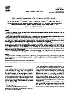
where: qk , qj are atomic charges, rkj denotes the distance between atoms k and j, ε0 is a dielectric constant [22]. Therefore, in our research, we have generated five different energy patterns for a single protein structure AN . They describe protein structures in terms of bonded and non-bonded interactions between atoms. In Fig. 1 we can see the spatial structure of the Human RAB5A, molecule 1N6H in the Protein Data Bank [21]. The molecule is visualized in the RasMol [24]. In Fig. 2 we can observe graphical representation of the van der Waals energy characteristic for the molecule 1N6H. The energy characteristic presented in Fig. 2 is retrieved from the Energy Distribution Data Bank (EDB). The EDB [25] is a special repository that we
Energy Properties in the Analysis of the Human RAB5A Cellular Activity
5
Fig. 1. Spatial structure of the Human RAB5A (1N6H): (a) ribbon representation, (b) sticks representation
have designed and developed to store so called energy profiles. Energy profiles are distributions of different potential energies over all atoms in protein structures. The EDB website (http://edb.aei.polsl.pl ) also allows to generate all five energy characteristics for chosen proteins. As of Wednesday 2008-12-31 there are 32 229 energy profiles in the EDB.
Fig. 2. Van der Waals energy characteristic for the Human RAB5A, molecule 1N6H
4 The RAB5A Cellular Activity Observed At the Level of Energy Characteristics Entire energy profiles and different component energy characteristics support a variety of studies on protein structures. In our research, we use them to search strong protein similarities [20]. We also examine protein structure modifications with the use of data stored in the Energy Distribution Data Bank. In the section, we show the use of energy characteristics in the analysis of the
6
Dariusz Mrozek, Bo˙zena MaÃlysiak-Mrozek, StanisÃlaw Kozielski, et al.
human RAB5A conformational switching and the influence of the switching on the RAB5A cellular activity. The following molecules retrieved from the Protein Data Bank were considered during our analysis: • • • • • • • •
Crystal Structure of Human RAB5A (molecule 1N6H) Crystal Structure of Human RAB5A A30P Mutant Complex with GDP (molecule 1N6I) Crystal Structure of Human RAB5A A30P Mutant Complex with GDP and Aluminum Fluoride (molecule 1N6K) Crystal Structure of Human RAB5A A30P Mutant Complex with GTP (molecule 1N6L) Crystal Structure of Human RAB5A A30R Mutant Complex with GPPNHP (molecule 1N6N) Crystal Structure of Human RAB5A A30K Mutant Complex with GPPNHP (molecule 1N6O) Crystal Structure of Human RAB5A A30E Mutant Complex with GPPNHP (molecule 1N6P) Crystal Structure of Human RAB5A A30L Mutant Complex with GPPNHP (molecule 1N6R)
The 1N6H molecule represents the crystal structure of human RAB5A GTPase domain and other molecules represent human RAB5A GTPase domain mutants in complex with different nucleotides. All molecules have similar overall folding. The mutation exists at the Ala30 residue of the amino acid chain in the specific region called P-loop (Fig. 3). The P-loop is a part of the RAB5A structure built up with residues 27 GESAVGKS34 . This part of the RAB5A plays a crucial role in GTP hydrolysis. Mutations in the P-loop have an associated influence on two other characteristic regions of the protein called switch region I (residues 47-65) and switch region II (residues 77-93). This kind of point mutation caused by the replacement of one amino acid with other amino acids can have significant biological consequences. In the active stage, the RAB5A exposes the switch region I and the switch region II. This enables the hydrolysis to occur. In the inactive stage, these two regions are not exposed enough to interact with other molecules. Therefore, conformational disorders in these two regions produced by the point mutation at the Ala30 residue have disadvantageous influence on the RAB5A activity. In the studied case, the mutation reduces catalytic abilities of the RAB5A molecule. Amino acid sequences of the human RAB5A (1N6H) and mutant 1N6O are presented in Fig. 4. Comparison of energy characteristics allows to observe the influences of the mutation and produced conformational changes in structures of studied proteins. In Fig. 5 we can see van der Waals and electrostatic energy characteristics for referential molecule 1N6H and mutant 1N6O. Replacements in the P-loop are visible in the form of changes in the distribution of energy in the P-loop region of energy characteristics. Associated conformational changes in the switch region I and switch region II are also visible as disorders in energy characteristics presented in Fig. 5.
Energy Properties in the Analysis of the Human RAB5A Cellular Activity
7
Fig. 3. Structure of the human RAB5A (1N6H) molecule with characteristic regions: P-loop, switch region I (SW1) and switch region II (SW2)
Fig. 4. Primary structure of the human RAB5A GTPase domain 1N6H (residues 15-181) and mutant 1N6O with the characteristic P-loop
Further analysis of energy characteristics presented in Fig. 5 confirms studies, observations and conclusions regarding structural mutants of the RAB5A presented in [8]. Point mutations at the Ala30 residue in the P-loop result in conformational changes in the characteristic switch region I and switch region II and also in the neighborhood of the Gln79 residue, and conformational change at the Lys33 residue. These changes are visible in the energy characteristics as energy discrepancies. Conformational disorders can be viewed by the observation of energy characteristics and further verified with the use of molecular viewers, like RasMol [14]. Spatial structures of specific regions of the RAB5A (1N6H) and its mutant (1N6O) are compared in Fig. 6 - Fig. 8. Referential structure and mutated structure of the P-loop are presented in Fig. 6. Replacement occurred at the 30th residue. In the case, the Ala amino acid was replaced by the Lys. Conformational deformations are visible near residues Glu28 , Ser29 , and Lys33 . In Fig. 7 we can observe conjugate deformations of the switch region I - visible changes near residues Ser51 , Asp65 , and Asp66 . In Fig. 8 we can see conformation deformations near residues Gln79 and Arg91 in the switch region II.
8
Dariusz Mrozek, Bo˙zena MaÃlysiak-Mrozek, StanisÃlaw Kozielski, et al.
Fig. 5. Comparison of electrostatic (a) and van der Waals (b) energy characteristics for molecules 1N6H and 1N6O. Only distinctive regions are presented
a)
b)
Fig. 6. Separated structure of the P-loop (27-34 residues) for molecules: a) 1N6H (Human RAB5A), b) 1N6O (Human RAB5A A30K Mutant Complex with GPPNHP)
5 Concluding Remarks Structural disorders caused by replacements of just a single residue in a protein amino acid chain can have very serious biological consequences. As a result of such a change mutant molecule may not be able to interact properly with other molecules in cellular reactions. Therefore, some biological processes can be impeded or completely disabled. However, not every conformational change
Energy Properties in the Analysis of the Human RAB5A Cellular Activity
a)
9
b)
Fig. 7. Separated structure of the switch region I (46-65 residues) for molecules: a) 1N6H (Human RAB5A), b) 1N6O (Human RAB5A A30K Mutant Complex with GPPNHP)
a)
b)
Fig. 8. Separated structure of the switch region II (76-94 residues) for molecules: a) 1N6H (Human RAB5A), b) 1N6O (Human RAB5A A30K Mutant Complex with GPPNHP)
causes so serious results as it was presented in previous section for the RAB5A molecule. Many deformations are effects of some natural biological processes, e.g. cellular reactions, different interactions, environmental influence, binding the molecule to another one. Moreover, some deformations caused by possible mutations can appear or influence regions that does not play a key role for any biological process, e.g. outside the active site of an enzyme. As a result, they do not influence the functioning of the whole particle. In Fig. 5 we can observe regions that indicated conformational deformations but were not directly involved in the hydrolysis process. Protein energy characteristics allow to observe such situations. They are generally supportive in the analysis of protein conformation switching and investigation of other structural deformations. Therefore, they support studies on the activity of proteins, such as human RAB5A.
10
Dariusz Mrozek, Bo˙zena MaÃlysiak-Mrozek, StanisÃlaw Kozielski, et al.
References 1. Lodish H, Berk A, Zipursky SL, et al.(2001) Molecular Cell Biology. Fourth Edition. W. H. Freeman and Company, NY 2. Dickerson RE, Geis I (1981) The Structure and Action of Proteins, 2nd ed. Benjamin/Cummings, Redwood City, Calif. Concise 3. Allen JP (2008) Biophysical Chemistry, Wiley-Blackwell 4. Branden C, Tooze J (1991) Introduction to Protein Structure, Garland 5. Creighton TE (1993) Proteins: Structures and Molecular Properties, 2nd ed. Freeman, San Francisco 6. Attwood TK, Parry-Smith DJ (1999) Introduction to Bioinformatics. Prentice Hall 7. Gibas C, Jambeck P (2001) Developing Bioinformatics Computer Skills. First Edition, O’Reilly 8. Zhu G, Liu J, Terzyan S, Zhai P, Li G, Zhang XC (2003) High Resolution Crystal Structures of Human Rab5a and Five Mutants with Substitutions in the Catalytically Important Phosphate-Binding Loop. J.Biol.Chem., Vol. 278: 2452–2460 9. Li G, and Liang Z (2001) Phosphate-Binding Loop and Rab GTPase Function: Mutations at Ser29 and Ala30 of Rab5 Lead to Loss-of-Function as well as Gain-of-Function Phenotype. Biochem. J. 355: 681–689 10. Liang Z, Mather T, and Li G (2000) GTPase Mechanism and Function: New Insights from Systematic Mutational Analysis of the Phosphate-Binding Loop Residue Ala30 of Rab5. Biochem. J. 346: 501–508 11. Thorner DA, Wild DJ, et al. (1996) Similarity Searching in Files of ThreeDimensional Chemical Structures: Flexible Field-Based Searching of Molecular Electrostatic Potentials. J. Chem. Inf. Comput. Sci.: 900–908 12. Rodrigo J, Barbany M, et al. (2002) Comparison of Biomolecules on the Basis of Molecular Interaction Potentials, J. Braz. Chem. Soc., Vol. 13, No. 6: 795–799 13. Ji H, Li H, et al. (2003) Computer Modeling of Selective Regions in the Active Site of Nitric Oxide Synthases: Implication for the Design of Isoform-Selective Inhibitors. J. Med. Chem.: 5700–5711 14. Goodford PJ (1985) A Computational Procedure for Determining Energetically Favorable Binding Sites on Biologically Important Macromolecules. J. Med. Chem., 28 (7), 849-857 15. Caceres M, Villa J, Lozano JJ, Sanz F (2000) MIPSIM: Similarity Analysis of Molecular Interaction Potentials. Bioinformatics Vol. 16 no. 6: 568–569 16. Liu HC, Lyu PC, Leong MK, Tsai KC, Hsiue GH. (2004) 3D-QSAR Studies on PU3 Analogues by Comparative Molecular Field Analysis. Bioorg Med Chem Lett. 14(3): 731–4 17. Hansch C, Verma RP (2007) 20-(S)-Camptothecin Analogues as DNA Topoisomerase I Inhibitors: a QSAR Study. ChemMedChem. 2(12): 1807–13 18. Trossini GH, Guido RV, et al. (2009) Quantitative Structure-Activity Relationships for a Series of Inhibitors of Cruzain from Trypanosoma Cruzi: Molecular Modeling, CoMFA and CoMSIA Studies. J Mol Graph Model. (to be published) 19. McNaught AD, Wilkinson A (1997) IUPAC. Compendium of Chemical Terminology, 2nd ed. (the ”Gold Book”). Blackwell Scientific Publications, Oxford 20. Maysiak B, Momot A, Kozielski S, Mrozek D (2008) On Using Energy Signatures in Protein Structure Similarity Searching. In: Rutkowski L, et al. (eds.)
Energy Properties in the Analysis of the Human RAB5A Cellular Activity
21. 22. 23.
24. 25.
11
Artificial Intelligence and Soft Computing, LNAI, Springer, Heidelberg, vol. 5097: 939–950 Berman HM, Westbrook J, Feng Z, Gilliland G, et al. (2000) The Protein Data Bank. Nucleic Acids Res. (28): 235–242 Leach A (2001) Molecular Modelling: Principles and Applications, 2nd Edition. Pearson Education EMA, UK Cornell WD, Cieplak P, et al. (1995) A Second Generation Force Field for the Simulation of Proteins. Nucleic Acids, and Organic Molecules. J.Am. Chem. Soc. (117): 5179-5197 Sayle R, Milner-White EJ (1995) RasMol: Biomolecular Graphics for All. Trends in Biochemical Sciences (TIBS), Vol. 20, No. 9, 374 ´ Mrozek D, MaÃlysiak-Mrozek B, Kozielski S, Swierniak A (2009) The Energy Distribution Data Bank: Collecting Energy Features of Protein Molecular Structures. In Proc. of the 9th IEEE International Conference on Bioinformatics and Bioengineering, IEEE, 1–6