Am. J. Trop. Med. Hyg., 69(1), 2003, pp. 45–52 Copyright © 2003 by The American Society of Tropical Medicine and Hygiene
ENDEMIC MALARIA IN THE PERUVIAN AMAZON REGION OF IQUITOS BABACK ROSHANRAVAN, ELINA KARI, ROBERT H. GILMAN, LILIA CABRERA, ELLEN LEE, JOHN METCALFE, MARITZA CALDERON, ANDRES G. LESCANO, SONIA H. MONTENEGRO, CARLOS CALAMPA, AND JOSEPH M. VINETZ Tulane University School of Public Health and Tropical Medicine, New Orleans, Louisiana; Johns Hopkins University School of Hygiene and Public Health, Baltimore, Maryland; Universidad Peruana Cayetano Heredia, Lima, Peru; Ochsner Clinic Foundation, New Orleans, Louisiana; Proyectos en Informática Salud, Medicina y Agricultura (Asociación Benéfica PRISMA), Lima, Peru; Dirección Regional de Salud de Loreto, Iquitos, Peru; World Health Organization Collaborating Center for Tropical Diseases, University of Texas Medical Branch, Galveston, Texas
Abstract. A cross-sectional study was conducted in the Peruvian Amazon to test the hypothesis that a reservoir of asymptomatic malaria parasitemic patients would form the basis for continuing malaria endemicity in the region. Active surveillance yielded a Plasmodium spp. slide-positive prevalence of 4.2% (43 of 1,023) and a polymerase chain reaction (PCR)–positive prevalence of 17.6% (144 of 819). Plasmodium vivax prevalence was 2.9% and 14.2% while Plasmodium falciparum prevalence was 1.3% and 2.6% by microscopy and PCR, respectively. Approximately two-thirds of slidepositive and one-fourth of PCR-positive people were symptomatic. Anemia was associated with slide positivity (P < 0.001) and PCR positivity for P. falciparum (P ⳱ 0.003). Sensitivity of field microscopy and agreement between field and reference laboratory microscopists were low, arguing for using PCR for epidemiologic investigation and malaria control. While these data confirm recent findings from the Brazilian Amazon suggesting that sufficient numbers of asymptomatic malaria parasitemic patients are present to form a persistent reservoir for continuous reinfection within the Peruvian Amazon region, these results also indicate that clinical immunity in human populations can be driven in malaria-endemic regions that do not have high intensity malaria transmission Anopheles darlingi currently accounts for 90% of the mosquito population in the villages around Iquitos during the wettest part of the year, and continues to be implicated as the major Anopheles spp. during the less wet seasons as well.1 Unpublished observations suggest that the proportion of Plasmodium-infected An. darlingi is low (on the order of less than 0.1–0.5%) (Flores-Mendoza C, Schoeler G, U.S. Naval Medical Research Center Detachment, Lima, Peru, unpublished data). In the present study, we used a cross-sectional approach to examine the prevalence of malaria parasitemia in four villages in the Peruvian Amazon region near Iquitos. We compared results from light microscopy examination of Giemsa-stained blood smears with results of a nested polymerase chain reaction assays (PCR) to detect and speciate parasites. The results obtained by slide microscopy were then compared with those obtained by the nested PCR. In anticipation of further investigations of the epidemiology of malaria transmission in the Peruvian Amazon, we sought to answer three specific questions: 1) How does the PCR compare with local microscopy readings of samples collected in the field?; 2) What is the prevalence of symptomatic malaria versus asymptomatic parasitemia that would suggest the epidemiology of reservoirs for malaria; and 3) What is the association between morbidity attributable to malaria and the PCR versus light microscopy diagnosis?
INTRODUCTION The emergence of chloroquine-resistant Plasmodium falciparum and the marked increase in the incidence of P. vivax malaria over the past decade in the Peruvian Amazon1 have made malaria control a major priority of Peruvian public health efforts. The lack of an effective and deployable vaccine, the regional spread of resistance of P. falciparum to pyrimethamine/sulfadiazine and other anti-malarial drugs,2 the potential for P. vivax to become resistant to chloroquine in Latin America,3,4 and limitations of anti-anopheline measures highlight the urgent need for new approaches to malaria control.5 An important part of such efforts is the precise characterization of the epidemiology of malaria in this region. Malaria transmission in the region of Loreto, Peru (total population of approximately 650,000) has increased since 1991, with cases in villages near the capital city of Iquitos (population approximately 400,000) accounting for most cases.1 In 1997, a year of epidemic malaria, Loreto reported 158,132 cases of malaria, 54,290 (34.3%) of which were caused by P. falciparum, and the remainder by P. vivax.6 In 1998, the number of reported cases decreased by approximately 40,000.6 The Peruvian Ministry of Health has used a strategy that includes vector control, epidemiologic surveillance, and community work. However, despite governmental interventions, malaria persists as a serious problem in the region, with cases of malaria occurring 12 months per year, ranging from 3,000 to 9,000 monthly, with the largest number of cases occurring from January to July, and decreasing by approximately half from August through December (Peruvian Ministry of Health, Dirección Regional Salud, Loreto, Iquitos, Peru, unpublished data). Changing vector dynamics seem to be associated with the changed epidemiology of malaria in the region.1 What is now the principal malaria vector in the Loreto region, Anopheles darlingi, was not reported to be present there until the mid1990s.1 The entry of An. darlingi into the ecology of the Loreto mosquito population has been temporally associated with the increase in P. falciparum transmission in the region.1
MATERIALS AND METHODS Site description. The study was conducted in four villages near Iquitos, the capital city of the Department of Loreto, Peru in the Amazon region. The city of Iquitos is a major tourist and shipping center situated 120 meters above sea level (73°W, 3°S) at the juncture of the Ucayali and Napo Rivers forming the Amazon River proper. Iquitos, with a population of approximately 400,000 and a surrounding rural population of 474,000, is accessible only by air or river. The target villages of Morallilo, El Nuevo Milagro, San Carlos, and Varillal are located 14–25 kilometers from the
45
46
ROSHANRAVAN AND OTHERS
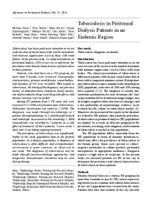
center of the city of Iquitos and are accessible via the paved road leading to Nauta (Figure 1). Moralillo is located approximately 15 km from the city of Iquitos and is accessible from the Iquitos-Nauta road. El Nuevo Milagro is located approximately 2 km from the road and 21 km from the city of Iquitos. San Carlos is approximately 10 km from the Iquitos-Nauta road and 21 km from the city of Iquitos, with direct access to the River Itaya. Varillal is located on the side of the IquitosNauta road approximately 14 km from the city of Iquitos. The study communities and the many rural communities that surround the city of Iquitos serve as ideal larval breeding areas with small pools on cleared land, fish farms, areas of poor sanitation, swamps, and the edges of small rivers. Adult A. darlingi biting hours peak at dusk and dawn. The populations in the four villages are of low socioeconomic status and ethnicities are primarily mestizo. Household economies are sustained by agriculture, fishing, or production of firewood, bricks, or aguardiente (a sugar cane liquor). None of these communities have electricity or basic sanitation services. The Ministry of Health of Peru operates health posts in various villages around Iquitos. Those that use a microscopist provide free same-day malaria diagnosis and treatment. Among the four study communities, only Varillal had a health post staffed by a microscopist; Varillal was the local reference center for the study communities. Individuals reporting to the health post with fever were entered into the Fever Registry (Registro de Febriles). Individuals diagnosed with malaria had diagnosis, prescribed course of treatment, and outcome recorded in the Treatment Log (Registro de Seguimientos). In the villages without a health post staffed with a microscopist,
FIGURE 1. Map of the study communities in relation to the city of Iquitos, the road to Nauta, and Rio Itaya.
trained volunteer health promoters collected blood samples for thick and thin blood smears and delivered them to the Varillal health post for diagnosis. These positive individuals were also registered in the health post fever and treatment logs. Active surveillance methods. Active surveillance for malaria in the study communities involved visiting all households in both remote and central parts of each community at random and collecting clinical data and samples from both symptomatic and asymptomatic individuals. People who agreed to participate were sampled once. The study team was comprised of a physician, a field nurse, and two local health promoters who returned to households to sample family members not available during initial surveillance. Fifty-one days of staggered sampling was conducted between April 27 and July 17, 1999, within the peak malaria transmission season.1 The majority of samples (548 of 1,023, 53.6%) were collected between April 27 and May 31 while the remainder was collected between June 1 and July 17. Case definitions. The determination of Plasmodium infection was based on presence of parasitemia on the thick and thin blood smears or by a nested PCR. Symptomatic cases were defined as PCR-positive and/or slide-positive cases with a documented fever or having a history of fever and chills with or without other symptoms within the preceding two weeks. Fever was defined as a patient having oral temperature ⱖ 37.5°C. Asymptomatic cases of malaria were defined as cases testing PCR-positive and/or slide-positive for Plasmodium spp. and presenting none of the above symptoms. Testing for anemia was done only during active surveillance using capillary hematocrit tubes. Anemia was defined as having a hematocrit < 33% for individuals < 2 years old, < 34% for individuals 2–5.9 years old, < 35% for individuals 6–11.9 years old, < 36% for individuals 12–17.9 years old, < 41% for males > 18 years old, and < 36% for females > 18 years old.7 Clinical data. A brief clinical history following a standardized questionnaire was carried out before obtaining blood samples. A history of fever, chills, general malaise, abdominal pain, muscle or joint pain, nausea, vomiting, and diarrhea within the past two weeks was recorded. The physical examination conducted by a physician also included assessment of body temperature and spleen size. Patient treatment and follow-up. Thick and thin blood smear diagnoses of malaria and hematocrit results were reported to individuals the following day. Individuals who tested slide-positive for Plasmodium spp. were provided with anti-malarial treatment according to Peruvian Ministry of Health guidelines. Anemic individuals were given ferrous sulfate supplements. Passive surveillance methods. Passive surveillance for symptomatic malaria consisted of cross-referencing the fever registries and treatment logs in Varillal with the censuses of the target communities to estimate symptomatic case prevalence over the study period. Data from registries and logs were analyzed for a period of time encompassing the period of active surveillance, as well as one week before and one week after. Passive symptomatic case prevalence was defined as the percentage of symptomatic individuals who reported to the health post with a fever and tested slide positive for malaria among the total number of febrile individuals who reported to the health post and received a malaria test during the same period. Active symptomatic case prevalence repre-
47
MALARIA IN IQUITOS, PERU
sented the proportion of malaria cases among documented febrile cases detected during the active surveillance. Sample processing. EDTA-anticoagulated blood (∼ 2 mL) was collected by venipuncture from individuals ⱖ 2 years of age. Finger stick blood samples were collected from children < 2 years of age and from children up to five years of age, depending on parental preference. Samples were prepared for thick and thin smear microscopy using 5% Giemsa (Sigma, St. Louis, MO), hematocrit determination, and a PCR assay. One hundred Giemsa-stained thick film fields were examined for sexual and asexual stage Plasmodium spp. prior to diagnosing a slide as negative. Two slides for each individual were prepared. The first examination was performed by the microscopist at the Varillal health post who had five years of experience, typical of experienced microscopists in the region. This diagnosis was considered as the standard used in the analysis. The same slide was then sent to a reference laboratory at Universidad Peruana Cayetano Heredia in Lima for a quality control reading by a professor of microbiology with more than 20 years of experience reading malaria slides. The second reading was used to determine inter-microscopist variation and as a reference for discrepancies between PCR and microscopy. Analysis of blood specimens for the presence of Plasmodium spp. by PCR. DNA was extracted from EDTAanticoagulated blood samples that had been stored at −20°C, using a commercial kit and following the manufacturer’s instructions (DNA Isolation Kit; Gentra, Minneapolis, MN). A nested PCR assay was modified from a method that amplifies conserved and variable sequences within the small subunit ribosomal RNA gene as previously described.8 The PCR amplification reaction mixtures contained the following: 50 mM KCl, 10 mM Tris-HCl, (pH 8.3, 1.5 mM MgCl2, 200 M each of dNTPs, 0.5 M of each primer, 1.25 units of Taq polymerase, and 3 L of the sample DNA in a total volume of 25 L. The cycling protocol was 94°C for one minute, 58°C for one minute, and 72°C for one minute for 25 cycles for genus-level primers and 35 cycles for species-specific primers. Amplification products were then analyzed by electrophoresis on 2% agarose gels. The genus primer sequences used were 5⬘CTTGTTGTTGCCTTAAACTTC-3⬘ and 5⬘-TTAAAATTGTTGCAGTTAAAACG-3⬘.9 The P. vivax primers used were 5⬘-CGCTTCTAGCTTAATCCACATAAC-3⬘ and 5⬘ACTTCCAAGCCGAAGCAAAGAAAGTCCTTA-3⬘. 10 The P. falciparum primers used were 5⬘-TTAAACTGGTTTGGGAAAACCAAATATATT-3⬘ and 5⬘ACACAATGAACTCAATCATGACTACCCGTC-3⬘.11 Specificity of the PCR. To verify specificity of the PCR assay, 50 control samples were processed from residents of Pampas, a peri-urban shantytown outside Lima where there is no malaria transmission.12 All control samples were negative. Statistical analysis. Comparison of the rates of positivity for microscopy and the PCR was limited to individuals who had both tests. Associations between test results and demographics were determined by calculating odds ratios (ORs) and 95% confidence intervals (CIs). Differences were detected using the chi-square test of independence. Epi-Info version 6.04 (Public Domain Software; Centers for Disease Control and Prevention, Atlanta, GA) was used to calculate agreement. Calculations of chi-square values and ORs were done using SPSS version 8 (SPSS, Inc., Chicago, IL). Ethical review and approval. The protocol was reviewed
and approved by the Peruvian Ministry of Health and passed ethical review at Asociación Benéfica PRISMA and the Johns Hopkins University Bloomberg School of Public Health. Verbal informed consent was obtained at both the individual level and from community leaders. RESULTS Demographics of the study population. A census of the target villages of Moralillo and Varillal was carried out in 1998, and a census of El Nuevo Milagro and San Carlos was done in 1999. The villages had an aggregate population of 1,447 with the following breakdown: Moralillo, 355; Varillal, 643; El Nuevo Milagro, 293; San Carlos, 156. Almost three-fourths of the populations (1,023, 71%) participated in the study. The age distribution of the study population reflected that of the total population of the four target villages (Table 1). Active surveillance for malaria and anemia in the communities. Sample collection. A total of 1,023 collected samples was analyzed by slide microscopy, of which 819 (80.0%) were available for the PCR. Due to the difficulty of collecting whole blood samples from children less than five years old, fewer samples were available for the PCR than for microscopy in this age group (Table 1). Interviews for symptom history were conducted with 998 of the 1,023 individuals sampled (97.6%), while 891 (87.1%) had physical examinations performed on them by a physician. Of the 819 PCR samples, 107 (13.1%) lacked an associated physical examination. Hematocrit analysis was performed in 1,010 samples (98.7%). There were significant differences in the age distribution of tested and non-tested individuals for both PCR and microscopy (P < 0.001); tested subjects were more frequently greater than 50 years old, while small children were less likely to be included in the study. Tested subjects were comparable regarding sex to those not included in the study. Prevalence of Plasmodium spp. Of the 1,023 samples, 43 (4.2%) were slide positive while 144 (17.6%) of 819 were PCR positive. Plasmodium falciparum gametocytes were detected in 2 cases (4.7%) among the 43 positive slides. Active surveillance yielded community-specific slide-positive rates of 12.1% (14 of 116) in San Carlos, 6.8% (19 of 280) in Moralillo, 1.9% (4 of 214) in Nuevo Milagro, and 1.6% (6 of 413) in Varillal. The PCR showed a consistently higher prevalence rate for P. falciparum and P. vivax than microscopy (P < 0.001; Table 2). The PCR also detected seven mixed infections missed by slide microscopy. Prevalence of anemia. Of the study participants, 44.9% (453 of 1,010) were anemic. The mean hematocrit value for 476 females was lower (35.4%) than for 534 males (37.5%) (P < 0.001). No association was observed between sex and anemia (P ⳱ 0.447). After stratifying by age, individuals between 0 and 9 years of age and individuals more than 40 years of age were found to have an increased likelihood of being anemic. Approximately half (141 of 277, 50.9%) of the individuals between 0 and 9 years of age (OR ⳱ 1.40, 95% CI ⳱ 1.06–1.85, P ⳱ 0.017) and 59.5% (100 of 168) of the individuals more than 40 years of age (OR ⳱ 2.04, 95% CI ⳱ 1.45–2.86, P < 0.001) were anemic. In addition, males between 15 and 39 years of age were less likely to be anemic (OR ⳱ 0.59, 95% CI ⳱ 0.45–0.77, P < 0.001). Passive surveillance for symptomatic malaria. The prevalence of acute malaria was determined by passive surveil-
48
Negative P. falciparum P. vivax Mixed infection All Plasmodium spp.
5.3 ± 2.9
26 (32) 39 (37) 38 (44) 39 (45) 64 (62) 50 (68) 19 (36) 44 (81) 319 (50) 57 (70) 54 (51) 47 (55) 45 (52) 71 (69) 62 (84) 23 (43) 54 (100) 413 (64) 81 106 86 86 103 74 53 54 643 127
Slide microscopy
4.8 ± 2.3 5.8 ± 3.0 5.2 ± 2.4
13 (52) 24 (83) 18 (86) 5 (56) 20 (91) 12 (60) 14 (100) 10 (63) 116 (74) 25 29 21 9 22 20 14 16 156 30
* For the 0–4 years age group there was a statistically significant difference in PCR vs. microscopy sampling.
51 (67) 65 (83)
8 (32) 21 (72) 14 (67) 5 (56) 18 (82) 10 (50) 13 (93) 9 (56) 98 (63)
62 60 54 33 52 37 27 30 355 69
43 (69) 51 (85) 47 (87) 18 (55) 35 (67) 29 (78) 27 (100) 30 (100) 280 (79)
24 (39) 49 (82) 40 (74) 15 (45) 30 (58) 27 (73) 25 (93) 27 (90) 237 (67)
25 28 59 90 33 27 17 14 293 34
17 (68) 19 (68) 52 (88) 73 (81) 16 (48) 16 (59) 11 (65) 10 (71) 214 (73)
7 (28) 14 (50) 41 (69) 65 (72) 12 (36) 13 (48) 7 (41) 6 (43) 165 (56)
171 (54) 148 (46) 210 (66) 203 (63) 316 322
TABLE 2 Comparison of microscopy and polymerase chain reaction (PCR) results obtained over a 51-day period between April 27 and July 15, 1999
Sex Male Female Age (years) 0–4.99 5–9.99 10–14.99 15–19.99 20–29.99 30–39.99 40–49.99 ⱖ50 Total No. of houses No. of persons/house, mean ± SD
76 78
42 (55) 56 (72)
177 171
139 (79) 141 (83)
116 (66) 121 (71)
192 101
141 (73) 73 (72)
113 (59) 52 (51)
No. (%)
PCR Microscopy
No. (%) No. (%) N
No. (%)
No. (%)
N
n (%)
No. (%)
N
No. (%)
Varillal
PCR Microscopy
Nuevo Milagro
PCR Microscopy
Moralillo
PCR Microscopy
San Carlos
TABLE 1 Age distribution along with polymerase chain reaction (PCR) and microscopy sampling distribution in the study communities*
N
ROSHANRAVAN AND OTHERS
PCR
(n ⳱ 819)
Rate
(n ⳱ 819)
Rate
781 12 26 0 38
95.8 1.5 3.2 0 4.6
675 21 116 7 144
82.4 2.6 14.2 0.9 17.6
lance, defined as febrile patients presenting to a Ministry of Health Health Post, having parasitemia detected on blood smear, and recorded in the Health Post log book. Passive case detection rates of acute malaria during study period was 5.7% (82 of 1,433) for all communities combined. Rates for each community were 9.7% (15 of 154) in San Carlos, 1.7% (6 of 348) in Moralillo, 3.8% (11 of 293) in Nuevo Milagro, and 7.8% (50 of 638) in Varillal. Passive versus active symptomatic case rates. Comparison of active and passive surveillance malaria rates of the communities showed the following symptomatic case prevalence rates (active/passive): San Carlos (75.0%/45.5%), Moralillo (24.0%/28.5%), El Nuevo Milagro (14.0%/23.9%), and Varillal (5.0%/18.1%). These differences in the proportion of active to passive cases parallel the differences in the active case detection prevalence for light microscopy-diagnosed malaria. Analysis of overall study population. Sensitivity of microscopy. Microscopy had an overall sensitivity of 24.3% (35 of 144), using the PCR as the gold standard. All fifty control samples were negative by PCR, indicating a specificity of 100.0%. The sensitivity for detecting P. vivax was 19.5% (24 of 123) compared with the PCR. Sensitivity for P. falciparum detection was 32.1% (9 of 28) when compared with the PCR. The percent agreement between the PCR and microscopy for detection of Plasmodium was 86.3%: 97.3% for P. falciparum and 87.7% for P. vivax (Table 3). Comparison between microscopists. The inter-microscopist agreement among paired observation of Plasmodium spp. slide-positives was 54.1% (33 of 61). The species-specific inter-microscopist agreement of positive slides ranged from 42.1% for P. falciparum to 47.6% for P. vivax. Demographic associations and presentation of signs and symptoms. Slide-positive individuals. Demographics, symptoms, anemia status, and malaria diagnosis were significantly associated when slide microscopy was used to define cases. Individuals ⱖ 16 years of age were more likely to test slide positive (OR ⳱ 2.64, 95% CI ⳱ 1.31–5.29, P ⳱ 0.005). Sex was not associated with slide-diagnosed malaria (P ⳱ 0.276). Of substantial significance, only two-thirds of slide-positive individuals had symptoms attributable to malaria (Table 4). Slide-positive status was associated with a wide range of symptoms (Table 5), as well as splenomegaly (P < 0.05) and anemia (P < 0.01). PCR-positive individuals. Less than one-third of the PCRpositive individuals were symptomatic (Table 4). There was no association between sex (P ⳱ 0.344), and symptom status among PCR-positive individuals. Similarly, there was no association between age and PCR-positive status. The prevalence ratios of the signs and symptoms associated with the
49
MALARIA IN IQUITOS, PERU
TABLE 3 Sensitivity, specificity, and agreement of the polymerase chain reaction (PCR) compared with the health post microscopist’s readings* All Plasmodium spp. PCR +
Microscopy + Microscopy − Sensitivity Specificity Overall % agreement
35 109
P. falciparum PCR −
PCR +
3† 672 24.3% (17.7–32.3) 99.6% (98.6–99.9)
9 19
86.3
P. vivax PCR −
3‡ 788 32.1% (16.6–52.4) 99.6% (98.8–99.9)
PCR +
24 99
PCR −
2§ 694 19.5% (13.1–27.8) 99.7% (98.8–100.0)
97.3
87.7
* Mixed infections were included in both P. falciparum and P. vivax sensitivity and specificity calculations. Values in parentheses are 95% confidence intervals. † Two slides lacked confirmatory readings and one was later confirmed as negative by the reference microscopist. ‡ Two slides were later confirmed as P. vivax by the reference microscopist and one lacked a confirmatory reading. § One slide was later confirmed as negative by the reference microscopist and the second lacked a confirmatory reading.
PCR are shown in Table 5. A negative association was observed between PCR-positive status and diarrhea. Stratified analysis showed that among males between the ages of 15 and 39, 46.7% (28 of 60) of those PCR positive were anemic (OR ⳱ 1.97, 95% CI ⳱ 1.10–3.50) versus 30.8% (73 of 237) of those who were PCR negative. Symptomatic presentation among PCR-positive and slidenegative individuals. Among the PCR-positive asymptomatic cases, 88.3% (83 of 94) were slide negative for malaria. The PCR-positive/slide-negative status was negatively associated with history of diarrhea (OR ⳱ 0.23, 95% CI ⳱ 0.06–0.97, P ⳱ 0.023) and abdominal pain (OR ⳱ 0.42, 95% CI ⳱ (0.21– 0.82, P ⳱ 0.007). A total of 23.8% (19 of 80) of P. vivax and 25.0% (3 of 12) of P. falciparum PCR-positive/slide-negative diagnosed infections had a documented fever or a history of a fever within the past two weeks. Among these PCR-positive/ slide-negative individuals, those who were anemic were more likely to have been symptomatic (OR ⳱ 5.10, 95% CI ⳱ 1.28–20.41, P < 0.01). The rate of splenomegaly among PCRpositive/slide-negative individuals was 1.8% (3 of 109). Symptomatic presentation of P. falciparum PCR-positive individuals. Only six (28.6%) of the 21 P. falciparum PCRpositive cases were detected as P. falciparum slide-positive. The PCR-positive status for P. falciparum was associated with anemia (Table 5). Eight (61.5%) of the 13 anemic P. falciparum PCR-positive individuals were also asymptomatic. Overall, 44.4% (8 of 18) of the P. falciparum PCR-positive individuals were asymptomatic. While no association was observed between a history of fever and P. falciparum PCR diagnosis, such an association was observed for P. vivax (P < 0.05).
TABLE 4 Rates of symptomatic infection defined as having reported fever and chills in the past two weeks or having fever during sample collection* Diagnosis
P. vivax P. falciparum Mixed infection All Plasmodium spp.
Slide† n/N (%)
PCR + (all) n/N (%)
PCR + / slide— n/N (%)
17/28 (60.7)† 8/11 (72.7)† 0
23/102 (22.5)‡ 6/18 (33.3)§ 4/7 (57.1)‡
9/79 (11.4) 2/12 (16.6) 1/4 (25)
25/39¶ (64.1)†
33/127# (26.0)†
12/95 (12.6)
* PCR ⳱ polymerase chain reaction. † P < 0.001. ‡ P < 0.01. § P < 0.05. ¶ Four microscopy-positive individuals did not receive clinical examinations. # Seventeen PCR-positive individuals did not receive clinical examinations.
DISCUSSION There are four major findings from this study. First, there was a substantial proportion of asymptomatic patients with patent P. falciparum and P. vivax infections in the villages in the Iquitos area of the Peruvian Amazon. Second, the very presence of a substantial proportion of parasitemic individuals without clinical symptoms referable to malaria indicates that the low intensity of transmission in this region may be sufficient to drive clinical immunity within this population, despite the fact that the point prevalence of malaria in this region is much lower than in regions frequently associated with extremely high rates of Plasmodium parasitemia, such as parts of sub-Saharan Africa.12 Third, there are likely to be sufficient numbers of asymptomatic parasitemic individuals in a reservoir for malaria transmission in the region. Fourth, screening for Plasmodium parasitemia using PCR-based methods rather than light microscopy appears to be an important tool for malaria control in the Peruvian Amazon region. Our data suggest that asymptomatic Plasmodium parasitemia in the Peruvian Amazon region of Iquitos, and thus clinical immunity to disease, occurred even in absence of intense malaria transmission. This finding is in contrast to holoendemic regions in Africa and elsewhere, where clinical immunity in the face of parasitemia is the norm, but is associated with the vast majority of the population harboring parasites, indicating intense transmission.13 Although we did not study the entomologic inoculation rate in the various villages here, the proportion of anopheline vectors infected with Plasmodium sporozoites in the region is less than 0.5% (Flores-Mendoza C, Schoeler G, U.S. Naval Medical Research Center Detachment, Lima, Peru, unpublished data), and the number of infectious bites per year has been estimated to be on the order of 10–20. Further studies will confirm the assumption that relatively low level malaria transmission is capable of engendering clinical immunity and asymptomatic Plasmodium parasitemia within the small village populations in the peri-Iquitos region. A high prevalence of asymptomatic P. falciparum and P. vivax infections has recently been reported from the western Brazilian Amazon region of Rondonia among native Amazonian riverine populations.14 In the communities studied, Alves and others14 found that asymptomatic infections were 4–5 times higher than symptomatic ones, and that the likelihood of symptoms associated with malaria (namely, fever and chills) decreased with age, which is consistent with other stud-
50
ROSHANRAVAN AND OTHERS
TABLE 5 Prevalence and odds ratios of symptoms among individuals testing positive by microscopy and polymerase chain reaction (PCR)* PCR +
Microscopy +
Symptom
N
n (%)
(N ⳱ 143)
OR (95% CI)
(N ⳱ 43)
OR (95% CI)
Fever Nausea Vomiting Diarrhea Malaise Shakes/chills Muscle pain Abdominal pain Joint pain Cough Splenomegaly Anemia
998 998 998 998 998 998 998 998 998 998 994 1,010
217 (21) 94 (9) 38 (4) 83 (8) 182 (18) 134 (13) 143 (14) 180 (18) 72 (7) 136 (13) 8 (1) 453 (44)
43 (30%)† 20 (14%) 8 (6%) 3 (2%)† 35 (25%)‡ 32 (22%)† 33 (23%)† 19 (13%) 20 (14%)† 17 (12%) 3 (2%) 71 (50%)‡
1.89 (1.25–2.84) 1.59 (0.93–2.73) 1.80 (0.78–4.14) 0.26 (0.08–0.83) 1.56 (1.01–2.40) 2.11 (1.33–3.34) 1.97 (1.25–3.08) 0.639 (0.38–1.08) 2.51 (1.42–4.44) 0.92 (0.53–1.60) 15.87 (0.78–16.67) 1.44 (1.00–2.07)
27 (63%)† 11 (26%)† 5 (12%)‡ 1 (2%) 23 (55%)† 22 (51%)† 24 (56%)† 11 (26%) 12 (28%)† 11 (26%)‡ 2 (5%)‡ 32 (72%)†
6.79 (3.59–12.87) 3.61 (1.76–7.43) 3.68 (1.36–9.94) 0.25 (0.03–1.87) 5.76 (3.09–10.73) 7.89 (4.20–14.80) 8.87 (4.72–16.70) 1.60 (0.79–3.24) 5.77 (2.82–11.81) 2.28 (1.12–4.64) 7.69 (1.49–33.33) 3.77 (1.88–7.57)
P. falciparum PCR + Symptom
(N ⳱ 21)
Fever Nausea Vomiting Diarrhea Malaise Shakes/chills Muscle pain Abdominal pain Joint pain Cough Splenomegaly Anemia
7 (33%) 2 (10%) 0 0 5 (24%) 6 (29%) 4 (19%) 3 (14%) 2 (10%) 3 (14%) 1 (5%) 16 (76%)†
OR (95% CI)
1.97 (0.78–4.95) 0.93 (0.21–4.08) 1.39 (0.501–3.86) 2.57 (0.97–6.77) 1.35 (0.45–4.07) 0.74 (0.22–2.55) 1.31 (0.30–5.76) 1.16 (0.34–4.00) 6.45 (0.74–55.56) 4.47 (1.62–12.32)
P. vivax Microscopy +
PCR +
Microscopy +
(N ⳱ 13)
OR (95% CI)
(N ⳱ 115)
OR (95% CI)
(N ⳱ 30)
OR (95% CI)
8 (62%)† 3 (23%) 1 (8%) 1 (8%) 8 (62%)† 7 (54%)† 8 (62%)† 6 (46%)‡ 4 (31%)† 5 (39%)‡ 1 (8%) 12 (92%)†
6.48 (1.88–22.33) 3.69 (0.96–14.16) 2.57 (0.33–19.23) 1.10 (0.14–8.73) 12.46 (3.27–47.44) 8.05 (2.42–26.77) 7.45 (2.24–24.73) 5.61 (1.69–18.58) 4.99 (1.29–19.23) 3.70 (1.07–12.82) 13.89 (1.57–125.00) 12.55 (1.60–98.42)
33 (29%)‡ 16 (14%) 7 (6%) 1 (1%)† 27 (24%) 24 (21%)‡ 28 (24%)† 12 (10%)‡ 16 (14%)‡ 12 (10%) 1 (1%) 51 (45%)
1.69 (1.08–2.64) 1.54 (0.86–2.77) 1.95 (0.82–4.68) 0.11 (0.01–0.76) 1.43 (0.89–2.30) 1.81 (1.10–3.00) 2.07 (1.26–3.35) 0.48 (0.26–0.90) 2.35 (1.28–4.33) 0.78 (0.41–1.48) 0.99 (0.12–8.33) 1.11 (0.74–1.65)
19 (63%)† 8 (27%)† 4 (13%)‡ 0 15 (50%)† 15 (50%)† 16 (53%)† 5 (17%) 8 (27%)† 6 (20%) 1 (3%) 20 (67%)†
6.72 (3.15–14.35) 3.73 (1.61–8.63) 4.23 (1.40–12.78) 4.17 (2.00–8.70) 8.23 (3.92–17.30) 8.74 (4.14–18.43) 0.91 (0.34–2.40) 6.16 (2.71–14.00) 1.61 (0.65–4.02) 4.72 (0.56–40.00) 2.25 (1.17–5.45)
* OR ⳱ odds ratio; CI ⳱ confidence interval. † P < 0.01. ‡ P < 0.05.
ies around the world.15–17 In this study, we found that the PCR was far more effective at identifying parasitemic individuals in the Amazon region than light microscopy. A relatively high rate of asymptomatic P. vivax infection has also been reported in areas of the Venezuelan Amazon among the Yanomamai Indians.18 In this region, transmission of P. vivax seems to be consistent and P. falciparum occurs in sporadic epidemic patterns.18 Although the prevalence of asymptomatic parasitemia in the Amazon regions of Peru, Brazil, and Venezuela is relatively low, less than 10%, malaria transmission is common enough to engender substantial clinical immunity in the population. These patterns of malaria epidemiology are substantially different than in holoendemic regions of Africa. There, P. falciparum is virtually the only malaria parasite affecting people, and in some places nearly 100% of people harbor parasites in their blood, as determined by PCR analysis of samples obtained by active surveillance.13 Our findings agree with other recent reports on the frequency of asymptomatic malaria parasitemia in the Amazon basin. Recent data collected from Zungarococha and Manacamiri, two other peri-Iquitos villages, indicated that approximately 5–8% of the population has asymptomatic malaria (Gillenwater K, Vinetz J, survey completed September 2002, unpublished data). Data from our studies in the Iquitos region of Peru are valuable because they examined mechanisms of clinical immunity and the possible relationship between clinical immunity and transmissibility of gametocytes to mosquitoes. Further studies will be conducted in the region to determine the relative infectivity of parasites for mosquitoes in
symptomatic versus asymptomatic malaria patients. We hypothesize that asymptomatic patients form a potent reservoir for maintaining malaria transmission P. vivax within the Amazon region. Our data, along with other studies from South America, suggest that much can be learned about the immunologic mechanisms that lead to the asymptomatic P. falciparum and P. vivax malaria in the Amazon region. In our study, there was poor agreement between microscopists in their readings of thick blood smears. Furthermore, the PCR detected far more asymptomatic infections than did light microscopy, primarily because of lower parasitemias in the patient groups studied. Our findings confirm those of numerous other studies from Africa,13,19–21 Asia,22,23 and South America.24,25 The PCR has far greater sensitivity in detecting infection and greater accuracy in detecting mixed infections. In addition, the PCR allows for bulk processing of samples, an essential quality for any test to be used in screening and control programs. The PCR is more expensive than microscopy in terms of reagents, equipment, and logistics, and microscopy has the advantage of cost and rapidity, with a shorter interval between sample collection and diagnosis. However, the effectiveness of microscopy is limited to acute cases or when a small number of samples are to be examined. Microscopy is also limited by the level of skill and experience of the microscopist, as demonstrated by the low percentage of paired observer agreement for positive diagnosis (54.1%) between our two highly experienced microscopists. In our study, the PCR showed a specificity of 100% and was much more sensitive in
51
MALARIA IN IQUITOS, PERU
its detection of asymptomatic malaria infections and mixed infections. The association between smear-positive malaria and anemia is well known,26 particularly in relationship to parasite density.27 The high rate of asymptomatic anemia among P. falciparum PCR-positive individuals would suggest that subclinical infections existing in regions of unstable transmission may contribute to the high prevalence of anemia. Our data show that a strong association was observed between PCR positivity for P. falciparum parasitemia and anemia, suggesting that the morbidity caused by malaria is greater than slide microscopy prevalence rates indicate. Immune control of parasitemia to such low levels that require PCR detection therefore seems to have the additional effect of causing anemia, consistent with previous reports.28,29 This study was a cross-sectional survey limited to four villages during a single period within the 1999 malaria season. Therefore, no conclusions can be drawn about the dynamics of Plasmodium parasitemia vis á vis the incidence of symptomatic malaria at the population level. Symptomatic status (namely, subjective fever within the past two weeks) was assessed at a single time point, and not prospectively. Our study was limited in this way because of local concerns that all parasitemic patients need to be treated for radical cure of malaria. Recent data from Africa suggest, but do not conclusively demonstrate, that curing asymptomatic parasitemic patients at risk for reinfection may in fact do more harm than good.30 Therefore, a carefully designed clinical trial is needed to determine whether all asymptomatic parasitemic people in a malaria-endemic region should be treated for radical cure or only for symptomatic malaria. Received October 21, 2002. Accepted for publication April 16, 2003. Acknowledgments: We thank the health promoters in Milagro and Moralillo, Manuel Silva Fasabi and Welinton Flores Flores, along with Yank Daniel Flores Bartra for their invaluable assistance, and the residents of Moralillo, Milagro, San Carlos, and Varillal for their gracious cooperation. We also thank Drs. C. Evans and M. A. James for their comments on the manuscript, and J. B. Phu, D. Sarah, S. Haji, and M. H. Ravan for their technical support. Financial support: This work was supported by the Fogarty International Center at the National Institutes of Health (Bethesda, MD) (International Training and Research in Emerging Infectious Diseases grant), the National Institute of Allergy and Infectious Diseases (grants AI-45999 and AI-50049) the U.S. Agency for International Development, the Doris Duke Charitable Foundation Innovations in Clinical Research Award, and the anonymous RG-ER fund. Authors’ addresses: Baback Roshanravan, Elina Kari, Lilia Cabrera, Ellen Lee, John Metcalfe, and Andres G. Lescano, Proyectos en Informática Salud, Medicina y Agricultura (Asociación Benéfica PRISMA), Calle Carlos Gonzales 251, Urbanización, Maranga, San Miguel, Lima 32, Peru, Telephone: 51-1-464-0221, Fax: 51-1-464-0781. Robert H. Gilman, Department of International Health, Johns Hopkins Bloomberg School of Public Health, 615 North Wolfe Street, Room W3503, Baltimore, MD 21205, Telephone: 410-614-3959, Fax: 410-614-6060, E-mail:
[email protected]. Maritza Calderon, Universidad Peruana Cayetano Heredia, Avenida Honorio Delgado, S/N San Martín de Porres, Lima 31, Peru, Telephone: 51-1-483 2942. Sonia H. Montenegro, Molecular Immunogenetics Laboratory, Division of Research, Ochsner Clinic Foundation, 1516 Jefferson Highway, Biomedical Research Building, Room 1N408, New Orleans, LA 70121, Telephone: 504-842-3768, Fax: 504-842-3381. Carlos Calampa, Dirección Regional de Salud de Loreto, Iquitos, Perú. Joseph M. Vinetz, University of California San Diego School of Medicine, Division of Infectious Diseases, 9500 Gilman Drive, 0640, Cellular
and Molecular Medicine-East, Room 2052, La Jolla, CA 92093-0640. Telephone: 858-822-4469, Fax: 858-534-6020, Email: jvinetz@ucsd. edu. Reprint requests: Robert H. Gilman, Department of International Health, Johns Hopkins School of Hygiene and Public Health, Room W3503, 615 North Wolfe Street, Baltimore, MD 21205.
REFERENCES 1. Aramburu Guarda J, Ramal Asayag C, Witzig R, 1999. Malaria reemergence in the Peruvian Amazon region. Emerg Infect Dis 5: 209–215. 2. Roper MH, Carrion Torres RS, Cava Goicochea CG, Andersen EM, Aramburu Guarda JS, Calampa C, Hightower AW, Magill AJ, 2000. The epidemiology of malaria in an epidemic area of the Peruvian Amazon. Am J Trop Med Hyg 62: 247–256. 3. Alecrim MdG, Alecrim W, Macedo V, 1999. Plasmodium vivax resistance to chloroquine (R2) and mefloquine (R3) in Brazilian Amazon region. Rev Soc Bras Med Trop 32: 67–68. 4. Alecrim Mdos G, Alecrim WD, Macedo V, Korves CT, Roberts DR, Li J, Sullivan M, McCutchan TF, 1999. Description of a possible clonal expansion of Plasmodium vivax in ManausAmazonas-Brazil. Rev Soc Bras Med Trop 32: 303–305. 5. Guerin PJ, Olliaro P, Nosten F, Druilhe P, Laxminarayan R, Binka F, Kilama WL, Ford N, White NJ, 2002. Malaria: current status of control, diagnosis, treatment, and a proposed agenda for research and development. Lancet Infect Dis 2: 564–573. 6. Ministerio de Salud, Dirección Regional de Salud de Loreto, 1998. Boletín de Malaria 1997-1998. Iquitos, Peru. 7. Johnson JB, 1993. The Harriet Lane Handbook. St. Louis: Mosby-Year Book, Inc. 8. Snounou G, Viriyakosol S, Jarra W, Thaithong S, Brown KN, 1993. Identification of the four human malaria parasite species in field samples y the polymerase chain reaction and detection of a high prevalence of mixed infections. Mol Biochem Parasitol 58: 283–292. 9. Li J, Wirtz RA, McConkey GA, Sattabongkot J, Waters AP, Rogers MJ, McCutchan TF, 1995. Plasmodium: genusconserved primers for species identification and quantitation. Exp Parasitol 81: 182–190. 10. Qari SH, Collins WE, Lobel HO, Taylor F, Lal AA, 1994. A study of polymorphism in the circumsporozoite protein of human malaria parasites. Am J Trop Med Hyg 50: 45–51. 11. McCutchan TF, de la Cruz VF, Lal AA, Gunderson JH, Elwood HJ, Sogin ML, 1988. Primary sequences of two small subunit ribosomal RNA genes from Plasmodium falciparum. Mol Biochem Parasitol 28: 63–68. 12. Checkley W, Gilman RH, Epstein LD, Suarez M, Diaz JF, Cabrera L, Black RE, Sterling CR, 1997. Asymptomatic and symptomatic cryptosporidiosis: their acute effect on weight gain in Peruvian children. Am J Epidemiol 145: 156–163. 13. Bottius E, Guanzirolli A, Trape J-F, Rogier C, Konate L, Druilhe P, 1996. Malaria: even more chronic in nature than previously thought; evidence for subpatent parasitaemia detectable by the polymerase chain reaction. Trans R Soc Trop Med Hyg 90: 15–19. 14. Alves FP, Durlacher RR, Menezes MJ, Krieger H, Silva LH, Camargo EP, 2002. High prevalence of asymptomatic Plasmodium vivax and Plasmodium falciparum infections in native Amazonian populations. Am J Trop Med Hyg 66: 641–648. 15. Vinetz JM, Gilman RH, 2002. Asymptomatic Plasmodium parasitemia and the ecology of malaria transmission. Am J Trop Med Hyg 66: 639–640. 16. Baird JK, Jones TR, Danudirgo EW, Annis BA, Bangs MJ, Basri H, Purnomo, Masbar S, 1991. Age-dependent acquired protection against Plasmodium falciparum in people having two years exposure to hyperendemic malaria. Am J Trop Med Hyg 45: 65–76. 17. Fryauff D, Tuti S, Mardi A, Masbar S, Patipelohi R, Leksana B, Kain K, Bangs M, Richie T, Baird J, 1998. Chloroquineresistant Plasmodium vivax in transmigration settlements of West Kalimantan, Indonesia. Am J Trop Med Hyg 59: 513–518. 18. Laserson KF, Wypij D, Petralanda I, Spielman A, Maguire JH, 1999. Differential perpetuation of malaria species among
52
19.
20.
21.
22.
23.
24.
ROSHANRAVAN AND OTHERS
Amazonian Yanomami Amerindians. Am J Trop Med Hyg 60: 767–773. Snounou G, Viriyakosol S, Zhu XP, Jarra W, Pinheiro L, do Rosario VE, Thaithong S, Brown KN, 1993. High sensitivity of detection of human malaria parasites by the use of nested polymerase chain reaction. Mol Biochem Parasitol 61: 315–320. Craig MH, Bredenkamp BL, Williams CH, Rossouw EJ, Kelly VJ, Kleinschmidt I, Martineau A, Henry GF, 2002. Field and laboratory comparative evaluation of ten rapid malaria diagnostic tests. Trans R Soc Trop Med Hyg 96: 258–265. Owusu-Agyei S, Smith T, Beck HP, Amenga-Etego L, Felger I, 2002. Molecular epidemiology of Plasmodium falciparum infections among asymptomatic inhabitants of a holoendemic malarious area in northern Ghana. Trop Med Int Health 7: 421–428. Brown AE, Kain KC, Pipithkul J, Webster HK, 1992. Demonstration by the polymerase chain reaction of mixed Plasmodium falciparum and P. vivax infections undetected by conventional microscopy. Trans R Soc Trop Med Hyg 86: 609–612. Tham JM, Lee SH, Tan TM, Ting RC, Kara UA, 1999. Detection and species determination of malaria parasites by PCR: comparison with microscopy and with ParaSight-F and ICT malaria Pf tests in a clinical environment. J Clin Microbiol 37: 1269–1273. Urdaneta L, Guevara P, Ramirez JL, 1998. Evaluation of DNA recombinant methodologies for the diagnosis of Plasmodium
25.
26. 27.
28.
29.
30.
falciparum and their comparison with the microscopy assay. Mem Inst Oswaldo Cruz 93: 639–646. Cavasini MT, Ribeiro WL, Kawamoto F, Ferreira MU, 2000. How prevalent is Plasmodium malariae in Rondonia, western Brazilian Amazon? Rev Soc Bras Med Trop 33: 489–492. Woodruff AW, Ansdell VE, Pettitt LE, 1979. Cause of anaemia in malaria. Lancet 1: 1055–1057. Ekvall H, Premji Z, Bennett S, Bjorkman A, 2001. Hemoglobin concentration in children in a malaria holoendemic area is determined by cumulated Plasmodium falciparum parasite densities. Am J Trop Med Hyg 64: 58–66. Kurtzhals JA, Addae MM, Akanmori BD, Dunyo S, Koram KA, Appawu MA, Nkrumah FK, Hviid L, 1999. Anaemia caused by asymptomatic Plasmodium falciparum infection in semiimmune African schoolchildren. Trans R Soc Trop Med Hyg 93: 623–627. Kurtzhals JA, Rodrigues O, Addae M, Commey JO, Nkrumah FK, Hviid L, 1997. Reversible suppression of bone marrow response to erythropoietin in Plasmodium falciparum malaria. Br J Haematol 97: 169–174. Owusu-Agyei S, Binka F, Koram K, Anto F, Adjuik M, Nkrumah F, Smith T, 2002. Does radical cure of asymptomatic Plasmodium falciparum place adults in endemic areas at increased risk of recurrent symptomatic malaria? Trop Med Int Health 7: 599–603.