n
Preliminary Communication
For reprint orders, please contact
[email protected]
Electromagnetic fields in the treatment of chronic lower back pain in patients with degenerative disc disease
Aim: To examine the effects of low-amplitude, low frequency electromagnetic field therapy (EMF) therapy in patients with persistent chronic lower back pain associated with degenerative disc disease. Design: Double-blind, randomized and placebo controlled. Intervention: EMF using a medical device resonator; control group underwent same procedures, except the device was turned off. Outcome measures: Pain reduction and mobility. Results: Improvements in overall physical health, social functioning and reduction in bodily pain were observed in the EMF group. The pain relief rating scale showed a higher level of pain relief at the target area in the EMF group. An increase in left lateral mobility was seen only in the EMF group. Conclusion: EMF treatment may be of benefit to patients with chronic nonresponsive lower back pain associated with degenerative disc disease. Lay abstract: In this preliminary study, we examined the possible value of using a medical device that generates low energy electromagnetic fields for reducing pain and increasing movement in individuals with persistent lower back pain associated with degeneration of the discs of the spine. After five treatment sessions (each lasting 1 h) over a 2-week period, it was found that as compared with those individuals that were not exposed, low energy electromagnetic fields at the pain area may not only improve physical functioning, but also may reduce pain.
Amarjit S Arneja1, Alan Kotowich2, Doug Staley3, Randy Summers 4 & Paramjit S Tappia*,5 1 Rehabilitation Hospital, Internal Medicine, Winnipeg, Manitoba, Canada 2 BioResonance Technology Inc., Winnipeg, Manitoba, Canada 3 St. Boniface Hospital Research, Office of Clinical Research, Winnipeg, Manitoba, Canada 4 National Research Council, Institute for Biodiagnostics, Winnipeg, Manitoba, Canada 5 St. Boniface Hospital Research, Asper Clinical Research Institute, CR3129-369 Tache Avenue, Winnipeg, Manitoba R2H 2A6, Canada *Author for correspondence: ptappia@ sbrc.ca
First draft submitted: 17 December 2015; Accepted for publication: 25 January 2016; Published online: 11 February 2016 Keywords: alternative and complimentary medicine • biotechnology • pain
It has been estimated that up to 84% of adults have low back pain at some point during their lives [1,2] . Intervertebral disc degeneration is a disease of the discs connecting adjoining vertebrae in which structural damage leads to degeneration of the disc and surrounding area [3] . The degeneration of the disc is considered to be a normal process of aging, but can accelerate or be precipitated by other factors [3] . Degenerative disc disease is a strong etiologic risk factor of chronic low back pain (LBP) [4] . Although a number of pharmacological treatment options are available for pain management, the occurrence of side effects, toler-
10.4155/fsoa-2015-0019 © Paramjit S Tappia
ance, noncompliance and contraindications have raised concerns over their use in some patients with LBP due to degenerative disc disease [5] . On the other hand, a number of nonpharmacological therapies for LBP have been employed [6] . In this regard, therapies with evidence of moderate efficacy for chronic or subacute low back pain were suggested to be cognitive-behavioral therapy, exercise, spinal manipulation and interdisciplinary rehabilitation [6] . While for acute low back pain, the only therapy with evidence of efficacy was suggested to be superficial heat [6] . However, electromagnetic field therapy (EMF) has now also emerged
Future Sci. OA (2016) 2(1) FSO105
part of
eISSN 2056-5623
Preliminary Communication Arneja, Kotowich, Staley, Summers & Tappia to further assess effectiveness of EMF in this cohort of subjects.
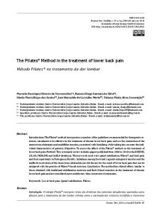
Helmholtz coil
Patients & methods Subject recruitment
Signal generator
Figure 1. Medical Device Resonator, showing (A) the Helmholtz coil and (B) signal generator. The Medical Device Resonator has Health Canada License and CE Mark (#2001010303CPON0318) and was developed using defined physical principles for electromagnetic field therapy production.
as an alternative, safe and effective treatment option for chronic pain in different clinical settings [7–10] . Presently, there is a paucity of information available in the literature that describes the clinical utility of very low EMF in the treatment of chronic LBP associated with degenerative disc disease. Accordingly, in this pilot study, a placebo-controlled, randomized, double-blind study design was employed to assess the efficacy and safety of low-amplitude/low-frequency EMF in the management of nonresponsive chronic LBP and functional capacity in patients with some degenerative disc disease. In addition, quality of life scores as well as mobility measurements were collected
Twenty one subjects were referred to participate in the study through family physicians, physical medicine and rehabilitation, sports medicine, orthopedics and rheumatology departments from the Health Sciences Centre, Winnipeg, Manitoba. The protocol was approved by the institutional review committee. Subjects were male and female, over 25 years of age who had documented chronic LBP persisting more than 6 months, which was not responsive to conservative therapy, MRI radiographic confirmation of diagnosis (minimum 25% and maximum 75% degeneration, and loss of some disc height) and no herniation. In these subjects, the disc degeneration resulted in discogenic low back pain (due to inflammation and abnormal micromotion instability [11] . In order to minimize confounds and to allow for a stringent study design, several exclusion criteria were applied. Exclusion criteria did not allow for the enrollment of patients with bone-on-bone cases, subjects taking steroids, weighing more than 210 lbs, subjects with pacemakers or with metallic prostheses, pregnancies or with epilepsy, diabetes, uncontrolled high blood pressure, history of congestive heart failure and/or cardiac arrhythmias, stroke, heart valve defects, thyroid conditions, subjects at risk of transient ischemic attacks, blood clots or aneurysms, subjects with past history of major head trauma or brain surgery and subjects with chronic low back pain due to sciatica. Subject randomization
Patients were randomly assigned to either the control (n = 7) or EMF treatment (n = 14) arm. Randomization was carried out by the subject drawing an envelope containing a preassigned number. The envelope was handed unopened to the operator of the device. The operator of the device opened the envelope in the control room where no other persons involved in the study were present. Inserts of envelopes containing even numbers were assigned to the treatment arm, whereas inserts
Table 1. Electromagnetic field pattern protocol. Magnetic field amplitude (G)
Frequency (Hz)
Duration (minutes)
3.3 × 10 -8 G
0.92
15
2.74 × 10 -7 G
7.7
12
3.43 × 10 G
9.6
10
3.3 × 10 -8 G
0.92
23
-7
G: Gauss; Hz: Hertz.
10.4155/fsoa-2015-0019
Future Sci. OA (2016) FSO105
future science group
EMF for chronic lower back pain associated with degenerative disc disease
Preliminary Communication
Table 2. Demographic and medical variable comparison of electromagnetic field group and control groups. Variable
EMF
Control
p-value
Age (years)
60.3 (9.7)
59.1 (13.4)
0.831
Systolic BP (mm Hg)
120.8 (9.9)
122.8 (15.5)
0.929
Diastolic BP (mm Hg)
81.4 (10.0)
75.5 (14.7)
0.216
Heart rate (beats/min)
71.9 (11.3)
77.0 (11.6)
0.387
Differences in the demographic and medical variables at baseline between control and EMF groups were determined by using the independent groups t-test for continuous variables. The level of statistical significance was set at p < 0.05. BP: Blood pressure; EMF: Electromagnetic field therapy.
of envelopes containing odd numbers were assigned to the control arm. Since the device operates in total silence and there are no other indicators of when the device is on or off, the study was blinded for the patient, and the clinical assistant doing the positioning of the patient. Subsequent visits to the center remained blind as only the operator of the device had the record of what the previously assigned subject number was and which arm of the study the subject was in. Intervention & study design
The magnetic fields to be used in this pilot study were generated by a medical device resonator. This resonator is a noninvasive device that does not utilize ionizing radiation and produces a uniform low-amplitude/lowfrequency EMF through a Helmholtz coil (Figure 1A) operated through a signal generator (Figure 1B) . The coil was positioned over the treatment area of the individual lying in horizontal position. This device has a Health Canada license and is listed as an approved device. Subjects assigned to the treatment group were exposed to EMF for 1 h according to the values defined in the Pattern Protocol (Table 1) . Subjects in the treatment group were exposed to five treatment sessions (visit 2 through to visit 6) during a 2-week period, for 60 min per treatment, whereas subjects assigned to the placebo group, underwent all procedures, as the treatment arm, except that the device remained off during
the session. We selected a room that was not close to items that generate significant environmental fields, such elevators, heating, ventilation and air conditioning (HVAC) systems, and electrical panels. Each subject rated their pain level before and after each treatment, 1 week and 1 month after the last treatment. Accordingly, post-treatment evaluation was conducted at visit 7 and visit 8 at the 5th and 9th week of the study period. Participants underwent a demographic questionnaire, medical history, the Roland Disability Questionnaire and the SF-36 Health Survey at baseline (visit 1, week 1). The Roland Disability Questionnaire and the SF-36 Health Survey were repeated at the end of the study following visit 8. On visits 2 through 8, participants completed the Pain Relief Rating Scale immediately following treatment. Participants completed the McGill Visual Analogue Pain Scale (VAS) and Present Pain Intensity Index before and after treatment on visits 2 through 8. It is pointed out that on visits 7 and 8 the McGill Pain Scales were only measured post-treatment. In addition, measurements of forward mobility (measured from waist to point on mid-upper back), right lateral mobility and left lateral mobility (side to side measure to floor) were obtained before and after treatment on visits 2 through 8. Pain scores were collected through a manual assessment by the subjects by placing a mark on a continuous line between 0 to 10, where 0 = no pain and 10 = worst pain.
Table 3. Roland disability questionnaire changes from baseline to end point. Variable
Baseline median values (first quartile, third quartile)
End point median values (first quartile, third quartile)
p-value
Control
12.0 (8.0, 19.0)
11.0 (11.0, 17.0)
0.917
Experimental
17.0 (12.0, 21.0)
17.0 (12.0, 20.0)
0.195
Control
6.0 (2.0, 8.0)
6.0 (1.0, 8.0)
0.657
Experimental
7.0 (2.0, 9.0)
7.0 (2.0, 8.0)
0733
Total score
Bothersome index
Differences in scores on the Roland Disability Questionnaire between baseline and end point for each group were analyzed by the Wilcoxon matched pairs test on ranks. The level of statistical significance was set at p < 0.05. High scores = poor functioning.
future science group
www.future-science.com
10.4155/fsoa-2015-0019
Preliminary Communication Arneja, Kotowich, Staley, Summers & Tappia
Table 4. SF-36 health survey changes from baseline to end point. Variable
Baseline median values (first quartile, third quartile)
End point median values (first quartile, third quartile)
p-value
Physical health total score
47 0 (45.0, 54.0)
49.0 (47.0, 52.0)
0.295
Physical functioning
20.0 (16.0, 23.0)
20.0(15.0, 20.0)
1.000
Role-physical
4.0 (4.0, 5.0)
4.0 (4.0, 5.0)
1.000
Bodily pain
8.0 (7.0, 9.0)
7.0 (7.0, 7.0)
0.173
General health
14.0 (14.0, 19.0)
16.0 (16.0, 22.0)
0.043
Mental health total score
44.0 (42.0, 49.0)
47.0 (46.0, 49.0)
0.447
Vitality
14.0 (13.0, 19.0)
16.0 (15.0, 18.0)
0.590
Social functioning
6.0 (4.0, 6.0)
6.0 (5.0, 7.0)
0.423
Role-emotional
3.0 (3.0, 4.0)
3.0 (3.0, 5.0)
1.000
Mental health
20.0 (18.0, 23.0)
20.0 (18.0, 22.0)
0.686
Physical health total score
41.5 (40.0, 45.0)
43.0 (39.0, 46.0)
0. 929
Physical functioning
15.5 12.0, 18.0)
16.5 (15.0, 22.0)
0.022
Role-physical
4.0 (4.0, 4.0)
4.0 (4.0, 5.0)
0.575
Bodily pain
9.0 (8.0, 10.0)
7.5 (6.0, 9.0)
0.045
General health
13.0 (11.0, 14.0)
13.0 (10.0,15.0)
0.906
Mental health total score
45.5 (42.0, 49.0)
46.9 (43.0, 50.0)
0.753
Vitality
15.5 (11.0, 18.0)
14.0 (13.0, 16.0)
0.563
Social functioning
6.5 (6.0, 7.0)
7.0 (6.0, 7.0)
0.799
Role-emotional
4.0 (3.0, 5.0)
4.0 (3.0, 6.0)
0.735
Mental health
20.5 (19, 21)
20.0 (19.0, 23)
0.347
Control group
EMF group
Differences in scores in the SF-36 Health Survey between baseline and end point for each group were analyzed by the Wilcoxon matched pairs test on ranks. The level of statistical significance was set at p