EDUCATION EXHIBIT
585
Imaging of Musculoskeletal Fibromatosis1 CME FEATURE See accompanying test at http:// www.rsna.org /education /rg_cme.html
LEARNING OBJECTIVES FOR TEST 2 After reading this article and taking the test, the reader will be able to: List the general 䡲 imaging characteristics and corresponding histopathologic features of the various superficial and deep musculoskeletal fibromatoses. 䡲 Describe the characteristic MR imaging features of musculoskeletal fibromatoses and use of these features in preoperative staging and treatment planning. 䡲 Discuss the various treatment options for musculoskeletal fibromatosis and imaging evaluation of masses after treatment and of postoperative recurrences.
Mark R. Robbin, MD ● Mark D. Murphey, MD ● H. Thomas Temple, MD ● Mark J. Kransdorf, MD ● James J. Choi, MD The musculoskeletal fibromatoses comprise a wide range of lesions with a common histopathologic appearance. They can be divided into two major groups: superficial and deep. The superficial fibromatoses are typically small, slow-growing lesions and include palmar fibromatosis, plantar fibromatosis, juvenile aponeurotic fibroma, and infantile digital fibroma. The deep fibromatoses are commonly large, may grow rapidly, and are more aggressive. They include infantile myofibromatosis, fibromatosis colli, extraabdominal desmoid tumor, and aggressive infantile fibromatosis. Radiographs typically reveal a nonspecific softtissue mass, and calcification is common only in juvenile aponeurotic fibroma. Advanced imaging (ultrasonography, computed tomography, and magnetic resonance [MR] imaging) demonstrates lesion extent. Involvement of adjacent structures is common, reflecting the infiltrative growth pattern often seen in these lesions. MR imaging may show characteristic features of prominent low to intermediate signal intensity and bands of low signal intensity representing highly collagenized tissue. However, fibromatoses with less collagen and more cellularity may have nonspecific high signal intensity on T2-weighted images. Local recurrence is frequent after surgical resection due to the aggressive lesion growth. It is important for radiologists to recognize the imaging characteristics of musculoskeletal fibromatoses to help guide the often difficult and protracted therapy and management of these lesions.
Index terms: Bones, fibroma, 40.313 ● Desmoid, 40.313 ● Fibromatosis, 40.1544, 40.313, 40.3132 ● Soft tissues, fibroma, 40.1544, 40.313 RadioGraphics 2001; 21:585– 600 1From
the Department of Radiology, University Hospitals of Cleveland, Case Western Reserve University School of Medicine, 10900 Euclid Ave, Cleveland, OH 44106 (M.R.R.); the Department of Radiologic Pathology, Armed Forces Institute of Pathology, Washington, DC (M.D.M., J.J.C.); the Departments of Radiology and Nuclear Medicine, Uniformed Services University of the Health Sciences, Bethesda, Md (M.D.M.); the Department of Radiology, University of Maryland School of Medicine, Baltimore (M.D.M.); the Department of Orthopedic Surgery, University of Miami School of Medicine, Miami, Fla (H.T.T.); and the Department of Radiology, Mayo Clinic, Jacksonville, Fla (M.J.K.). Recipient of a Certificate of Merit award for a scientific exhibit at the 1997 RSNA scientific assembly. Received June 5, 2000; revision requested July 14; final revision received February 15, 2001; accepted February 15. Address correspondence to M.R.R. (e-mail:
[email protected]). The opinions and assertions contained herein are the private views of the authors and are not to be construed as official or as reflecting the views of the Departments of the Army, Navy, or Defense.
©
RSNA, 2001
586
May-June 2001
RG f Volume 21
●
Number 3
Introduction The fibromatoses are a diverse group of soft-tissue lesions that occur at different ages and anatomic locations and that have common histopathologic features. They are composed of spindle-shaped fibrous cells that are separated and surrounded by abundant collagen material with rare mitoses. Their biologic behavior is intermediate in aggressiveness between benign fibrous lesions and fibrosarcoma. There is a strong tendency toward local recurrence; however, these lesions never metastasize. The lesions have a wellcircumscribed or infiltrative margin (Fig 1), and the degree of cellularity is widely variable (Fig 2). The fibromatoses are commonly divided into two major groups: superficial (fascial) and deep (musculoaponeurotic). The superficial fibromatoses are usually small, slow-growing lesions that arise from fascia and aponeuroses. The superficial group includes palmar fibromatosis (Dupuytren disease), plantar fibromatosis (Ledderhose disease), juvenile aponeurotic fibroma, and infantile digital fibromatosis. The deep fibromatoses are larger and may grow rapidly. They are more aggressive and likely arise from the deep fascia about muscle and aponeurotic tissue. The most common types of deep fibromatoses are infantile myofibromatosis, fibromatosis colli, extraabdominal desmoid tumor, and aggressive infantile fibromatosis (1,2).
Superficial Fibromatoses
Figure 1. Histopathologic features of fibromatosis. Photographs of gross specimens show examples of welldefined margins in fibromatosis (a) as well as a lesion with infiltrative margins (arrows in b).
Palmar Fibromatosis Palmar fibromatosis (Dupuytren disease) is the most common type of fibromatosis, with a prevalence of 1%–2% in the general population (1,3). Older patients are most frequently affected, with 24% of people over the age of 65 years demonstrating such lesions. Palmar fibromatosis is bilateral in 42%– 60% of cases (3). These lesions are three to four times more common in men and are rarely seen in children (3). The clinical presentation is subcutaneous nodules on the palmar surface at the level of the distal crease of the hand (Fig 3). These nodules slowly progress to fibrous cords or bands that attach to and cause traction on the adjacent flexor tendons, resulting in flexion contractures of the digits. The fourth and fifth rays are most commonly affected, followed by the second and third rays (3). Surgery is often purposely delayed owing to the high rate of
recurrence (30%– 40%) for lesions in the early proliferative phase (4). This delay in definitive treatment may result in relatively severe flexion contractures. Palmar fibromatosis in the proliferative phase demonstrates a high degree of cellularity. These lesions typically mature over time with increased collagen content and reduced cellularity. Highly collagenized lesions have a much lower prevalence of local recurrence following surgical resection. Although these lesions have traditionally been diagnosed and treated clinically, MR imaging has recently become valuable in planning surgical treatment. Yacoe and co-workers (4) correlated the cellularity of the lesions with their MR imaging appearance. These authors reported that predominantly low signal intensity on T2-weighted spin-echo MR images and low to intermediate signal intensity on T1-weighted MR images corresponded to lesions with relative hypocellularity caused by predominant dense collagen at histopathologic analysis. Those lesions that demon-
RG f Volume 21
●
Number 3
Robbin et al
587
Figure 2. Histopathologic features of fibromatosis. (a) Photomicrograph (original magnification, ⫻200; hematoxylin-eosin stain) shows a lesion that is predominantly cellular with relatively less fibrosis. (b) Photomicrograph (original magnification, ⫻250; hematoxylin-eosin stain) shows a lesion that is predominantly fibrotic with less cellularity.
Figure 3. Palmar fibromatosis. (a) Clinical photograph shows multiple subcutaneous cords and nodules (arrowheads) at the bases of the second through fifth digits, which resulted in flexion contractures. (b, c) Axial (b) and sagittal (c) T1-weighted magnetic resonance (MR) images (repetition time msec/echo time msec ⫽ 633/20) obtained in another patient show the fibromatosis as a low-signal-intensity band (arrowheads) superficial to and paralleling the flexor tendon (arrow) of the fifth digit.
strated intermediate signal intensity on T2weighted spin-echo MR images were more cellular and thus more likely to locally recur. This information may be useful in guiding the timing of surgical intervention. If a lesion demonstrates intermediate or high signal intensity on T2-
weighted spin-echo MR images, surgery may be delayed until the lesion matures and becomes more hypocellular, thus helping decrease the rate of postoperative recurrence (4).
588
May-June 2001
RG f Volume 21
●
Number 3
Figure 4. Plantar fibromatosis in a 65-year-old woman. (a) Clinical photograph shows a nodular soft-tissue mass along the medial plantar surface (arrows). (b, c) Sagittal T1-weighted (500/20) (b) and T2-weighted (2,000/80) (c) MR images show the mass (arrowheads). The high signal intensity on the T2-weighted image (c) corresponded to a more cellular lesion with less collagen than many fibromatoses (cf Fig 2a).
Plantar Fibromatosis Plantar fibromatosis (Ledderhose disease) occurs most frequently between the ages of 30 and 50 years, with bilateral involvement seen in 20%– 50% of cases. Concomitant palmar fibromatosis is seen in 10%– 65% of patients (5). These lesions develop most commonly in the medial aspect of the plantar aponeurosis. They usually manifest as one or multiple firm, fixed, subcutaneous nodules, which can extend to involve the skin or invade the deep structures of the foot (5). Plantar fibromatosis is often asymptomatic until the lesion enlarges and causes mass effect or invades adjacent muscles or neurovascular structures (5,6). Surgery with wide excision is reserved for larger, painful lesions and those demonstrating neurovascular involvement. Adjunctive radiation therapy may be used to decrease the rate of recurrence (5). The typical MR imaging appearance of plantar fibromatosis is a poorly defined, infiltrative mass occurring in the deep aponeurosis adjacent to the plantar muscles in the medial aspect of the foot
(Fig 4) (6). These lesions typically have heterogeneous signal intensity equal to or less than that of skeletal muscle on both T1- and T2-weighted spin-echo MR images. Lesions with high signal intensity on T2-weighted images reflect a more cellular lesion with relatively less collagen. The enhancement with gadolinium contrast material is variable, with marked enhancement seen in approximately 50% of lesions (6,7). Lesions with minimal symptoms are usually treated conservatively by means of orthopedic footwear.
Juvenile Aponeurotic Fibroma Juvenile aponeurotic fibroma is a rare, locally aggressive fibroblastic lesion located in the palms of the hands and soles of the feet in young children (8). Male patients are twice as commonly affected as female patients, and most of the lesions manifest within the first 2 decades of life (9). Juvenile aponeurotic fibroma is uncommon in older children and adults (9). This type of fibromatosis is one of the few to frequently calcify and is also referred to as calcified aponeurotic fibroma (10). The typical clinical presentation is a slow-growing, asymptomatic soft-tissue mass. Most of the le-
RG f Volume 21
●
Number 3
Robbin et al
589
Figure 5. Juvenile aponeurotic fibroma in a 12-yearold boy. Radiographs of the long finger show a calcified soft-tissue mass (arrow) with mild extrinsic erosion of the middle phalanx (arrowhead).
Figure 6. Infantile digital fibromatosis in a 4-year-old girl. Radiograph shows a soft-tissue mass (arrowhead) involving the fifth finger without bone involvement.
sions (67%) arise in the deep palmar fascia of the hand (9,10). The lesions are locally aggressive and have a recurrence rate of greater than 50% following surgical resection (10). Radiographs show a nonspecific soft-tissue mass, which can demonstrate fine stippled calcifications and occasional scalloping of the adjacent bone (Fig 5) (8,10,11). Biopsy is important in evaluation of these lesions and differentiation from synovial sarcoma, which can occur in young children, commonly calcifies, and may demonstrate extrinsic erosion of bone.
lateral aspect of the distal or middle phalanx (13). The most common treatment is surgical excision. Spontaneous regression has been reported in approximately 8% of cases (15). However, as with other fibromatoses, there is a high recurrence rate of approximately 60% (15). Radiographs commonly show a nonspecific soft-tissue mass involving a digit with infrequent underlying bone involvement (Fig 6) (15).
Deep Fibromatoses Infantile Myofibromatosis
Infantile Digital Fibromatosis Infantile digital fibromatosis occurs in the fingers and toes of infants (12). This lesion is distinguished from other forms of infantile fibromatosis by the characteristic patient age and location and histologic identification of a specific intracytoplasmic perinuclear inclusion body (13). This lesion has also been referred to as Reye tumor, infantile digital fibroma, and infantile digital myofibroblastoma (13,14). Most cases (80%) are diagnosed within the 1st year of life, and approximately 30% of cases are congenital (14). The fingers are more frequently affected (60% of cases) than the toes (40% of cases) (15). These lesions usually manifest as single or multiple nodules involving the extensor surfaces, most commonly the dorsal and
Infantile myofibromatosis is a solitary or multicentric proliferation of fibrous tissues and represents a distinct subtype of the fibromatoses (1). At histopathologic analysis, infantile myofibromatosis is characterized by spindle cells with histologic features of both smooth muscle and fibroblasts (16). At gross examination, lesions may be well circumscribed or have an infiltrative margin. Solitary and multicentric forms occur with approximately equal frequency. Multicentric disease often affects multiple organ systems including skeletal muscle, bone, soft tissue, skin, and viscera such as the lung and liver (17). The vast majority of cases occur before the age of 2 years. However, rare cases affecting adolescents or
590
May-June 2001
RG f Volume 21
●
Number 3
Figure 7. Infantile myofibromatosis in a 5-month-old girl. (a, b) Clinical photograph (a) and anteroposterior skull radiograph (b) show a large, exophytic soft-tissue mass (arrow) extending from the left side of the neck. (c) Lowerextremity radiograph shows multiple geographic, symmetric, lytic lesions in the metaphyses of the long bones (arrowheads). Lytic lesions were also seen in the ribs, right scapula, and clavicles.
adults with primary or recurrent lesions have been reported (18). Both solitary and multicentric disease is more common in male patients (male-tofemale ratio, 1.6 –1.7:1) (19). In patients with solitary disease but without visceral involvement, the prognosis is quite favorable because many lesions undergo spontaneous regression (17,19). The recurrence rate after surgical excision of solitary myofibromatosis is low (approximately 10%) compared with that of other forms of fibromatosis. Spontaneous regression of multicentric disease is seen in approximately 30% of cases (16,19). Identification of visceral involvement is an important prognostic indicator. Although the overall mortality rate is less than 15%, approximately 75% of patients with visceral involvement (particularly of the cardiopulmonary or gastrointestinal system) die of the disease (16,17,19). Skin lesions often have prominent vascularity, thus resembling hemangiomas (Fig 7) (19). Frequent locations of soft-tissue masses in infantile myofibromatosis include the head, neck, and trunk (18). Common areas of bone involvement include the femora, tibiae, ribs, spine, and pelvis (16,21).
In patients with myofibromatosis with osseous involvement, the metaphyseal regions of the long bones are frequently affected. Radiographs of bone lesions typically show eccentric, geographic lesions that may have a rim of sclerosis (21). Extremity lesions are often bilateral and may be symmetric (16,21). Bone lesions usually heal during disease regression (22–24). The differential diagnosis of multiple lytic skeletal lesions in young children also includes Langerhans cell histiocytosis, hematogenous osteomyelitis (particularly with unusual organisms), enchondromatosis, metastatic neuroblastoma, and angiomatosis (2). At computed tomography (CT), soft-tissue masses may have increased attenuation compared with that of skeletal muscle both before and after intravenous administration of contrast material (25). Small foci of calcification may be seen within skeletal or soft-tissue lesions (22). MR imaging best demonstrates the soft-tissue extent of lesions and particularly visceral involvement (17). Pulmonary involvement may have a variable radiographic appearance, including pulmonary fibrosis, reticulonodular infiltrates, and generalized bronchial pneumonia (25). Gastrointestinal tract involvement typically appears as diffuse narrowing of the intestine or multiple filling defects on images from barium studies.
RG f Volume 21
●
Number 3
Robbin et al
591
Figure 8. Fibromatosis colli in a 6-week-old girl. (a) Transverse US scan shows a diffusely enlarged right sternocleidomastoid muscle (arrowheads) relative to the normal left sternocleidomastoid muscle (not shown). (b, c) Coronal T1-weighted (800/15) (b) and T2-weighted (2,000/90) (c) MR images show an enlarged right sternocleidomastoid muscle (arrows in b). A poorly defined mass with mild increased signal intensity (arrowheads) is seen on the long repetition time image (c).
Fibromatosis Colli Fibromatosis colli is a rare form of infantile fibromatosis that occurs solely in the sternocleidomastoid muscle. Most cases show no abnormality at birth but manifest between the 2nd and 4th weeks of life as a firm soft-tissue mass in the lower onethird of the sternocleidomastoid muscle. The disease is usually unilateral (slightly more common on the right side) and affects both the sternal and clavicular heads of the muscle (1). Bilateral involvement is rare. Fibromatosis colli affects male patients slightly more often than female patients (1). Torticollis is seen in 14%–20% of patients due to contraction of the sternocleidomastoid muscle (26,27). In fact, cases of congenital torticollis, even those lacking a focal soft-tissue mass, likely represent unrecognized fibromatosis colli (28). Although the exact cause is unclear, it is likely related to birth trauma, with greater than 90% of cases associated with a difficult or forceps delivery (26). Davids and co-workers (26) have suggested that the lesions result from an in utero fetal head position, which causes selective injury to the sternocleidomastoid muscle. Such injury leads to development of a secondary compartment syndrome and resultant pressure necrosis and fibrosis within the muscle (26). Initially, the mass may grow rapidly, although subsequently
growth slows and ultimately ceases. Lesions regress by the age of 2 years in approximately twothirds of cases (27). Ultrasonography (US) is the best imaging modality for diagnosis due to its relative low cost, its lack of radiation, and the proximity of the lesion to the skin (29). Various US findings have been described, ranging from a homogeneously enlarged sternocleidomastoid muscle without a focal mass (Fig 8) to a hypoechoic mass with ill-defined or well-defined margins in the substance of the sternocleidomastoid muscle (29). The MR imaging appearance has been described by Davids and co-workers (26). All cases in their series demonstrated mild enlargement of the lower one-third of the sternocleidomastoid muscle with diffuse abnormal high signal intensity (greater than that of fat) on T2-weighted images. None of the cases in their series demonstrated a focal discrete mass at MR imaging (26). CT shows a homogeneous enlarged sternocleidomastoid muscle, also without a focal soft-tissue mass (30). Although radiographs are usually normal, lytic lesions within the head of the clavicle at the attachment of the sternocleidomastoid muscle have rarely been reported (31).
592
May-June 2001
RG f Volume 21
●
Number 3
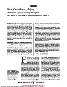
Figure 9. Desmoid tumor of the abdominal wall in a 29-year-old woman taking birth control pills. (a) Contrast material– enhanced CT scan shows an intensely enhanced mass arising from the lateral abdominal wall (arrowheads). (b) Axial T1-weighted MR image (544/18) shows that the mass (arrows) has intermediate signal intensity. (c) Axial T2-weighted MR image (1,800/180) shows that the mass (arrows) has heterogeneous high signal intensity. (d) Photograph of the gross surgical specimen shows the well-defined mass (arrows).
Extraabdominal Desmoid Tumor Extraabdominal desmoid tumors, which occur outside the peritoneal cavity, are a relatively frequent cause of a soft-tissue mass in adults (Figs 9 –11). Young adults are most commonly affected, with a peak prevalence between the ages of 25 and 35 years. Lesions in patients younger than 10 years are uncommon (32). The extremities account for approximately 70% of cases. Frequent sites of involvement include the shoulder (20%), chest wall and back (15%), thigh (12%), mesentery (10%), neck (10%), and knee (7%) (32– 45). Most cases manifest as solitary lesions; however, synchronous multicentric lesions in the same extremity occur in approximately 10%–15% of cases (34 –36). The prognosis appears to be related to the age of the patient. Patients under the age of 30 years tend to have more aggressive lesions. The local recurrence rate is high in these patients (up to 87%) (35,36,45). The recurrence rate in patients older than 20 years is approximately 50% (36). In patients with multicentric fibromatosis, a metaphyseal dysplasia similar to that of Gaucher or Pyle disease—with an Erlen-
meyer flask deformity and undertubulation of the extremity long bones—is seen infrequently (34). Desmoid tumors of the abdominal wall, although identical to other types of extraabdominal desmoid tumors at histopathologic analysis, are considered a distinct entity due to their predilection to develop in female patients taking birth control pills and following or during pregnancy. Abdominal wall desmoid tumors occur in female patients of childbearing age (87% of cases), most commonly in the rectus abdominis and internal oblique musculoaponeurotic structures (Fig 9) (1,34).
RG f Volume 21
●
Number 3
Robbin et al
593
Figure 10. Extraabdominal desmoid tumor involving the paraspinal region in a 57-year-old man. (a) Sagittal T1weighted MR image (550/20) shows a large paraspinal mass (arrows) with signal intensity similar to that of muscle. (b) Axial T2-weighted MR image (2,000/90) shows that the mass (black arrows) has heterogeneous high signal intensity with low-signal-intensity bands (arrowheads). The ill-defined infiltrative margins (white arrows) correspond to those of the gross specimen shown in Figure 1b.
Figure 11. Extraabdominal desmoid tumor of the right shoulder in a 20-year-old woman. (a) Coronal T1weighted MR image (400/15) shows a vague mass lateral to the humerus (arrowheads). (b) Axial fat-suppressed T1weighted MR image (500/20) obtained after intravenous administration of gadolinium contrast material shows that the mass (large arrowheads) is intermuscular and invades the subcutaneous fat (arrow). The mass demonstrates marked enhancement and irregular infiltrative margins. (c) Axial T2-weighted MR image (2,500/80) shows that the mass has heterogeneous intermediate signal intensity (less than that of fat) with low-signal-intensity collagenized bands (arrows); these bands lack prominent enhancement on the postcontrast image (small arrowheads in b).
Extraabdominal desmoid tumors typically demonstrate an infiltrative growth pattern at histologic examination. The lesion is composed of parallel halo arrays of uniform-appearing fibroblasts surrounded by highly variable amounts of
collagen fibers. Areas of myxoid tissue, hemorrhage, and inflammation can be seen, although these are unusual (1).
594
May-June 2001
RG f Volume 21
●
Number 3
Figure 12. Aggressive infantile fibromatosis in a newborn. (a) CT scan of the thorax shows a large, posterior softtissue mass (arrowheads) invading the thoracic spine and posterior chest wall (arrows). (b, c) Axial T1-weighted (550/20) (b) and sagittal T2-weighted (2,000/90) (c) MR images show that the mass has heterogeneous intermediate signal intensity and low-signal-intensity bands (arrows) with both sequences. There is invasion of the spinal canal and posterior chest wall (arrowheads).
Aggressive Infantile Fibromatosis Aggressive infantile fibromatosis is the childhood equivalent of deep fibromatosis (Fig 12). It usually manifests in the first 2 years of life and rarely occurs after the age of 5 years (46,47). Male patients are affected slightly more commonly than female patients. The lesion typically manifests as a firm, nodular soft-tissue mass, usually within skeletal muscle, adjacent fascia, or periosteum (1,47). The most common location is the head and neck, often with involvement of the tongue, mandible, and mastoid process. Other sites frequently affected include the shoulder, thigh, and
foot (1,46,47). Aggressive lesions may be highly cellular at histologic analysis and may mimic an infantile fibrosarcoma (1).
Imaging of Extraabdominal Desmoid Tumor and Aggressive Infantile Fibromatosis Imaging and treatment of extraabdominal desmoid tumor and of aggressive infantile fibromatosis are similar and therefore are discussed together. Radiographs may be normal or may show a nonspecific soft-tissue mass. Calcification is uncommon. Underlying bone involvement is seen in 6%–37% of patients, typically with pressure erosion and cortical scalloping but without invasion of the medullary canal (37,38). Bone scintigraphy
RG f Volume 21
●
Number 3
usually demonstrates increased uptake on blood pool and static images (39). Angiograms are variable in appearance, often showing marked hypervascularity, although some lesions demonstrate no vascular blush (40). CT scans of the deep fibromatoses are also usually nonspecific. Lesions may be hypoattenuating relative to skeletal muscle but are typically isoattenuating or even hyperattenuating (Fig 9) (40,41). The latter finding may be related to lesions with more extensive collagen. Lesions usually demonstrate enhancement after intravenous administration of iodinated contrast material; the enhancement is sometimes marked (40,41). Owing to the infiltrative growth pattern and the attenuation similar to that of skeletal muscle, the margins of the lesion are often indistinct at CT unless it is separated from normal tissue by a fat plane (40). Subtle pressure erosions of bone are often better evaluated on radiographs owing to beam-hardening artifact at CT (40). The best imaging modality for evaluation and staging of the deep fibromatoses is MR imaging (34). Extraabdominal desmoid tumors and aggressive infantile fibromatosis are typically intermuscular lesions, although muscle invasion is common. In addition, linear extension along fascial planes is a frequent manifestation and is uncommon with other soft-tissue neoplasms. Initial reports suggested that the lesions have decreased signal intensity on both T1- and T2-weighted spin-echo images (41,42). Sundaram and coworkers (41) compared the signal intensity patterns at MR imaging with the histopathologic findings in three cases. In two cases, the lesions had decreased signal intensity on T2-weighted images, which represented hypocellularity and abundant collagen. In one case with high signal
Robbin et al
595
intensity on T2-weighted images, the lesion demonstrated marked cellularity (42). In subsequent larger series, the MR imaging pattern of the deep fibromatoses has been highly variable. The most common signal intensity pattern is heterogeneous, with intermediate signal intensity (similar to that of fat on T2-weighted images and similar to that of skeletal muscle on T1-weighted images) seen with standard pulse sequences (Fig 10) (2,43– 45). The heterogeneous signal intensity pattern likely corresponds to the varying proportions of cellular tissue, myxoid tissue (high water content and high signal intensity on T2-weighted images), and collagen (low signal intensity with all pulse sequences) in the lesion (33,42). Prominent low-signal-intensity bands are often seen with all pulse sequences and are likely related to the dense areas of collagen (Fig 11) (33,42). Areas of low signal intensity with all pulse sequences are characteristic of fibromatosis but not specific for it. Other types of soft-tissue masses with prominent low signal intensity on T2weighted images include giant cell tumor of tendon sheath (a localized form of pigmented villonodular synovitis), calcified masses, and malignancies such as fibrosarcoma or malignant fibrous histiocytoma (33,42). The deep fibromatoses typically demonstrate moderate to marked enhancement after administration of gadolinium contrast material (Fig 11), particularly in less collagenized and more cellular regions (44). Only 10% of lesions lack significant enhancement at MR imaging (45). Lesion margins at MR imaging may be well defined or infiltrative.
596
May-June 2001
RG f Volume 21
●
Number 3
Figure 13. Recurrent aggressive fibromatosis of the foot in a 17-year-old girl. (a) Radiograph of the foot shows a recurrent soft-tissue mass that causes extrinsic erosion of the first and second metatarsals. (b) Sagittal T1-weighted MR image (400/15) shows that the mass (arrows) is large, with extensive invasion of the deep plantar tissues. (c) Clinical photograph shows the mass along the medial aspect of the foot (arrow). Surgical resection was performed. (d, e) Sagittal T1-weighted (550/25) (d) and fat-saturated T2-weighted (5,000/80) (e) MR images obtained 2 years later show an aggressive recurrent soft-tissue mass (arrowheads). The lesion has heterogeneous high signal intensity on the T2-weighted image (e), which corresponds to increased cellularity. (f ) Clinical photograph shows the mass and secondary ulceration of the skin (arrowheads) from secondary infection. Amputation was performed due to the extensive infiltrative nature of the lesion. (g) Photograph of the sagittally sectioned gross specimen shows that the lesion represents recurrent aggressive fibromatosis (arrowheads).
RG f Volume 21
●
Number 3
Robbin et al
597
Figure 14. Recurrent extraabdominal desmoid tumor in a 19-year-old woman. (a, b) Sagittal T1-weighted (800/ 20) (a) and T2-weighted (2,000/100) (b) MR images show a heterogeneous intermuscular soft-tissue mass in the popliteal fossa (arrowheads). Bands of low signal intensity (white arrows in a, solid arrows in b) are prominent. Linear extension along fascial planes inferiorly (black arrows in a, open arrow in b) was not recognized at surgical resection. (c) Sagittal T1-weighted MR image (500/20) obtained 16 months after surgical resection shows a recurrent intermuscular soft-tissue mass (arrows) along the gastrocnemius muscle and soleus aponeurosis at the site of previously identified fascial extension. The mass has signal intensity characteristics similar to those of the original lesion, with bands of low signal intensity (arrowheads).
Treatment of Extraabdominal Desmoid Tumor and Aggressive Infantile Fibromatosis Several treatment options are used in management of the deep fibromatoses. Although deep or aggressive fibromatosis is not considered a malignancy and does not metastasize, these lesions are locally aggressive and can invade or encase neurovascular structures and involve an entire extremity in multicentric disease. As discussed earlier, the prevalence of local recurrence is high and is related to the infiltrative margins seen at pathologic analysis. Local recurrence is typically seen within 12–18 months after surgical resection (47). Wide excision is the treatment of choice for lesions that are relatively small and favorably located (47– 49). Although amputations are rare, they may be necessary in patients with multiple recurrences (Fig 13). If wide excision cannot be achieved without functional loss, marginal excision and postoperative radiation therapy are often used for treatment (50). In some cases, particularly in lesions that invade major neurovascular structures such as the brachial plexus, radiation therapy and chemotherapy are the sole methods of treatment (50, 51). Because of the potential morbidity of disfiguring surgery and the complications of radiation therapy, particularly when applied to young children, chemotherapy alone has been used with
variable success (52,53). In a small study (10 patients) by Skapek and co-workers (52), five patients had clinical evidence of response to therapy, with complete resolution in 30% and partial resolution in 20%. An additional 30% of the patients had stable disease at up to 35 months after treatment, with 20% showing disease progression. MR imaging should be used for preoperative staging, particularly to evaluate for neurovascular and bone involvement. In extremity lesions, the entire limb should be imaged to rule out multicentric disease. MR imaging is also the best imaging modality to evaluate for postsurgical local recurrence. Recurrent deep fibromatosis shows intrinsic MR imaging characteristics similar to those of the original lesion. The site of recurrence is frequently at the lesion margins at areas of fascial extension where surgical resection is incomplete, leaving residual tumor (Fig 14). In patients who undergo radiation therapy or chemotherapy alone without surgery, MR imaging is useful in evaluating the effectiveness of therapy (Fig 15) (33,43,44). In our experience, effective therapy (good lesion response) is indicated by a reduction in size and an increasing degree of low signal intensity on T2-weighted images, which reflects increased collagenization in response to therapy.
598
RG f Volume 21
May-June 2001
●
Number 3
Figure 15. Radiation therapy of aggressive fibromatosis in an 18-year-old man. (a) Sagittal T2-weighted MR image (2,500/100) obtained before radiation therapy shows a large mass (arrowheads) involving the infraspinatus and subscapularis muscles. (b) Sagittal T2-weighted MR image (2,500/120) obtained 2 months later shows a decrease in both the size and signal intensity of the lesion (arrows). (c, d) Photomicrographs (original magnification, ⫻200; hematoxylin-eosin stain) obtained before (c) and after (d) treatment show that decreased cellularity and increased fibrosis (pink material) from a good response to the radiation therapy are the cause of the imaging findings.
Conclusions The fibromatoses represent a wide spectrum of lesions that can involve both superficial and deep musculoskeletal structures. These lesions can be difficult to manage clinically because of their infiltrative growth, leading to frequent local recurrence. Because of these characteristics, imaging plays a pivotal role in assessing the fibromatoses throughout their clinical course. MR imaging is usually the modality of choice for evaluation of these soft-tissue lesions. Common characteristics of the fibromatoses include infiltrative margins, low or intermediate signal intensity with all pulse sequences, and bands of low signal intensity rep-
resenting highly collagenized tissue. However, in lesions with a higher degree of cellularity and less collagen, high signal intensity may be apparent at MR imaging. It is important for radiologists to recognize these characteristics and imaging spectrum to help guide the often difficult and protracted therapy and management of these lesions.
References 1. Enzinger FM, Weiss SW. Soft tissue tumors. 3rd ed. St Louis, Mo: Mosby, 1995. 2. Kransdorf MJ, Murphey MD. Imaging of soft tissue tumors. Philadelphia, Pa: Saunders, 1997; 149 –175. 3. Laskin WB, Weiss SW. Benign fibrous lesions. In: Bogumill GP, ed. Tumors of the hand and upper limb. Edinburgh, Scotland: Churchill Livingstone, 1993; 224 –243. 4. Yacoe ME, Bergman AG, Ladd AL, Hellman BH. Dupuytren’s contracture: MR imaging findings and correlation between MR signal intensity and
RG f Volume 21
5. 6.
7. 8.
9.
10. 11. 12. 13.
14.
15. 16. 17. 18. 19.
20.
21. 22. 23. 24.
●
Number 3
cellularity of lesions. AJR Am J Roentgenol 1993; 160:813– 817. Lee TH, Wapner KL, Hecht PJ. Plantar fibromatosis. J Bone Joint Surg Am 1993; 75:1080 –1084. Morrison WB, Schweitzer ME, Wapner KL, Lackman RD. Plantar fibromatosis: a benign aggressive neoplasm with a characteristic appearance on MR images. Radiology 1994; 193:841– 845. Wetzel LH, Levine E. Soft-tissue tumors of the foot: value of MR imaging for specific diagnosis. AJR Am J Roentgenol 1990; 155:1025–1030. Keasbey LE. Juvenile aponeurotic fibroma (calcifying fibroma): a distinctive tumor arising in the palms and soles of young children. Cancer 1953; 6:338 –346. Goldman RL. The cartilage analogue of fibromatosis (aponeurotic fibroma): further observations based on 7 new cases. Cancer 1970; 26:1325– 1331. Carroll RE. Juvenile aponeurotic fibroma. Hand Clin 1987; 3:219 –224. Karasick D, O’Hara AE. Juvenile aponeurotic fibroma: a review and report of a case with osseous involvement. Radiology 1977; 123:725–726. Reye RDK. Recurrent digital fibrous tumors of childhood. Arch Pathol 1965; 80:228 –231. Fringes B, Thais H, Bohm N, Altmannsberger M, Osborn M. Identification of actin microfilaments in the intracytoplasmic inclusions present in recurring infantile digital fibromatosis (Reye tumor). Pediatr Pathol 1986; 6:311–324. Bhawan J, Bacchetta C, Joris I, Majno G. A myofibroblastic tumor: infantile digital fibroma (recurrent digital fibrous tumor of childhood). Am J Pathol 1979; 94:19 –36. Beckett JH, Jacobs AH. Recurring digital fibrous tumors of childhood: a review. Pediatrics 1977; 59:401– 406. Chung EB, Enzinger FM. Infantile myofibromatosis. Cancer 1981; 48:1807–1818. Wiswell TE. Infantile myofibromatosis and the use of magnetic resonance imaging (letter). Am J Dis Child 1988; 142:486. Stout AP. Juvenile fibromatosis. Cancer 1954; 7:953–978. Wiswell TE, Davis J, Cunningham BE, Solenberger R, Thomas PJ. Infantile myofibromatosis: the most common fibrous tumor of infancy. J Pediatr Surg 1988; 23:315–318. Jennings TA, Duray PH, Collins FS, Sabetta J, Enzinger FM. Infantile myofibromatosis: evidence for an autosomal-dominant disorder. Am J Surg Pathol 1984; 8:529 –538. Morettin LB, Mueller E, Schreiber M. General hamartomatosis (congenital generalized fibromatosis). AJR Am J Roentgenol 1972; 114:722–734. Chateil JF, Brun M, Lebail B, Perel Y, Castell JF, Diard F. Infantile myofibromatosis. Skeletal Radiol 1995; 24:629 – 632. Gold RH, Mirra JM. Case report 339: congenital multiple fibromatosis. Skeletal Radiol 1985; 14: 309 –311. Present DA, Abdelwahab IF, Zwass A, Klein MJ. Case report 575: infantile myofibromatosis. Skeletal Radiol 1989; 18:557–560.
Robbin et al
599
25. Baer JW, Radkowski MA. Congenital multiple fibromatosis: a case report with review of the world literature. Am J Roentgenol Radium Ther Nucl Med 1973; 118:200 –205. 26. Davids JR, Wenger DR, Mubarak SJ. Congenital muscular torticollis: sequela of intrauterine or perinatal compartment syndrome. J Pediatr Orthop 1993; 13:141–147. 27. Coventry MB, Harris LE, Bianco AJ, Bulbulian AH. Congenital muscular torticollis (wryneck). Postgrad Med 1960; 28:383–391. 28. Whyte AM, Lufkin RB, Bredenkamp J, Hoover L. Sternocleidomastoid fibrosis in congenital muscular torticollis: MR appearance. J Comput Assist Tomogr 1989; 13:163–164. 29. Kraus R, Han BK, Babcock DS, Oestreich AE. Sonography of neck masses in children. AJR Am J Roentgenol 1986; 146:609 – 613. 30. Crawford SC, Harnsberger HR, Johnson L, Aoki JR, Giley J. Fibromatosis colli of infancy: CT and sonographic findings. AJR Am J Roentgenol 1988; 151:1183–1184. 31. Sartoris DJ, Mochizuki RM, Parker BR. Lytic clavicular lesions in fibromatosis colli. Skeletal Radiol 1983; 10:34 –36. 32. Taylor LJ. Musculoaponeurotic fibromatosis: a report of 28 cases and review of the literature. Clin Orthop 1987; 224:294 –302. 33. Kransdorf MJ, Jelinek JS, Moser RP Jr, et al. Magnetic resonance appearance of fibromatosis: a report of 14 cases and review of the literature. Skeletal Radiol 1990; 19:495– 499. 34. Disler DG, Alexander AA, Mankin HJ, O’Connell JX, Rosenberg AE, Rosenthal DI. Multicentric fibromatosis with metaphyseal dysplasia. Radiology 1993; 187:489 – 492. 35. Rock MG, Pritchard DJ, Reiman HM, Soule EH, Brewster RC. Extra-abdominal desmoid tumors. J Bone Joint Surg Am 1984; 66:1369 –1374. 36. Abramowitz D, Zornoza J, Ayala AG, Romsdahl MM. Soft-tissue desmoid tumors: radiographic bone changes. Radiology 1983; 146:11–13. 37. Griffiths HJ, Robinson K, Bonfiglio TA. Aggressive fibromatosis. Skeletal Radiol 1983; 9:179 – 184. 38. Terui S, Terauchi T, Abe H, et al. Role of technetium-99m pertechnetate scintigraphy in the management of extra-abdominal fibromatosis. Skeletal Radiol 1995; 24:331–336. 39. Hudson TM, Vandergriend RA, Springfield DS, et al. Aggressive fibromatosis: evaluation by computed tomography and angiography. Radiology 1984; 150:495–501. 40. Francis IR, Dorovini-Zis K, Glazer GM, Lloyd RV, Amendola MA, Martel W. The fibromatoses: CT-pathologic correlation. AJR Am J Roentgenol 1986; 147:1063–1066. 41. Sundaram M, McGuire MH, Schajowicz F. Softtissue masses: histologic basis for decreased signal (short T2) on T2-weighted MR images. AJR Am J Roentgenol 1987; 148:1247–1250.
600
May-June 2001
42. Quinn SF, Erickson SJ, Dee PM, et al. MR imaging in fibromatosis: results in 26 patients with pathologic correlation. AJR Am J Roentgenol 1991; 156:539 –542. 43. Hawnaur JM, Jenkins JP, Isherwood I. Magnetic resonance imaging of musculoaponeurotic fibromatosis. Skeletal Radiol 1990; 19:509 –514. 44. Feld R, Burk DL Jr, McCue P, Mitchell DG, Lackman R, Rifkin MD. MRI of aggressive fibromatosis: frequent appearance of high signal intensity on T2-weighted images. Magn Reson Imaging 1990; 8:583–588. 45. Romero JA, Kim EE, Kim CG, Chung WK, Isiklar I. Different biologic features of desmoid tumors in adult and juvenile patients: MR demonstration. J Comput Assist Tomogr 1995; 19:782– 787. 46. Cintora E, del Cura JL, Ruiz JC, Grau M, Ereno C. Case report 807: infantile desmoid-type fibromatosis. Skeletal Radiol 1993; 22:533–535. 47. Khorsand J, Karakousis CP. Desmoid tumors and their management. Am J Surg 1985; 149:215–218.
RG f Volume 21
●
Number 3
48. Easter DW, Halasz NA. Recent trends in the management of desmoid tumors: summary of 19 cases and review of the literature. Ann Surg 1989; 210: 765–769. 49. Higaki S, Tateishi A, Ohno T, et al. Surgical treatment of extra-abdominal desmoid tumours (aggressive fibromatoses). Int Orthop 1995; 19:383– 389. 50. Kamath SS, Parsons JT, Marcus RB, Zlotecki RA, Scarborough MT. Radiotherapy for local control of aggressive fibromatosis. Int J Radiat Oncol Biol Phys 1996; 36:325–328. 51. Stockdale AD, Cassoni AM, Coe MA, et al. Radiotherapy and conservative surgery in the management of musculo-aponeurotic fibromatosis. Int J Radiat Oncol Biol Phys 1988; 15:851– 857. 52. Skapek SX, Hawk BJ, Hoffer FA, et al. Combination chemotherapy using vinblastine and methotrexate for the treatment of progressive desmoid tumor in children. J Clin Oncol 1998; 16:3021– 3027. 53. Rao BN, Horowitz ME, Parham DM, et al. Challenges in the treatment of childhood fibromatosis. Arch Surg 1987; 122:1296 –1298.
This article meets the criteria for 1.0 credit hour in category 1 of the AMA Physician’s Recognition Award. To obtain credit, see accompanying test at http://www.rsna.org/education/rg_cme.html.